Article

Structural Analysis of Acidic Glycosphingolipids in the Adductor Muscle of the Japanese Giant Scallop (Patinopecten yessoensis)

1
Faculty of Chemistry, Materials, and Bioengineering, Kansai University, 3-3-35, Yamate-cho, Suita 564-8680, Osaka, Japan
2
Faculty of Liberal Arts and Education, Shiga University, 2-5-1, Shiga, Hiratsu 520-0862, Otsu, Japan
3
Faculty of Food and Nutritional Sciences, Toyo University, 48-1, Oka, Asaka 351-8510, Saitama, Japan
*
Authors to whom correspondence should be addressed.
Fishes 2025, 10(9), 460; https://doi.org/10.3390/fishes10090460
Submission received: 24 July 2025 / Revised: 3 September 2025 / Accepted: 10 September 2025 / Published: 12 September 2025

Abstract

Structural analysis of glycosphingolipids provides novel insights into organismal classification and reveals conserved functional roles that transcend taxonomic boundaries. To elucidate the structural characteristics of acidic glycosphingolipids (AGLs) in the adductor muscle of the Japanese giant scallop (Patinopecten yessoensis), AGLs were isolated and purified by column chromatography using anion exchange resin and silica gel. Structural characterization was performed using mass spectrometry, proton nuclear magnetic resonance spectroscopy, and immunological techniques. The sugar chain structure was identified as GlcA4Meβ1-4(GalNAc3Meα1-3)Fucα1-4GlcNAcβ1-2Manα1-3Manβ1-4Glcβ1-Cer, consistent with the mollu-series core reported for mollusks. In addition to uronic acid, the structure was distinguished by internal fucose and methylated sugars, features commonly found in bivalves. The presence of xylose in the sugar chains of AGLs was also suggested. In contrast, the ceramide moiety was composed primarily of fatty acids C16:0 and C18:0 and the long-chain base d16:1. This chemical structure provides valuable insights into the biological classification of P. yessoensis and the mollu-series glycolipids containing fucose and methylated sugars, which may serve as bioactive components shared across species in the phylum Mollusca and class Bivalvia.
Key Contribution: The sugar chain structure of acidic glycosphingolipids in the adductor muscle of Patinopecten yessoensis was identified as GlcA4Meβ1-4(GalNAc3Meα1-3)Fucα1-4GlcNAcβ1-2Manα1-3Manβ1-4Glcβ1-Cer.

Graphical Abstract

1. Introduction

The Japanese giant scallop (Patinopecten yessoensis) is a marine bivalve mollusk belonging to the phylum Mollusca. P. yessoensis is one of Japan’s major marine exports, with primary production areas including the coast of the Sea of Okhotsk in Hokkaido, Uchiura Bay and its vicinity, and Mutsu Bay in Aomori Prefecture [1,2]. In addition, aquaculture is actively conducted along the Sanriku coast, where seedlings and juvenile scallops are transplanted from these regions. Morphologically, the body of P. yessoensis can be divided into three major parts: the adductor muscle, mantle, and viscera [3]. The adductor muscles account for approximately 35% of the total soft body weight [4]. On the periphery of the mantle are the pallial tentacle and pallial eye [5], which detect external enemies. In response to environmental stress or predators, P. yessoensis rapidly contracts its adductor muscles, closes its shell, and propels itself by expelling water, thereby enabling active swimming and escape [6,7,8].
Glycosphingolipids (GSLs) are composed of glycosidic linkages between monosaccharides or sugar chains and ceramides, which consist of amide-linked fatty acids and long-chain bases. GSLs are localized in the outer layers of the cell membrane [9] and form clusters on the plasma membrane through hydrogen bonding and van der Waals interaction driven by their physicochemical properties and chemical structures [10], resulting in the formation of domains called lipid rafts in the plasma membrane [11,12]. Acidic glycosphingolipids (AGLs) are classified into four types: sialic acid-containing (ganglioside), uronic acid-containing, sulfate-containing, and inositol phosphate-containing [13]. AGLs exhibit physiological functions related to cell adhesion and proliferation. In vertebrates, including mammals, gangliosides are abundant in central nervous system tissues, and their functions have been elucidated [14,15]. For example, GM3 ganglioside deficiency has been associated with hearing loss, abnormalities in immune cell populations, and enhanced insulin sensitivity [16,17].
Previous studies have reported on the GSLs of invertebrates, particularly focusing on their sugar chain structures. To date, the sugar chain structures of GSLs have been characterized in bivalves, such as Hyriopsis schlegelii [18,19], Corbicula sandai [20], and Meretrix lusoria [21]. A common core sugar chain structure identified in these species, GlcNAcβ1-2Manα1-3Manβl-4Glcβl-Cer, is known as the mollu-series. Recent findings suggest that core sugar chain structures vary across taxonomic groups. Previous studies have also identified GSL species containing lipid moieties that are unique to specific organisms, indicating their biosynthetic origin. These observations suggest that GSL structures can serve as novel taxonomic markers. Although the sugar chain structures of GSLs in H. schlegelii, C. sandai, and M. lusoria have been reported, no detailed analysis has been conducted on the sugar chain structure of GSLs in the adductor muscle of P. yessoensis. Given the high abundance of AGLs in the adductor muscle of P. yessoensis, this study involved their extraction, fractionation, and purification, culminating in a detailed structural characterization of their sugar chain and ceramide components. This study also examined the characteristics of the identified structures.

2. Materials and Methods

2.1. Preparation of Crude Sphingolipids

One hundred and fifty P. yessoensis (approximately 27 kg shelled mass) were purchased from Marunaka Fisheries Ltd. (Aomori, Japan) in 2007. Samples were selected in late September when the adductor muscle was growing. Crude sphingolipids were fractionated as described by Ito et al. [22]. After boiling P. yessoensis, the adductor muscle was collected, dehydrated with acetone, and air-dried. Approximately 1600 g of dried muscle was ground using a mixer and extracted twice with 3.5 L of chloroform/methanol (C/M; 2:1, v/v), followed by a single extraction with 3.0 L of C/M (1:1, v/v). The C/M extracts were combined, and the solvent was evaporated to yield 122 g of the total lipid fraction. One liter of 0.5 M NaOH in methanol was added to the total lipid fraction, and saponification to break down glycerolipids was performed at 37 °C for 6 h. Upon completion of the reaction, 11.3 M HCl was added while cooling on ice to adjust the pH to 1, and the solution was left undisturbed for 1 h. The solution was transferred to a dialysis membrane (UC24-32-100, Cut off molecular weight 12,000~16,000, Size 24/32; Viskase Companies, Inc., Lombard, IL, USA) and dialyzed against running water for two days. Following dialysis, 8.6 g of the crude sphingolipids was obtained via cold acetone precipitation.

2.2. Fractionation of AGL

2.2.1. QAE-Sephadex Column Chromatography

AGL fractionation was performed as described by Kojima et al. [23]. Crude sphingolipids were applied to a QAE-Sephadex A-25 column (OH form; GE Healthcare Bio-Sciences AB, Chicago, IL, USA). Elution was carried out sequentially using the following solvents: (1) chloroform/methanol/water (C/M/W) 30:60:8 (v/v/v), (2) methanol, (3) 0.05 M CH3COONH4 in methanol, (4) 0.15 M CH3COONH4 in methanol, and (5) 0.45 M CH3COONH4 in methanol. Eluates from steps (1) and (2) were combined, concentrated using a rotary evaporator, precipitated with cold acetone, and the resulting precipitate was dried. Eluates from steps (3) to (5) were concentrated under reduced pressure, dialyzed against running water for two days, and subsequently concentrated and precipitated with cold acetone to yield crude AGL.

2.2.2. Folch Partition

Ten milliliters of C/M 2:1 and 2.0 mL of water were added to approximately 50 mg of crude AGL. After stirring, the mixture was centrifuged at 1200× g for 5 min. The upper layer from the Folch partition [24] was transferred to a separate vessel. Subsequently, 5.0 mL of methanol/water (M/W; 1:1, v/v) was added to the remaining lower layer, followed by stirring and centrifugation under the same conditions. The resulting upper layer was transferred to the same vessel as the initial extract, and the lower layer was collected separately. The upper and lower layers were then concentrated under reduced pressure, dried under a stream of nitrogen, and weighed.

2.2.3. DEAE-Sephadex Column Chromatography

The upper fraction obtained from the Folch partition was applied to a DEAE-Sephadex A-25 column (CH3COO form; GE Healthcare Bio-Sciences AB). Elution was performed sequentially using the following solvents: (i) C/M/W (30:60:8, v/v/v), (ii) methanol, (iii) 0.05 M CH3COONH4 in methanol, (iv) 0.15 M CH3COONH4 in methanol, and (v) 0.45 M CH3COONH4 in methanol. Eluates (i) and (ii) were combined, concentrated using a rotary evaporator, precipitated with cold acetone, and the resulting precipitate was dried. Eluates (iii) to (v) were concentrated under reduced pressure and dialyzed against running water for two days. The dialysates were then concentrated under reduced pressure, precipitated with cold acetone, and the resulting precipitates were dried to obtain solid fractions (semi-purified AGL).

2.2.4. Iatrobeads Column Chromatography

Semi-purified AGL was further purified by Iatrobeads column chromatography (6RS-8060; Mitsubishi Kagaku Iatron, Inc., Tokyo, Japan) using a stepwise C/M/W elution method. The solvents used for elution were as follows: (I) C/M (80:20, v/v), (II) C/M (70:30, v/v), (III) C/M (60:40, v/v), (IV) C/M/W (50:50:5, v/v/v), (V) C/M/W (60:40:10, v/v/v), and (VI) C/M/W (20:80:10, v/v/v). Eluted fractions were concentrated under reduced pressure, dried under a stream of nitrogen, and weighed. The lipids obtained from the elution fraction (V) were designated Intact-AGL. The extraction and purification experiment was carried out only once.

2.2.5. Reduction of AGL

To reduce uronic acid in Intact-AGL, a modified procedure based on the method described by Taylor and Conrad [25] was employed. Approximately 100 mg of Intact-AGL was treated with 5.0 mL of a 0.03% aqueous solution of 1-ethyl-3-(3-dimethylaminopropyl) carbodiimide and adjusted to pH 4.7–5.0 with 0.01 M HCl. The mixture was then incubated at room temperature for 2 h. Following the reaction, 0.01 M ammonia water was added to increase the pH to approximately 8. Subsequently, 500 mg of NaBH4 and a few drops of n-butanol were added, and the mixture was stirred at 55 °C for 18 h. The reduction reaction was terminated by adding acetic acid, and the solution was dialyzed against running water using a dialysis membrane. QAE-Sephadex column chromatography was performed to remove unreduced substances from the fraction obtained by concentrating the dialysate under reduced pressure. The non-adsorbed fraction eluted with C/M/W (30:60:8, v/v/v) was collected and concentrated under reduced pressure to yielded 32.8 mg of reduced AGL. This fraction was further purified by Iatrobeads column chromatography using a stepwise elution with C/M/W (50:50:5 to 20:80:10, v/v/v). The eluted components were separated into five fractions. The lipid fraction obtained by concentrating the third elution fraction was designated Reduced-AGL.

2.2.6. Partial Hydrolysis of Sugar Chain

Partial hydrolysis of the sugar chains with HCl was performed to analyze the sugar chain structure. Approximately 10 mg of Intact-AGL was treated with 0.2 mL of 0.2 M HCl and incubated in a hot water bath at 100 °C for 1 h. The reaction mixture was then transferred to a dialysis membrane (UC8-32-25, Cut off molecular weight 14,000, Size 8/32; Viskase Companies, Inc.) and dialyzed against running water for approximately 24 h, yielding 17.8 mg of the hydrolyzed product. The hydrolyzed product was then subjected to Iatrobeads column chromatography using a stepwise elution method with C/M/W (80:20:1 to 50:50:5, v/v/v). This process yielded AGL-monosaccharide (AGL-M), AGL-disaccharides (AGL-D), AGL-trisaccharides (AGL-T), and AGL-tetrasaccharides (AGL-Q). Sugar chain lengths were confirmed by thin-layer chromatography (TLC) analysis.

2.3. Analysis of AGL

2.3.1. TLC Analysis

Lipid samples were spotted onto silica gel 60 TLC plates (Merck KGaA, Darmstadt, Germany) and developed using a solvent mixture of C/M/W (60:40:10, v/v/v). Sugar, phosphate, and amino groups were detected using orcinol-H2SO4 reagent, Dittmer–Lester reagent, and ninhydrin reagent, respectively.

2.3.2. Analysis of Sugar Chain Composition

AGL was converted to trimethylsilyl (TMS)-methylglycoside derivatives and analyzed by gas chromatography (GC; Shimadzu GC-18A, Shimadzu Co., Kyoto, Japan) [26]. Analysis was performed using Shimadzu Hi Cap-CBP5 columns (0.22 mm × 25 m) with a temperature program from 140 °C to 230 °C at a rate of 2 °C/min. Compounds were identified by comparison with authentic standards, and the molar ratio of each sugar was calculated based on peak area ratios.
Constituent sugar analysis was also performed using alditol acetate derivatives. A mixture of CH3COOH/HCl/Water (16:1:3, v/v/v) was added to the samples, followed by heating at 80 °C for 18 h. After drying under a stream of nitrogen, 0.25 mL of 0.01 M NaOH and then 0.25 mL of 2% NaBH4 in 0.01 M NaOH was added, and the mixture was allowed to react at room temperature for approximately 12 h. The reaction was terminated by the addition of acetic acid, and the mixture was dried under nitrogen with successive additions of methanol. Subsequently, 0.25 mL of pyridine and 0.25 mL of acetic anhydride were added sequentially, and acetylation was carried out in a hot water bath at 100 °C for 10 min. The resulting alditol acetate derivatives were analyzed by GC and GC–mass spectrometry (GC–MS; GCMS-QP5050, Shimadzu Co.), using the same capillary column as described above. GC analysis was performed with a temperature program from 170 °C to 230 °C at 4 °C/min, and GC–MS analysis used a temperature program of 80 °C (2 min) → 160 °C (20 °C/min) → 240 °C (4 °C/min). The interface temperature was set to 250 °C, injection port temperature to 240 °C, helium pressure to 100 kPa, ionization voltage to 70 eV, and ionization current to 60 mA. Peaks in the chromatograms were identified by comparing retention times and physicochemical properties with those in the NIST/EPA/NIH Mass Spectral Library (NIST 11; Shimadzu Co.).

2.3.3. Analysis of Sugar Linkage

Sugar linkage analysis was performed by preparing partially methylated alditol acetates (PMAAs) according to the method described by Bajwa et al. [27]. Intact-AGL was dissolved in 0.2 mL dimethyl sulfoxide (DMSO) and methylated with NaOH and CH3I [26]. Subsequently, 0.3 mL of CH3COOH/HCl/water (3:16:1, v/v/v) was added, and hydrolysis was carried out at 80 °C for 18 h. The reaction mixture was dried under a stream of nitrogen, followed by reduction with 1% NaBH4 in 0.01 M NaOH overnight. After drying under nitrogen, the sample was acetylated using acetic anhydride/pyridine (1:1, v/v) at 100 °C for 10 min [28].
The resulting PMAAs were analyzed by GC and GC–MS using a Hi Cap-CBP5 column. GC and GC–MS temperature program was as follows: 80 °C (2 min) → 160 °C (20 °C/min) → 240 °C (4 °C/min). The interface temperature was set to 250 °C, injection port temperature to 240 °C, helium pressure to 100 kPa, ionization voltage to 70 eV, and ionization current to 60 mA. GC–MS peaks were identified by comparing retention times and physicochemical properties with those in the NIST/EPA/NIH Mass Spectral Library (NIST 11) [29].

2.3.4. Analyses of Fatty Acids and Long-Chain Bases (LCBs)

Fatty acids and LCBs were analyzed according to our previously reported method [26], using GC and GC-MS. Fatty acids and LCBs were analyzed as fatty acid methyl ester derivatives and TMS-LCB, respectively. The composition ratios of fatty acids and LCBs were calculated based on the peak area ratios obtained from the GC analysis. Each fatty acid methyl ester and TMS-LCB was identified by comparing relative retention times with those of standard compounds (FAME; Supelco 37 Component FAME Mix [Merck KGaA]. LCB; each authentic LCB) and by evaluating the mass spectra obtained from GC–MS analysis.

2.3.5. Proton Nuclear Magnetic Resonance Spectroscopy (1H-NMR) Analysis

The Intact-AGL and Lipid IV (GSL derived from schlegelii spermatozoa with already determined chemical structures [30]) sample (1 mg) was dissolved in DMSO-d6/D2O (98:2, v/v) and analyzed using a JEOL ECS400 instrument (JEOL Ltd., Tokyo, Japan). The analysis conditions were as follows: observation frequency, 400 MHz; sample temperature, 60 °C; integrated cycles, 1024; and chemical shift reference value, 2.49 ppm in DMSO.

2.3.6. TLC-Immunostaining Assay

AGL was developed on a Polygram Sil G plate (Macherey-Nagel GmbH&Co. KG, Düren, Germany) using a solvent mixture of C/M/W (60:40:10, v/v/v). Subsequently, the plates were sprayed with phosphate-buffered saline (PBS, pH 7.2), immersed in a PBS-based blocking solution (pH 7.2; Nacalai Tesque Inc., Kyoto, Japan), and incubated overnight with shaking.
After discarding the blocking solution, the plates were treated with the primary antibody (anti-H. schlegelii spermatozoa Lipid IV antiserum from rabbit) [30], diluted to 1:100 in blocking solution, and incubated at room temperature for 2 h with gentle shaking. After washing with PBS, a secondary antibody (Peroxidase-conjugated AffiniPure Goat Anti-Rabbit IgG [H + L]; Jackson ImmunoResearch, Inc., West Grove, PA, USA), diluted 1:500 in blocking solution, was added and incubated at room temperature for 1 h with shaking. Following the final PBS wash, color development was performed using a substrate mixture containing 4-chloro-1-naphthol in methanol (3 mg/mL), 5 mM Tris-HCl buffer (pH 7.2), and 30% hydrogen peroxide, mixed at a ratio of 1:5:0.005 (4-chloro-1-naphthol-based coloring reagent).

2.3.7. Matrix-Assisted Laser Desorption/Ionization Time-of-Flight Mass Spectrometry (MALDI-TOF MS) Analysis

MALDI-TOF MS analysis was performed using a Voyager-DE STR instrument (Applied Biosystems, Framingham, MA, USA). AGL was dissolved in C/M (2:1, v/v) at a concentration of 1 mg/mL. The matrix solution was prepared by dissolving α-cyano-4-hydroxycinnamic acid in 50% ethanol at 1 mg/mL. Measurements were carried out in negative reflector mode using a 337 nm nitrogen laser. Mass calibration was performed with angiotensin I ([m/z] 1296.69; product no. 012-18153, Fujifilm Wako Pure Chemical Co., Osaka, Japan). Data were processed using Workstation version 5 software.

3. Results

3.1. Fractionation of AGL

3.1.1. QAE-Sephadex Column Chromatography

The yields obtained from each elution step were as follows: 121.5 mg from the combined C/M/W (30:60:8, v/v/v) and methanol fractions; 5.8 g from the 0.05 M CH3COONH4 in methanol fraction; 384.6 mg from the 0.15 M CH3COONH4 in methanol fraction; and 231.4 mg from the 0.45 M CH3COONH4 in methanol fraction.
Figure 1 shows the TLC color development results for these fractions. Orcinol–sulfuric acid staining revealed that bands corresponding to GSL were present in the QAE-adsorbed fractions (lanes 3 and 4). Lane 3 corresponded to the fraction in which AGL was eluted from GSL and showed the highest yield. Therefore, the chemical structure of AGL was analyzed using this fraction in the present study. A clear difference in band positions was observed between those stained with the Dittmer–Lester and ninhydrin reagents and those stained with orcinol–sulfuric acid in the middle region of the plate, suggesting that partial separation could be achieved by Folch partition.

3.1.2. Folch Partition

Folch partition was performed to separate polar lipids and AGLs using 5.0 g of the 0.05 M CH3COONH4 in methanol fraction. The upper and lower layers obtained from the Folch partition were concentrated under reduced pressure, yielding 0.8 g and 4.1 g, respectively. The TLC analysis results are shown in Figure S1. AGLs were detected in the upper layer of the Folch partition. This fraction also exhibited positive staining with Dittmer–Lester and ninhydrin reagents on TLC, indicating the presence of phospholipids and amino group-containing lipids.

3.1.3. DEAE-Sephadex Column Chromatography

To remove these contaminants, further purification was performed using DEAE-Sephadex ion-exchange column chromatography. The procedure was conducted using 0.8 g of the upper layer from the Folch partition. The yields obtained from each elution step were as follows: 132.4 mg from the combined C/M/W (30:60:8, v/v/v) and methanol fractions, 590.0 mg from the 0.05 M CH3COONH4 in methanol fraction, 18.8 mg from the 0.15 M CH3COONH4 in methanol fraction, 6.0 mg from the 0.45 M CH3COONH4 in methanol fraction. TLC analysis revealed that, in addition to AGLs, the 0.05 M CH3COONH4 in methanol fraction contained phospholipids that reacted positively with the Dittmer–Lester reagent (Figure S2).

3.1.4. Iatrobeads Column Chromatography

Iatrobeads column chromatography was performed using a stepwise elution with C/M/W solvent mixtures. A total of 500.0 mg of semi-purified AGL from the DEAE-Sephadex column was fractionated using a column packed with Iatrobeads.
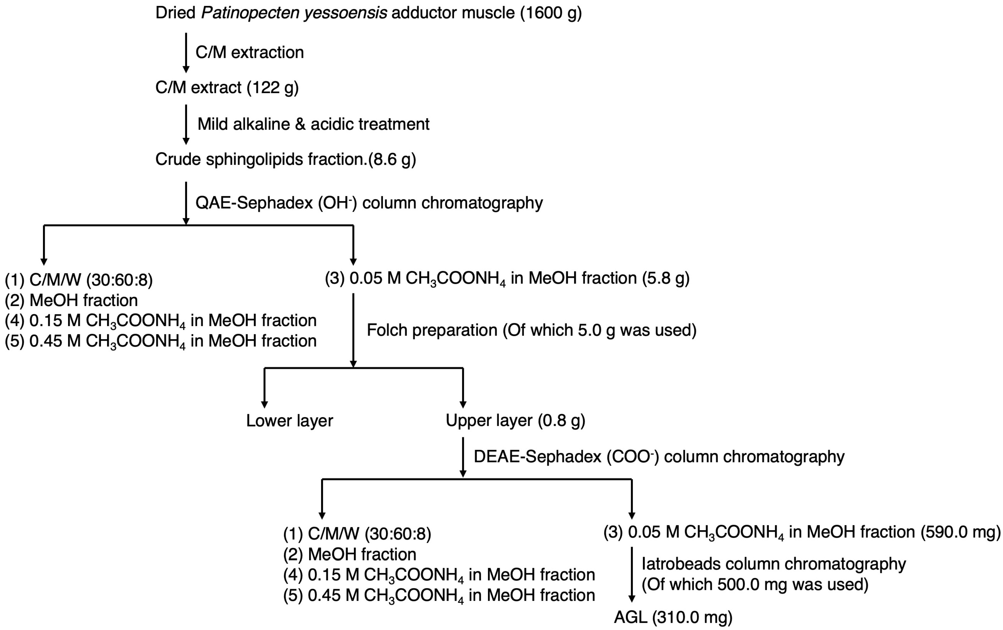
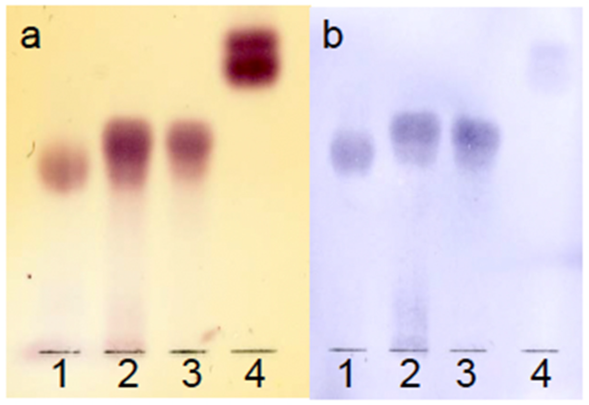
The yields from each elution step were as follows: 34.4 mg from the C/M (80:20, v/v) fraction; 22.1 mg from the C/M (70:30, v/v) fraction; 14.0 mg from the C/M (60:40, v/v) fraction; 24.1 mg from the C/M/W (50:50:5, v/v/v) fraction; 310.0 mg from the C/M/W (60:40:10, v/v/v) fraction; and 48.5 mg from the C/M/W (20:80:10, v/v/v) fraction. The C/M/W (60:40:10, v/v/v) fraction was negative for the Dittmer–Lester reagent and positive for orcinol–sulfuric acid staining, indicating the presence of AGLs (Figure S3). This fraction was designated as Intact-AGL. An overview of AGL preparation is shown in Figure 2.

3.1.5. Reduction of AGL

The reduction of uronic acid was performed to elucidate the structure of the sugar chain. After the reduction of Intact-AGL (100 mg), 32.8 mg of the reduced AGL fraction was obtained as the non-adsorbed fraction on a QAE-Sephadex column. This fraction was further separated and purified by Iatrobeads column chromatography, yielding five subfractions (Fr.1–Fr.5) with the following quantities: 0.6 mg for Fr.1, 6.1 mg for Fr.2, 4.5 mg for Fr.3, 6.7 mg for Fr.4, and 2.4 mg for Fr.5. The TLC analysis results are shown in Figure S4. All five fractions were identified as TMS-methylglycoside derivatives. Among them, Fr.3 exhibited the most prominent peak, corresponding to an unknown sugar (designated as sugar X), which was presumed to be the reduced form of uronic acid (Figure S5). Accordingly, Fr.3 was designated as Reduced-AGL and used in subsequent analyses.

3.2. Structural Analysis of AGL

3.2.1. Sugar Composition of AGL

To determine the sugar composition of AGL, both Intact-AGL and Reduced-AGL were analyzed by GC as TMS-methylglycoside derivatives. The results are shown in Figure 3A. The sugar composition detected in Intact-AGL was glucose (Glc), mannose (Man), N-acetyl glucosamine (GlcNAc), fucose (Fuc), and 3-O-methyl-N-acetyl galactosamine (GalNAc3Me), with a molar ratio of Glc:Man:GlcNAc:Fuc:GalNAc3Me = 1.0:2.0:1.0:1.1:0.8. In Reduced-AGL, the same sugars were detected along with an additional unknown sugar (designated as sugar X), which was presumed to be derived from reduced uronic acid. The molar ratio was Glc:Man:GlcNAc:Fuc:GalNAc3Me:X = 1.0:1.7:0.9:1.0:0.7:1.0. A small amount of xylose (Xyl) was also detected in Intact- and Reduced-AGL.
To identify the unknown sugar X, sugar composition analysis using alditol acetate derivatives was performed on both Intact- and Reduced-AGL (Figure 3B). The GC-MS spectrum of the peak derived from the unknown sugar X, which was generated by the reduction treatment, is shown in Figure S6. The GC-MS fragment of the peak derived from the unknown sugar X was identified as 1,2,3,5,6-penta-O-acetyl-4-methylhexitol (4-O-Me-Hexitol).

3.2.2. Sugar Linkage Analysis

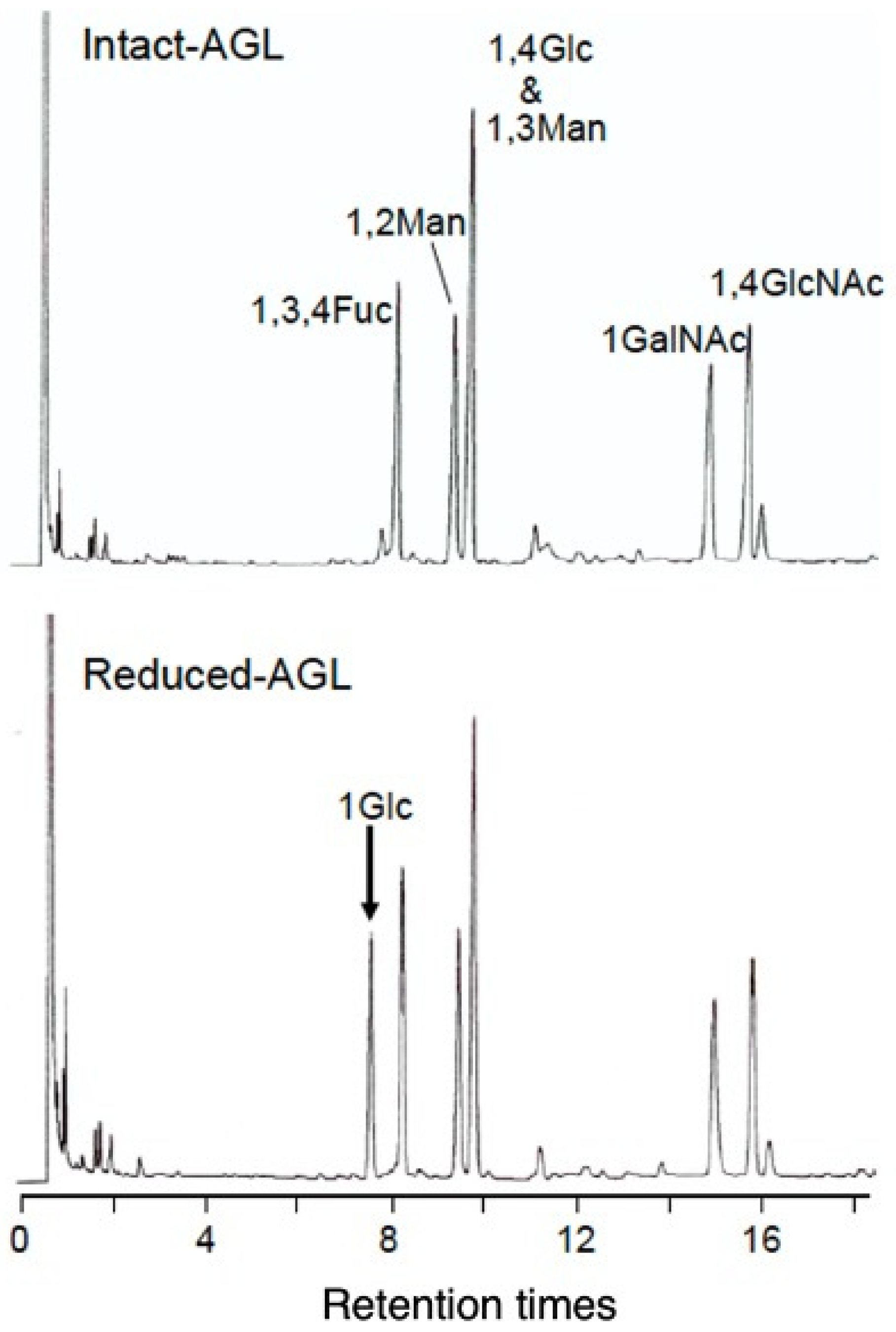
To elucidate the sugar linkages in AGL, linkage analysis was performed on both Intact- and Reduced-AGL (Figure 4). In Intact-AGL, 1,3,4,5-tetra-O-acetyl-2-O-methylfucitol (1,3,4Fuc), 1,2,5-tri-O-acetyl-3,4,6-tri-O-methylmannitol (1,2Man), 1,3,5-tri-O-acetyl-2,4,6-tri-O-methylmannitol (1,3Man), 1,4,5-tri-O-acetyl-2,3,6-tri-O-methylglucitol (1,4Glc), 1,5-di-O-acetyl-3,4,6-tri-O-methyl-N-acetylgalactosaminitol (1GalNAc), and 1,4,5-tri-O-acetyl-3,6-di-O-methyl-N-acetylglucosaminitol (1,4GlcNAc) were detected. Additionally, in Reduced-AGL, 1,5-di-O-acetyl-2,3,4,6-tetra-O-methylglucitol (1Glc) was detected. The detection of 1Glc in Reduced-AGL confirmed that the unknown sugar X was 4-O-methyl-glucuronic acid (4-O-Me-GlcA; Figure S6).
To determine the sugar residues, Intact-AGL was hydrolyzed with HCl and fractionated by Iatrobeads column chromatography based on sugar chain length (Figure S7). The hydrolysis products exhibited different mobilities on TLC and were tentatively designated as AGL-M, AGL-D, AGL-T, and AGL-Q, in descending order of mobility. The total yield of the degradants was 9.1 mg: AGL-M; tr, AGL-D; 1.0 mg, AGL-T; 2.3 mg, and AGL-Q; 6.6 mg.
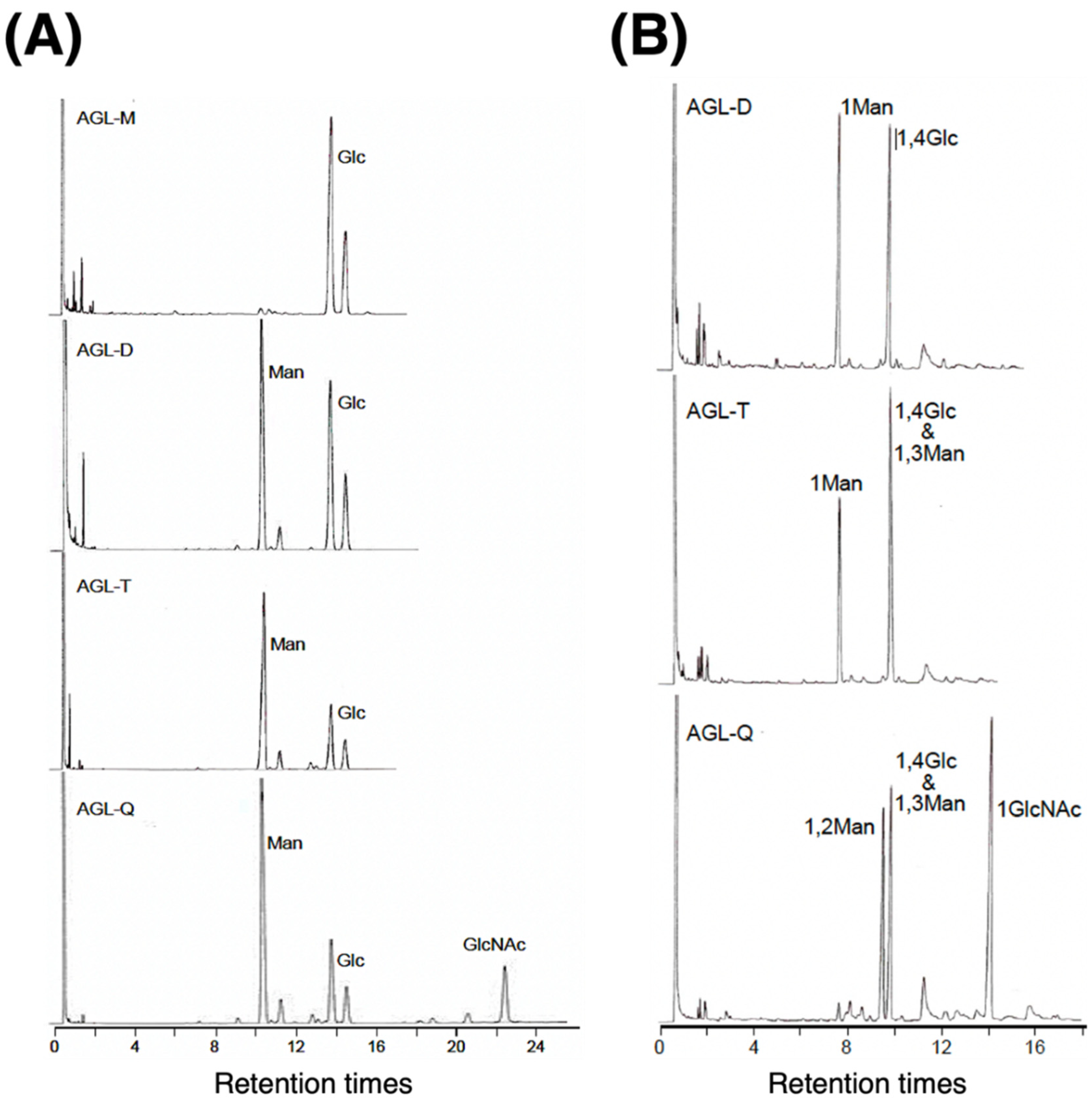
AGL-M, AGL-D, AGL-T, and AGL-Q were analyzed by GC as TMS-methylglycoside derivatives (Figure 5A). Glc was detected in AGL-M, Glc and Man in AGL-D and -T, and Glc, Man, and GlcNAc in AGL-Q. Although the sugar species detected in -D and -T were the same, the peak of Man was larger in -T than in -D, indicating an increase in Man residues. Based on these results, the sugar chain structures were determined to be Glc-Cer for AGL-M, Man-Glc-Cer for AGL-D, Man-Man-Glc-Cer for AGL-T, and GlcNAc-Man-Man-Glc-Cer for AGL-Q, respectively. Thus, the sequence of the four sugar residues at the reducing end of Intact-AGL was identified as GlcNAc-Man-Man-Glc-Cer. In addition, AGL-D, AGL-T, and AGL-Q were analyzed by GC as PMAAs (Figure 5B). In AGL-D, 1Man and 1,4Glc were detected; in AGL-T, 1Man, 1,4Glc, and 1,3Man; and in AGL-Q, 1,2Man, 1,4Glc, 1,3Man, and 1GlcNAc. Therefore, the sugar chain structure considering the binding position is Man1-4Glc1-Cer for AGL-D, Man1-3Man1-4Glc1-Cer for AGL-T, and GlcNAc1-2Man1-3Man1-4Glc1-Cer for AGL-Q. Based on these results, the sugar chain structure up to tetrasaccharide was determined to be GlcNAc1-2Man1-3Man1-4Glc1-Cer. Comparing Figure 4 and Figure 5B, the sugar chain structure beyond the pentasaccharide is determined by the order of 1GlcA4Me, 1,3,4Fuc, and 1GalNAc. 1,3,4Fuc is the fifth branched sugar, and 1GlcA4Me and 1GalNAc are bound at position 3 or 4 here. Based on the PMAA analysis of sugar linkages, two possible sugar linkage structures for Intact-AGL were proposed: GlcA4Me1-4(GalNAc3Me1-3)Fuc1-4GlcNAc1-2Man1-3Man1-4Glc1-Cer or GalNAc3Me1-4(GlcA4Me1-3)Fuc1-4GlcNAc1-2Man1-3Man1-4Glc1-Cer.

3.2.3. 1H-NMR Analysis

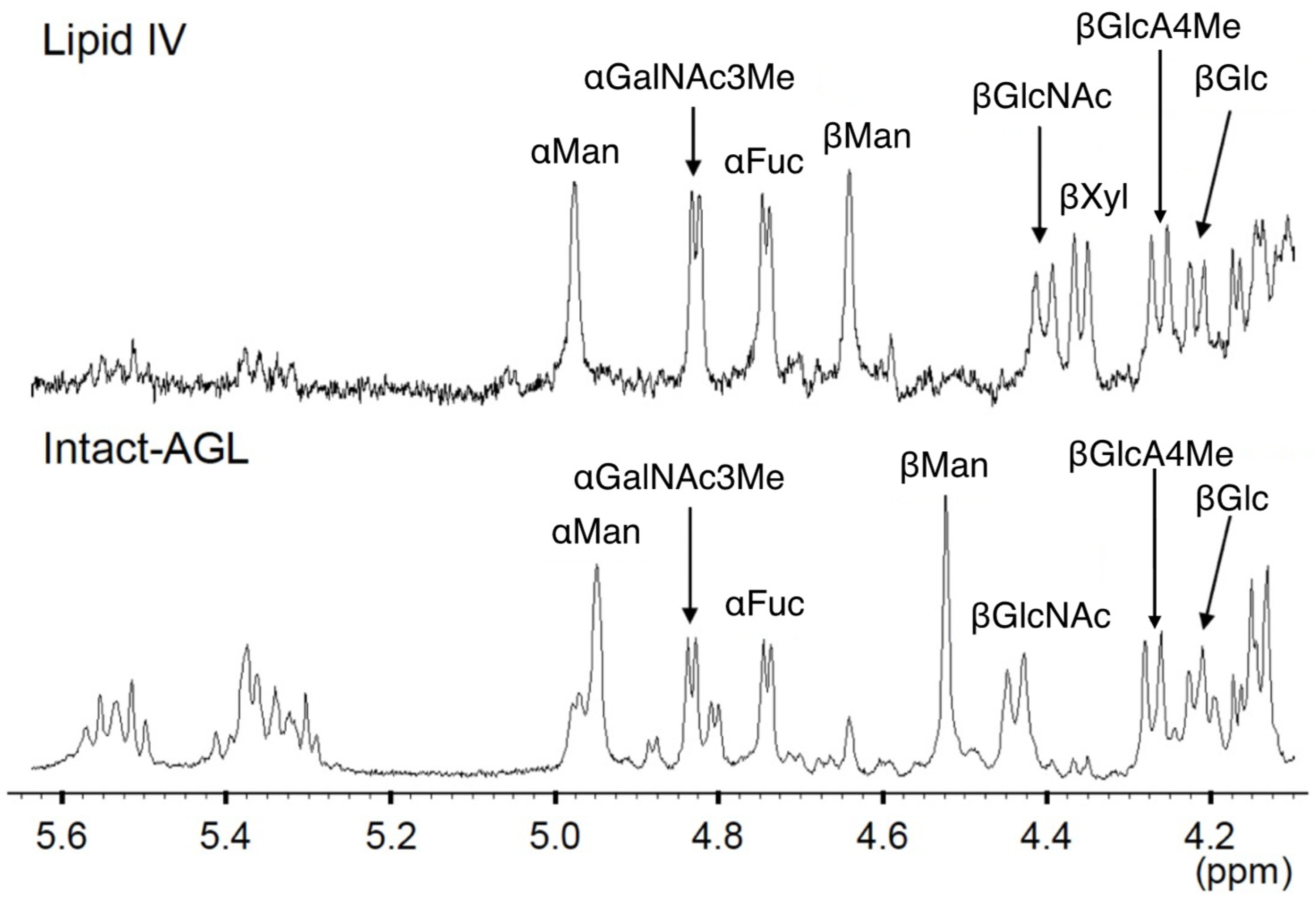
To determine the anomeric configuration of the sugar residue, the 1H-NMR spectrum of Intact-AGL was acquired. The anomeric configuration of each glycosidic bond was assigned based on the obtained spectra and by comparing with Lipid IV, a GSL whose sugar chain structure is already known [30], as shown in Figure 6. The chemical shifts (ppm) and J1,2 coupling constants (Hz) are listed in Table 1. However, unlike Lipid IV, Intact-AGL does not have Xyl that binds to Man. Therefore, Man is detected at a higher field than Xyl-bound Man in Lipid IV. From these results, the proposed sugar chain structures are as follows: GlcA4Meβ1-4(GalNAc3Meα1-3)Fucα1-4GlcNAcβ1-2Manα1-3Manβ1-4Glcβ1-Cer or GalNAc3Meα1-4(GlcA4Meβ1-3)Fucα1-4GlcNAcβ1-2Manα1-3Manβ1-4Glcβ1-Cer. Signals observed around 5.3 ppm and 5.5 ppm were attributed to the double bond of the LCB.

3.2.4. TLC-Immunostaining Assay

The TLC-immunostaining assay was performed using an antibody that specifically recognizes the GlcA4Meβ1-4Fuc epitope to determine which of the two proposed structures corresponds to Intact-AGL. The antibody used was a rabbit-derived anti-H. schlegelii antiserum, prepared using Lipid IV [GlcA4Meβ1-4(GalNAc3Meα1-3)Fucα1-4GlcNAcβ1-2Manα1-3(Xylβ1-2)Manβ1-4Glcβ1–Cer], isolated from H. schlegelii, as the immunogen [30] (Figure 7). Lipid IV standard, semi-purified AGL, and Intact-AGL (lanes 1–3 in Figure 7) were positive for both the orcinol-H2SO4 reagent and immunostaining (anti-Lipid IV antiserum). In contrast, Reduced-AGL was positive for the orcinol-H2SO4 reagent but poorly reactive in immunostaining (lane 4 in Figure 7). The sugar chain structure of Intact-AGL was determined to be GlcA4Meβ1-4(GalNAc3Meα1-3)Fucα1-4GlcNAcβ1-2Manα1-3Manβ1-4Glcβ1-Cer because the major recognition site of the anti-Lipid IV antiserum is GlcA4Meβ1-4Fuc.

3.2.5. Ceramide Composition Analysis

The fatty acid and LCB components of AGL are summarized in Table 2. The major fatty acids were C16:0 and C18:0, accounting for 43.9% of total fatty acids. In addition, unsaturated fatty acids such as C20:1 and C22:1 were detected, comprising 22.7%. The predominant LCB was d16:1, followed by d18:2.

3.2.6. MALDI-TOF/MS Analysis

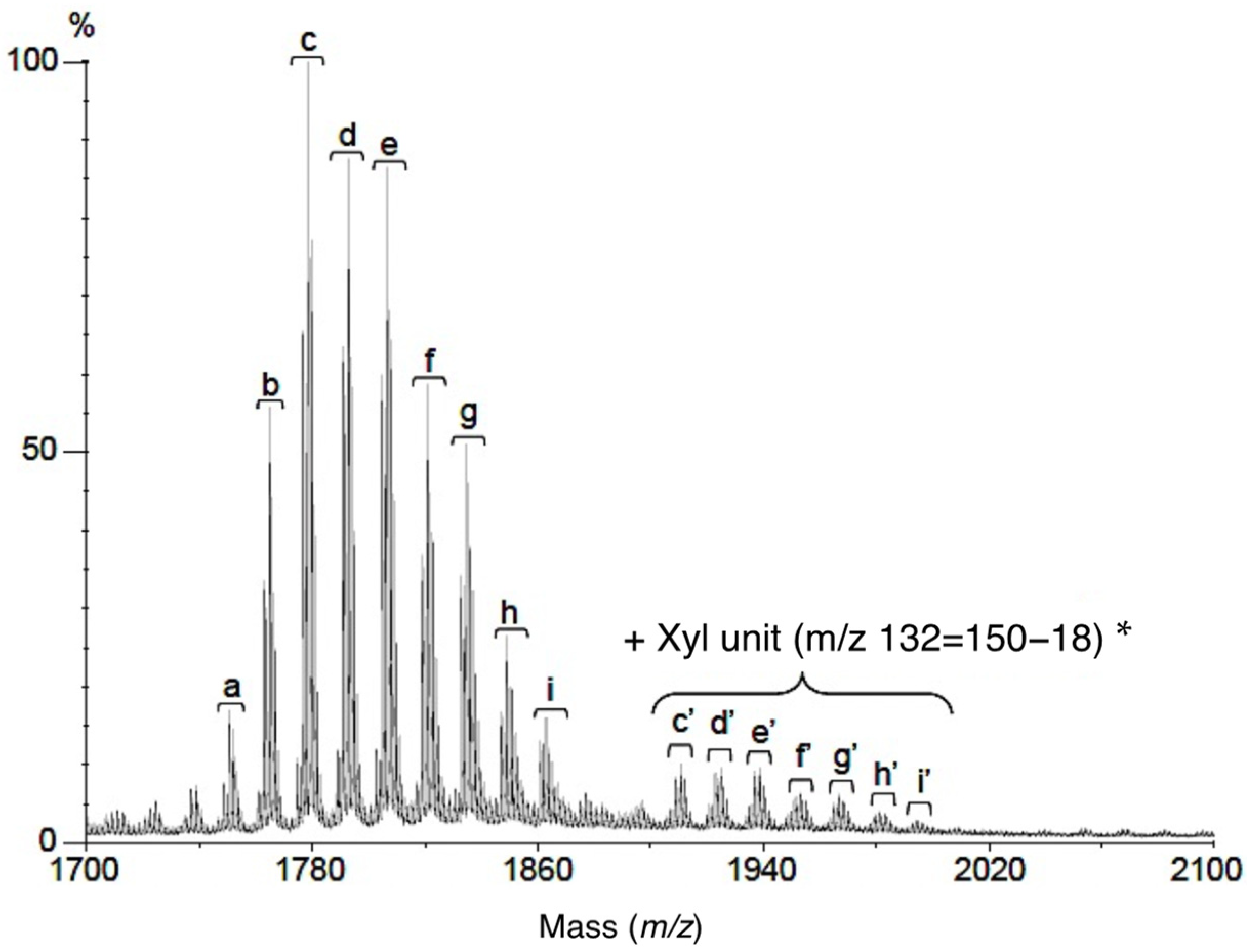
Figure 8 shows the MALDI-TOF/MS spectrum of AGL, and the measured and calculated [M-H] values are summarized in Table 3. These results indicate that the ceramide moieties of AGL are composed of various combinations of fatty acids and LCBs. Several major peaks differing by 14 u were observed in the AGL spectrum. These peaks were assigned as [M-H] ions corresponding to differing combinations of aliphatic components: peak c at m/z 1778.88 (C16:0-d18:1, C17:0-d17:1, C18:0-d16:1), peak d at m/z 1792.89 (C17:0-d18:1, C18:0-d17:1), peak e at m/z 1806.90 (C18:0-d18:1, C20:0-d16:1) for major AGL species, consistent with HexAMe-HexNAcMe-Fuc-HexNAc-(Hex)3-ceramide. The measured and calculated values for peaks c′ to i′ were consistent with theoretical masses corresponding to the addition of Xyl residues to Intact-AGL. GC analysis of TMS-methylglycoside derivatives and MALDI-TOF MS results suggested that Xyl residues were incorporated into the sugar chain of AGL. However, because of the low abundance of Xyl-containing AGL, structural determination by 1H-NMR or GC analysis of sugar linkage was not possible.

4. Discussion

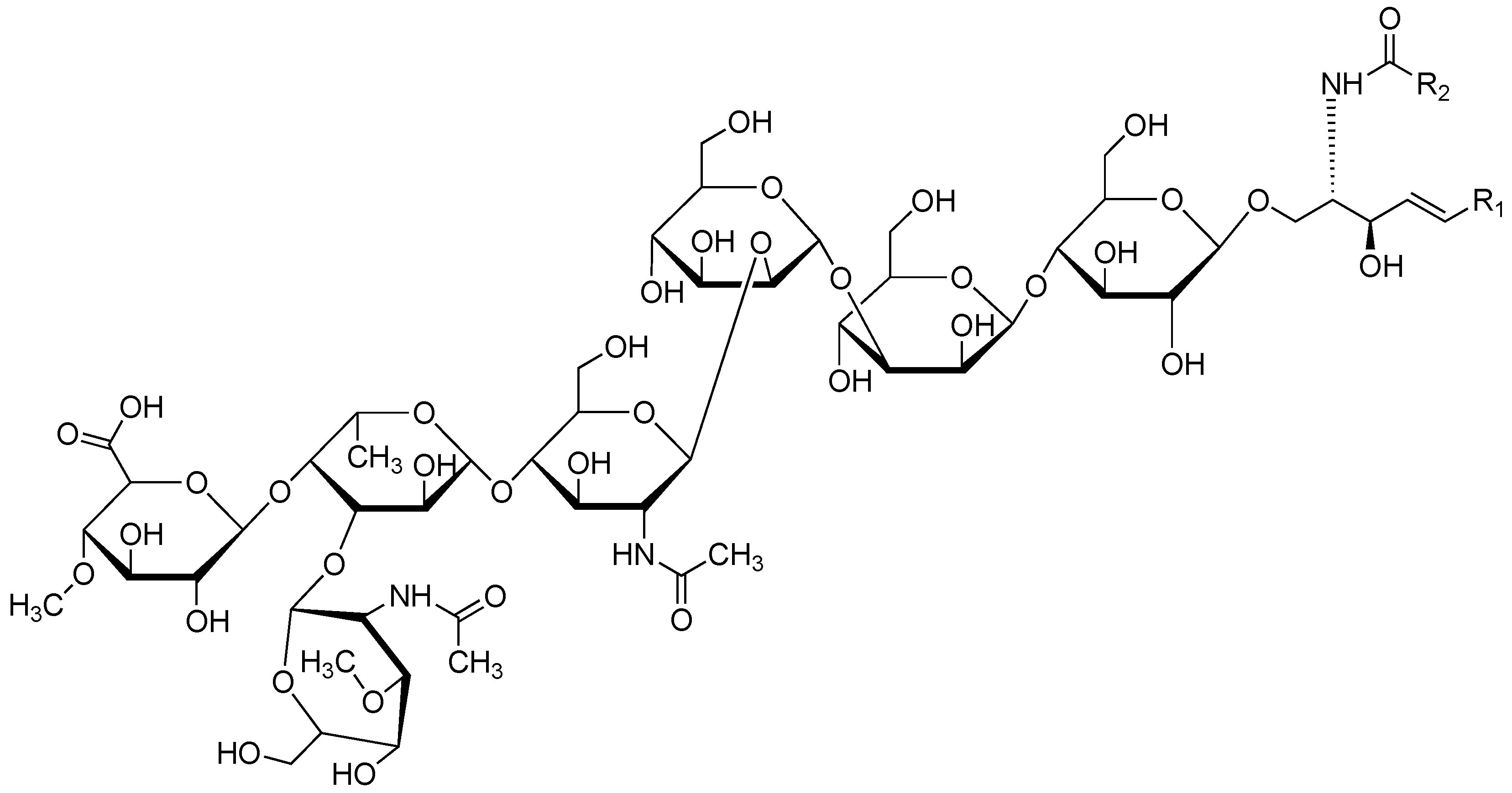
This study revealed that the sugar chain structure of AGL in the adductor muscle of P. yessoensis is GlcA4Meβ1-4(GalNAc3Meα1-3)Fucα1-4GlcNAcβ1-2Manα1-3Manβ1-4Glcβ1-Cer (Figure 9). This structure is represented as a novel sugar chain that has not been reported previously and is absent from databases of various sphingolipid chemical structures (https://lipidbank.jp/wiki/Category:LBSP, accessed on 1 September 2025). It belongs to the mollu-series and contains two mannose residues, a characteristic feature of neutral GSL identified in mollusks and the brachiopod Lingula unguis [31]. In addition, the presence of methylated sugars, such as 3-O-methyl galactosamine and Fuc, located internally within the sugar chain, is also characteristic of species within these phyla [19]. These findings suggest that mollu-series GSL-bearing Fuc and methyl sugars may represent conserved biochemical components across the phyla Mollusca and Brachiopoda members. Notably, L. unguis exhibits morphological similarity to bivalves and is closely related to Mollusca in phylogenetic analyses.
AGL from the adductor muscle of P. yessoensis was found to contain uronic acid, a rare component of GSLs. Uronic acid-containing GSLs have previously been reported in H. schlegelii, Anodonta woodiana, and Cristaria plicata using immunochemical detection [18,30]. These are freshwater bivalves and taxonomically related species to the saltwater bivalves P. yessoensis. Other uronic acid-containing GSLs have been identified in insects such as Lucilia caesar and Drosophila melanogaster [32,33], with structures such as GlcAβ1-3Galβ1-3GalNAcα1-4GalNAcβ1-4GlcNAclβ1-3Manβ1-4Glcβ1-Cer and related analogues with/without phosphate derivatives. Other uronic acid-containing GSLs have also been identified in the ascidian Halocynthia roretzi and H. aurantium [22,23], with the structure Galβ1-4(Fucα1-3)GlcAcβ1-Cer. Although these AGLs are phylogenetically scattered, they may provide an indication of AGL distribution in future studies.
Trace amounts of Xyl were consistently detected in the AGL fraction. One known Xyl-containing AGL is Lipid IV, with the structure GlcA4Meβ1-4(GalNAc3Meα1-3)Fucα1-4GlcNAcβ1-2Manα1-3(Xylβ1-2)Manβ1-4Glcβ1-Cer. Lipid IV was originally isolated from the spermatozoa of H. schlegelii but not from its eggs, suggesting a role in gamete recognition during fertilization [30]. Furthermore, immunochemical detection studies have reported the presence of Lipid IV in the spermatozoa of several bivalves, including species from the Unionidae, Veneridae, and Mytilidae families, as well as in H. schlegelii, but not in gastropods [30]. The detection of the Lipid IV-like structure in the adductor muscle of P. yessoensis is notable and may indicate a potential functional role in reproduction or species specificity. Currently, there are no findings regarding the biological role of AGL in mollusks, making it challenging to understand the function of this sugar chain structure.
Fatty acid analysis of the ceramide moiety revealed that only normal fatty acids were detected, with C16:0 and C18:0 accounting for 43.9% of the total fatty acids. In addition, odd- and branched-chain fatty acids, particularly C17:0 and its branched isomers, were present at notable levels (14.8%). In comparison, previous studies on cerebrosides from P. yessoensis organs reported that C16:0, C18:1, and C18:0 accounted for 52.0% of the fatty acid composition, whereas C17:0 and branched C17:0 were detected at lower levels (5.5%) [34]. Lipid IV, isolated from H. schlegelii spermatozoa, has been reported to have a sugar chain structure similar to that of AGL determined in this study. However, the sum of C16:0 and C18:0 accounts for a higher percentage of the fatty acid composition (approximately 60%), whereas the odd chain C17:0 accounts for a lower percentage (5.1%) [19]. The composition of the LCB was dominated by d16:1 (43.4%) and followed by d18:1 (22.6%). In M. lusoria, a marine bivalve similar to P. yessoensis, the LCB composition isolated from whole tissues was approximately 18–20% d18:1, 28–31% d18:2, and 7–9% d16:1 [21]. The compositions of d18:1 and d18:2 were similar; however, P. yessoensis had a relatively high d16:1 ratio. Furthermore, Lipid IV, isolated from H. schlegelii spermatozoa, has a sugar chain structure similar to that observed in this study. However, the LCB composition was considerably different, with d18:1 accounting for approximately 97% of the LCB composition [18].
A limitation of this study is that the absolute configuration of Intact-AGL has not been determined. Monosaccharides exist in chiral forms. Therefore, there is interest in determining whether the monosaccharides that constitute the sugar chain structure of Intact-AGL are D-forms or L-forms. However, in previous studies, the distinction between D-forms or L-forms of monosaccharides constituting the sugar chain structure was not made in the classification of sphingolipids. Therefore, at this stage, it is considered unnecessary to clarify the chiral forms of the monosaccharides; however, this may lead to new discoveries in the future.

5. Conclusions

This study identified the sugar chain structure of novel GSL in the adductor muscle of P. yessoensis as GlcA4Meβ1-4(GalNAc3Meα1-3)Fucα1-4GlcNAcβ1-2Manα1-3Manβ1-4Glcβ1-Cer. This chemical structure provides valuable insights into the biological classification of P. yessoensis and the functional roles of GSL.

Supplementary Materials

The following supporting information can be downloaded at https://www.mdpi.com/article/10.3390/fishes10090460/s1, Figure S1: TLC analysis of GSL from the adductor muscle of Patinopecten yessoensis obtained by Folch partition; Figure S2: TLC analysis of AGL from the adductor muscle of Patinopecten yessoensis obtained by DEAE-Sephadex column chromatography; Figure S3: TLC analysis of AGL from the adductor muscle of Patinopecten yessoensis obtained by Iatrobeads column chromatography; Figure S4: TLC analysis of Reduced-AGL from the adductor muscle of Patinopecten yessoensis obtained by Iatrobeads column chromatography; Figure S5: Gas chromatogram of TMS derivatives of Fr. 1-5 in reduced AGL; Figure S6: Mass spectrum of the alditol acetate derivatives from the unknown sugar X obtained using GC-MS analysis; Figure S7: TLC analysis of AGL from the adductor muscle of Patinopecten yessoensis obtained by partial hydrolysis.

Author Contributions

Conceptualization, S.I. and M.S.; methodology, S.I. and M.S.; validation, S.S., S.I., M.S., and A.H.; formal analysis, S.S., S.I., M.S., and A.H.; investigation, S.S., S.I., M.S., and A.H.; resources, S.I. and M.S.; data curation, S.S., S.I., M.S., A.H., and K.F.; writing—original draft preparation, S.S., S.I., K.S., R.H., and K.F.; writing—review and editing, S.S., S.I., K.S., R.H., and K.F.; visualization, S.S., S.I., K.S., R.H., and K.F.; supervision, S.I., M.S., and K.F.; project administration, S.I. and K.F.; funding acquisition, S.I., M.S., R.H., and K.F. All authors have read and agreed to the published version of the manuscript.

Funding

This research was funded by the Kansai University Grant-in-Aid for Progress of Research in Graduate Course 2025.

Institutional Review Board Statement

Not applicable.

Informed Consent Statement

Not applicable.

Data Availability Statement

The data presented in this study are available upon request from the corresponding authors. These data are not publicly available because of the lack of a public archive platform for data sharing.

Conflicts of Interest

The funders had no role in the design of the study; in the collection, analyses, or interpretation of data; in the writing of the manuscript; or in the decision to publish the results.

Abbreviations

1,2Man1,2,5-tri-O-acetyl-3,4,6-tri-O-methylmannitol
1,3,4Fuc1,3,4,5-tetra-O-acetyl-2-O-methylfucitol
1,3Man1,3,5-tri-O-acetyl-2,4,6-tri-O-methylmannitol
1,4Glc1,4,5-tri-O-acetyl-2,3,6-tri-O-methylglucitol
1,4GlcNAc1,4,5-tri-O-acetyl-3,6-di-O-methyl-N-acetylglucosaminitol
1GalNAc1,5-di-O-acetyl-3,4,6-tri-O-methyl-N-acetylgalactosaminitol
1Glc1,5-di-O-acetyl-2,3,4,6-tetra-O-methylglucitol
1H-NMRproton nuclear magnetic resonance
4-O-Me-GlcA4-O-methyl-glucuronic acid
4-O-Me-Hexitol1,2,3,5,6-penta-O-acetyl-4-methylhexitol
AGLsacidic glycosphingolipids
AGL-Dacidic glycosphingolipids-disaccharides
AGL-Macidic glycosphingolipids-monosaccharide
AGL-Qacidic glycosphingolipids-tetrasaccharides
AGL-Tacidic glycosphingolipids-trisaccharides
C/Mchloroform/methanol
C/M/Wchloroform/methanol/water
Cerceramide
Fucfucose
Galgalactose
GalNAcN-acetyl galactosamine
GalNAc3Me3-O-methyl-N-acetyl galactosamine
GCgas chromatography
GC-MSgas chromatograph-mass spectrometry
Glcglucose
GlcAglucuronic acid
GlcNAcN-acetyl glucosamine
GSLsglycosphingolipids
Hexhexose
HexAMeO-methyl hexuronic acid
HexNAcN-acetyl hexosamine
HexNAcMeO-methyl-N-acetyl hexosamine
LCBlong-chain bases
M/Wmethanol/water
MALDI-TOF MSmatrix-assisted laser desorption/ionization time-of-flight mass spectrometry
PMAApartially methylated alditol acetates
TLCthin-layer chromatography
TMStrimethylsilyl
Xylxylose

References

  1. Enda, H.; Sagane, Y.; Nakazawa, Y.; Sato, H.; Yamazaki, M. Data on color and chemical composition of dried scallop (Mizuhopecten yessoensis) produced in different areas of Hokkaido, Japan. Data Brief 2018, 16, 635–638. [Google Scholar] [CrossRef]
  2. Baba, K.; Miyazono, A.; Matsuyama, K.; Kohno, S.; Kubota, S. Occurrence and detrimental effects of the bivalve-inhabiting hydroid Eutima japonica on juveniles of the Japanese scallop Mizuhopecten yessoensis in Funka Bay, Japan: Relationship to juvenile massive mortality in 2003. Mar. Biol. 2007, 151, 1977–1987. [Google Scholar] [CrossRef]
  3. Chen, J.H.; Yu, R.C.; Gao, Y.; Kong, F.Z.; Wang, Y.F.; Zhang, Q.C.; Kang, Z.J.; Yan, T.; Zhou, M.J. Tracing the origin of paralytic shellfish toxins in scallop Patinopecten yessoensis in the northern Yellow Sea. Food Addit. Contam. Part A Chem. Anal. Control. Expo. Risk Assess. 2013, 30, 1933–1945. [Google Scholar] [CrossRef]
  4. Kimura, M. Studies on the quality control of the Japanese scallop adductor muscle. Sci. Rep. Hokkaido Fish. Exp. Stn. 2003, 65, 1–47. [Google Scholar]
  5. Sasaki, T. Anatomy of the Mollusca: Sepia esculenta (Cephalopoda: Sepiidae), Turbo cornutus (Gastropoda: Turbinidae) and Patinopecten yessoensis (Bivalvia: Pectinidae). Fossils 2008, 84, 86–95. [Google Scholar] [CrossRef]
  6. Sun, X.; Liu, Z.; Wu, B.; Zhou, L.; Wang, Q.; Wu, W.; Yang, A. Differences between fast and slow muscles in scallops revealed through proteomics and transcriptomics. BMC Genomics 2018, 19, 377. [Google Scholar] [CrossRef]
  7. Gutsell, S.J. Natural history of the bay scallop (Pecten irradians). U.S. Bur. Fish. 1930, 46, 569–632. [Google Scholar]
  8. Winter, M.A.; Hamilton, P.V. Factors influencing swimming in bay scallops, Argopectenirradians (Lamarck, 1819). J. Exp. Mar. Biol. Ecol. 1985, 88, 227–242. [Google Scholar] [CrossRef]
  9. Murate, M.; Abe, M.; Kasahara, K.; Iwabuchi, K.; Umeda, M.; Kobayashi, T. Transbilayer distribution of lipids at nano scale. J. Cell Sci. 2015, 128, 1627–1638. [Google Scholar] [CrossRef]
  10. Sonnino, S.; Prinetti, A.; Mauri, L.; Chigorno, V.; Tettamanti, G. Dynamic and structural properties of sphingolipids as driving forces for the formation of membrane domains. Chem. Rev. 2006, 106, 2111–2125. [Google Scholar] [CrossRef]
  11. Iwabuchi, K.; Nakayama, H.; Masuda, H.; Kina, K.; Ogawa, H.; Takamori, K. Membrane microdomains in immunity: Glycosphingolipid-enriched domain-mediated innate immune responses. BioFactors 2012, 38, 275–283. [Google Scholar] [CrossRef]
  12. Hakomori, S.; Handa, K.; Iwabuchi, K.; Yamamura, S.; Prinetti, A. New insights in glycosphingolipid function: “glycosignaling domain,” a cell surface assembly of glycosphingolipids with signal transducer molecules, involved in cell adhesion coupled with signaling. Glycobiology 1998, 8, xi–xix. [Google Scholar] [CrossRef]
  13. Kawahara, K.; Seydel, U.; Matsuura, M.; Danbara, H.; Rietschel, E.T.; Zähringer, U. Chemical structure of glycosphingolipids isolated from Sphingomonas paucimobilis. FEBS Lett. 1991, 292, 107–110. [Google Scholar] [CrossRef] [PubMed]
  14. Wennekes, T.; van den Berg, R.J.; Boot, R.G.; van der Marel, G.A.; Overkleeft, H.S.; Aerts, J.M. Glycosphingolipids--nature, function, and pharmacological modulation. Angew. Chem. Int. Ed. Engl. 2009, 48, 8848–8869. [Google Scholar] [CrossRef]
  15. Lingwood, C.A. Glycosphingolipid functions. Cold Spring Harb Perspect. Biol. 2011, 3. [Google Scholar] [CrossRef]
  16. Yoshikawa, M.; Go, S.; Takasaki, K.; Kakazu, Y.; Ohashi, M.; Nagafuku, M.; Kabayama, K.; Sekimoto, J.; Suzuki, S.; Takaiwa, K.; et al. Mice lacking ganglioside GM3 synthase exhibit complete hearing loss due to selective degeneration of the organ of Corti. Proc. Natl Acad. Sci. USA 2009, 106, 9483–9488. [Google Scholar] [CrossRef] [PubMed]
  17. Yamashita, T.; Hashiramoto, A.; Haluzik, M.; Mizukami, H.; Beck, S.; Norton, A.; Kono, M.; Tsuji, S.; Daniotti, J.L.; Werth, N.; et al. Enhanced insulin sensitivity in mice lacking ganglioside GM3. Proc. Natl Acad. Sci. USA 2003, 100, 3445–3449. [Google Scholar] [CrossRef] [PubMed]
  18. Hori, T.; Sugita, M.; Ando, S.; Tsukada, K.; Shiota, K.; Tsuzuki, M.; Itasaka, O. Isolation and characterization of a 4-O-methylglucuronic acid-containing glycosphingolipid from spermatozoa of a fresh water bivalve, Hyriopsis schlegelii. J. Biol. Chem. 1983, 258, 2239–2245. [Google Scholar] [CrossRef]
  19. Sugita, M.; Nakano, Y.; Nose, T.; Itasaka, O.; Hori, T. Neutral glycosphingolipids of ova of the fresh-water bivalve, Hyriopsis schlegelii. J. Biochem. 1984, 95, 47–55. [Google Scholar] [CrossRef]
  20. Itasaka, O.; Kosuga, M.; Okayama, M.; Hori, T. Characterization of a novel ceramide octasaccharide isolated from whole tissue of a fresh-water bivalve, Corbicula sandai. Biochim. Biophys. Acta 1983, 750, 440–446. [Google Scholar] [CrossRef]
  21. Sugita, M.; Nakae, H.; Yamamura, T.; Takamiya, Y.; Itasaka, O.; Hori, T. The occurrence of glycosphingolipids containing mannose in the sea-water bivalve, Meretrix lusoria (Hamaguri). J. Biochem. 1985, 98, 27–34. [Google Scholar] [CrossRef]
  22. Ito, M.; Matsumuro, Y.; Yamada, S.; Kitamura, T.; Itonori, S.; Sugita, M. Isolation and characterization of a novel uronic acid-containing acidic glycosphingolipid from the ascidian Halocynthia roretzi. J. Lipid Res. 2007, 48, 96–103. [Google Scholar] [CrossRef]
  23. Kojima, H.; Shinohara, R.; Itonori, S.; Ito, M. Characterization of a novel rhamnose-containing acidic glycosphingolipid from the ascidian Halocynthia aurantium. J. Oleo Sci. 2017, 66, 285–295. [Google Scholar] [CrossRef] [PubMed]
  24. Nagasawa, H.; Miyazaki, S.; Kyogashima, M. Simple separation of glycosphingolipids in the lower phase of a Folch’s partition from crude lipid fractions using zirconium dioxide. Glycoconj J. 2022, 39, 789–795. [Google Scholar] [CrossRef] [PubMed]
  25. Taylor, R.L.; Conrad, H.E. Stoichiometric depolymerization of polyuronides and glycosaminoglycuronans to monosaccharides following reduction of their carbodiimide-activated carboxyl groups. Biochemistry 1972, 11, 1383–1388. [Google Scholar] [CrossRef]
  26. Itonori, S.; Takahashi, M.; Kitamura, T.; Aoki, K.; Dulaney, J.T.; Sugita, M. Microwave-mediated analysis for sugar, fatty acid, and sphingoid compositions of glycosphingolipids. J. Lipid Res. 2004, 45, 574–581. [Google Scholar] [CrossRef][Green Version]
  27. Bajwa, B.; Xing, X.; Terry, S.A.; Gruninger, R.J.; Abbott, D.W. Methylation-GC-MS/FID-based glycosidic linkage analysis of unfractionated polysaccharides in red seaweeds. Mar. Drugs 2024, 22, 192. [Google Scholar] [CrossRef]
  28. Kimura, K.; Itonori, S.; Kajiwara, C.; Hada, N.; Takeda, T.; Sugita, M. Structural Elucidation of the Neutral glycosphingolipids, Mono-, di-, tri- and tetraglycosylceramides from the Marine Crab Erimacrus isenbeckii. J. Oleo Sci. 2014, 63, 269–280. [Google Scholar] [CrossRef]
  29. Oberacher, H.; Whitley, G.; Berger, B. Evaluation of the sensitivity of the ‘Wiley registry of tandem mass spectral data, MSforID’ with MS/MS data of the ‘NIST/NIH/EPA mass spectral library’. J. Mass Spectrom. 2013, 48, 487–496. [Google Scholar] [CrossRef]
  30. Sugita, M.; Inoue, T.; Itasaka, O.; Hori, T. The localization of 4-O-methylglucuronic acid-containing glycosphingolipid on the cell surface of bivalve spermatozoa by use of an immunological method. J. Biochem. 1984, 95, 737–742. [Google Scholar] [CrossRef] [PubMed]
  31. Aoki, K.; Sugiyama, S.; Dulaney, J.T.; Itonori, S.; Sugita, M. Classification into a Novel Mollu-Series of Neutral glycosphingolipids from the Lamp Shell, Lingula unguis. J. Oleo Sci. 2002, 51, 463–472. [Google Scholar] [CrossRef]
  32. Sugita, M.; Itonori, S.; Inagaki, F.; Hori, T. Characterization of two glucuronic acid-containing glycosphingolipids in larvae of the green-bottle fly, Lucilia caesar. J. Biol. Chem. 1989, 264, 15028–15033. [Google Scholar] [CrossRef] [PubMed]
  33. Seppo, A.; Moreland, M.; Schweingruber, H.; Tiemeyer, M. Zwitterionic and acidic glycosphingolipids of the Drosophila melanogaster embryo. Eur. J. Biochem. 2000, 267, 3549–3558. [Google Scholar] [CrossRef] [PubMed]
  34. Makita, A.; Sato, A.; Sugita, M.; Itonori, S.; Ito, M.; Arakawa, Y. Chemical characterization of two main sphingolipids of scallop. Bull. Tenshi Coll. 2010, 10, 1–9. [Google Scholar]
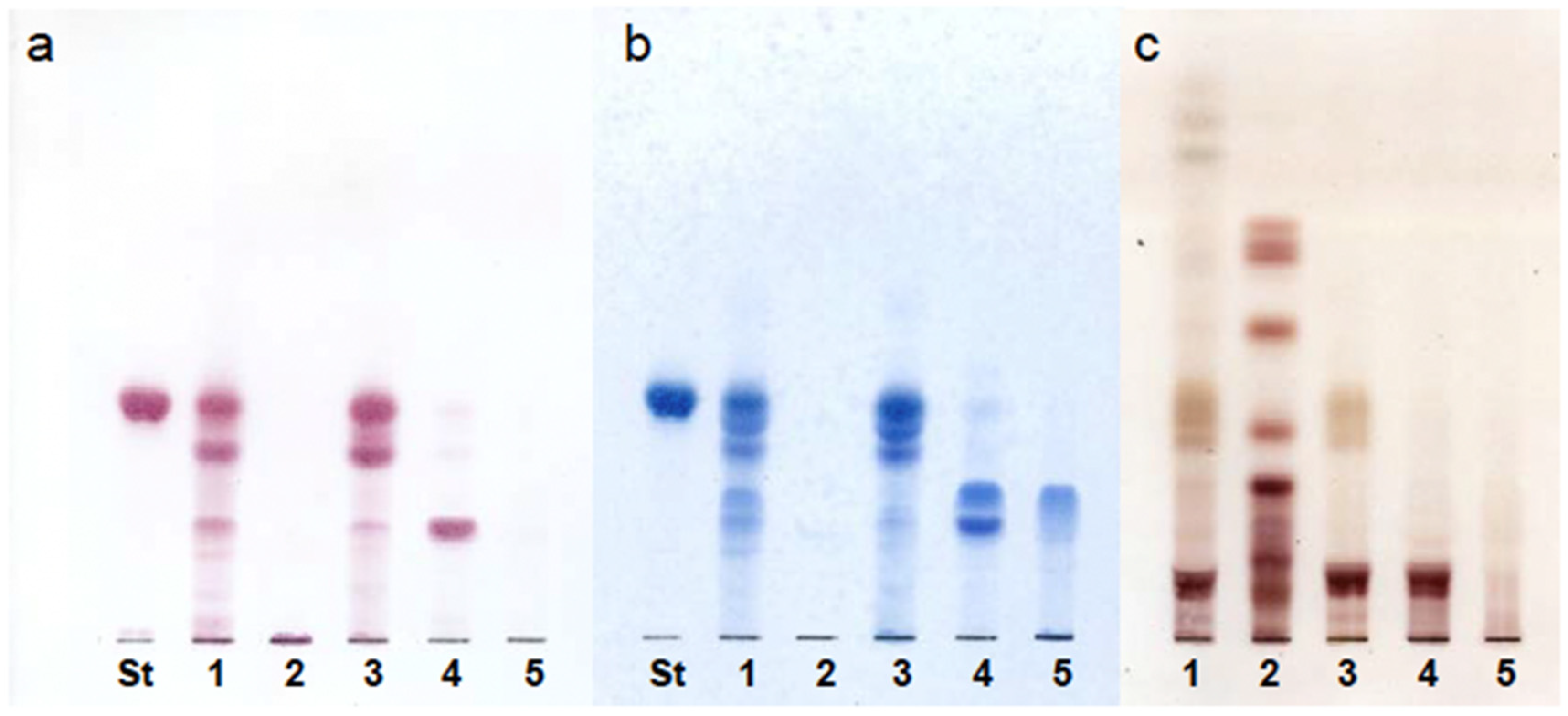
Figure 1. TLC analysis of GSL from the adductor muscle of Patinopecten yessoensis obtained by QAE-Sephadex column chromatography. Lanes: St. CAEPn (Corbicula sandai), 1. Crude sphingolipids, 2. combined C/M/W (30:60:8, v/v/v) and methanol fractions, 3. 0.05 M CH3COONH4 in methanol fraction, 4. 0.15 M CH3COONH4 in methanol fraction, 5. 0.45 M CH3COONH4 in methanol fraction. The plate was developed with C/M/W (60:35:8, v/v/v), and the spots were visualized using (a) ninhydrin, (b) Dittmer–Lester, and (c) orcinol-H2SO4 reagents. CAEP, ceramide 2-aminoethylphosphonate; C/M/W, chloroform/methanol/water; St, standard; GSL, glycosphingolipids; TLC, thin-layer chromatography.
Fishes 10 00460 g001
Figure 2. Scheme for preparation and fractionation of acidic glycolsphingolipid (AGL) from the adductor muscle of Patinopecten yessoensis. C/M, chloroform/methanol; C/M/W, chloroform/methanol/water; MeOH, methanol.
Fishes 10 00460 g002
Figure 3. GC analysis of TMS-methylglycoside derivatives (A) and alditol acetate derivatives (B) of Intact-AGL and Reduced-AGL. Peaks: a. fucose, b. xylose, c. unknown sugar X, d. mannose, e. glucose f. 3-O-methyl N-acetyl galactosamine, g. N-acetyl glucosamine. AGL, acidic glycosphingolipid, GC, gas chromatography; TMS, trimethylsilyl.
Fishes 10 00460 g003
Figure 4. GC analysis of PMAAs derived from Intact- and Reduced-AGL. AGL, acidic glycosphingolipid; Fuc, fucose; Man, mannose; Glc; glucose; GalNAc, N-acetyl galactosamine; GC, gas chromatography; GlcNAc, N-acetyl glucosamine; PMAAs, partially methylated alditol acetates.
Fishes 10 00460 g004
Figure 5. GC analysis of TMS-methylglycoside derivatives (A) and PMAAs of partially hydrolyzed-Intact-AGL (B). AGL, acidic glycosphingolipid; Glc; glucose; GlcNAc, N-acetyl glucosamine; GC, gas chromatography; Man, mannose; PMAAs, partially methylated alditol acetates; TMS, trimethylsilyl.
Fishes 10 00460 g005
Figure 6. 1H-NMR analysis of Lipid IV and Intact-AGL. The spectrum was obtained in dimethylsulfoxide-d6 containing 2% D2O at 60 °C. Identification of each peak in Intact-AGL was performed using Lipid IV as the standard. AGLs, acidic glycosphingolipids; Fuc, fucose; Glc; glucose; GalNAc3Me, 3-O-methyl N-acetyl galactosamine; GlcNAc, N-acetyl glucosamine; GlcA4Me, 4-O-methyl glucuronic acid; 1H-NMR, proton nuclear magnetic resonance spectroscopy; Man, mannose.
Fishes 10 00460 g006
Figure 7. TLC-immunostaining using anti-Lipid IV antiserum. Lanes: 1. Lipid IV, 2. semi-purified AGL, 3. Intact-AGL, 4. Reduced-AGL. The TLC plate was developed with C/M/W (60:40:10, v/v/v), and spots were visualized using (a) orcinol-H2SO4 and (b) immunostaining. AGL, acidic glycosphingolipid; C/M/W, chloroform/methanol/water; TLC, thin-layer chromatography.
Fishes 10 00460 g007
Figure 8. Negative-ion mode MALDI-TOF MS analysis of the Intact-AGL from the adductor muscle of Patinopecten yessoensis. The details of each peak indicated by the alphabet are shown in Table 3. * The measured and calculated values for peaks c′ to I′ were consistent with theoretical masses corresponding to the addition of Xyl residues to Intact-AGL. AGL, acidic glycosphingolipid; MALDI-TOF MS, matrix-assisted laser desorption/ionization time-of-flight mass spectrometry.; Xyl, xylose.
Fishes 10 00460 g008
Figure 9. Chemical structure of the AGL from the adductor muscle of Patinopecten yessoensis. R1: alkyl of long chain base, R2: alkyl of fatty acid. For combinations of R1 and R2, refer to Table 3. AGL, acidic glycosphingolipid.
Fishes 10 00460 g009
Table 1. Chemical shifts (ppm) and J1,2 coupling constants (Hz) of protons in the anomeric region of isolated Intact-AGL in 1H-NMR analysis.
Chemical Shifts [ppm]
(Coupling Constants [Hz])
βGlc4.23 (6.63)
βMan4.53 (Singlet)
αMan4.96 (Singlet)
βGlcNAc4.45 (8.04)
αFuc4.75 (3.74)
αGalNAc3Me4.84 (3.74)
βGlcA4Me4.28 (7.82)
AGLs, acidic glycosphingolipids; Fuc, fucose; Glc, glucose; GlcNAc, N-acetyl glucosamine; GalNAc3Me, 3-O-methyl N-acetyl galactosamine; GlcA4Me, 4-O-methyl glucuronic acid; 1H-NMR, proton nuclear magnetic resonance; Man, mannose.
Table 2. Ceramide composition of Intact-AGL.
NamesIntact-AGL
Fatty acid (wt%)
Saturated fatty acid
C16:025.0
iso-C17:06.9
C17:07.9
C18:018.9
C 20:010.6
C 22:08.0
Unsaturated fatty acid
C20:112.4
C22:110.3
Long-chain bases (wt%)
Sphingenine
d16:143.4
d17:18.4
d18:122.6
Sphingadienine
d18:225.6
AGLs, acidic glycosphingolipids.
Table 3. Measured and calculated values by MALDI-TOF MS.
Measured Value
[m/z]
Calculated Value
[M-H]
Fatty Acid-LCB
a1750.851750.91C16:0-d16:1
b1764.871764.93C16:0-d17:1, C17:0-d16:1
c1776.861776.93C16:0-d18:2
1778.881778.94C16:0-d18:1, C17:0-d17:1, C18:0-d16:1
d1790.881790.94C17:0-d18:2
1792.891792.96C17:0-d18:1, C18:0-d17:1
e1804.891804.96C18:0-d18:2, C20:1-d16:1
1806.901806.97C18:1-d18:1, C20:0-d16:1
f1818.901818.97C20:1-d17:1
1820.901820.99C20:0-d17:1
g1830.881830.97C20:1-d18:2
1832.911832.99C20:1-d18:1, C20:0-d18:2, C22:1-d16:1
1834.921835.01C20:0-d18:1, C22:0-d16:1
h1846.931847.01C22:1-d17:1
1848.931849.02C22:0-d17:1
i1858.881859.01C22:1-d18:2
1860.951861.02C22:1-d18:1, C22:0-d18:2
1862.951863.04C22:0-d18:1
c′1908.911908.97C16:0-d18:2
1910.921910.96C16:0-d18:1, C17:0-d17:1, C18:0-d16:1
d′1922.921922.99C17:0-d18:2
1924.921925.00C17:0-d18:1, C18:0-d17:1
e′1936.931937.00C18:0-d18:2, C20:1-d16:1
1938.941939.02C18:0-d18:1, C20:0-d16:1
f′1950.931951.02C20:1-d17:1
1952.941953.03C20:0-d17:1
g′1964.961965.03C20:1-d18:1, C20:0-d18:2, C22:1-d16:1
1966.961967.05C20:0-d18:1, C22:0-d16:1
h′1978.971979.05C22:1-d17:1
1980.971981.06C22:0-d17:1
i′1990.931991.05C22:1-d18:2
1992.961993.06C22:1-d18:1, C22:0-d18:2
1994.981995.08C22:0-d18:1
Each letter of the alphabet corresponds to a peak in Figure 8. The ceramide composition was added to the molecular weight of the sugar chain portion, HexAMe (190.0477) + HexNAcMe (217.0951) + Fuc (146.0579) + HexNAc (203.0794) + Hex (162.0528) × 3 = 1242.4390. Fuc, fucose; Hex, hexose; HexAMe, O-methyl hexuronic acid; HexNAc, N-acetyl hexosamine; HexNAcMe, O-methyl-N-acetyl hexosamine; MALDI-TOF MS, matrix-assisted laser desorption/ionization time-of-flight mass spectrometry.
Disclaimer/Publisher’s Note: The statements, opinions and data contained in all publications are solely those of the individual author(s) and contributor(s) and not of MDPI and/or the editor(s). MDPI and/or the editor(s) disclaim responsibility for any injury to people or property resulting from any ideas, methods, instructions or products referred to in the content.