Article

Use of Essential Oil from Aloysia citrodora Paláu in Anesthesia and Simulated Transport of Tambaqui Colossoma macropomum (Cuvier 1826) at Two Different Cargo Densities

by
Orlando Pinto de Almeida Castro Neto
1,
Patrick Jordan Correia-Silva
1,
Isabelle Santos Silva
2,
Aline dos Anjos Santos
2,
Aline da Silva Rocha
2,
Ricardo David Couto
3,
Erick dos Santos Silva
4,
Denise Schmidt
4 and
Carlos Eduardo Copatti
1,2,*
1
Post-Graduate Program in Biodiversity and Evolution, Universidade Federal da Bahia (UFBA), Rua Barão de Jeremoabo, 668, Ondina, Salvador 40170-115, Brazil
2
Post-Graduate Program in Zootechnic, Universidade Federal da Bahia (UFBA), Av. Milton Santos, 500, Ondina, Salvador 40170-110, Brazil
3
Department of Clinical and Toxicological Analysis, Faculty of Pharmacy, Universidade Federal da Bahia (UFBA), Rua Barão de Jeremoabo, 147, Ondina, Salvador 40170-115, Brazil
4
Department of Agronomical and Environmental Sciences, Universidade Federal de Santa Maria (UFSM), Frederico Westphalen Campus, Rua Sete de Setembro, Frederico Westphalen 98400-000, Brazil
*
Author to whom correspondence should be addressed.
Fishes 2025, 10(9), 448; https://doi.org/10.3390/fishes10090448
Submission received: 15 July 2025 / Revised: 17 August 2025 / Accepted: 26 August 2025 / Published: 3 September 2025
(This article belongs to the Special Issue Fish Health and Welfare in Aquaculture and Research Settings)

Abstract

This study evaluated the effectiveness of essential oil from Aloysia citrodora (EOAC) (48% citral and 19% limonene) for use in anesthesia and simulated transport of tambaqui (Colossoma macropomum) juveniles at two cargo densities (CDs). Concentrations of 0 (control), 10, 25, 50, 100, 150, 200, and 250 µL EOAC L−1 were tested for use in anesthesia induction and recovery, while 0 (control) and 20 µL EOAC L−1 were tested for their effects on the ventilatory rate (VR) and during 6 h simulated transport at a low CD (LCD, 65 g L−1) and standard CD (SCD, 130 g L−1). Fish were anesthetized at EOAC concentrations above 50 µL L−1, with the optimal anesthesia (141.83 s) and recovery times (160.00 s) at 250 µL L−1. The water unionized ammonia was lowest in the EOAC-LCD group. Using 20 µL EOAC L−1 during transport minimized changes in the hematological parameters (erythrocytes, hemoglobin, hematocrit, total leukocytes, and heterophils) and reduced the liver aspartate aminotransferase activity at both CDs. Transport at an SCD, regardless of EOAC use, increased the plasma glucose, hepatic glycogen, and alanine transaminase activity. The VR was higher with 20 µL EOAC L−1 than in the control group. In conclusion, our findings confirm that 20 µL EOAC L−1 can effectively be used to transport tambaqui for up to 6 h without impairing fish health.
Key Contribution: For rapid anesthetic induction and recovery in tambaqui, a concentration of 250 µL EOAC L−1 is recommended. For fish sedation and transport, 20 µL EOAC L−1 can be used, particularly under an LCD, because it may improve the water quality, hematological, and biochemical responses.

1. Introduction

The tambaqui [Colossoma macropomum (Cuvier 1816)] belongs to the family Serrasalmidae, order Characiformes, and is native to the Amazon and Orinoco River basins. In Brazil, tambaqui was the second most exported species in 2024, with exports totaling 226 tons (valued at USD 648,865) [1]. It is an omnivorous species with traits favorable for farming, such as efficient nutrient absorption, good reproductive performance, and high resistance to hypoxia, supported by labial extensions that aid in capturing oxygen at the water’s surface [2,3].
The production and commercialization of reared fish, such as tambaqui, involve handling and transportation, which can induce stress [4,5,6,7]. Optimal cargo densities (CDs) are prioritized during transport to increase the economic benefits; however, the physiological stress and mortality caused by inadequate CDs can negatively impact fish welfare [8]. During transport, the CD affects water quality parameters (e.g., oxygen consumption and nitrogenous waste production and accumulation), animal physiology, and metabolism [9,10]. Therefore, lower CDs during fish transportation could improve welfare and survival.
Among the methods used to reduce stress from routine procedures in fish farming, such as transportation, essential oils (EOs) have proven to be viable anesthetics due to their low cost, availability, and biodegradability, thus helping to reduce stress during routine aquaculture activities [11,12,13]. Additionally, during transport, using EOs to promote sedation has shown benefits for maintaining stable water quality parameters [14,15] and supporting fish health [4,16,17].
Aloysia citrodora Paláu (a synonym of Aloysia tryphilla) is commonly used in traditional medicine because of its anesthetic, anxiolytic, neuroprotective, and antimicrobial properties, and its EO has been investigated for reducing stress in handled fish [5,6]. The EO from A. citrodora (EOAC) typically contains citral, limonene, and 1,8-cineole as its major components [18]. EOAC has shown promise for use in fish anesthesia and handling in species such as Nile tilapia [Oreochromis niloticus (L. 1758)], tambaqui, silver catfish [Rhamdia quelen (Quoy & Gaimard 1824)], and grass carp [Ctenopharyngodon idella (Valenciennes 1844)] [5,19,20]. Benefits have also been reported for sedation during transport in Nile tilapia [6], silver catfish [21], and rainbow trout [Oncorhynchus mykiss (Walbaum, 1792)] [22]. However, no studies have yet evaluated the use of EOAC as a sedative for transporting tambaqui.
Therefore, the current study evaluated the anesthetic potential of EOAC and its effects on water quality, biochemical, and hematological parameters during the simulated transport of tambaqui juveniles at two CDs. An additional trial assessed the ventilatory rate (VR) in sedated and non-sedated fish.

2. Materials and Methods

2.1. Acquisition and Maintenance Conditions of the Animals

The Committee on Ethics in the Use of Animals of the Institute of Biology at the Federal University of Bahia (IBIO-UFBA), Bahia, Brazil, approved the experimental procedure under Protocol Number 01/2024 (9 April 2024). Vouchers were deposited in the ichthyological collection of the Natural History Museum of the UFBA (catalog number UFBA 7256-PEI).
The fish (n = 193 fish; males and females were randomly distributed between the different treatments) used in the experiments were obtained from the Bahia Pesca Fish Farming Station, Camaçari, BA, Brazil. The animals were acclimated for 2 weeks in three 250 L tanks in a semi-static system with constant aeration and physical and biological filters at the Laboratory for the Study and Physiology of Aquatic Fauna, IBIO-UFBA. During acclimation, they were fed daily with commercial feed (Nutripiscis TR—Presence, Franca, Brazil; 36.0% crude protein and 3042 kcal kg−1 digestible energy).
During acclimation, the following water quality parameters were kept stable: the dissolved oxygen (6.65 mg O2 L−1), temperature (28.05 °C) (using a 200A oxygen meter; YSI, Yellow Springs, OH, USA), pH (6.95) (using a HmPA-210P pH meter; Piracicaba, Brazil), total ammonia (0.15 mg N-NH3 L−1), nitrite (0.02 mg N-NO2 L−1), hardness (120 mg CaCO3 L−1), and alkalinity (40 mg CaCO3 L−1) (using an Alfatecnoquímica kit; Florianópolis, SC, Brazil).

2.2. Acquisition of the Essential Oil from Aloysia citrodora

EOAC was extracted from fresh leaves of plants cultivated at the Federal University of Santa Maria, Frederico Westphalen, RS, Brazil. Extraction was performed by steam distillation for 4 h, followed by chromatographic analysis to determine the chemical composition. The analysis was performed using gas chromatography–mass spectrometry (GC-MS) to obtain a total ion chromatogram, performed with a gas chromatograph (Agilent Technologies 6890N GC-FID, Santa Clara, CA, USA) coupled to an inert mass-selective detector (Agilent Technologies 5973, Santa Clara, CA, USA) and using an HP5-MS column and 70 eV EI-MS. The conditions were a 1:100 split injection; a temperature program set to increase from 40 to 320 °C at a rate of 4 °C min−1; He carrier gas; a flow rate of 1 mL min−1; and injector and detector temperatures of 250 °C. The EO constituents were identified based on the retention indices and mass spectrum fragmentation patterns using the Agilent ChemStation program and the NIST database [23]. The EOAC was stored in amber vials and kept in a freezer at −20 °C until use.

2.3. Anesthetic Induction and Recovery Experiment

The EOAC was diluted in ethanol (99.8%) at a 1:10 ratio to facilitate solubilization in water, and concentrations of 10, 25, 50, 100, 150, 200, and 250 µL L−1 were tested. Two control treatments were used: one with only water and another with water and ethanol at the highest concentration used in the dilution (2250 µL L−1). Six fish were used per treatment (4.32 ± 0.61 g, 6.62 ± 0.30 cm; total n = 54 fish).
Two fish were introduced at a time into 2 L aquariums without aeration. They remained in the aquariums until they were considered anesthetized or until 30 min had elapsed. The fish were considered to be in a light sedation stage when they showed reduced sensitivity to external stimuli, in deep sedation when they exhibited erratic swimming and a partial loss of balance, and in anesthesia when they showed a total loss of balance in addition to no responses to stimuli, as adapted from [24]. Fish in the control group underwent the same procedures.
After reaching the anesthesia stage, the fish were transferred to 4 L aquariums with clean, EOAC-free water and no aeration to assess their recovery. The fish were considered to have partially recovered when they regained balance and fully recovered when they resumed normal swimming behavior, similar to that of the control groups. The fish were then placed in 20 L aquariums with constant aeration to evaluate their survival for up to 72 h after recovery. The sample size, EOAC concentrations, and procedures adopted in this trial were based on [4,5,11,15].

2.4. Simulated Transport Experiment

A 20 µL EOAC L−1 concentration was selected for the simulated transport experiment. This concentration was based on the anesthetic induction and recovery experiment results and a pilot test (using fish not previously included in other experiments). In the pilot test, to assess whether the fish reached a stage of light or deep sedation or anesthesia, juveniles were placed in 20 L aquariums with constant aeration and exposed for 6 h to 10, 15, 20, and 25 µL EOAC L−1 (n = 6 fish per concentration; total n = 24). The highest concentration that promoted only light sedation without causing deep sedation or anesthesia after 6 h was 20 µL EOAC L−1.
Fish not used in previous experiments were selected for the simulated transport experiment. They were placed in plastic bags (100 × 45 cm) containing 5 L of water and 10 L of oxygen, with or without the addition of 20 µL EOAC L−1. Two CDs were evaluated: 65 g L−1 (n = 5 fish) as a low CD (LCD) and 130 g L−1 (n = 10 fish) as a standard CD (SCD). These CDs were chosen based on [25], which recommended 78 g L−1 for transporting tambaqui for up to 10 h, as well as other studies on fish transport using EOs, where SCDs of between 100 and 170 g L−1 were applied [4,6,11,26]. The SCD was also chosen to meet the demand of the aquaculture sector, where a higher CD corresponds to lower costs [27].
Thus, a control treatment with only water and another with water + 20 µL EOAC L−1 were evaluated for each CD, resulting in four distinct treatments: Control–LCD, Control–SCD, EOAC-LCD, and EOAC-SCD. Each treatment was performed in triplicate, totaling 12 bags. Additionally, 9 fish that were not transported served as a negative control group, resulting in a total of 99 fish in this experiment.
To simulate conventional fish transport, the containers holding the plastic bags with the fish in were moved for 5 min every 20 min (to imitate vibrational disturbances caused by vehicle movement on roads), followed by a 15 min rest period, as adapted from [9,28]. At the end of the transport, the fish were transferred to 30 L aquariums with clean, EOAC-free water and aeration to assess their survival over 72 h.
Water quality parameters were measured before (0 h) and after transportation (6 h) in triplicate. The dissolved oxygen and temperature were measured with an oxygen meter (YSI 200A, Yellow Springs, OH, USA), and the pH was measured with a pH meter (HmPA-210P, Piracicaba, Brazil). The hardness, total ammonia, and nitrite were measured using a commercial kit (Alfatecnoquímica, Florianópolis, Brazil). The unionized ammonia levels were calculated using a conversion table.

2.5. Sample Collection

For the simulated transport experiment, two blood aliquots were collected from 3 fish per bag (n = 9 per treatment), along with an additional 9 non-transported fish. Fish were randomly removed from the plastic bags, and blood was collected from the caudal vasculature using a 2.0 mL syringe containing 10 µL of heparin 5000 IU (Blau Farmacêutica S.A., Cotia, SP, Brazil) as an anticoagulant. The first 1.0 mL aliquot was used for plasma preparation for biochemical analyses, while the second aliquot (0.5 mL) was used for hematological analysis. Following blood collection, the fish were euthanized by stunning and spinal cord sectioning [29], and liver samples were collected.
Blood samples for the hematological analyses were refrigerated at 2 °C and analyzed immediately after collection. For the biochemical analyses, the blood was centrifuged at 4000× g at 4 °C for 5 min to obtain the plasma. The plasma and liver samples were then stored at −20 °C until analysis.

2.6. Hematological, Plasmatic, and Liver Analysis

The hematological analyses were based on [30]. A Neubauer counting chamber (Sigma Aldrich, São Paulo, SP, Brazil) and a microscope (400× magnification) (Marte científica, Santa Rita do Sapucaí, MG, Brazil) were used to count the erythrocytes (1 × 10⁶ μL–1). Centrifuged (12,000× g for 5 min at 4 °C) blood and heparinized capillary tubes were used to determine the hematocrit levels. The cyanmethemoglobin method (spectrophotometer at 540 nm) was used to determine the hemoglobin concentration. Hematimetric indices were then calculated for the mean corpuscular volume (MCV, fL) = Hct × 10/Ery (×106 μL); mean corpuscular hemoglobin (MCH, pg) = Hb × 10/Ery (×106 μL); and mean corpuscular hemoglobin concentration (MCHC, g dL–1) = Hb × 100/Hct.
The total leukocyte count was determined using the colorimetric method. Blood smears were prepared and stained with MGG dye (May–Grunwald–Giemsa) (Sigma Aldrich, São Paulo, SP, Brazil). Then, from each smear, 2000 cells were examined to obtain the differential leukocyte count, determining the percentage of each cell type of interest (heterophils, lymphocytes, eosinophils, and monocytes).
The plasma glucose (mg dL–1), total cholesterol (mg dL–1), high-density lipoprotein (HDL) (mg dL–1), triglyceride (mg dL–1), total protein (g dL–1), and albumin (g dL–1) levels and aspartate aminotransferase (AST) (U L–1) and alanine aminotransferase (ALT) (U L–1) activities were determined using an enzymatic colorimetric method with the Wiener® commercial kit (São Paulo, SP, Brazil). Calculations of the plasma very-low-density lipoprotein (VLDL = Triglycerides/5) and low-density lipoprotein (LDL = total cholesterol—(HDL + VLDL)) were performed.
For the determination of the AST activity, the homogenization of the liver (100 mg) was performed in 1.0 mL of a buffer solution (10 mM phosphate/20 mM Tris at a neutral pH) using a mechanical homogenizer (Marconi MA-039, São Paulo, Brazil). The liver mass/buffer volume ratio was adjusted according to the AST concentration in the liver. The sample (20 µL) was mixed with Biuret reagent (1.0 mL) (Sigma Aldrich, São Paulo, SP, Brazil). Then, the sample was placed in a water bath at 37.0 °C for 10 min. The liver total protein levels and AST activity were determined using a commercial kit (Labtest®, Vista Alegre, MG, Brazil) and a spectrophotometer with 340 and 545 nm wavelengths (Thermo Scientific Genesys-10S UV Vis, Waltham, MA, USA). The liver glycogen levels were quantified as described in [31].

2.7. Ventilatory Rate (VR) Experiment

To evaluate the VR, a third experiment was conducted using fish (n = 16) that had not been used in the previous experiments. Eight fish were assigned to the treatment with 20 µL EOAC L−1 (the same concentration used in the simulated transport experiment), and another eight fish were assigned to the control group, which were only exposed to clean water. Two fish at a time were introduced into a 2 L aquarium with constant aeration. Filming was conducted for later evaluation of the VR with the following exposure times: 0, 0.5, 1, 2, 3, 4, 5, and 6 h. The VR was quantified by visually counting the buccal/opercular movements over 60 s. This trial’s sample size and procedures were based on those in [6,7,16].

2.8. Statistical Analysis

The results are expressed as the mean ± the standard error of the mean. Levene’s test (with significance at 5%) was used to verify the homoscedasticity of the variances, and the Shapiro–Wilk test (with significance at 5%) confirmed normality. Data from the anesthetic induction and recovery experiment were analyzed using power regression between the EOAC concentrations and the stages of light sedation, deep sedation, anesthesia, and partial and total anesthetic recovery. A two-way analysis of variance was conducted for the simulated transport and VR experiments (the CD × treatment and time × treatment, respectively). Significant differences were determined using Duncan’s multiple range test (p < 0.05). In the transport experiment, comparisons with non-transported fish were performed using Dunnett’s test. The minimum significance level was 95% (p < 0.05).

3. Results

No clinical signs of disease or fish mortality were observed during or after 72 h of EOAC exposure in the trials.

3.1. Composition of the Essential Oil from Aloysia citrodora

The main chemical compounds in the EOAC were citral (B-citral and Cis-citral) (48.34%) and limonene (19.00%). The remaining chemical compounds made up 29.31% of the EOAC (Figure 1).

3.2. Experiment 1: Anesthetic Induction and Recovery

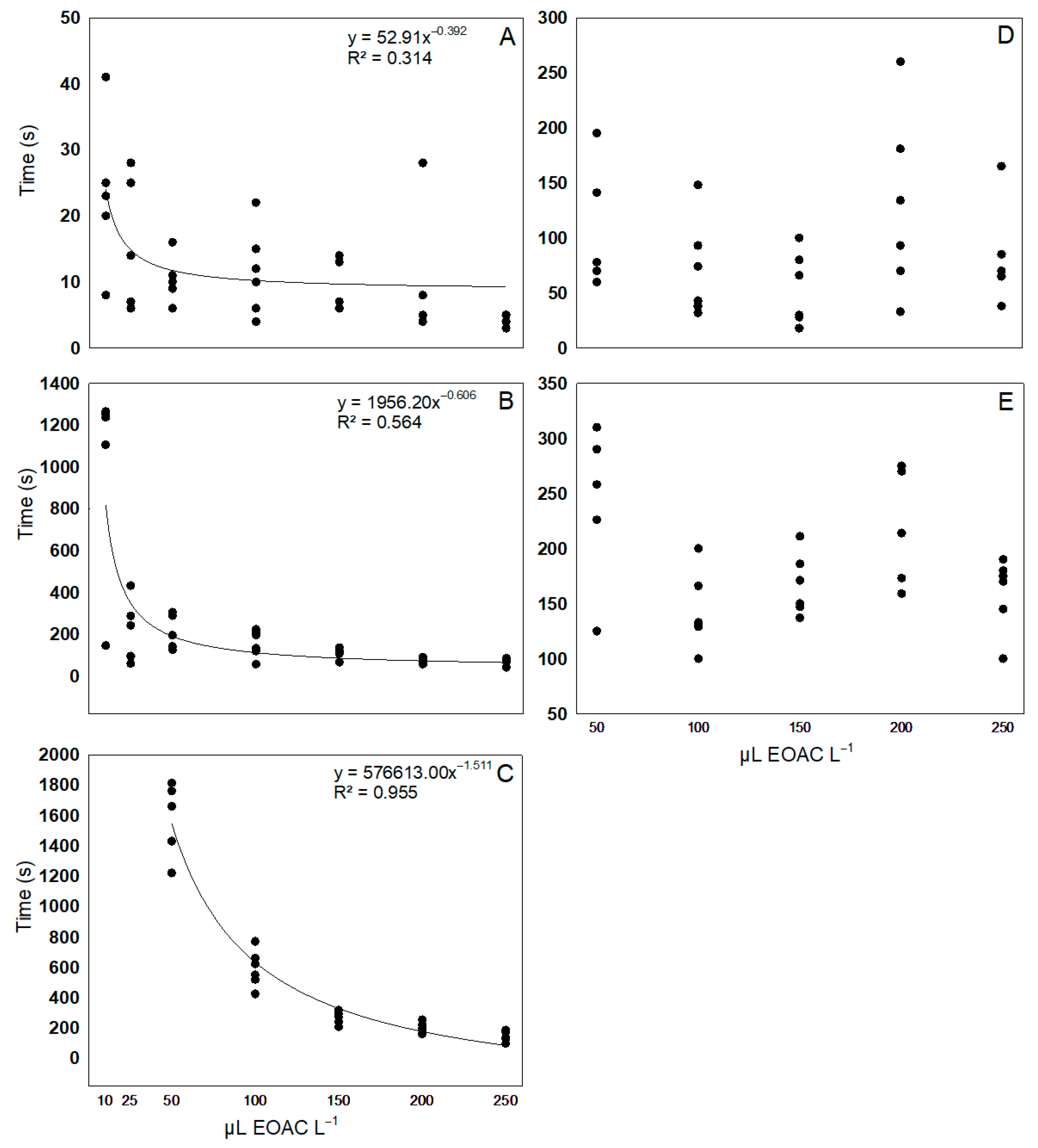
Tambaqui exposed to 10 and 25 μL EOAC L−1 reached only the light and deep sedation stages. Anesthesia was observed at concentrations of 50 μL L−1 and higher, with 250 μL L−1 resulting in rapid anesthesia and recovery (141.83 and 160.00 s, respectively) (Figure 2). No sedation or anesthesia was observed in the control groups.
Power regression was identified for the stages of light sedation (Figure 2A), deep sedation (Figure 2B), and anesthesia (Figure 2C), indicating that as the concentration increased, the time required to reach these stages decreased. However, no regression was observed between the EOAC concentrations and the times taken for partial or total recovery (Figure 2D,E).

3.3. Experiment 2: Simulated Transport

3.3.1. Water Quality

The non-transported group had significantly lower dissolved oxygen, total ammonia, and unionized ammonia levels (except for in EOAC-LCD) and significantly higher pH values than the transported groups (p < 0.05). Among the transported groups, the alkalinity was significantly lower in EOAC-LCD than in the Control–LCD and EOAC-SCD treatments. Additionally, the total ammonia values were significantly lower in EOAC-LCD than in EOAC-SCD (p < 0.05). The unionized ammonia values were significantly lower in EOAC-LCD and higher in Control–SCD than in the other groups at the same CD or EOAC concentration (p < 0.05). There were no significant changes in the hardness, nitrite, or temperature across the treatments (p > 0.05) (Table 1).

3.3.2. Hematological Analysis

All the transported treatments showed reduced heterophil and increased lymphocyte percentages compared with the non-transported group (p < 0.05). The hemoglobin levels in Control–LCD and Control–SCD; the erythrocyte, hematocrit, and total leukocyte counts in Control–SCD; and the MCHC in EOAC-LCD were significantly higher than those in the non-transported group (p < 0.05) (Table 2).
The erythrocyte count was significantly higher in Control–SCD than in the other treatments (p < 0.05). Additionally, the erythrocyte and heterophil counts in EOAC-LCD were significantly lower than those in Control–LCD (p < 0.05). The hemoglobin, hematocrit, and total leukocyte counts in both EOAC treatments were significantly lower than those in the other treatments (p < 0.05). The MCHC values were significantly higher in EOAC-LCD than in EOAC-SCD (p < 0.05). The lymphocyte counts in EOAC-LCD and Control–SCD were significantly higher than those in Control–LCD (p < 0.05). No significant changes were observed in the MCV, HCM, eosinophils, or monocytes across the treatments (p > 0.05) (Table 2).

3.3.3. Biochemical Analysis

Compared with the non-transported group, the plasma glucose and liver glycogen levels were significantly increased in Control–SCD and EOAC-SCD, and the plasma HDL levels were significantly higher in EOAC-LCD. The plasma albumin and ALT values were significantly decreased in Control–LCD (p < 0.05). Transported juveniles showed significantly higher liver total protein levels and lower AST activity (except for those in Control–SCD) compared with non-transported juveniles (p < 0.05) (Table 3).
The plasma glucose, total protein (except for in EOAC-LCD), and ALT values were significantly lower in both LCD groups than in the other treatments (p < 0.05). The plasma albumin and liver glycogen levels were significantly lower in Control–LCD than in Control–SCD and EOAC-LCD (p < 0.05). The liver total protein levels were significantly higher in the EOAC treatments than in the control treatments, with EOAC-SCD showing significantly higher levels than EOAC-LCD (p < 0.05). The liver AST activity was significantly lower in the EOAC treatments than in the control treatments (p < 0.05). There were no significant changes across treatments in the plasma triglycerides, total cholesterol, LDL, VLDL, or AST (p > 0.05) (Table 3).

3.4. Experiment 3: Ventilatory Rate (VR)

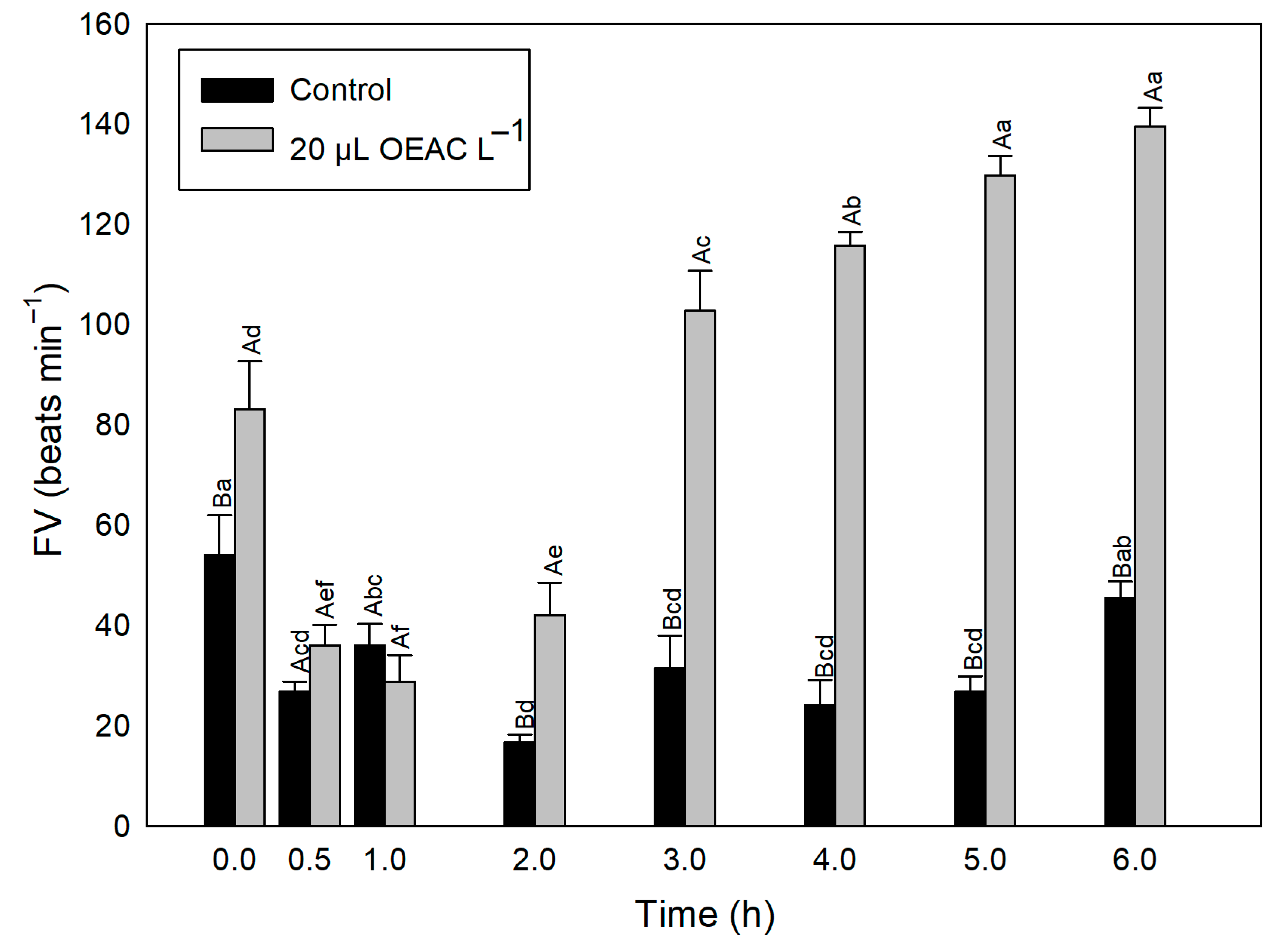
The VR was significantly higher in the group sedated with EOAC (20 μL L−1) than in the control group at 0 h and between 3 and 6 h (p < 0.05). In the control group, the VR at 0 h was significantly higher than that at other times, except at 6 h, at which it was higher than that at all other times (except 1 h) (p < 0.05). In the EOAC treatment, the VR was significantly higher at 5 and 6 h than at other times (p < 0.05). Additionally, the VR at 4 h was significantly higher than that between 0 and 3 h, and the VR at 3 h was higher than that between 0 and 2 h (p < 0.05). In sedated fish, the VR at 0 h was also significantly higher than that between 0.5 and 2 h (p < 0.05) (Figure 3).

4. Discussion

The EOAC used in this study was primarily composed of citral (48.34%), followed by limonene (19.00%). Both compounds are commonly found in EOs from various plant species, with demonstrated anesthetic potential in fish. For example, EO from Lippia alba [(Mill.) N.E.Br. ex Britton & P. Wilson], containing 55.28% citral, effectively promoted anesthesia in tambaqui at a concentration of 100 µL L−1 [32]. In silver catfish, EOs from Aloysia citrodora (50.19% citral) and Cymbopogon flexuosus [(Nees) Will. Watson], with 86.37% citral, promoted anesthesia at 300 µL L−1 [33]. Similarly, Cymbopogon citratus [(DC.) Stapf] EO, containing 73.56% citral, induced anesthesia in freshwater angelfish [Pterophyllum scalare (Schultze, 1823)] at 250 µL L−1 [17]. For tambacu [Piaractus mesopotamicus (Holmberg 1887) × Colossoma macropomum], a concentration of 300 µL L−1 of C. flexuosus EO (90.45% citral) was suitable for inducing anesthesia [34]. Anesthesia was also achieved with EO from Citrus × aurantium × latifolia (L.) (49.73–93.89% limonene) in silver catfish at 500 µL L−1 [35] and with EO from Citrus sinensis (L.) Osbeck (93.89% limonene) in betta [Betta splendens (Regan 1910)] at 300 µL L−1 [36].
These concentrations, along with those found in the current study, are considered the indicated concentrations suitable for fish use because they promote an anesthetic effect in less than 180 s, followed by anesthetic recovery within 600 s [7,17]. In our study, the 250 µL EOAC L−1 concentration achieved induction (141.83 s) and anesthetic recovery (160.00 s) times that best met these criteria. Conversely, if fish farmers prefer slower anesthesia, perhaps due to more time-intensive handling, they can use EOs at lower concentrations. The present study also achieved anesthesia and recovery with lower EOAC concentrations, with induction and recovery times of 1597 and 242 s and 591 and 143 s at 50 and 100 µL L−1, respectively. Additionally, differences in the experimental conditions may explain variations in the recommended EO concentrations between our study and others. Key factors include the animal’s species and size, the plant’s species and origin, and the proportions of chemical compounds in the EOs [37].
The anesthetic effect of EOAC may stem from its action on the central nervous system. It has been suggested that EO constituents may exert biological effects by modulating the GABAergic system and inhibiting Na+ channels [38]. In Swiss mice [Mus musculus (L.)], EO from C. citratus (71.29% citral) demonstrated an effect on the GABAergic system, acting on gamma-aminobutyric acid (GABA) by binding to GABAA receptors to enhance the anticonvulsant, anxiolytic, and anesthetic effects of these receptors [39]. This effect is likely associated with the anesthetic potential of citral, the main compound in the EOAC used in this study, as GABAA receptors are the primary targets of anesthetic action in animals, resulting in central nervous system inhibition [38,40]. Additionally, in zebrafish [Danio rerio (Hamilton, 1822)], citral likely plays a role in modulating GABAA receptors and could potentially be used for anxiety disorder treatment [41]. Similarly, a study using EO from L. alba (44.52% citral) demonstrated the involvement of the GABAergic system in silver catfish anesthesia by causing neuronal hyperpolarization and central nervous system depression [42].
EOs from L. alba with limonene and citral chemotypes exhibit similar pharmacological properties [43], as demonstrated by the anti-stress effect of limonene, which also acts via the GABAergic system and influences dopaminergic regulation [44]. This action induces the release of GABA, leading to an anxiolytic effect [45]. Thus, limonene may act in a complementary manner to citral by stimulating GABA release and enhancing its effect on receptor cells.
In this study, these chemical compounds (citral and limonene), along with the minor compounds in EOAC, only caused sedation (without affecting fish’s balance, swimming, or breathing) in transported tambaqui at a concentration of 20 µL L−1. Similar concentrations have been reported for sedation in other studies using EOAC in species such as Nile tilapia (30 µL L−1; 45.09% citral) [6] and tambacu (25 µL L−1; 90.45% citral) [33].
During fish transportation, variations in the water quality parameters commonly occur due to physiological activities (e.g., respiration and excretion); such parameter alterations include a reduction in the dissolved oxygen and an increase in nitrogen compound concentrations [4,6]. EOs may help reduce metabolism and, consequently, the VR of these animals [26]. As a result, oxygen consumption and the production of ammonia and nitrite would decrease, improving the water quality during transport [16,46]
EOs can have a vasodilatory effect, which affects fish osmoregulation and can trigger hyperventilation that typically stabilizes over time [7,47]. However, contrary to our study’s expectations, a VR increase was observed in tambaqui sedated with 20 µL EOAC L−1 even 6 h after the initial exposure. The VR is influenced by the species, developmental stage, and EO characteristics, such as the composition, concentration, and exposure time, making it challenging to establish a general response model [48,49].
In the present study, at 0 h, an increase in the VR was observed as a reaction to the presence of EOAC. Immediately after this initial exposure (0.5–2 h), the VR decreased in these fish, then increased again at 3 h, likely influenced by a reduction in the sedative potential of EOAC. Notably, sedated fish showed a higher VR than the non-sedated group. An increase in the VR is associated with greater fish activity [50] as they regain their swimming speed, suggesting that the rise may have been a response to transient stress induced by the presence of EO in the water [7]. Therefore, this increase in the VR likely contributed to the reoxygenation of tissues post-sedation [51]; however, it did not alter the dissolved oxygen consumption of tambaqui in the simulated transport experiment. Finally, due to the increased RV, a greater metabolic demand must have occurred in these animals, which is in agreement with our findings regarding the plasma metabolic variables.
Similarly to our findings, an increase in the VR has been reported in previous studies with other fish species. For example, studies have noted increased VRs in silver catfish with eugenol [26], EO from L. alba [26], and EO from Citrus [35]; pacamã [Lophiosilurus alexandri (Steindachner 1876)] with EO from Ocimum gratissimum [50]; common carp with EO from Mentha spicata [48]; Nile tilapia with eugenol and EO from O. basilicum [49]; and fat snook [Centropomus parallelus (Poey 1860)] with EO from L. alba [7]. On the contrary, other studies have demonstrated a reduced VR in fish during the sedation stage. This was verified for Nile tilapia exposed to EOs from Aloysia triphylla and L. alba [6,16], freshwater angelfish exposed to EOs from C. citratus and Lippia sidoides [17], and silver catfish with EO from L. alba [26]. It is possible to hypothesize that the different VR responses found in our study and previous studies are species-specific but also dependent on the sedative compound and its concentration.
Another finding of the present study regarding water quality changes was a reduction in the pH across all the treatments involving fish transport. This may have been due to an accumulation of CO2 produced by the fish during respiration, leading to acidification of the transport water [52]. Tambaqui are well adapted to variations in the water pH, so this reduction does not adversely affect them [53]. A slight reduction in the transport water pH can be beneficial because it decreases the amount of unionized ammonia, facilitating ammonia excretion through the gills [54].
In the current study, the total ammonia increased in all the treatments after transport, but the unionized ammonia did not increase in the EOAC-LCD group. Notably, the EOAC-LCD treatment led to a lower accumulation of ammonia compared with the other groups, likely due to the combination of EOAC with a lower CD. The water alkalinity was also lower in this treatment, possibly due to reduced CO2 accumulation and organic matter decomposition (due to regurgitation) [21,26], despite all fish having been fasted for 24 h. Thus, the lower water alkalinity and ammonia in this treatment may reflect an improvement in the water quality, with reduced excreta and decomposing organic matter. Conversely, the Control–SCD group showed the highest levels of unionized ammonia, compromising the NH3 plasma–water gradient and resulting in ionoregulatory and metabolic disturbances that could negatively impact fish welfare [55]. In the Control–SCD treatment, the stress triggered by increasing the CD without using EOAC may have compromised gill functions and increased ammonia secretion. Similarly, a previous study [17] found that freshwater angelfish transported without sedative substances had gill lesions associated with higher levels of unionized ammonia.
In addition to assessing the VR and water quality, analysis of hematological parameters is commonly used as a non-lethal method to detect physiological changes and even diagnose diseases in fish [56]. In the current study, the Control–SCD group exhibited greater hematological parameter variations than the non-transported group. Specifically, the erythrocyte, hemoglobin, hematocrit, and total leukocyte levels increased in this group, potentially indicating increased energy expenditure due to transport stress [57].
Additionally, a change in the differential leukocyte profile was observed in all the transported groups. There was a decrease in the relative percentage of heterophils and an increase in lymphocytes, with no changes in the total leukocyte count, except in the Control–SCD group, which showed an increase in these cells. Lymphocytes are the most abundant defense cells among the total leukocytes and are associated with immunoglobulin production and immune response modulation [58]. Under stress, animals typically show a decrease in lymphocytes and an increase in heterophils, which are involved in the inflammatory response [57,59]. Thus, in this study, the reduced heterophil-to-lymphocyte ratio suggests that the transport stress did not significantly impact the immunological response of these fish [58]. Finally, it is noteworthy that overall, the hematological parameters were more stable in tambaqui transported with EOAC than those transported without it.
In addition to the hematological parameters, analyzing the biochemical parameters is highly relevant for assessing fish health. When exposed to transport stress, fish tend to expend more energy to maintain homeostasis, thereby increasing their energy demand [5]. In the present study, this likely occurred in fish transported at an SCD (with or without EOAC), as indicated by a hyperglycemic response in these animals. This hyperglycemia could be attributed to the release of catecholamines into the bloodstream in response to external stress stimuli, such as transport [12]. Catecholamines promote an increase in the plasma glucose through glycogenolysis, primarily in the liver, which involves breaking down hepatic glycogen to meet the heightened energy demands of animals under stress [16,60]. This may indicate that the observed increase in the plasma glucose levels was driven by gluconeogenesis and glycogenolysis (secondary stress responses) in these fish [61].
Furthermore, these processes are associated with a decrease in the liver glycogen levels. Interestingly, in our study, the hepatic glycogen levels also increased in the SCD groups. Under stress, glucose can be mobilized into the blood faster than it is used, and surplus glucose may be reconverted to glycogen in the liver, especially if swimming activity is limited. This “overshoot” effect can paradoxically increase the liver glycogen [60,62].
Another possible explanation is that this increase in both the plasma glucose and liver glycogen levels is related to the presence of glucosensor systems. These systems have already been identified in the hypothalamus, hindbrain, and Brockmann bodies of rainbow trout and are activated when the glucose levels increase while the food intake decreases and are characterized by changes in the glycogen levels [63]. They are also related to increased cortisol levels under stress conditions [64]. In our study, the main stress condition for the fish was a higher CD, and the increase in both parameters in the SCD groups could indicate an intensified response of their glucose-suppressing systems. A similar behavior was verified in rainbow trout exposed to hyperglycemic conditions, with increased liver glycogen levels [64]. To cope with stressful situations, the primary energy substrate used is carbohydrates (e.g., glucose), although lipid molecules are also essential energy sources, especially when carbohydrate supplementation is needed [65]. In fish, cholesterol synthesis primarily occurs in the liver; however, in the present study, the plasma total cholesterol and triglyceride levels showed no change. An increase in the plasma HDL levels was only observed in the EOAC-LCD group compared with those of the non-transported group, suggesting the homeostatic functioning of hepatocytes [3].
Additionally, increased activity of the ALT and AST enzymes may indicate liver tissue damage or hepatic hyperactivity [3,66]. Based on an integrative view of the plasma and hepatic responses, the EOAC-LCD treatment resulted in reduced transaminase activity, supporting an improved anabolic state in hepatic tissue in tambaqui. Furthermore, fish transported with EOAC showed higher hepatic total protein levels than those in the control groups, suggesting that this substrate may be more readily available for other bodily functions [14]. Albumin can also serve as an energy reserve, and the reduction in the plasma albumin levels in the Control–LCD group could indicate a decrease in metabolism in these animals [65]. Thus, under the conditions tested in this study, using 20 µL EOAC L−1 could help maintain the metabolic state of tambaqui after exposure to transport stress, especially when transported at an LCD, suggesting that despite changes in the VR, the fish energy reserves remained unaffected.

5. Conclusions

For rapid anesthetic induction and recovery in tambaqui, a concentration of 250 µL EOAC L−1 is recommended. For fish sedation and transport, 20 µL EOAC L−1 can be used, particularly under an LCD, because it may improve the hematological (erythrocytes, hematocrit, and total leukocytes) and biochemical (plasma glucose, ALT, hepatic AST, and glycogen) responses, as well as minimize changes in the water quality parameters. We suggest that further studies be conducted to investigate the sedative effect of citral and limonene, the main compounds found in EOAC, in fish under different CDs during transport. This will allow for a more precise assessment of each compound’s contribution, clarifying how these compounds interact and result in the effect of EOAC for use in the aquaculture industry.

Author Contributions

Conceptualization: C.E.C. Methodology: O.P.d.A.C.N., I.S.S., E.d.S.S. and C.E.C. Validation: R.D.C. and D.S. Formal analysis: O.P.d.A.C.N., A.d.A.S., A.d.S.R. and P.J.C.-S. Investigation: O.P.d.A.C.N., I.S.S., A.d.A.S., A.d.S.R. and P.J.C.-S. Resources: C.E.C., R.D.C. and D.S. Data curation: E.d.S.S. and D.S. Writing—original draft preparation: O.P.d.A.C.N. and C.E.C. Writing—review and editing: C.E.C. Supervision and project administration: C.E.C. All authors have read and agreed to the published version of the manuscript.

Funding

The authors are grateful to the CNPq (Conselho Nacional de Desenvolvimento Científico e Tecnológico, Brazil) for the research fellowships provided to C.E. Copatti (# PQ 303337/2025-7) and D. Schmidt (# PQ 314827/2021-8) and the FAPESB (Fundação de Amparo à Pesquisa do Estado da Bahia, BA, Brazil) for a research grant provided to O.P.A. Castro Neto (2206/2022).

Institutional Review Board Statement

This study was conducted in accordance with the requirements of and approved by the Committee on Ethics in the Use of Animals of the Institute of Biology at the Federal University of Bahia (IBIO-UFBA), Bahia, Brazil. (Approval code: Protocol Number 01/2024; approval date: 9 April 2024.)

Data Availability Statement

Data are contained within the article.

Conflicts of Interest

The authors declare no conflict of interest.

References

  1. Embrapa CIAqui—Centro de Inteligência e Mercado em Aquicultura: Comércio exterior—Exportação 2025. Available online: https://www.embrapa.br/cim-centro-de-inteligencia-e-mercado-em-aquicultura (accessed on 27 August 2025).
  2. Neves, L.; Favero, G.C.; Beier, S.L.; Ferreira, N.S.; Palheta, G.D.A.; Melo, N.F.A.C.; Luz, R.K. Physiological and metabolic responses in juvenile Colossoma macropomum exposed to hypoxia. Fish. Physiol. Biochem. 2020, 46, 2157–2167. [Google Scholar] [CrossRef]
  3. Chung, S.; Ribeiro, K.; Teixeira, D.V.; Copatti, C.E. Inclusion of essential oil from ginger in the diet improves physiological parameters of tambaqui juveniles (Colossoma macropomum). Aquaculture 2021, 543, 736934. [Google Scholar] [CrossRef]
  4. Sena, A.C.; Teixeira, R.R.; Ferreira, E.L.; Heinzmann, B.M.; Baldisserotto, B.; Caron, B.O.; Schmidt, D.; Couto, R.D.; Copatti, C.E. Essential oil from Lippia alba has anaesthetic activity and is effective in reducing handling and transport stress in tambacu (Piaractus mesopotamicus × Colossoma macropomum). Aquaculture 2016, 465, 374–379. [Google Scholar] [CrossRef]
  5. Teixeira, R.R.; Souza, R.C.; Sena, A.C.; Baldisserotto, B.; Heinzmann, B.M.; Couto, R.D.; Copatti, C.E. Essential oil of Aloysia triphylla in Nile tilapia: Anaesthesia, stress parameters and sensory evaluation of fillets. Aquac. Res. 2017, 48, 3383–3392. [Google Scholar] [CrossRef]
  6. Teixeira, R.R.; de Souza, R.C.; Sena, A.C.; Baldisserotto, B.; Heinzmann, B.M.; Copatti, C.E. Essential oil of Aloysia triphylla is effective in Nile tilapia transport. Bol. Inst. Pesca 2018, 44, 17–24. [Google Scholar] [CrossRef]
  7. Simões-Bueno, L.N.; Copatti, C.E.; Gomes, L.C.; Val, A.L.; Amanajás, R.D.; Caron, B.O.; Heinzmann, B.M.; Baldisserotto, B. Linalool chemotype essential oil from Lippia alba in the anesthesia of fat snook (Centropomus parallelus): Ventilatory rate, biochemical, antioxidant, and oxidative status parameters. Neotrop. Ichthyol. 2024, 22, e230114. [Google Scholar] [CrossRef]
  8. Fang, D.; Mei, J.; Xie, J.; Qiu, W. The effects of transport stress (temperature and vibration) on blood biochemical parameters, oxidative stress, and gill histomorphology of pearl gentian groupers. Fishes 2023, 8, 218. [Google Scholar] [CrossRef]
  9. Hong, J.; Chen, X.; Liu, S.; Fu, Z.; Han, M.; Wang, Y.; Gu, Z.; Ma, Z. Impact of fish density on water quality and physiological response of golden pompano (Trachinotus ovatus) fingerlings during transportation. Aquaculture 2019, 507, 260–265. [Google Scholar] [CrossRef]
  10. Lima, A.F.; Oliveira, H.J.B.; Pereira, A.S.; Sakamoto, S.S. Effect of density of fingerling and juvenile pirarucu during transportation on water quality and physiological parameters. Acta Amaz. 2020, 50, 223–231. [Google Scholar] [CrossRef]
  11. Hohlenwerger, J.C.; Copatti, C.E.; Sena, A.C.; Couto, R.D.; Baldisserotto, B.; Heinzmann, B.M.; Caron, B.O.; Schmidt, D. Could the essential oil of Lippia alba provide a readily available and cost-effective anaesthetic for Nile tilapia (Oreochromis niloticus)? Mar. Freshw. Behav. Physiol. 2016, 49, 119–126. [Google Scholar] [CrossRef]
  12. Taheri Mirghaed, A.; Yasari, M.; Mirzargar, S.S.; Hoseini, S.M. Rainbow trout (Oncorhynchus mykiss) anesthesia with myrcene: Efficacy and physiological responses in comparison with eugenol. Fish. Physiol. Biochem. 2018, 44, 919–926. [Google Scholar] [CrossRef] [PubMed]
  13. Aydın, B.; Barbas, L.A.L. Sedative and anesthetic properties of essential oils and their active compounds in fish: A review. Aquaculture 2020, 520, 734999. [Google Scholar] [CrossRef]
  14. Becker, A.G.; Parodi, T.V.; Zeppenfeld, C.C.; Salbego, J.; Cunha, M.A.; Heldwein, C.G.; Loro, V.L.; Heinzmann, B.M.; Baldisserotto, B. Pre-sedation and transport of Rhamdia quelen in water containing essential oil of Lippia alba: Metabolic and physiological responses. Fish. Physiol. Biochem. 2016, 42, 73–81. [Google Scholar] [CrossRef] [PubMed]
  15. Oliveira, C.P.B.; Lemos, C.H.P.; Felix e Silva, A.; Souza, S.A.; Albinati, A.C.L.; Lima, A.O.; Copatti, C.E. Use of eugenol for the anaesthesia and transportation of freshwater angelfish (Pterophyllum scalare). Aquaculture 2019, 513, 734409. [Google Scholar] [CrossRef]
  16. Hohlenwerger, J.C.; Baldisserotto, B.; Couto, R.D.; Heinzmann, B.M.; Silva, D.T.; Caron, B.O.; Schmidt, D.; Copatti, C.E. Essential oil of Lippia alba in the transport of Nile tilapia. Ciência Rural 2017, 47, e20160040. [Google Scholar] [CrossRef]
  17. Oliveira, I.C.; Oliveira, R.S.M.; Lemos, C.H.P.; Oliveira, C.P.B.; Felix e Silva, A.; Lorenzo, V.P.; Lima, A.O.; Cruz, A.L.; Copatti, C.E. Essential oils from Cymbopogon citratus and Lippia sidoides in the anesthetic induction and transport of ornamental fish Pterophyllum scalare. Fish. Physiol. Biochem. 2022, 48, 501–519. [Google Scholar] [CrossRef]
  18. Bahramsoltani, R.; Rostamiasrabadi, P.; Shahpiri, Z.; Marques, A.M.; Rahimi, R.; Farzaei, M.H. Aloysia citrodora Paláu (Lemon verbena): A review of phytochemistry and pharmacology. J. Ethnopharmacol. 2018, 222, 34–51. [Google Scholar] [CrossRef]
  19. Brandão, F.R.; Farias, C.F.S.; Souza, D.C.M.; Oliveira, M.I.B.; Matos, L.V.; Majolo, C.; Oliveira, M.R.; Chaves, F.C.M.; O’Sullivan, F.L.A.; Chagas, E.C. Anesthetic potential of the essential oils of Aloysia triphylla, Lippia sidoides and Mentha piperita for Colossoma macropomum. Aquaculture 2021, 534, 736275. [Google Scholar] [CrossRef]
  20. Santos, A.C.; Bianchini, A.E.; Bandeira Junior, G.; Garlet, Q.I.; Brasil, M.T.B.; Heinzmann, B.M.; Baldisserotto, B.; Caron, B.O.; Cunha, M.A. Essential oil of Aloysia citriodora Paláu and citral: Sedative and anesthetic efficacy and safety in Rhamdia quelen and Ctenopharyngodon idella. Vet. Anaesth. Analg. 2022, 49, 104–112. [Google Scholar] [CrossRef]
  21. Parodi, T.V.; Cunha, M.A.; Becker, A.G.; Zeppenfeld, C.C.; Martins, D.I.; Koakoski, G.; Barcellos, L.G.; Heinzmann, B.M.; Baldisserotto, B. Anesthetic activity of the essential oil of Aloysia triphylla and effectiveness in reducing stress during transport of albino and gray strains of silver catfish, Rhamdia quelen. Fish. Physiol. Biochem. 2014, 40, 323–334. [Google Scholar] [CrossRef]
  22. Ali, H.B.; Masoumeh, M.; Mohammad, S.; Mohammad, M. The effect of ethanol extract of Aloysia triphylla on anesthesia and improve the physiological parameters of rainbow trout (Oncorhynchus mykiss) after transfer. J. Aquat. Anim. Nutr. 2023, 9, 1–14. [Google Scholar]
  23. Burgess, D.R.; Linstrom, P.J.; Mallard, W.G. NIST Chemistry WebBook; Linstrom, PJ: Gaithersburg, MA, USA, 2016. [Google Scholar]
  24. Small, B.C. Anesthetic efficacy of metomidate and comparison of plasma cortisol responses to tricaine methanesulfonate, quinaldine and clove oil anesthetized channel catfish Ictalurus punctatus. Aquaculture 2003, 218, 177–185. [Google Scholar] [CrossRef]
  25. Gomes, L.C.; Araujo-Lima, C.A.R.M.; Roubach, R.; Chippari-Gomes, A.R.; Lopes, N.; Urbinati, E.C. Effect of fish density during transportation on stress and mortality of juvenile tambaqui Colossoma macropomum. J. World Aquac. Soc. 2003, 34, 76–84. [Google Scholar] [CrossRef]
  26. Becker, A.G.; Parodi, T.V.; Heldwein, C.G.; Zeppenfeld, C.C.; Heinzmann, B.M.; Baldisserotto, B. Transportation of silver catfish, Rhamdia quelen, in water with eugenol and the essential oil of Lippia alba. Fish. Physiol. Biochem. 2012, 38, 789–796. [Google Scholar] [CrossRef] [PubMed]
  27. Sampaio, F.D.F.; Freire, C.A. An overview of stress physiology of fish transport: Changes in water quality as a function of transport duration. Fish Fish. 2016, 17, 1055–1072. [Google Scholar] [CrossRef]
  28. Pramod, P.K.; Ramachandran, A.; Sajeevan, T.P.; Thampy, S.; Pai, S.S. Comparative efficacy of MS-222 and benzocaine as anaesthetics under simulated transport conditions of a tropical ornamental fish Puntius filamentosus (Valenciennes). Aquac. Res. 2010, 41, 309–314. [Google Scholar] [CrossRef]
  29. CONCEA. Diretrizes da Prática de Eutanásia do Conselho Nacional de Controle de Experimentação Animal; Ministério da Ciência, Tecnologia e Inovação: Brasília, Brazil, 2018. [Google Scholar]
  30. Blaxhall, P.C.; Daisley, K.W. Routine haematological methods for use with fish blood. J. Fish. Biol. 1973, 5, 771–781. [Google Scholar] [CrossRef]
  31. Bidinotto, P.M.; Souza, R.H.S.; Moraes, G. Hepatic glycogen in eight tropical freshwater teleost fish: A procedure for field determinants of microsamples. Bol. Téc. CEPTA 1997, 10, 53–60. [Google Scholar]
  32. Silva, H.N.P.; Carvalho, B.C.F.; Maia, J.L.D.S.; Becker, A.G.; Baldisserotto, B.; Heinzmann, B.M.; Mourão, R.H.V.; Silva, L.V.F. Anesthetic potential of the essential oils of Lippia alba and Lippia origanoides in tambaqui juveniles. Ciência Rural 2019, 49, e20181059. [Google Scholar] [CrossRef]
  33. Santos, A.C.; Bandeira Junior, G.; Zago, D.C.; Zeppenfeld, C.C.; Silva, D.T.; Heinzmann, B.M.; Baldisserotto, B.; Cunha, M.A. Anesthesia and anesthetic action mechanism of essential oils of Aloysia triphylla and Cymbopogon flexuosus in silver catfish (Rhamdia quelen). Vet. Anaesth. Analg. 2017, 44, 106–113. [Google Scholar] [CrossRef]
  34. Limma-Netto, J.D.; Sena, A.C.; Copatti, C.E. Essential oils of Ocimum basilicum and Cymbopogon flexuosus in the sedation, anesthesia and recovery of tambacu (Piaractus mesopotamicus male x Colossoma macropomum Female). Bol. Inst. Pesca 2016, 42, 727–733. [Google Scholar] [CrossRef]
  35. Lopes, J.M.; Souza, C.F.; Schindler, B.; Pinheiro, C.G.; Salbego, J.; Siqueira, J.C.; Heinzmann, B.M.; Baldisserotto, B. Essential oils from Citrus x aurantium and Citrus x latifolia (Rutaceae) have anesthetic activity and are effective in reducing ion loss in silver catfish (Rhamdia quelen). Neotrop. Ichthyol. 2018, 16, e170152. [Google Scholar] [CrossRef]
  36. Silva, R.C.; Silva, L.R.; França, I.F.; Lopes, J.M.; Pantoja, B.T.S.; Pereira, M.M.; Ramos, L.R.V. Anesthetic effect and acute toxicity of Citrus sinensis essential oil in betta. Bol. Inst. Pesca 2023, 49, e816. [Google Scholar] [CrossRef]
  37. Tarkhani, R.; Imani, A.; Jamali, H.; Farsani, H.G. Anaesthetic efficacy of eugenol on various size classes of angelfish (Pterophyllum scalare Schultze, 1823). Aquac. Res. 2017, 48, 5263–5270. [Google Scholar] [CrossRef]
  38. Wang, Z.J.; Heinbockel, T.T. Essential oils and their constituents targeting the GABAergic system and sodium channels as treatment of neurological diseases. Molecules 2018, 23, 1061. [Google Scholar] [CrossRef] [PubMed]
  39. Costa, C.A.R.D.A.; Kohn, D.O.; Lima, V.M.; Gargano, A.C.; Flório, J.C.; Costa, M. The GABAergic system contributes to the anxiolytic-like effect of essential oil from Cymbopogon citratus (lemongrass). J. Ethnopharmacol. 2011, 137, 828–836. [Google Scholar] [CrossRef]
  40. Nesterkina, M.; Kravchenko, I. Synthesis and pharmacological properties of novel esters based on monocyclic terpenes and GABA. Pharmaceuticals 2016, 9, 32. [Google Scholar] [CrossRef]
  41. Hacke, A.C.M.; Miyoshi, E.; Marques, J.A.; Pereira, R.P. Anxiolytic properties of Cymbopogon citratus (DC.) Stapf extract, essential oil and its constituents in zebrafish (Danio rerio). J. Ethnopharmacol. 2020, 260, 113036. [Google Scholar] [CrossRef] [PubMed]
  42. Heldwein, C.G.; Silva, L.L.; Reckziegel, P.; Barros, F.M.C.; Bürger, M.E.; Baldisserotto, B.; Mallmann, C.A.; Schmidt, D.; Caron, B.O.; Heinzmann, B.M. Participation of the GABAergic system in the anesthetic effect of Lippia alba (Mill.) N.E. Brown essential oil. Braz. J. Med. Biol. Res. 2012, 45, 436–443. [Google Scholar] [CrossRef]
  43. Vale, T.G.; Matos, F.J.A.; Lima, T.C.M.; Viana, G.S.B. Behavioral effects of essential oils from Lippia alba (Mill.) N.E. Brown chemotypes. J. Ethnopharmacol. 1999, 167, 127–133. [Google Scholar] [CrossRef] [PubMed]
  44. Zhou, W.; Yoshioka, M.; Yokogoshi, H. Sub-chronic effects of s-limonene on brain neurotransmitter levels and behavior of rats. J. Nutr. Sci. Vitaminol. 2009, 55, 367–373. [Google Scholar] [CrossRef]
  45. Song, Y.; Seo, S.; Lamichhane, S.; Seo, J.; Hong, J.T.; Cha, H.J.; Yun, J. Limonene has anti-anxiety activity via adenosine A2A receptor-mediated regulation of dopaminergic and GABAergic neuronal function in the striatum. Phytomedicine 2021, 83, 153474. [Google Scholar] [CrossRef]
  46. He, R.; Lei, B.; Su, Y.; Wang, A.; Cui, K.; Shi, X.; Chen, X. Effectiveness of eugenol as an anesthetic for adult spotted sea bass (Lateolabrax maculatus). Aquaculture 2020, 523, 735180. [Google Scholar] [CrossRef]
  47. Lam, P.H.; Vo, H.D.N.; Truong, L.M.T.; Dang, D.M.T.; Dang, C.M.; Doan, T.C.D.; Mollaamin, F.; Monajjemi, M. Anesthetic effects of clove basil essential oil (Ocimum gratissimum) microemulsion on asian redtail catfish (Hemibagrus wyckioides) and Its Biochemical Stress Indicators. Fishes 2025, 10, 104. [Google Scholar] [CrossRef]
  48. Roohi, Z.; Imanpoor, M.R. The efficacy of the oils of spearmint and methyl salicylate as new anesthetics and their effect on glucose levels in common carp (Cyprinus carpio L., 1758) juveniles. Aquaculture 2015, 437, 327–332. [Google Scholar] [CrossRef]
  49. Ventura, A.S.; Corrêa Filho, R.A.C.; Teodoro, G.C.; Laice, L.M.; Barbosa, P.T.L.; Stringhetta, G.R.; Jerônimo, G.T.; Povh, J.A. Essential oil of Ocimum basilicum and eugenol as sedatives for Nile tilapia. J. Agric. Stud. 2020, 8, 657–665. [Google Scholar] [CrossRef]
  50. Boaventura, T.P.; Souza, C.F.; Ferreira, A.L.; Favero, G.C.; Baldissera, M.D.; Heinzmann, B.M.; Baldisserotto, B.; Luz, R.K. Essential oil of Ocimum gratissimum (Linnaeus, 1753) as anesthetic for Lophiosilurus alexandri: Induction, recovery, hematology, biochemistry and oxidative stress. Aquaculture 2020, 529, 735676. [Google Scholar] [CrossRef]
  51. Janssen, G.J.A.; Jerrett, A.R.; Black, S.E.; Forster, M.E. The effects of progressive hypoxia and re-oxygenation on cardiac function, white muscle perfusion and haemoglobin saturation in anaesthetised snapper (Pagrus auratus). J. Comp. Physiol. B 2010, 180, 503–510. [Google Scholar] [CrossRef]
  52. Luz, R.K.; Favero, G.C. Use of salt, anesthetics, and cargo density in transport of live fish: A review. Fishes 2024, 9, 286. [Google Scholar] [CrossRef]
  53. Aride, P.H.R.; Roubach, R.; Val, A.L. Tolerance response of tambaqui Colossoma macropomum (Cuvier) to water pH. Aquac. Res. 2007, 38, 588–594. [Google Scholar] [CrossRef]
  54. Zimmer, A.M. Ammonia excretion by the fish gill: Discoveries and ideas that shaped our current understanding. J. Comp. Physiol. B 2024, 194, 697–715. [Google Scholar] [CrossRef] [PubMed]
  55. Wang, C.; Kou, N.; Liu, X.; Yang, D. Oxygen Consumption and Ammonia Excretion of Marphysa sanguinea (Polychaeta: Eunicidae) in relation to body mass and temperature. Fishes 2021, 6, 52. [Google Scholar] [CrossRef]
  56. Fazio, F. Fish hematology analysis as an important tool of aquaculture: A review. Aquaculture 2019, 500, 237–242. [Google Scholar] [CrossRef]
  57. Witeska, M.; Kondera, E.; Ługowska, K.; Bojarski, B. Hematological methods in fish—Not only for beginners. Aquaculture 2022, 547, 737498. [Google Scholar] [CrossRef]
  58. Davis, A.K.; Maney, D.L.; Maerz, J.C. The use of leukocyte profiles to measure stress in vertebrates: A review for ecologists. Funct. Ecol. 2008, 22, 760–772. [Google Scholar] [CrossRef]
  59. Dinesh, R.; Daniel, N.; Stephen, J.; Kumar, S. Haematological parameters as reliable stress indicators in fish. J. Agric. Environ. 2021, 2, 48–52. [Google Scholar]
  60. Wendelaar-Bonga, S.E. The stress response in fish. Physiol. Rev. 1997, 77, 591–625. [Google Scholar] [CrossRef]
  61. Dai, Y.; Shen, Y.; Gu, J.; Yang, H.; Chen, F.; Zhang, W.; Wu, W.; Xu, X.; Li, J. Glycolysis and gluconeogenesis are involved of glucose metabolism adaptation during fasting and re-feeding in black carp (Mylopharyngodon piceus). Aquac. Fish. 2024, 9, 226–233. [Google Scholar] [CrossRef]
  62. Milligan, C.L. A regulatory role for cortisol in muscle glycogen metabolism in rainbow trout Oncorhynchus mykiss Walbaum. J. Exp. Biol. A 2003, 206, 3167–3173. [Google Scholar] [CrossRef]
  63. Polakof, S.; Míguez, J.M.; Moon, T.W.; Soengas, J.L. Evidence for the presence of a glucosensor in hypothalamus, hindbrain, and Brockmann bodies of rainbow trout. Am. J. Physiol. Regul. Integr. Comp. Physiol. 2007, 292, R1657–R1666. [Google Scholar] [CrossRef]
  64. Conde-Sieira, M.; Aguilar, A.J.; López-Patiño, M.A.; Míguez, J.M.; Soengas, J.L. Stress alters food intake and glucosensing response in hypothalamus, hindbrain, liver, and Brockmann bodies of rainbow trout. Physiol. Behav. 2010, 101, 483–493. [Google Scholar] [CrossRef] [PubMed]
  65. Javed, M.; Usmani, N. Stress response of biomolecules (carbohydrate, protein and lipid profiles) in fish Channa punctatus inhabiting river polluted by thermal power plant effluent. Saudi J. Biol. Sci. 2015, 22, 237–242. [Google Scholar] [CrossRef] [PubMed]
  66. Wang, R.; Lei, C.; Li, Z.; Lei, Y.; Luo, C.; Shao, L.; Huang, C.; Yang, P. Effects of a diet of Phragmites australis instead of Triticum aestivum L. on immune performance and liver tissue structure of Ctenopharyngodon idellus. Fishes 2022, 7, 378. [Google Scholar] [CrossRef]
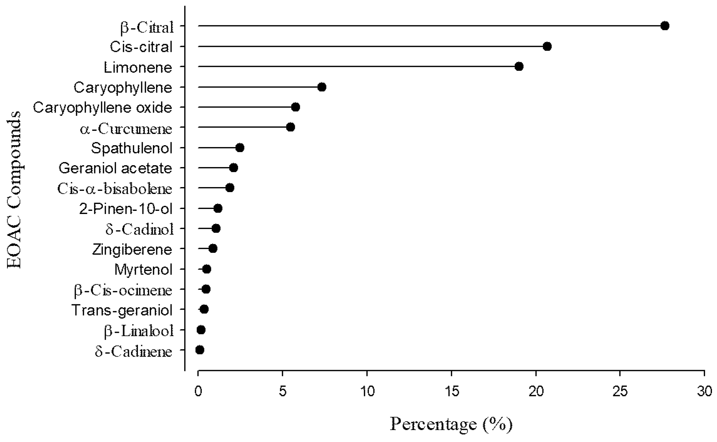
Figure 1. Chemical composition (%) of the essential oil from Aloysia citrodora (EOAC).
Fishes 10 00448 g001
Figure 2. Time (s) required to reach light sedation (A), deep sedation (B), anesthesia (C), and partial (D) and total (E) anesthetic recovery in tambaqui (Colossoma macropomum), according to the concentration of the essential oil from Aloysia citrodora (EOAC) (n = 6 fish per concentration).
Fishes 10 00448 g002
Figure 3. Ventilatory rate of tambaqui (Colossoma macropomum) in presence or absence of essential oil from Aloysia citrodora (EOAC) (20 μL L−1) for 6 h. Data are presented as mean ± standard error of mean ± SEM (n = 6 fish per treatment). Uppercase letters indicate significant differences between treatments at same time point. Lowercase letters indicate significant differences between time points for same treatment. Two-way ANOVA and Duncan’s tests were used to determine statistical significance (p < 0.05).
Fishes 10 00448 g003
Table 1. Water quality parameters after simulated transport (6 h) of tambaqui (Colossoma macropomum) with or without the addition of the essential oil from Aloysia citrodora (EOAC) (20 μL L−1) at low (LCD) and standard (SCD) cargo densities.
VariablesNon-TransportedControl–LCDControl–SCDEOAC-LCDEOAC-SCD
DO4.50 ± 1.228.63 ± 0.20 Aa*8.54 ± 0.22 Aa*8.71 ± 0.18 Aa*8.75 ± 0.20 Aa*
Alkalinity42.50 ± 2.0445.00 ± 4.08 Aa45.00 ± 4.08 Aa35.00 ± 4.08 Bb45.00 ± 4.08 Aa
Hardness115.00 ± 4.08120.00 ± 8.16 Aa125.00 ± 4.08 Aa125.00 ± 4.08 Aa115.00 ± 4.08 Aa
Total ammonia0.13 ± 0.021.50 ± 0.14 Aa*1.75 ± 0.14 Aa*1.00 ± 0.25 Ab*1.75 ± 0.25 Aa*
UIA0.72 ± 0.061.68 ± 0.15 Ab*2.21 ± 0.17 Aa*1.15 ± 0.16 Bb1.67 ± 0.21 Ba*
Nitrite0.013 ± 0.010.013 ± 0.01 Aa0.025 ± 0.004 Aa0.013 ± 0.010 Aa0.025 ± 0.004 Aa
Temperature28.63 ± 0.1028.83 ± 0.14 Aa28.88 ± 0.10 Aa28.90 ± 0.08 Aa28.85 ± 0.12 Aa
pH6.90 ± 0.086.20 ± 0.20 Aa*6.25 ± 0.15 Aa*6.21 ± 0.23 Aa*6.13 ± 0.10 Aa*
Dissolved oxygen (DO) is in mg O2 L−1. Alkalinity and hardness are in mg CaCO3 L−1. Total ammonia is in mg N-NH3 L−1. UIA (unionized ammonia) is in µg N-NH3 L−1. Nitrite is in mg N-NO2 L–1. Temperature is in °C. Data are presented as mean ± standard error of mean ± SEM (n = 3 plastic bags per treatment). Uppercase letters indicate significant differences between treatments with same cargo density. Lowercase letters indicate significant differences between cargo densities within same treatment. Two-way ANOVA and Duncan’s tests were used to determine statistical significance (p < 0.05). Asterisk (*) indicates significant difference compared to non-transported group (Dunnett’s test).
Table 2. Hematological parameters after simulated transport (6 h) of tambaqui (Colossoma macropomum) with or without the addition of the essential oil from Aloysia citrodora (EOAC) (20 μL L−1) at low (LCD) and standard (SCD) cargo densities.
VariablesNon-TransportedControl–LCDControl–SCDEOAC-LCDEOAC-SCD
Erythrocytes1.60 ± 0.041.77 ± 0.05 Ab1.96 ± 0.06 Aa*1.53 ± 0.08 Ba1.50 ± 0.03 Ba
Hemoglobin7.33 ± 0.228.40 ± 0.22 Aa*9.01 ± 0.39 Aa*7.15 ± 0.34 Ba6.90 ± 0.19 Ba
Hematocrit21.33 ± 0.8023.33 ± 0.71 Aa25.50 ± 1.33 Aa*19.50 ± 1.18 Ba20.16 ± 0.60 Ba
MCV133.83 ± 2.46131.16 ± 1.93 Aa129.50 ± 3.53 Aa126.83 ± 2.83 Aa134.50 ± 1.72 Aa
HCM46.16 ± 0.6547.17 ± 0.94 Aa45.83 ± 0.65 Aa46.83 ± 0.70 Aa46.00 ± 0.68 Aa
MCHC34.66 ± 0.4936.00 ± 0.51 Aa35.50 ± 0.61 Aa37.00 ± 0.81 Aa*34.00 ± 0.25 Ab
Total leukocytes2.66 ± 0.253.26 ± 0.28 Aa3.83 ± 0.22 Aa*2.45 ± 0.23 Ba2.31 ± 0.39 Ba
Heterophils54.66 ± 2.2747.17 ± 0.94 Aa*44.33 ± 0.84 Aa*42.00 ± 1.18 Ba*42.17 ± 0.83 Aa*
Lymphocytes42.83 ± 2.1050.33 ± 0.88 Bb*53.33 ± 0.99 Aa*55.16 ± 0.91 Aa*55.16 ± 0.87 Aa*
Eosinophils1.16 ± 0.161.33 ± 0.21 Aa1.16 ± 0.16 Aa1.50 ± 0.22 Aa1.33 ± 0.21 Aa
Monocytes1.33 ± 0.211.16 ± 0.16 Aa1.16 ± 0.16 Aa1.13 ± 0.21 Aa1.33 ± 0.21 Aa
Erythrocyte concentration (Ery) is in ×106 µL−1. Hematocrit (Hct), heterophil, lymphocyte, eosinophil, and monocyte levels are in %. Hemoglobin concentration (Hb) and MCHC (mean corpuscular hemoglobin concentration) are in g dL−1. MCV (mean corpuscular volume) is in fL. MCH (mean corpuscular hemoglobin) is in pg. Total leukocytes are in ×104 μL−1. Data are presented as mean ± standard error of mean ± SEM (n = 9 fish per treatment). Uppercase letters indicate significant differences between treatments with same cargo density. Lowercase letters indicate significant differences between cargo densities within same treatment. Two-way ANOVA and Duncan’s tests were used to determine statistical significance (p < 0.05). Asterisk (*) indicates significant difference compared to non-transported group (Dunnett’s test).
Table 3. Biochemical parameters of plasma and liver after simulated transport (6 h) of tambaqui (Colossoma macropomum) with or without addition of essential oil from Aloysia citrodora (EOAC) (20 μL L−1) at low (LCD) and standard (SCD) cargo densities.
VariablesNon-TransportedControl–LCDControl–SCDEOAC-LCDEOAC-SCD
Plasma
Glucose73.80 ± 2.5777.80 ± 8.80 Ab127.70 ± 7.81 Aa*74.90 ± 9.82 Ab107.30 ± 5.85 Aa*
Triglycerides59.30 ± 2.6753.00 ± 1.46 Aa46.50 ± 6.25 Aa45.60 ± 5.61 Aa48.10 ± 6.54 Aa
Total protein2.18 ± 0.052.10 ± 0.11 Ab2.33 ± 0.08 Aa2.10 ± 0.02 Aa2.24 ± 0.06 Aa
Albumin0.60 ± 0.030.32 ± 0.08 Bb*0.50 ± 0.09 Aa0.49 ± 0.07 Aa0.52 ± 0.04 Aa
Total cholesterol101.50 ± 2.6099.70 ± 3.92 Aa111.20 ± 3.86 Aa97.40 ± 12.66 Aa94.30 ± 10.85 Aa
HDL4.00 ± 0.525.80 ± 0.48 Aa4.80 ± 0.79 Aa6.10 ± 0.46 Aa*5.60 ± 0.53 Aa
LDL85.60 ± 2.0283.20 ± 3.68 Aa97.00 ± 4.05 Aa82.20 ± 11.51 Aa79.10 ± 9.69 Aa
VLDL11.90 ± 0.5310.60 ± 0.29 Aa9.30 ± 1.25 Aa9.10 ± 1.12 Aa9.60 ± 1.3 Aa
ALT10.70 ± 0.616.40 ± 1.05 Ab*9.50 ± 0.89 Aa8.30 ± 0.42 Ab11.90 ± 1.47 Aa
AST73.70 ± 5.9464.30 ± 7.83 Aa73.30 ± 9.66 Aa55.00 ± 4.42 Aa70.70 ± 11.16 Aa
Liver
Glycogen16.90 ± 1.1715.83 ± 1.79 Bb25.54 ± 2.00 Aa*23.28 ± 2.26 Aa30.44 ± 3.78 Aa*
Total protein0.48 ± 0.030.76 ± 0.07 Ba*0.62 ± 0.08 Ba*1.10 ± 0.06 Ab*1.31 ± 0.07 Aa*
AST54.75 ± 2.8834.45 ± 3.66 Aa*40.92 ± 3.98 Aa22.08 ± 1.63 Ba*18.74 ± 1.51 Ba*
Glucose, triglycerides, total cholesterol, high-density lipoprotein (HDL), very-low-density lipoprotein (VLDL), and low-density lipoprotein (LDL) are in mg dL−1. Total protein and albumin are in g dL−1. Aspartate aminotransferase (AST) and alanine aminotransferase (ALT) are in U L−1. Liver AST is in U mg protein−1. Glycogen is in nmol glucose g tissue−1. Data are presented as mean ± standard error of mean ± SEM (n = 9 fish per treatment). Uppercase letters indicate significant differences between treatments with same cargo density. Lowercase letters indicate significant differences between cargo densities within same treatment. Two-way ANOVA and Duncan’s tests were used to determine statistical significance (p < 0.05). Asterisk (*) indicates significant difference compared to non-transported group (Dunnett’s test).
Disclaimer/Publisher’s Note: The statements, opinions and data contained in all publications are solely those of the individual author(s) and contributor(s) and not of MDPI and/or the editor(s). MDPI and/or the editor(s) disclaim responsibility for any injury to people or property resulting from any ideas, methods, instructions or products referred to in the content.