Article

The Complete Mitochondrial Genome of Acrossocheilus spinifer (Osteichthyes: Cyprinidae) and Its Phylogenetic Analysis

1
College of Life Sciences, China Jiliang University, Hangzhou 310018, China
2
Zhejiang Museum of Natural History, Hangzhou 310012, China
3
Biodiversity Research Center of Zhejiang Province, Hangzhou 310014, China
4
Institute of Hydrobiology, Chinese Academy of Sciences, Wuhan 430072, China
*
Authors to whom correspondence should be addressed.
These authors contributed equally to this work.
Fishes 2025, 10(6), 296; https://doi.org/10.3390/fishes10060296
Submission received: 20 May 2025 / Revised: 12 June 2025 / Accepted: 13 June 2025 / Published: 18 June 2025
(This article belongs to the Section Taxonomy, Evolution, and Biogeography)

Abstract

In this study, we sequenced and characterized the complete mitochondrial genome of Acorssocheilus spinifer, an endemic stream-dwelling cyprinid species from South China first described in 2006. The mitogenome is 16,591 bp in length and contains the standard set of 37 genes (13 protein-coding genes, 22 tRNA genes, 2 rRNA genes) plus a control region. The genome exhibits typical cyprinid characteristics, with most genes encoded on the H-strand and a nucleotide composition biased toward A + T (55.9%). All tRNA genes display the typical cloverleaf secondary structure, except for tRNASer (GCU), which lacks the dihydrouridine (DHU) arm. Phylogenetic analysis using complete mitogenomes from 14 Acrossocheilus species revealed that 12 species form a monophyletic assemblage with three distinct clades. Within this framework, A. spinifer clusters closely with A. beijiangensis, supporting previous morphological observations. Our findings provide valuable genetic data for further taxonomic refinement and conservation efforts for Chinese barred species of Cyprinidae.
Key Contribution: In this study, the whole mitogenome sequence of A. stenotaeniatus was completed. In addition, the phylogenetic relationship within genus Acorssocheilus was investigated.

1. Introduction

Cyprinidae is the largest family of freshwater fishes, with over 3000 species distributed worldwide [1]. The genus Acrossocheilus Oshima, 1919, belongs to this family and consists of small to medium-sized stream-dwelling fishes distributed primarily in South China (including Taiwan Island), Vietnam and Laos [2,3,4,5,6,7]. The genus Acrossocheilus has been the subject of taxonomic studies recently. According to the data from the FishBase database (version 10/2024), eight new species of Acrossocheilus have been described, ten species of the genus have recorded as the synonyms of other congeneric species and several species have been move to the other genera, e.g., Poropuntius, Neolissochilus, Onychostoma, etc, in the past thirty years [8]. Currently, there are 26 species of this genus Acrossocheilus that have been recorded on the FishBase website, and there are also two newly described species—A. furongjiangensis (2024) and A. dabieensis (2025)—awaiting confirmation and inclusion [9,10].
Many species within this genus are characterized by the presence of 5–7 vertical black bars on their flanks, a longitudinal stripe along the lateral line, a medially interrupted lower lip with two thick lateral lobes anteriorly separated from the lower jaw and a thick or slender last simple dorsal-fin ray with strong or reduced serrations along its posterior margin [2,3,4,5,6,7]. Whereas sexual dimorphism in color pattern and ontogenetic color transformation often led to taxonomic confusion and misidentification in species of Acrossocheilus [11]. A. spinifer had long been misidentified as A. wenchowensis by the presence of 6 vertical black bars on the flank, a developed lower lip, and a stout last simple dorsal fin ray. This misidentification persisted until Yuan et al. (2006) described A. spinifer as a new species [12]. In fact, A. spinifer is morphologically most similar to A. beijiangensis in morphology and the phylogenetic tree constructed based on mitochondrial gene sequences indicates that A. spinifer and A. beijiangensis are two sister species of the genus [1,11,13].
The mitochondrial genome (mitogenome) is one of the most widely used molecular markers for resolving taxonomic uncertainties and inferring phylogenetic relationships among closely related fish species [14,15]. Fish mitogenomes typically contain 37 genes, including 13 protein-coding genes (PCGs), 22 transfer RNA (tRNA) genes, 2 ribosomal RNA (rRNA) genes, and a non-coding control region (D-loop), with sizes ranging from 15 to 19 kb [16,17]. To date, several mitogenomes of Acrossocheilus species have been sequenced and analyzed, providing valuable insights into the phylogeny of this genus [18]. However, the mitogenome of A. spinifer has not been reported. In this study, we sequenced and characterized the complete mitogenome of A. spinifer and compared it with other known mitogenomes from the genus Acrossocheilus. Additionally, we conducted a phylogenetic analysis to clarify the evolutionary relationships within this genus. Our findings will provide a genetic foundation for further investigations into the taxonomy, population genetics, and conservation management of A. spinifer and phylogenetic relationships of the species in Acrossocheilus and related genera.

2. Materials and Methods

2.1. Sample Collection

The sample of A. spinifer collected from Changting County (Ting-Jiang River flowing to the East China Sea) was deposited in the collection of the Zhejiang Museum of Natural History (ZMNH) under voucher number ZMNH 2014112201. Muscle tissues were used for total genomic DNA extraction. All the fresh samples were placed in absolute alcohol, preserved at −20 °C, and stored at the Zhejiang Museum of Natural History. Total genomic DNA was extracted from the muscle tissue of individual specimens using the Universal Genomic DNA Kit (Hangzhou, China), following manufacturer instructions. The procedure included tissue lysis, DNA binding, washing, and elution steps. The quality and quantity of extracted DNA were assessed using a NanoDrop 2000 spectrophotometer (Thermo Scientific, Waltham, MA, USA) and 1% agarose gel electrophoresis, with one of the high-quality samples (A260/A280 ratios between 1.8–2.0) used for sequencing. DNA was stored for sequencing at −20 °C.

2.2. Mitogenome Sequencing, Assembly, and Annotation

The Illumina Platform (Illumina Inc., San Diego, CA, USA) was used to sequence the genome of A. spinifer. The TrueSeq® Nano DNA Kit (San Diego, CA, USA) was used to produce the DNA libraries in accordance with the manufacturer’s instructions. Library preparation included DNA fragmentation, end-repair, A-tailing, and adapter ligation, with quality assessment performed using a Qubit fluorometer (Thermo Fisher Scientific, Waltham, MA, USA) and Agilent 2100 Bioanalyzer (Agilent Technologies, Santa Clara, CA, USA). The HiSeq 2000 (llumina, San Diego, CA, USA) was used for paired-end 150-bp mode of sequencing, according to previous protocol [19]. To achieve clean reads, raw data first passed quality control before moving on to subsequent processing. The trimmed reads were randomly sampled in order to assemble the mitochondrial genome. In this case, only sampled reads were used for de novo assembly. The quality of the generated sequencing reads was assessed using FastQC v0.11.5 (Babraham Institute, Bioinformatics, Babraham, UK). The high-quality reads (about 0.10% raw reads 26,551 out of 26,630,120) of the mitochondrial genome were de novo assembled by using commercial software (Geneious V9, Auckland, New Zealand) to produce a single, circular form of complete mitogenome with about an average 236× coverage. After the complete genome was assembled, BLAST analysis was carried out to identify contigs containing mitogenome sequences in the NCBI database (https://blast.ncbi.nlm.nih.gov/Blast.cgi, accessed on 20 October 2024).
The MITOS [20] web server was used to determine 37 genes and all tRNA secondary structures under default parameters. According to the MITOS predictions, secondary structures for tRNAs were manually drawn with Microsoft PowerPoint 2017. The remaining PCGs and rRNA were manually corrected in DOGMA [21] and ARWEN [22]. The nucleotide composition and relative synonymous codon usage (RSCU) were calculated by MEGA 11 software [23]. To draw the mitogenome circular map, we used Organellar Genome DRAW (OGDRAW) version 1.3.1 [24]. The locations of protein-coding genes were determined by identifying open reading frames and comparing them with previously published cyprinid mitogenomes. The tRNA genes were identified based on their cloverleaf secondary structures. The rRNA genes were identified by sequence comparison with other cyprinid fishes.

2.3. Genome Analysis

The nucleotide composition and codon usage of the mitogenome were analyzed using MEGA11 software [23]. The relative synonymous codon usage (RSCU) was calculated to investigate codon usage patterns. Strand asymmetry was analyzed according to the formulas: AT-skew = (A − T)/(A + T) and GC-skew = (G − C)/(G + C). Potential secondary structures of tRNA genes were predicted using the MITOS Web Server [20]. The tandem repeats, microsatellites (simple sequence repeats, SSRs), and dispersed repeats in the control region were identified using Tandem Repeats Finder [25], MISA [26], and Dispersed Repeats [27], respectively.

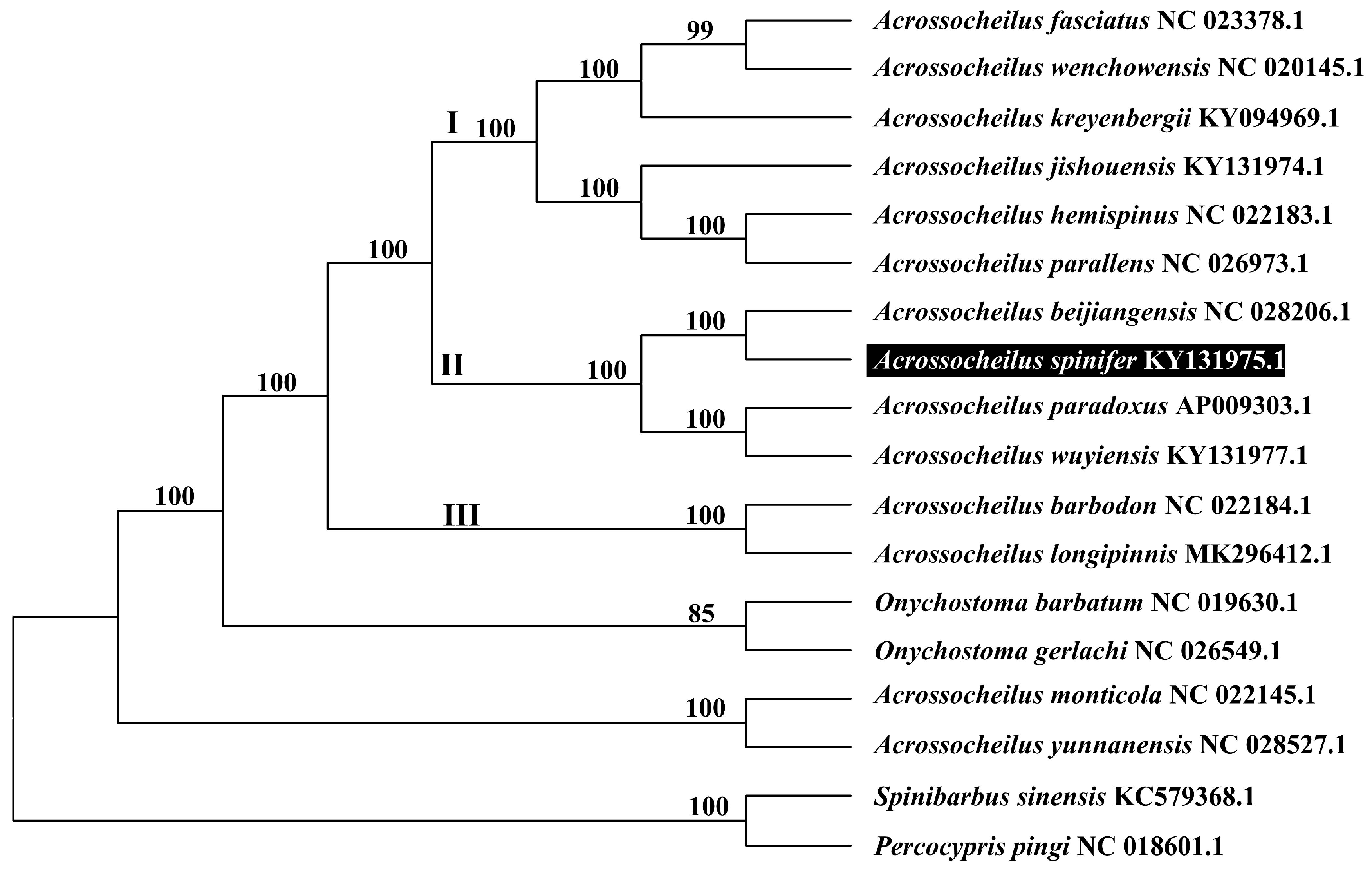
2.4. Phylogenetic Analysis

To validate the phylogenetic position of A. spinifer, we used MEGA11 software [23] to construct a Maximum likelihood tree (with 1000 bootstrap replicates) containing complete mitogenomes of 13 species derived from genus Acrossocheilus. Onychostoma barbatum, O. gerlachi, Spinibarbus sinensis, and Percocypris pingi were used as outgroup for tree rooting. The selected sequences used in this study were downloaded from the NCBI database. The phylogenetic analysis used a series of concatenated nucleotide sequences from 13 PCG datasets. These datasets were arranged in the specific order of ND1, ND2, CO1, CO2, ATP8, ATP6, CO3, ND3, ND4L, ND4, ND5, ND6, and Cyt b. The process of multiple sequence alignment was executed in MEGA11, using the ClustalW algorithm. Prior to constructing the phylogenetic tree, we conducted model testing using ModelTest to determine the most appropriate substitution model for our dataset. The GTR model was selected based on the Akaike Information Criterion (AIC) as the best-fit model for our data. Subsequently, a phylogenetic tree was generated utilizing the Maximum likelihood (ML) method. ML analysis was performed using default parameters in the GTR model with 1000 bootstrap replications.

3. Results

3.1. Genome Organization and Base Composition

The complete mitochondrial genome of A. spinifer was determined to be 16,591 bp in length (GenBank accession number KY131975). As typically found in vertebrates, the mitogenome contained a standard set of 37 genes, including 13 protein-coding genes (PCGs), 22 transfer RNA (tRNA) genes, two ribosomal RNA (rRNA) genes, and a non-coding control region (D-loop) (Figure 1). The gene arrangement followed the canonical pattern observed in most teleost fishes, with no gene rearrangements detected when compared to other cyprinid species.
Among the 22 tRNA genes, sizes ranged from 67 bp in tRNACys to 76 bp in tRNALeu and tRNALys. The two rRNA genes consisted of a 12S rRNA gene (959 bp) and a 16S rRNA gene (1683 bp), which were located between tRNAPhe and tRNALeu (UUR), separated by tRNAVal. The control region (D-loop), responsible for regulating mitochondrial replication and transcription, spanned 932 bp and was situated between tRNAPro and tRNAPhe.
The majority of genes (12 PCGs, 14 tRNAs, and both rRNAs) were encoded on the heavy strand (H-strand), while only ND6 and eight tRNA genes (tRNAGln, tRNAAla, tRNAAsn, tRNACys, tRNATyr, tRNASer (UCN), tRNAGlu, and tRNAPro) were located on the light strand (L-strand). This asymmetric distribution of genes between the two strands is a characteristic feature of vertebrate mitochondrial genomes and has functional implications for replication and transcription mechanisms.
The nucleotide composition analysis revealed a strong bias toward adenine and thymine, with the overall base composition being A: 31.2%, C: 28.0%, G: 16.1%, and T: 24.7%. The resulting A + T content (55.9%) was comparable to that observed in other cyprinid fishes. This nucleotide bias was evident across all genomic regions but was particularly pronounced in the control region, which typically exhibits the highest A + T content in vertebrate mitogenomes. The skewed nucleotide composition (with high A + T and low G content) likely influences codon usage patterns in the protein-coding genes and the secondary structures of RNA genes, reflecting the evolutionary constraints on mitochondrial genome evolution in this species.

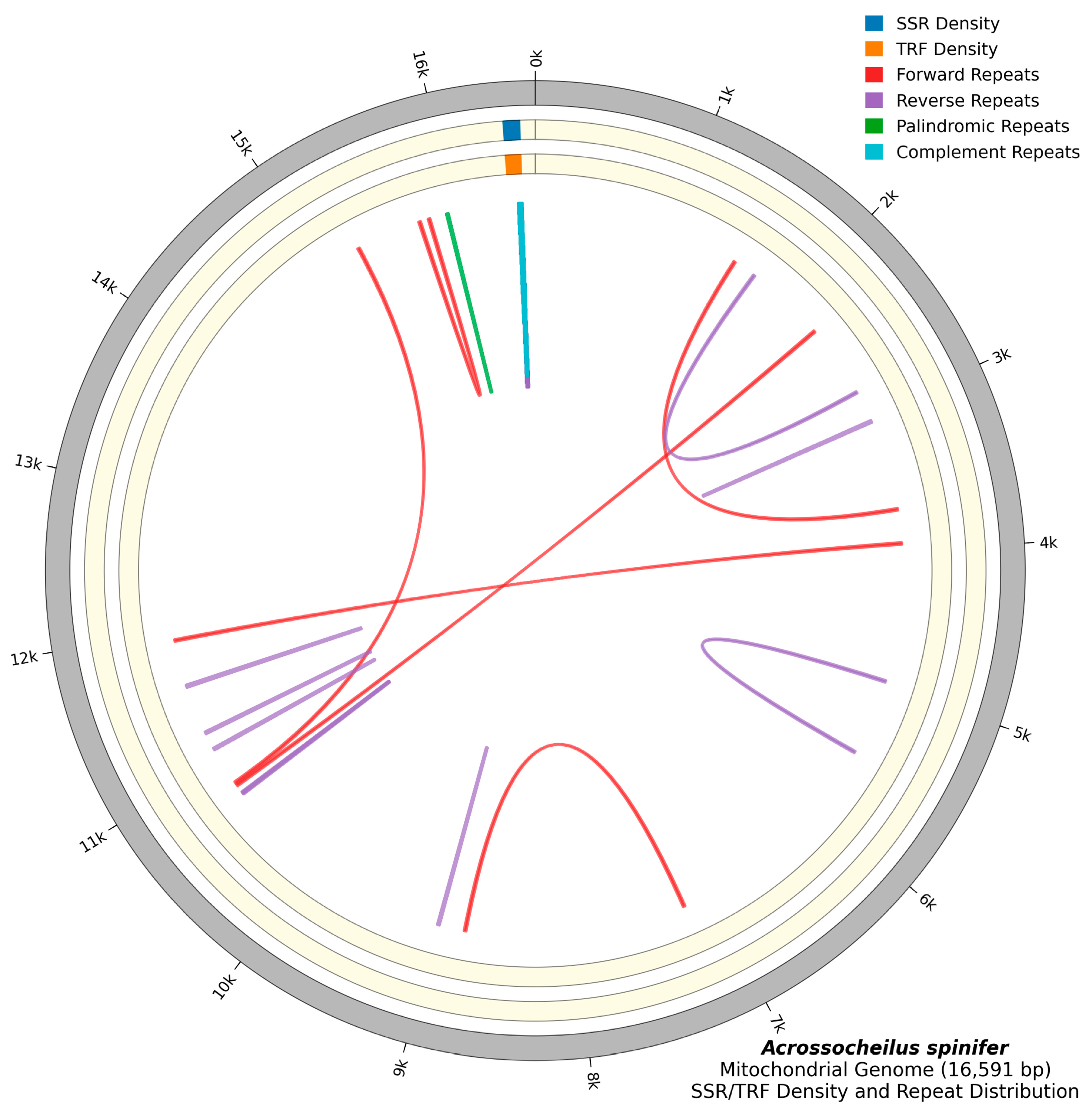
3.2. Repeat Sequence Analysis

The mitochondrial genome of A. spinifer was systematically analyzed for the presence and distribution of different types of repetitive elements, including microsatellites (simple sequence repeats or SSRs), tandem repeats, and dispersed repeats, all of which contribute to genomic complexity and may influence mitochondrial function and evolution [25,26,27].
Microsatellites, also known as simple sequence repeats (SSRs), are short, tandemly repeated DNA motifs of 1–6 base pairs that are widely distributed throughout eukaryotic genomes. In the mitochondrial genome of A. spinifer, we identified one SSR located in the control region (Figure 2). This microsatellite consisted of a poly-T stretch, which is a common feature in the control regions of cyprinid fishes and may play a role in the regulation of mitochondrial replication and transcription. The presence of only one SSR in the mitogenome indicates a relatively conservative pattern compared to nuclear genomes, which typically contain thousands of microsatellites.
Tandem repeats, also referred to as satellite DNA, consist of longer repeated sequences arranged in a head-to-tail fashion. Our analysis detected one tandem repeat in the A. spinifer mitochondrial genome, specifically within the control region (Figure 2). This tandem repeat comprises a 23-bp motif repeated 2.9 times, resulting in a total length of 67 bp. Such tandem repeats are frequently observed in the control regions of fish mitogenomes and are thought to be involved in the formation of secondary structures that regulate mitochondrial DNA replication.
A comprehensive examination of dispersed repeats in the A. spinifer mitochondrial genome revealed a total of 50 repetitive sequences with lengths greater than or equal to 30 bp (Figure 2). These dispersed repeats could be categorized into four types based on their orientations: 11 pairs of forward (direct) repetitive sequences, 20 pairs of reverse repetitive sequences, 13 pairs of palindromic repetitive sequences, and 6 pairs of complement repetitive sequences. The forward repeats were primarily distributed within protein-coding genes, while reverse, palindromic, and complement repeats were scattered throughout the mitogenome, including both coding and non-coding regions.
The abundance and diversity of these dispersed repeats suggest potential roles in mitochondrial genome stability, replication, and recombination. Furthermore, the distribution pattern of these repeats, particularly their concentration in certain regions like the control region and ND2 gene, implies functional constraints or selective pressures on repeat formation and maintenance. Comparative analysis with other cyprinid species indicates that while the overall repeat content is similar, the specific patterns and locations of repeats in A. spinifer exhibit unique characteristics that may reflect its evolutionary history and phylogenetic position within the genus Acrossocheilus.

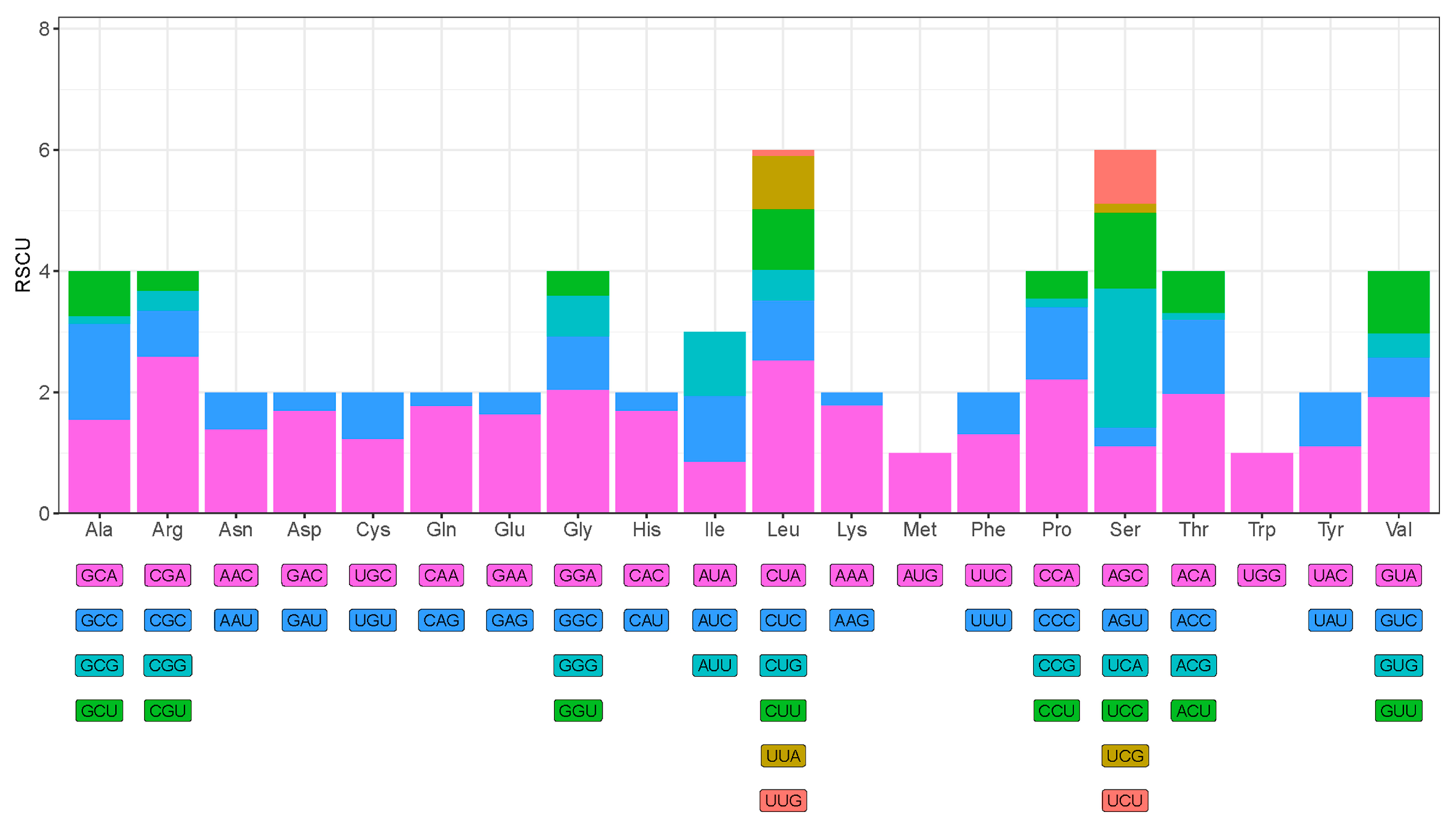
3.3. Protein-Coding Genes and Codon Usage

All 13 protein-coding genes in the A. spinifer mitogenome were identified. Only ND6 was encoded on the L-strand, while the remaining 12 PCGs were encoded on the H-strand. The total length of PCGs was 11,429 bp, accounting for 68.9% of the entire mitogenome.
All PCGs initiated with the typical start codon ATG, except for CO1 and ND3, which began with GTG, and ND2, which began with ATC. Eight of the PCGs (ATP6, CO1, CO3, Cyt b, ND1, ND4L, ND5, and ND6) used TAA as the stop codon, three PCGs (ATP8, ND2, and ND3) used TAG, and CO2 and ND4 terminated with AGA and AGG, respectively (Table 1). The use of alternative start and stop codons is common in fish mitogenomes and is thought to be related to post-transcriptional modifications [26].
Analysis of codon usage revealed that Leu, Ala, and Thr were the most frequently used amino acids, while Cys, His, and Arg were the least used. The relative synonymous codon usage (RSCU) analysis showed a preference for A or C at the third codon position (Figure 3, Table 2). This pattern of codon usage is consistent with those of other cyprinid fishes and reflects the nucleotide bias in the mitogenome [28].

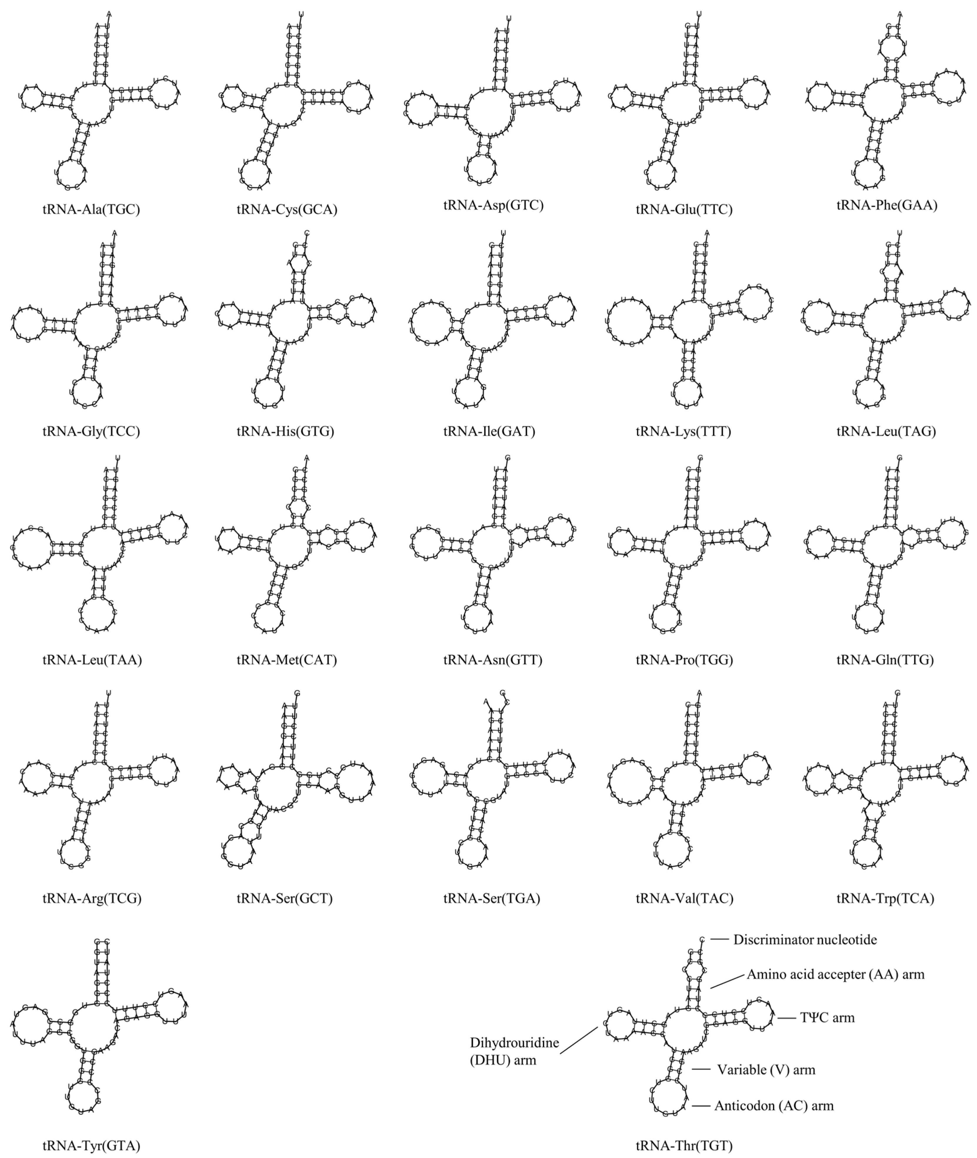
3.4. Transfer RNA and Ribosomal RNA Genes

The A. spinifer mitogenome contained 22 tRNA genes, ranging from 67 bp (tRNACys) to 76 bp (tRNALeu and tRNALys) in length. Most tRNA genes were encoded on the H-strand, with the exception of tRNACys, tRNAAla, tRNAGlu, tRNAPro, tRNAGln, tRNASer, tRNATyr, and tRNAAsn, which were encoded on the L-strand.
All 22 tRNA genes could be folded into the typical cloverleaf secondary structure, except for tRNASer (GCU), which lacked the dihydrouridine (DHU) arm (Figure 4). The predicted secondary structures showed features commonly observed in vertebrate mitochondrial tRNAs. The acceptor arms of all tRNAs were 7 bp in length and the anticodon arms were 5 bp long with an anticodon loop of 7 nucleotides. The D-loops ranged from 4 to 8 nucleotides, while the variable loops contained 4–5 nucleotides. The TΨC arms were typically 5 bp in length with TΨC loops of 3–7 nucleotides. Several non-canonical base pairs were observed in the stems of various tRNAs, including G-U wobble pairs and other mismatches. The presence of the mismatched base pairs seen in tRNA sequences may be corrected by the RNA-editing process, which has been extensively studied in vertebrate mitogenomes [29]. Overall, the secondary structure of the tRNA in A. spinifer exhibited the normal Watson–Crick pairing seen in vertebrate mitogenomes [16].
The two rRNA genes, 12S rRNA and 16S rRNA, were 959 bp and 1683 bp in length, respectively. They were located between tRNAPhe and tRNALeu, separated by tRNAVal, which is the typical arrangement in vertebrate mitogenomes.

3.5. Phylogenetic Tree

To determine the phylogenetic position of A. spinifer, we constructed a Maximum Likelihood tree based on the complete mitogenomes of 14 species from the genus Acrossocheilus, with Onychostoma barbatum, O. gerlachi, Spinibarbus sinensis, and Percocypris pingi as outgroups (Figure 5). The phylogenetic analysis revealed that 12 of 14 Acrossocheilus species constituted a monophyletic assemblage supported by 100% bootstrap values, and the 12 species fell into three major clades (I, II, and III). In clade II, A. spinifer clustered together with A. beijiangenisis and another two species of Acrossocheilus with high bootstrap support (100%).

4. Discussion

In this study, we sequenced and characterized the complete mitogenome of A. spinifer, providing valuable genetic information for this endemic species. The mitogenome of A. spinifer was composed of 37 genes and one control region with a length of 16,591 bp (Figure 1; GenBank No. KY131975.1) and showed typical features of cyprinid fishes, including gene content, order, and nucleotide composition. The gene structure and arrangement of these species were identical to those of other vertebrate mitogenomes, and none of them exhibited genetic rearrangements.
The genomic organization and base composition of the A. spinifer mitogenome aligned with the general patterns observed in other cyprinid fishes. The A + T bias (55.9%) is a common characteristic of fish mitogenomes and influences various aspects of mitochondrial function, including replication, transcription, and translation efficiency. The protein-coding genes exhibited the expected start and stop codon usage patterns, with some variation that likely reflects evolutionary adaptations specific to the Acrossocheilus lineage.
Our repeat sequence analysis revealed interesting patterns of repetitive elements in the A. spinifer mitogenome. The identification of one SSR and one tandem repeat in the control region is consistent with the role of this region in regulating mitochondrial replication and transcription. The presence of 50 dispersed repeats of various types (forward, reverse, palindromic, and complement) distributed throughout the mitogenome suggests potential functional roles in genome stability and organization. The concentration of certain repeat types in specific genomic regions, such as the forward repeats in protein-coding genes, may indicate selective constraints on repeat distribution. These patterns could potentially serve as additional markers for comparative analyses among closely related species within Cyprinidae.
The codon usage analysis revealed a preference for A or C at the third codon position, consistent with the overall nucleotide bias in the mitogenome. This biased codon usage is a common feature of mitochondrial genomes and likely reflects selection for translational efficiency within the mitochondrial environment. The most frequently used amino acids (Leu, Ala, and Thr) and least used (Cys, His, and Arg) in A. spinifer are similar to patterns observed in other fish species, suggesting evolutionary conservation of these preferences.
The tRNA genes in A. spinifer displayed the typical cloverleaf secondary structures with the exception of tRNASer (GCU), which lacked the DHU arm—a feature commonly observed in metazoan mitochondrial tRNASer genes. The presence of non-canonical base pairs in the stems of various tRNAs is also a common feature of mitochondrial tRNAs and may be corrected through post-transcriptional RNA editing mechanisms. These structural features of tRNAs are important for their function in mitochondrial protein synthesis and provide insights into the evolutionary constraints on mitochondrial tRNA structure.
In previous studies of phylogenetic analysis based on mitochondrial gene data, the genus Acrossocheilus has been proven to be polyphyletic and species in genus Acrossocheilus and its closely related genus Onychostoma formed a paraphyletic topology in the phylogenetic trees [18,30,31,32,33,34]. The genus Acrossocheilus sensu stricto should be restricted to the barred species with 5–7 vertical black bars on the flanks including A. fasciatus, A. wenchowensis, A. kreyenbergii, A. jishouensis, A. hemispinus, A. parallens, A. paradoxus, A. beijiangensis, A. wuyiensis, and A. spinifer [1,11]. The barred species A. monticola and A. clivosius may belong to their own genus for the characters of vertical black bars and lower lip lobes. The four species, A. longipinnis, A. iridescens, A. microstomus, and A. lamus, sharing the same body color pattern likely belong in the same genus Masticbarbus Tang, 1942 [13,34].
Phylogenetic analysis results based on complete mitogenomes were basically consistent with the findings of Yuan et al. (2015) [13]. This result shows that the close relationship between A. spinifer and A. beijiangensis was strongly confirmed (100% bootstrap value) and 12 of 14 Acrossocheilus species constituted a monophyletic assemblage supported by 100% bootstrap values, and the 12 species fell into three major clades (I, II, and III). In this monophyletic assemblage, Clade I and II included 6 lineages and 4 lineages supported by 100% bootstrap values, respectively, and Clade III composed of A. barbodon and A. longipinnis (100% bootstrap value) located at the basal position. The other two species, A. yunnanensis and A. monticola (100% bootstrap value) made the genus Acrossocheilus polyphyletic and may well be classified into new genera.
In terms of morphology, the three phylogenetic clades (I, II, and III) can be distinguished from one another and A. spinifer and A. beijiangensis are also the most closely related species. The two species in clade III differed from the ten species in Clade I and Clade II in having 5 (vs. 5–7) thin vertical black bars on their bodies exclusively during the juvenile stage (vs. lifetime), while the vertical black bars disappear and the body color between the bars darkens (vs. no identical changes occur) in adults. The six species in Clade I distinguished from those species in Clade II in having 6 (vs. 5–7) vertical black bars on the flank, a longitudinal stripe along the lateral line (vs. absent), the second vertical bar positioned anterior to (vs. posterior to) the origin of the dorsal fin, and the body color pattern shows significant sexual dimorphism in the sub-adult and adult stage (vs. absent). The two species, A. spinifer and A. beijiangensis, differed from other two species in clade II in having a stout (vs. slender) last simple dorsal-fin ray with a strongly serrated (vs. smooth or weakly serrated) posterior edge and having outer two-thirds (vs. entirety) of the dorsal fin membrane black. A. spinifer differs from A. beijiangensis in having the vertical bars 1–2 (vs. 2–5) scales wide.
Our phylogenetic analysis clarifies the taxonomic status of A. spinifer within the genus Acrossocheilus and provides additional support for the previous morphological classification. The high bootstrap support (100%) for the clustering of A. spinifer with A. beijiangensis confirms their close evolutionary relationship, which is consistent with their morphological similarities. This phylogenetic framework, based on complete mitogenomes, provides a robust foundation for further taxonomic revisions within the genus Acrossocheilus.
In summary, we suggest that A. yunnanensis, A. monticola, and A. clivosius may well be classified into new genera and the genus Acrossocheilus should be represented by the species of the clade I, II, and III with Gymnostomus formosanus Regan, 1908 (=A. paradoxus) as the type species of the genus. The three monophyletic clades (I, II, and III) represent three groups with different morphological characters, respectively: (1) species of this group have six vertical black bares on the flanks, with the second vertical bar positioned anterior to the origin of the dorsal fin; have a longitudinal stripe along the lateral line; and usually exhibiting remarkable sexual color dimorphism. (2) species of this group have 5–7 vertical black bars, with the second (sometimes the third) vertical bar positioned posterior to the origin of the dorsal fin; have no longitudinal stripe along the lateral line and have no obvious sexual color dimorphism; (3) species of this group only have 5 thin vertical black bars on their bodies during the juvenile stage, with the second vertical bar positioned posterior to the origin of the dorsal fin; the color of the vertical black bars becomes lighter and disappear during ontogeny, while the body color between the bars darkens; species of this group have no longitudinal stripe along the lateral line, and have no obvious sexual color dimorphism.
Our findings provide a foundation for further investigations into the taxonomy, population genetics, and conservation management of A. spinifer and related species. The complete mitogenome data will contribute to the database of cyprinid mitogenomes and facilitate future comparative genomic studies in this diverse fish family.

5. Conclusions

In conclusion, the current study presented the first complement mitogenome assembly and annotation of A. spinifer. We described the characterization of the mitochondrial genome of A. spinifer using genetic and phylogenetic approaches and discussed the classification of fishes in the genus Acrossocheilus. The complete mitogenome of A. spinifer exhibits the typical gene content, order, and structural features found in other cyprinid fishes, but with unique characteristics in its repetitive elements and nucleotide composition. The phylogenetic analysis based on complete mitogenomes strongly supports the close relationship between A. spinifer and A. beijiangensis and provides a robust framework for the taxonomic classification of species within the genus Acrossocheilus. These results can help to advance understanding and collect fundamental genetic data for the Cyprinidae family.

Author Contributions

Conceptualization, E.Z. and L.-Y.Y.; methodology, all authors; software, J.G., S.-Q.S. and X.-X.Z.; investigation, J.G., G.-F.L. and L.-Y.Y.; writing—original draft preparation, L.-Y.Y., E.Z. and S.-Q.S.; writing—review and editing, L.-Y.Y.; visualization, S.-Q.S. and L.-Y.Y.; supervision, L.-Y.Y. All authors have read and agreed to the published version of the manuscript.

Funding

This study was supported by grants from the National Natural Sciences Foundation of China (No. 31401974).

Institutional Review Board Statement

The sample used for this study was the dead body of a fish, we did not require approval from the Ethics Committee.

Informed Consent Statement

Not applicable.

Data Availability Statement

The mitogenome sequence data that support the findings of this study are openly available in the GenBank of NCBI at https://www.ncbi.nlm.nih.gov/ (accessed on 6 May 2025) under accession number KY131975.

Acknowledgments

Our sincere thanks are given to Zhuo-Cheng Zhou and Xiao-Lu Yu for helping with field work. We also thank Chung-Der Hsiao from Chung Yuan Christian University in Taiwan for his help on Bioinformatic analysis of NGS data.

Conflicts of Interest

The authors declare no conflict of interest.

References

  1. Nelson, J.S.; Grande, T.C.; Wilson, M.V.H. Fishes of the World, 5th ed.; John Wiley & Sons: Hoboken, NJ, USA, 2016. [Google Scholar]
  2. Yue, P.Q. Fauna Sinica, Osteichthyes: Cypriniformes III; Science Press: Beijing, China, 2000. [Google Scholar]
  3. Kottelat, M. Fishes of the Nam Theun and Xe Bangfai basins, Laos, with diagnoses of twenty-two new species (Teleostei: Cyprinidae, Baltoridae, Cobitidae, Coiidae and Odontobutidae). Ichthyol. Explor. Freshw. 1998, 9, 1–128. [Google Scholar]
  4. Kottelat, M. Diagnoses of a new genus and 64 new species of fishes from Laos (Teleostei: Cyprinidae, Balitoridae, Bagridae, Syngnathidae, Chauhuriidae and Tetraodontidae). J. South Asian Nat. Hist. 2000, 5, 37–82. [Google Scholar]
  5. Kottelat, M. Fishes of Laos; Wildlife Heritage Trust Publications: Colombo, Sri Lanka, 2001; pp. 1–198. [Google Scholar]
  6. Kottelat, M. Freshwater Fishes of Northern Vietnam. A Preliminary Check-List of the Fishes Known or Expected to Occur in Northern Vietnam with Comments on Systematics and Nomenclature; The World Bank: Washington, DC, USA, 2001; pp. 1–122. [Google Scholar]
  7. Kottelat, M. The fishes of the inland waters of Southeast Asia: A catalogue and core bibliography of the fishes known to occur in freshwaters, mangroves and estuaries. Raffles Bull. Zool. 2013, 27, 1–663. [Google Scholar]
  8. Froese, R.; Pauly, D. FishBase. World Wide Web Electronic Publication. 2025. Available online: https://www.fishbase.org (accessed on 6 May 2025).
  9. Xiao, G.B.; Lu, W.J.; Liao, L.; Mao, Q.; Wang, L.J.; Zhang, H.R.; Zhou, L.; Bao, J. A new Species of Acrossocheilus (Teleostei: Cyprinidae): A. furongjiangensis sp. nov. from Wujiang River Basin in Northern Guizhou. Sichuan J. Zool. 2024, 43, 205–214. [Google Scholar] [CrossRef]
  10. Chen, T.E.; Xu, J.X.; Li, P.J.; Hu, H.F.; Gao, K.; Zhao, H.P. A New Species of the Genus Acrossocheilus Oshima, 1919 (Cypriniformes: Cyprinidae) from the Dabie Mountains. Animals 2025, 15, 734. [Google Scholar] [CrossRef]
  11. Yuan, L.Y. Monophyly, Affinity and Taxonomic Revision of the Cyprinid Genus Acrossocheilus Oshima, 1919. Ph.D. Thesis, Graduate School of the Chinese Academy of Sciences, Beijing, China, 2009. [Google Scholar]
  12. Yuan, L.Y.; Wu, Z.Q.; Zhang, E. Acrossocheilus spinifer, a new species of barred cyprinid fish from south China (Pisces: Teleostei). J. Fish Biol. 2006, 8, 163–173. [Google Scholar] [CrossRef]
  13. Yuan, L.Y.; Liu, X.X.; Zhang, E. Mitochondrial phylogeny of Chinese barred species of the cyprinid genus Acrossocheilus Oshima, 1919 (Teleostei: Cypriniformes) and its taxonomic implications. Zootaxa 2015, 4059, 151–168. [Google Scholar] [CrossRef]
  14. Miya, M.; Friedman, M.; Satoh, T.P.; Takeshima, H.; Sado, T.; Iwasaki, W.; Yamanoue, Y.; Nakatani, M.; Mabuchi, K.; Inoue, J.G.; et al. Evolutionary origin of the Scombridae (tunas and mackerels): Members of a paleogene adaptive radiation with 14 other pelagic fish families. PLoS ONE 2013, 8, e73535. [Google Scholar] [CrossRef]
  15. Near, T.J.; Eytan, R.I.; Dornburg, A.; Kuhn, K.L.; Moore, J.A.; Davis, M.P.; Wainwright, P.C.; Friedman, M.; Smith, W.L. Resolution of ray-finned fish phylogeny and timing of diversification. Proc. Natl. Acad. Sci. USA 2012, 109, 13698–13703. [Google Scholar] [CrossRef]
  16. Boore, J.L. Animal mitochondrial genomes. Nucleic Acids Res. 1999, 27, 1767–1780. [Google Scholar] [CrossRef]
  17. Saccone, C.; Pesole, G.; Sbisa, E. The main regulatory region of mammalian mitochondrial DNA: Structure-function model and evolutionary pattern. J. Mol. Evol. 1991, 33, 83–91. [Google Scholar] [CrossRef] [PubMed]
  18. Hou, X.J.; Lin, H.D.; Tang, W.Q.; Liu, D.; Han, C.C.; Yang, J.Q. Complete mitochondrial genome of the freshwater fish Acrossocheilus longipinnis (Teleostei: Cyprinidae): Genome characterization and phylogenetic analysis. Biologia 2020, 75, 1871–1880. [Google Scholar] [CrossRef]
  19. Shen, K.N.; Yen, T.C.; Chen, C.H.; Li, H.Y.; Chen, P.L.; Hsiao, C.D. Next generation sequencing yields the complete mitochondrial genome of the flathead mullet, Mugil cephalus cryptic species NWP2 (Teleostei: Mugilidae). Mitochondrial DNA Part A 2016, 27, 1758–1759. [Google Scholar] [CrossRef] [PubMed]
  20. Bernt, M.; Donath, A.; Juhling, F.; Externbrink, F.; Florentz, C.; Fritzsch, G.; Putz, J.; Middendorf, M.; Stadler, P.F. MITOS: Improved de novo metazoan mitochondrial genome annotation. Mol. Phylogenet. Evol. 2013, 69, 313–319. [Google Scholar] [CrossRef]
  21. Wyman, S.K.; Jansen, R.K.; Boore, J.L. Automatic annotation of organellar genomes with DOGMA. Bioinformatics 2004, 20, 3252–3255. [Google Scholar] [CrossRef]
  22. Laslett, D.; Canback, B. ARWEN: A program to detect tRNA genes in metazoan mitochondrial nucleotide sequences. Bioinformatics 2008, 24, 172–175. [Google Scholar] [CrossRef]
  23. Tamura, K.; Stecher, G.; Kumar, S. MEGA11: Molecular Evolutionary Genetics Analysis Version 11. Mol. Biol. Evol. 2021, 38, 3022–3027. [Google Scholar] [CrossRef]
  24. Greiner, S.; Lehwark, P.; Bock, R. OrganellarGenomeDRAW (OGDRAW) version 1.3.1: Expanded toolkit for the graphical visualization of organellar genomes. Nucleic Acids Res. 2019, 47, W59–W64. [Google Scholar] [CrossRef]
  25. Benson, G. Tandem Repeats Finder: A program to analyze DNA sequences. Nucleic Acids Res. 1999, 27, 573–580. [Google Scholar] [CrossRef]
  26. Beier, S.; Thiel, T.; Munch, T.; Scholz, U.; Mascher, M. MISA-web: A web server for microsatellite prediction. Bioinformatics 2017, 33, 2583–2585. [Google Scholar] [CrossRef]
  27. Kurtz, S.; Choudhuri, J.V.; Ohlebusch, E.; Schleiermacher, C.; Stoye, J.; Giegerich, R. REPuter: The manifold applications of repeat analysis on a genomic scale. Nucleic Acids Res. 2001, 29, 4633–4642. [Google Scholar] [CrossRef] [PubMed]
  28. Ponce, M.; Infante, C.; Jimenez-Cantizano, R.M.; Perez, L.; Manchado, M. Complete mitochondrial genome of the blackspot seabream, Pagellus bogaraveo (Perciformes: Sparidae), with high levels of length heteroplasmy in the WANCY region. Gene 2008, 409, 44–52. [Google Scholar] [CrossRef]
  29. Janke, A.; Paabo, S. Editing of a tRNA anticodon in marsupial mitochondria changes its codon recognition. Nucleic Acids Res. 1993, 21, 1523–1525. [Google Scholar] [CrossRef] [PubMed]
  30. Yang, L.; Mayden, R.L. Phylogenetic relationships, subdivision, and biogeography of the cyprinid tribe Labeonini (sensu) (Teleostei: Cypriniformes), with comments on the implications of lips and associated structures in the labeonin classification. Mol. Phylogenet. Evol. 2010, 54, 254–265. [Google Scholar] [CrossRef]
  31. Wang, X.Z.; Li, J.B.; He, S.P. Molecular evidence for the monophyly of East Asian groups of Cyprinidae (Teleostei: Cypriniformes) derived from the nuclear recombination activating gene 2 sequences. Mol. Phylogenet. Evol. 2007, 42, 157–170. [Google Scholar] [CrossRef] [PubMed]
  32. Li, J.B.; Wang, X.Z.; Kong, X.H.; Zhao, K.; He, S.P.; Mayden, R.L. Variation patterns of the mitochondrial 16S rRNA gene with secondary structure constraints and their application to phylogeny of cyprinine fishes (Teleostei: Cypriniformes). Mol. Phylogenet. Evol. 2008, 47, 472–487. [Google Scholar] [CrossRef]
  33. Yang, L.; Sado, T.; Vincent Hirt, M.; Pasco-Viel, E.; Arunachalam, M.; Li, J.; Wang, X.; Freyhof, J.; Saitoh, K.; Simons, A.M.; et al. Phylogeny and polyploidy: Resolving the classification of cyprinine fishes (Teleostei: Cypriniformes). Mol. Phylogenet. Evol. 2015, 85, 97–116. [Google Scholar] [CrossRef]
  34. Tang, D.S. Fishes of Kweiyang, with descriptions of two new genera and five new species. Lingnan Sci. J. (Canton) 1942, 20, 147–166. [Google Scholar]
Figure 1. Circular map of the complete mitogenome of Acrossocheilus spinifer. Different colors indicate different types of genes and regions. Genes in the outer circle are located on the H-strand, and genes in the inner circle are located on the L-strand.
Fishes 10 00296 g001
Figure 2. Repeat analysis of the mitochondrial genome in Acrossocheilus spinifer. The colored lines on the innermost circle connect the two repetitive sequences of the scattered repeats, with the red lines representing forward match, the purple lines representing reverse match, the green line representing palindromic match and the cyan line representing complementary match. The orange line on the second circle represents the tandem repeat sequence, and the blue line on the outermost circle represents the microsatellite or simple sequence repeat (SSR) sequence.
Fishes 10 00296 g002
Figure 3. Relative synonymous codon usage (RSCU) of Acrossocheilus spinifer. The ordinate represents the RSCU (the number of times a certain synonymous codon is used/the average number of times that all codons encoding the amino acid are used). The abscissa represents different amino acids.
Fishes 10 00296 g003
Figure 4. Predicted secondary structures for 22 tRNA genes in the mitogenome of Acrossocheilus spinifer.
Fishes 10 00296 g004
Figure 5. Molecular phylogeny of Acrossocheilus spinifer and related species based on complete mitogenome. The complete mitogenomes were downloaded from GenBank and the phylogenic tree was constructed by the Maximum likelihood method with 1000 bootstrap replicates. The gene accession numbers for tree construction are listed behind the species name.
Fishes 10 00296 g005
Table 1. Sequence characteristics of Acrossocheilus spinifer mitogenome.
GroupGroup of GenesGeneThree Letter CodeSequenceSize (bp)StrandNo. of
Amino Acids
Start CodonStop CodonAnti-Codon
StartEnd
NADH dehydrogenase subunitND1-28703829960H319ATGTAA-
ND2-405250891038H345ATCCCT-
ND3-965910,006348H115GTGAAT-
ND4-10,36811,7411374H457ATGACT-
ND4L-10,07810,371294H97ATGTAA-
ND5-11,97013,7751806H601ATGTAA-
ND6-13,78414,302519L172ATGTAA-
PCGsCytochrome c oxidase subunitCO1-549070221533H510GTGTAA-
CO2-71937873681H226ATGCCT-
CO3-88029584783H260ATGATA-
ATP synthase subunitATP6-81198796678H225ATGTAA-
ATP8-79618122162H53ATGTAG-
Cytochrome bCyt b-14,37815,5111134H377ATGCTT-
Transfer RNA genestrnAAla5170523869L---TGC
trnCCys5346541267L---GCA
trnDAsp7107717872H---GTC
RNAstrnEGlu14,30314,37169L---TTC
trnFPhe16969H---GAA
trnGGly9587965872H---TCC
trnHHis11,74911,81769H---GTG
trnIIle3840391172H---GAT
trnKLys7884795976H---TTT
trnLLeu11,88811,96073H---TAG
trnLLeu2784285976H---TAA
trnMMet3983405169H---CAT
trnNAsn5240531273L---GTT
trnPPro15,59015,65970L---TGG
trnQGly3910398071L---TTG
trnRArg10,00810,07770H---TCG
trnSSer11,81811,88669H---GCT
trnSSer7035710571L---TGA
trnTThr15,51915,59072H---TGT
trnVVal1029110072H---TAC
trnWTrp5097516771H---TCA
trnYTyr5413548270L---GTA
12S rRNArrnS-701026957H----
16S rRNArrnL-110127831683H----
D-loopControl regionD-loop-15,66016,591932H----
Table 2. Codon number and RSCU of Acrossocheilus spinifer mitochondrial PCGs.
Amino_AcidCodonCountRSCUAmino_AcidCodonCountRSCU
LysAAA671.787AspGAC622.548
AsnAAC842.083GluGAG190.362
LysAAG80.213AspGAT110.452
AsnAAT370.917AlaGCA1321.935
ThrACA1572.476AlaGCC1351.979
ThrACC971.530AlaGCG110.161
ThrACG90.142AlaGCT630.924
ThrACT540.852GlyGGA1242.562
SerAGC432.039GlyGGC531.095
SerAGT120.569GlyGGG410.847
IleATA1201.718GlyGGT240.496
IleATC1522.177ValGTA1063.855
MetATG402.000ValGTC361.309
IleATT1472.105ValGTG220.800
GlnCAA911.784ValGTT562.036
HisCAC902.547TyrTAC642.226
GlnCAG110.216TyrTAT511.774
HisCAT160.453SerTCA894.220
ProCCA1182.770SerTCC482.276
ProCCC641.502SerTCG60.284
ProCCG70.164SerTCT341.612
ProCCT240.563CysTGC162.462
ArgCGA484.541TrpTGG102.000
ArgCGC141.324CysTGT101.538
ArgCGG60.568LeuTTA931.763
ArgCGT60.568PheTTC1442.618
LeuCTA2675.062LeuTTG100.190
LeuCTC1051.991PheTTT761.382
LeuCTG531.005StopTAA60.308
LeuCTT1051.991StopTAG10.051
GluGAA861.638StopTGA1105.641
Disclaimer/Publisher’s Note: The statements, opinions and data contained in all publications are solely those of the individual author(s) and contributor(s) and not of MDPI and/or the editor(s). MDPI and/or the editor(s) disclaim responsibility for any injury to people or property resulting from any ideas, methods, instructions or products referred to in the content.

