Article

Transcriptomic Responses of Gonadal Development to Photoperiod Regulation in Amur Minnow (Phoxinus lagowskii)

College of Animal Science and Veterinary Medicine, Shenyang Agricultural University, Shenyang 110065, China
*
Author to whom correspondence should be addressed.
Fishes 2025, 10(3), 137; https://doi.org/10.3390/fishes10030137
Submission received: 18 February 2025 / Revised: 14 March 2025 / Accepted: 18 March 2025 / Published: 20 March 2025
(This article belongs to the Special Issue Rhythms and Clocks in Aquatic Animals)

Abstract

:
Photoperiod regulates reproductive physiology in many fishes, but its sex-specific molecular effects under artificial manipulation remain unclear, especially in cold-water species. In this study, we investigated whether photoperiod manipulation during the reproductive season could modulate the rate and efficiency of gonadal development in the Amur minnow (Phoxinus lagowskii). High-throughput RNA sequencing was used to analyze transcriptomic responses of gonadal tissues under three photoperiod regimes: natural light (12L:12D), continuous light (24L:0D), and continuous darkness (0L:24D) over a 9-week experimental period. Our results revealed distinct sex-specific gonadal responses to photoperiodic changes. In males, continuous light significantly promoted spermatogenesis by upregulating meiosis-related genes (REC114 and syp3) and steroid biosynthesis. In females, prolonged light exposure induced ovarian stress, evidenced by vitellogenin (Vtg3) upregulation and retinoic acid suppression, whereas continuous darkness promoted lipid storage via downregulation of gluconeogenesis (PC and Fbp2) and fatty acid oxidation (ACSL1a). Additionally, immune activation, marked by IL1RAPL1-A upregulation, was observed in all groups except continuous-light males, with females exhibiting broader immune pathway engagement. These findings provide novel insights into the regulatory mechanisms of photoperiod-induced gonadal development and highlight potential strategies for optimising photoperiod management in cold-water fish aquaculture.
Key Contribution: The present study constitutes an analysis of the regulatory mechanisms governing gonadal development in male and female P. lagowskii, with a particular focus on prolonged photoperiod induction during winter gonadal development. The findings of this study serve to elucidate the impact of photoperiod on the male–female differences observed in fish, thereby establishing a biological foundation for subsequent research and providing a significant basis for the scientific regulation of P. lagowskii reproductive management.

1. Introduction

Photoperiod is a crucial environmental cue that regulates the hypothalamic–pituitary–gonadal (HPG) axis, controlling gonadal development and reproductive timing in fish [1,2,3]. In natural environments, seasonal changes in photoperiod synchronise gonadal development to optimise reproductive success. Artificial photoperiod manipulation is increasingly used in aquaculture to regulate spawning, growth, and reproductive timing [4]. For example, only females of rabbit fish (Siganus guttatus) after 24 h of continuous light were able to spawn successfully after human chorionic gonadotropin (hCG) injection [5]. Providing light at night significantly improved sperm quality in dominant males of Japanese medaka (Oryzias latipes), altering inter-male competitiveness [6]. Despite its importance, the precise molecular mechanisms underlying photoperiod regulation of gonadal development, especially sex-specific responses, remain inadequately understood.
Recent advances in high-throughput omics technologies, particularly RNA sequencing (RNA-seq), have provided unprecedented insights into gene expression profiles during gonadal development in fish. In combination with photoperiod manipulation, RNA-seq has been used to investigate differential gene expression in gonadal tissues, revealing key genes associated with reproductive processes such as meiosis, steroidogenesis, and immune responses. In the study of half-smooth tongue sole (Cynoglossus semilaevis), the time-dependent expression patterns of several sex-related genes, including Dmrt1, Amh, Foxl2, aromatase-encoding gene, Esr, and the Sox gene family, during gonad differentiation has been found [7]. In addition, the female sapphire devil (Chrysiptera cyanea) IGF3 plays a role in reproductive development and metabolic shifts from growth mode to reproductive mode in peripheral tissues under suitable photoperiod and water temperature conditions [8]. However, most studies have focused on general physiological responses, and sex-specific molecular mechanisms remain underexplored, especially in response to artificial light cycles.
The Amur minnow (Phoxinus lagowskii) is an economically valuable species native to northeastern China, characterised by its rapid sexual maturation, adaptability to cold water, and omnivorous diet [9]. These features make it an ideal model for studying reproductive physiology and photoperiodic regulation in cold-water fish. Notably, the fish attains sexual maturity at an age of two and, during the annual period of May to June, when the water temperature reaches between 20 and 26 °C, it becomes capable of engaging in oestrus and spawning. The spawning process involves the laying of eggs in batches on sand, gravel, or within aquatic grasses, and these eggs subsequently hatch within a period of two to three days [10]. Additionally, the overfishing of wild populations and genetic decline in Amur minnow populations highlight the importance of developing effective breeding and aquaculture strategies [11]. Despite its potential in aquaculture, the molecular mechanisms of gonadal development and the effects of photoperiod on this species remain poorly understood, making it an ideal subject for further investigation.

2. Materials and Methods

2.1. Ethics Statement

All experiments were performed according to the guidelines for scientific purposes, animal care, and use formulated by the Animal Ethics Committee of Shenyang Agricultural University (approval code: 2023090802 and Approval Date: 10 September 2023). The research team underwent comprehensive training to ensure appropriate handling and care of animals.

2.2. Experimental Animals

A total of 360 healthy P. lagowskii (mean weight: 11.42 ± 1.01 g) were obtained from a local fish farm and transported to the Aquatic Animal Physiology Laboratory at Shenyang Agricultural University. The fish underwent a 2-week acclimation period in blue plastic buckets equipped with a water circulation system, maintained at 9.73 ± 0.93 °C. During this period, they were fed commercial feed once daily at 16:00 at a rate of 3% of their total body weight. The feeding practices and environmental conditions were standardised across all groups. To prevent any potential effects on metabolism, the final feeding was performed 24 h before the start of the experiment.
Before the experiment, 1-year-old P. lagowskii, acclimated for 2 weeks, were sexed and separately allocated to 18 circular blue plastic buckets (diameter: 1 m, height: 1 m, water depth: 0.5 m) at 20 fish per bucket density to simulate the actual aquaculture environment. The buckets were exposed to three photoperiods: natural light (12L:12D; control), continuous light (24L:0D; experimental), and continuous darkness (0L:24D; experimental). Light intensity was 608.67 ± 161.59 lx at the surface and 66.69 ± 21.15 lx at the bottom. Incandescent lamps provided lighting with timers that regulated natural light conditions (6:00–18:00). All buckets were fully enclosed in a double-layer blackout cloth to prevent light interference, and each treatment included three replicates. Feeding was conducted daily at 16:00 with lights off, and the buckets were resealed immediately after feeding to minimise light exposure. The duration of the light treatment was 9 weeks.

2.3. Sample Collection

At 6:00 a.m. on day 7, three females and three males were randomly selected from each light treatment group. The fish were anaesthetised using a 100 mg/L MS-222 solution for 3 min. The gonads were dissected using sterilised scissors and forceps, with collection into 1.5 mL centrifuge tubes. Liquid nitrogen flash freezing was used and they were stored at −80 °C prior to analysis. The sampling was conducted on the ice. The continuous darkness group was sampled under dim red light to minimise stress from sudden light exposure. Samples were coded as N1 (natural light males), N2 (natural light females), L1 (continuous light males), L2 (continuous light females), D1 (continuous darkness males), and D2 (continuous darkness females).

2.4. RNA Isolation and RNA-Seq Library Preparation

Gonads were frozen with liquid nitrogen and pulverised in a grinding bowl, and total RNA was isolated using the Trizol Reagent (Invitrogen Life Technologies, Carlsbad, CA, USA). Three replicates were performed for each treatment group. The integrity of the extracted RNA was monitored using 1% agarose gels. The purity and concentration of RNA samples were assessed using a NanoDrop spectrophotometer (Thermo Scientific, Shanghai, China). Transcriptome sequencing was performed by next-generation sequencing (NGS) technology using the Illumina Truseq 2000 sequencing platform from Shanghai Personal Biotechnology Co., Ltd. Shanghai Biotechnology Corporation (Shanghai, China) performed the transcriptome sequencing of the target samples.

2.5. De Novo Assembly and Gene Annotation

The original FASTQ data were filtered to remove reads with connectors, lengths less than 50 bp, and average sequence quality less than Q20. The high-quality sequences were then spliced from scratch to obtain transcripts. The transcripts were clustered, and the longest transcripts were selected as unigenes. The gene function of each unigene was then annotated. The databases utilised for gene function annotation included the Non-Redundant Protein Sequence Database (NR), the Gene Ontology (GO), the Kyoto Encyclopedia of Genes and Genomes (KEGG), the evolutionary genealogy of genes: Non-supervised Orthologous Groups (eggNOG), Swiss-Prot, and Pfam.

2.6. Expression Analysis

Expression analysis was conducted utilising the transcription group expression quantitative software, RSEM version 1.3.3, and a comparison was made between the clean reads of each sample and the reference sequence, with the transcriptional sequence serving as the reference. Principal components analysis (PCA) was employed with the R language software package (version 4.0.3) to analyse the main components of each sample according to the expression quantity.

2.7. Differential Gene Expression and Enrichment Analysis

In this study, we employed the DESeq software version 2.12 to integrate RNA-seq data from three biological replicates per group and analyse gene expression disparities. We then proceeded to screen the condition of differentially expressed genes (DEGs) with multiple expression differences (|log2FoldChange| > 1, significant p-value < 0.05). Statistical significance was calculated using one-way analysis of variance (ANOVA), followed by Bonferroni’s post hoc test to determine significant peak–nadir differences in the means of the expression levels. The R language Pheatmap software package version 1.0.12 was used to cluster the union of different genes and samples for all groups being compared. The distance between these samples was calculated using the Euclidean method, and the hierarchical clustering method was used for the clustering process. Differentially expressed genes (DEGs) were then subjected to Kyoto Encyclopedia of Genes and Genomes (KEGG) and Gene Ontology (GO) analyses. The enrichment results were manually checked using public databases and literature searches based on Non-Redundant Protein Database (NR) annotation. All KEGG and GO enrichment results were obtained from the combined data of three biological replicates.

2.8. qRT-PCR Validation

Six genes were selected for validation using qRT-PCR. 3β-HSD, 17β-HSD, CYP19A1a, and CYP17A1a are key enzymes in steroid hormone synthesis, belonging to the oxidoreductase, short-chain dehydrogenase/reductase (SDR), and cytochrome P450 families, respectively. STS and EBP regulate sulphated steroid metabolism and cholesterol transport, directly influencing the function of steroidogenic acute regulatory (StAR) protein. For each photoperiod treatment group, three male and three female individuals were euthanised, and gonadal tissues were immediately dissected for RNA extraction. Gene-specific primers (Table 1) were designed using Primer Premier 6.0. β-actin served as the internal control. PCR was performed using Hiscript® III RT SuperMix for qPCR (+gDNA wiper) (Nanjing Vazyme Biotech Co., Ltd., Nanjing, China) in a 20 µL reaction system, following standard cycling conditions. Relative expression levels were calculated using the 2−∆∆Ct method, which was employed to determine relative changes in gene expression levels.

2.9. Statistical Analysis

All data were analysed by one-way analysis of variance (ANOVA) using SPSS 22.0. The significance level for all analyses was set at p-value < 0.05 to indicate statistically significant and significant differences between groups. The data are expressed as mean ± standard error.

3. Results

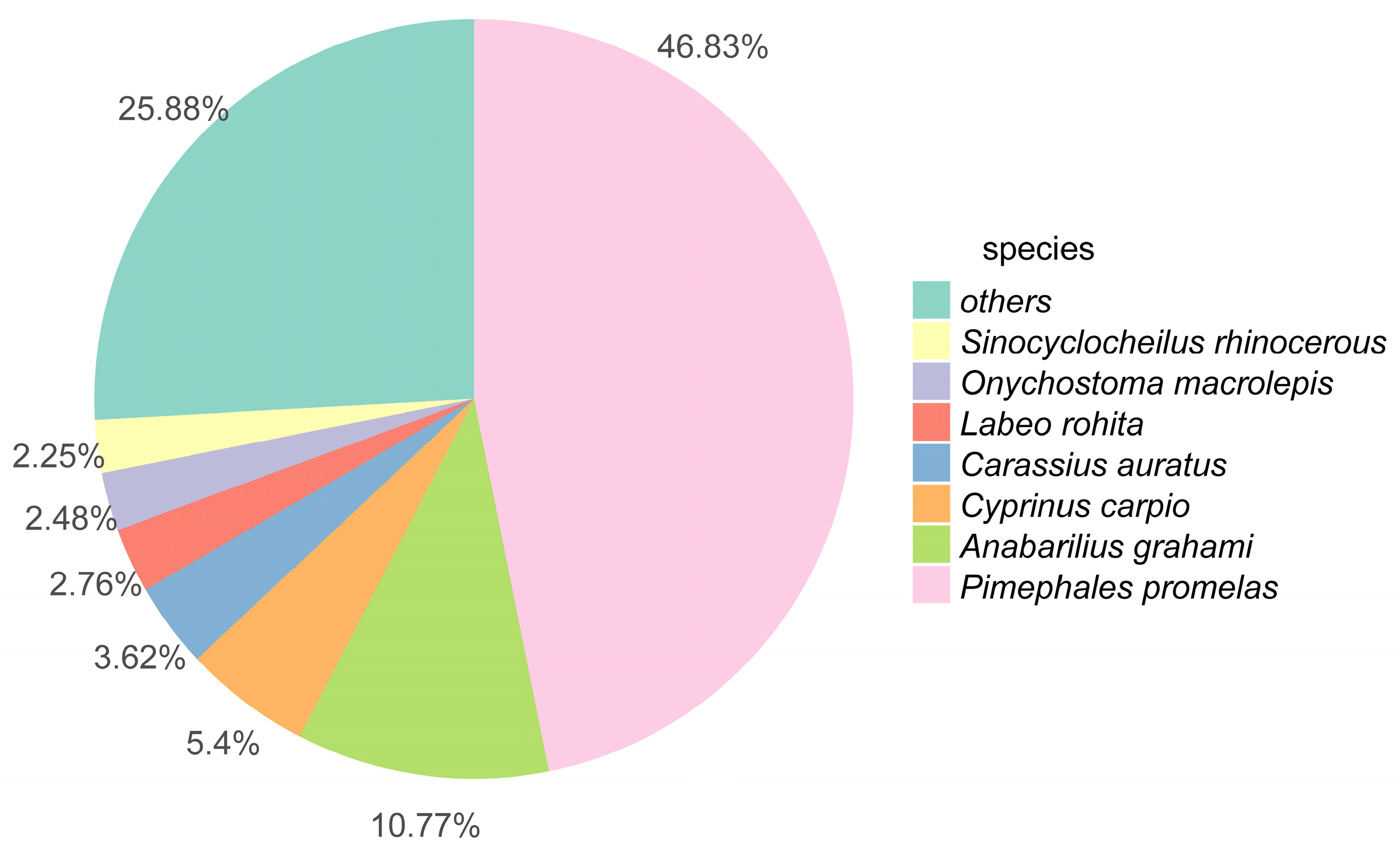
3.1. Transcriptome Sequence Assessment and Annotation

A total of 836,424,322 raw sequencing reads were obtained from the gonad of P. lagowskii (Table S1). After raw reads were filtered and cleaned, 822,156,896 (98.29%) high-quality reads were used for assembly (Table S2). As shown in Table 2, those sequence numbers resulted in 486,908 and 191,205 transcripts and unigenes, respectively. Annotations against major databases revealed 749,523, 27,263, 19,363, 27,275, and 43,584 unigenes mapped to NR, GO, KEGG, SwissProt, and eggNOG, respectively (Table S3). The top five species that contributed the most significant number of gene annotations from BLASTx were Pimephales promelas (46.83%), Anabarilius grahami (10.77%), Cyprinus carpio (5.4%), Carassius auratu (3.62%), and Labeo rohita (2.76%) (Figure 1).

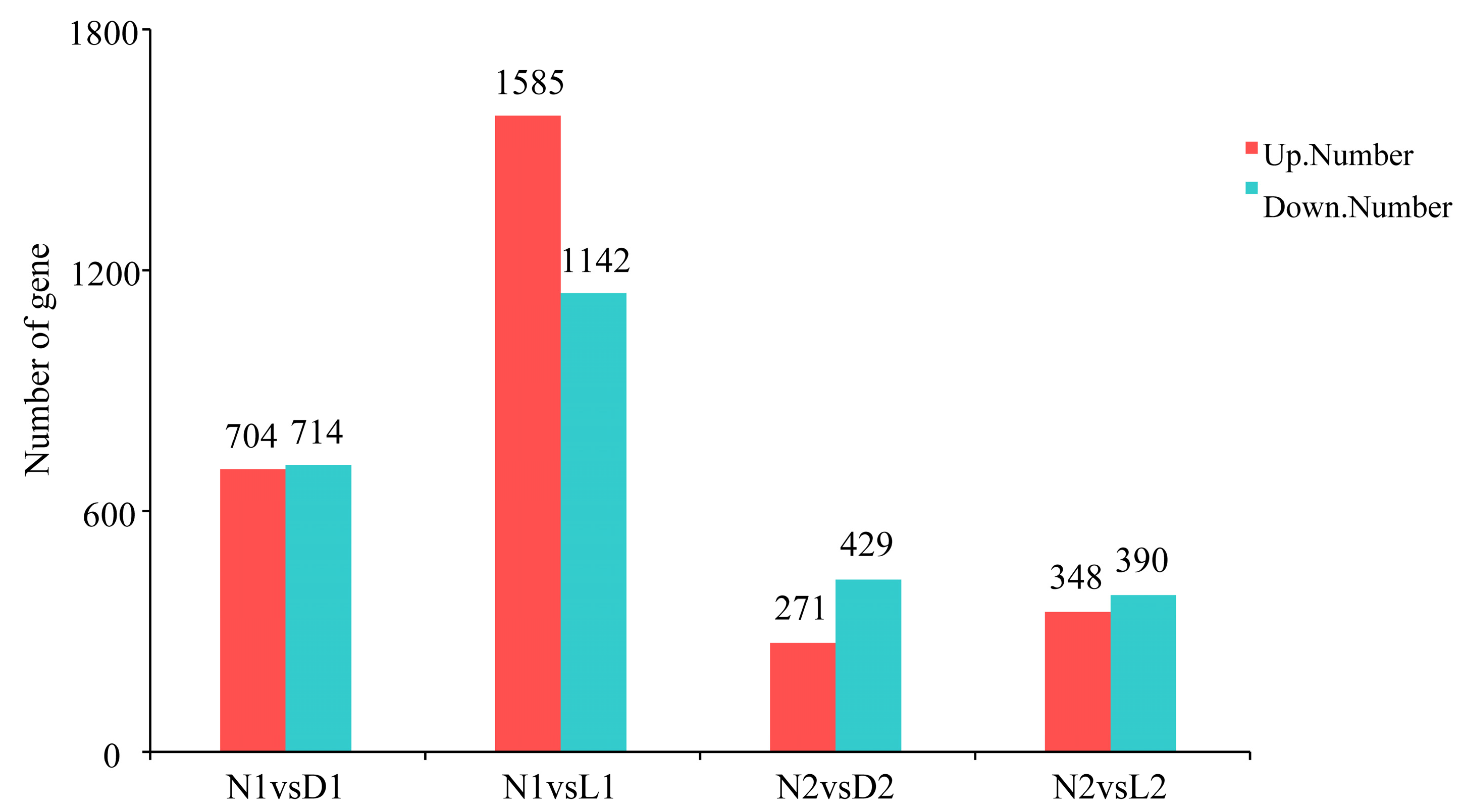
3.2. Differential Expression Analysis

DESeq analysis of gonads at different photoperiods differentially expressed genes (DEGs). The results showed that there were more DEGs in the male group than in the female group. Among the comparisons, N1vsL1 had the most DEGs (2727), with 1585 upregulated and 1142 downregulated. The number of upregulated genes and downregulated genes in N1vsD1 were close to each other, 704 and 714, respectively, whereas the number of DEGs in N2vsL2 and N2vsD2 was lower, 738 and 700, respectively (Figure 2).
DEGs related to reproduction, metabolism, and immunity were selectively screened. Reproductive-related DEGs include meiotic recombination protein REC114 (REC114), synaptonemal complex protein 3 (syp3), meiotic nuclear division protein 1 homolog (mnd1), outer dense fibre protein 2 (odf2), etc. Immune-related DEGs included interleukin-1 receptor accessory protein-like 1-A (IL1RAPL1-A), etc. (Table 3). Reproduction-related DEGs were most abundant in the continuous light group of males, while a large number of immune-related genes were found in females.

3.3. GO Functional Classification of Differentially Expressed Genes

GO enrichment analysis revealed sex-specific responses to photoperiod (Figure S1). In males, the continuous light group (24L:0D) exhibited significant enrichment in cell cycle processes (e.g., mitotic and meiotic cycles), indicating enhanced spermatogenesis. Conversely, the continuous darkness group (0L:24D) showed membrane-related enrichment, suggesting a shift in gonadal metabolic processes. In females, continuous light primarily affected bile acid metabolism and asparagine metabolism, whereas continuous darkness led to enrichment in MHC protein and interleukin-related immune pathways, implying a delayed reproductive state (Figure 3). Meanwhile, the continuous darkness group DEGs were mainly enriched in MHC protein (MHC protein complex, MHC class I receptor activity, and MHC class I protein complex) with interleukin (interleukin-1 production, interleukin-1 beta production, and interleukin-17 production) (Figure 4).

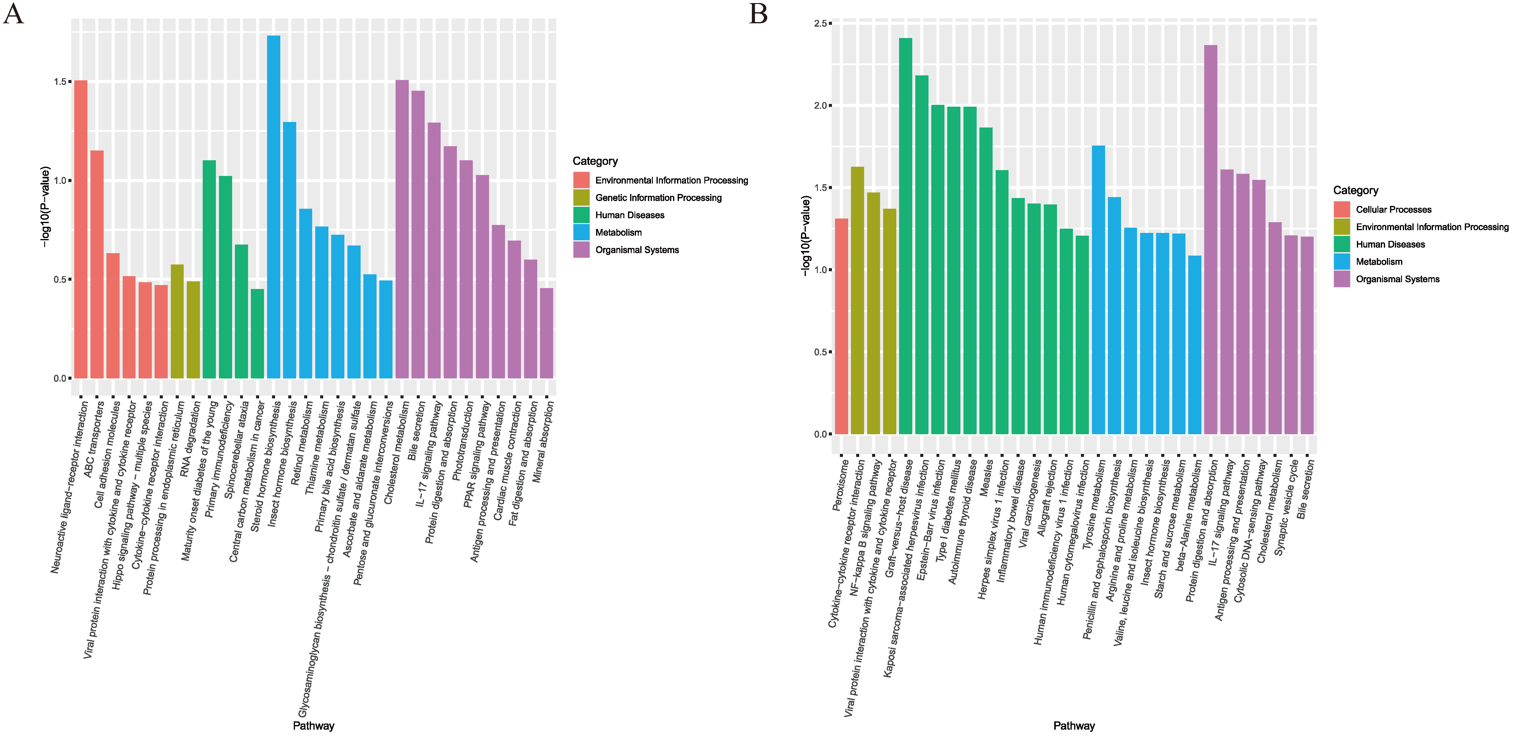
3.4. Significant Enrichment Analysis of KEGG Pathway

To elucidate the effect of light exposure on these pathways, we conducted a KEGG pathway analysis of the DEGs (Figure S2). In male fish, DEGs were significantly enriched in cellular processes in the continuous light group, especially in the cell cycle and p53 signalling pathway, and most genes were significantly upregulated in reproduction-associated oocyte meiosis, progesterone-mediated oocyte maturation, and steroid biosynthesis. Steroid biosynthesis was significantly upregulated. Steroid biosynthesis was only found in the male group and was also significantly upregulated in the darkness group (Figure 5).
In the continuous light group of females, steroid hormone biosynthesis was the main pathway in which metabolism was most abundant, and mineral absorption was found only in the continuous light group. In the continuous darkness group, DEGs acted mainly through graft-versus-host disease and protein digestion and absorption, which was only found in the female group, and were significantly downregulated in both light and dark groups (Figure 6).

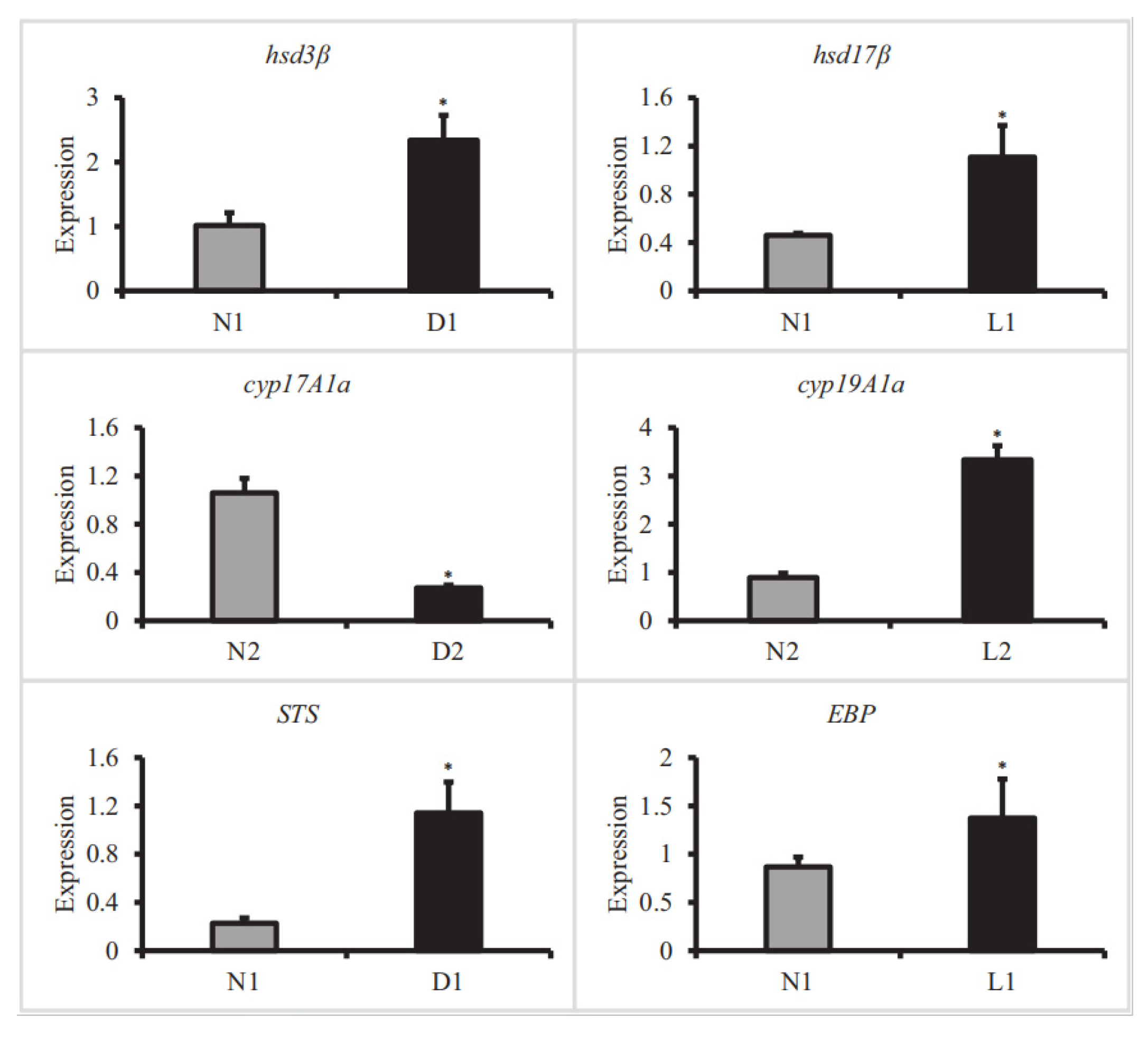
3.5. qRT-PCR Verification

To validate the RNA-Seq data, six DEGs with significant differences in expression—3beta-hydroxysteroid-dehydrogenase/decarboxylase (hsd3β), 17-beta-hydroxysteroid dehydrogenase type (hsd17β), gonad-type aromatase (cyp19A1a), steroid 17-alpha-hydroxylase/17,20 lyase (cyp17A1a), steryl-sulfatase (STS), and 3-beta-hydroxysteroid-Delta (8), Delta (7)-isomeras (EBP)—were selected for qRT-PCR analysis. The expression levels of these DEGs were significantly correlated with the RNA-seq results (Figure 7).

4. Discussion

Photoperiod plays a critical role in the reproductive cycle of aquatic species, influencing gonadal development via the HPG axis [12]. Our study demonstrates that photoperiod manipulation during the reproductive season can differentially regulate gonadal maturation in P. lagowskii, with continuous light accelerating spermatogenesis and continuous darkness delaying ovarian development. These findings suggest that optimising photoperiod regimes could serve as a practical strategy in cold-water fish aquaculture.

4.1. Male Responses to Photoperiod

Oocyte meiosis, progesterone-mediated oocyte maturation, and steroid biosynthesis were found to be enriched and mostly upregulated in the three gonadal-development-related genes in the continuous light group of males (N1 vs. L1). Notably, the validated genes 3β-HSD, 17β-HSD, and EBP are core components of steroid biosynthesis, suggesting that continuous light may promote steroid hormone secretion in male fish. Concurrently, it was ascertained that genes associated with meiosis exhibited substantial upregulation, including meiotic recombination protein REC114 (REC114), synaptonemal complex protein 3 (syp3), and meiotic nuclear division protein 1 homolog (mnd1). REC114 regulates the formation and positioning of DNA double-strand breaks during meiosis and promotes homologous recombination and chromosome segregation [13]. Syp3 promotes association complex assembly and chromosome association and is required for fertility in males [14]. Recent research in zebrafish has revealed the function of outer dense fibre protein 2 (odf2), a microtubule-associated protein. This protein has been found to form fibrous structures connected to the microtubule network, which is the maintenance cytoskeleton in sperm tails and centrosomes. This network is important for the structure and function of the sperm tail [15,16,17]. This finding suggests that prolonged exposure to light enhances the process of meiosis in the male spermatheca and that spermatozoa function is thereby enhanced, providing further evidence of enhanced gonadal function downstream.
Steroidogenic acute regulatory protein (StaR) was found to be significantly downregulated with cytochrome P450 26B1 (cyp26B1) in continuous darkness group males (N1 vs. D1). The protein known as steroidogenic acute regulatory protein (StAR) has been identified as a rate-limiting regulatory protein during the process of steroid hormone synthesis. The primary function of this protein is to promote the transport of cholesterol from the outer mitochondrial membrane to the inner mitochondrial membrane. This process marks the initiation point of steroid hormone synthesis [18]. A decline in the expression of StAR in the gonads may be indicative of a reduction in the synthesis of sex hormones, including testosterone. In the male Nile tilapia (Oreochromis niloticus), it was determined that StAR was closely related to syp3 and, when StAR2 was knocked down, it prevented meiotic initiation and downregulated expression of meiosis-related genes retinoic acid vasa, syp3, and deleted in azoospermia-like (dazl) expression was down-regulated and affected spermatogonia development and spermatogenesis [19]. Retinoic acid (RA) has been demonstrated to play a pivotal role in the processes of germ cell proliferation and sex determination [20]. In the male spermary, RA signalling assumes a more pivotal role during the differentiation of spermatogonia (spg) and the initial stages of meiosis in spermatocytes (spc) [21]. All-trans retinoic acid (at-RA) is the active metabolite of vitamin A. Cyp26B1 is responsible for the conversion of at-RA into inactive metabolites (e.g., 4-hydroxy retinoic acid), thus regulating intracellular RA levels and ensuring that retinoic acid levels do not become excessive [22]. A study of mouse testes revealed that the aberrant development of germ cells was attributable to excess retinoic acid (RA) rather than to a deficiency in RA metabolites produced by cyp26B1 [23]. It has been established that constant darkness leads to disruption of retinoic acid metabolism, which, in turn, disrupts the development of germ cells. Concurrently, the synthesis of steroid hormones is inhibited, which further inhibits sperm production.

4.2. Female Responses to Photoperiod

In the ovary, the RA signalling pathway plays a pivotal role in folliculogenesis and steroidogenesis. This pathway exerts a significant influence on oocyte maturation and follicular degeneration [24,25]. Retinol binding protein 7 (rbp7) is subject to the regulatory influence of peroxisome proliferator-activated receptor gamma (PPARγ), the principal regulator of adipogenesis. The interaction between these two factors governs the metabolic processes associated with retinol in adipocytes [26]. Retinoid isomerohydrolase (RPE65) converts all-trans retinyl ester to 11-cis retinol in the visual cycle [27]. Vtg3 was significantly upregulated in continuous light-exposed females (N2 and L2), while rbp7 and RPE65 were significantly downregulated. Vitellogenin (Vtg) is a precursor of vitellogenin, which is synthesised by the liver and transported through the bloodstream to the ovaries, where it provides nutrients to the oocytes and is extremely oestrogen-sensitive [28,29,30]. This finding indicates that fish may demonstrate a preference for utilising resources for yolk protein synthesis under conditions of continuous light, suggesting a potential benefit of lower levels of RA in maintaining ovarian health. Concurrently, it was determined that both light and environmental stress induce the activation of Vtg3 in gonadal tissue [3132]. It is hypothesised that the upregulation of Vtg may be attributable to the necessity for greater nutrient reserves in the ovary in order to respond to injury or to repair cells. Concurrently, genes associated with calcium homeostasis and cellular repair were found to be significantly upregulated by activation, including Progressive Ankylosis Protein Homolog B (ANKH), Parathyroid Hormone-like Hormone a (PTHa), and Stanniocalcin-1 (STC1) [33,34,35,36]. This finding lends further support to our hypothesis that continuous light exposure may have stimulated ovarian development in females but concomitantly induced stress and ovarian damage, resulting in reproductive abnormalities.
We found many differential genes related to metabolism in the continuous darkness group females (N2 vs. D2). Phosphatidate phosphatase LPIN1(Lipin-1) functions as a phosphatidate phosphatase (PAP) enzyme in the glycerol 3-phosphate pathway for triglyceride storage and as a transcriptional coactivator/corepressor for metabolic nuclear receptors, for which modulation of phosphatidic acid levels is required for early steps in adipogenesis [37]. Meanwhile, long-chain-fatty-acid–CoA ligase 1a isoform X2 (ACSL1a) was significantly downregulated, which plays a crucial role in directing fatty acids toward beta-oxidation in adipocytes and regulating fatty acid oxidation [38]. Furthermore, the level of expression of pyruvate carboxylase, mitochondrial (PC) and fructose-1,6-bisphosphatase isozyme 2 isoform X1 (Fbp2) was found to be significantly reduced, with these enzymes being associated with gluconeogenesis [39,40]. It is hypothesised that the absence of light signalling results in females in dark environments preferentially storing lipids as the main source of energy in low light and low temperature conditions during winter, rather than allocating energy to reproductive development. This may be considered an adaptive resource allocation strategy.

4.3. Aquaculture Applications of Photoperiod Regulation

Diametrically different results were observed after photoperiod treatment of P. lagowskii males and females, suggesting a biphasic effect of photoperiod on gonadal development in P. lagowskii. Males tend to be “opportunistic”, responding quickly to environmental signals (such as light) to maximise sperm production. For example, higher temperatures significantly increase the body size and sperm swimming speed of male mosquitofish (Gambusia holbrooki) [41]. Females, on the other hand, favour a “conservative” strategy, ensuring offspring quality through energy storage and ovarian repair. It is evident that such findings are of significant importance with regard to the management of P. lagowskii culture. For instance, the photoperiodic stimulation of males with extended periods of light (long photoperiods) can facilitate rapid sperm acquisition and augmented sperm production, which is conducive to both parenting and artificial insemination. Dynamic photoperiods (e.g., alternating “long-short”) can be employed to regulate yolk synthesis and maintain ovarian health, while short photoperiods can be utilised to delay the reproductive window. In the present study, interleukin-1 receptor accessory protein-like 1-A (IL1RAPL1-A) was significantly upregulated in all groups except males in the continuous light group. The IL1RAPL1-A is a member of the interleukin-1 receptor family and functions as an important cofactor of the interleukin-1 signalling pathway. Similar to the classical interleukin-1 (IL-1) receptor accessory protein (IL-1RAP), IL1RAPL1-A may promote proinflammatory cytokine production by modulating inflammation-related signalling pathways (e.g., NF-κB and MAPK pathways), thus contributing to immune regulation [42,43]. In the future, further optimisation of the photoperiodic scheme is required to reduce the stimulation of light to fish and weaken the stress response. Concurrently, it has the potential to substitute, at least in part, for exogenous hormones (e.g., hCG and LHRH) in the reproduction of P. lagowskii. This approach would reduce the cost and ecological risk of culture, whilst also enabling more precise control of the energy distribution of females (e.g., fat storage and vitellogenesis) through the regulation of photoperiods. The result would be improved reproductive efficiency and fish health and an increase in the benefits of culture.

5. Conclusions

In this study, we performed comparative transcriptome analyses of the gonads of male and female P. lagowskii under different photoperiods, providing comprehensive insights into the molecular adaptations of P. lagowskii to different photoperiods and revealing the complex pathways and genes involved in these processes. We found that continuous photoperiod promotes gonadal development only in males and impairs ovarian development in females. In contrast, continuous darkness resulted in unfavourable gonadal development in both females and males. All immune-related genes were upregulated in all groups except males in the continuous light group, suggesting that the photoperiod treatment was stressful for P. lagowskii.

Supplementary Materials

The following supporting information can be downloaded at: https://www.mdpi.com/article/10.3390/fishes10030137/s1, Figure S1: The GO annotation of assembled genes; Figure S2: The KEGG annotation of assembled genes; Table S1: The summary information of transcriptomic sequences; Table S2: Evaluation of RNA-Seq data of P. lagowskii; Table S3: The annotation statistics results.

Author Contributions

Conceptualisation, M.Z. and Y.L.; methodology, M.Z. and Y.L.; software, M.Z.; validation, M.Z.; formal analysis, M.Z.; investigation, M.Z.; resources, Y.L.; data curation, M.Z.; writing—original draft preparation, M.Z.; writing—review and editing, Y.L.; visualisation, Y.L.; supervision, Y.L.; project administration, Y.L.; funding acquisition, Y.L. All authors have read and agreed to the published version of the manuscript.

Funding

This research received no external funding.

Institutional Review Board Statement

This study was approved by the Animal Ethics Committee of Shenyang Agriculture University permit number 2023090802, approval date: 10 September 2023.

Informed Consent Statement

Not applicable.

Data Availability Statement

The raw reads have been deposited in the NCBI database (BioSample number SAMN39511903).

Acknowledgments

We thank Dongnan Star (Shenyang) Ecological Technology Co., Ltd. for providing the fishes.

Conflicts of Interest

The authors declare no conflicts of interest.

References

  1. Adolfi, M.C.; Depincé, A.; Wen, M.; Pan, Q.; Herpin, A. Development of Ovaries and Sex Change in Fish: Bringing Potential into Action. Sex. Dev. 2023, 17, 84–98. [Google Scholar] [CrossRef] [PubMed]
  2. Wang, N.; Teletchea, F.; Kestemont, P.; Milla, S.; Fontaine, P. Photothermal control of the reproductive cycle in temperate fishes. Rev. Aquac. 2010, 2, 209–222. [Google Scholar]
  3. Imamura, S.; Hur, S.P.; Takeuchi, Y.; Badruzzaman, M.; Mahardini, A.; Rizky, D.; Takemura, A. Effect of short- and long-term melatonin treatments on the reproductive activity of the tropical damselfish Chrysiptera cyanea. Fish Physiol. Biochem. 2022, 48, 253–262. [Google Scholar] [CrossRef] [PubMed]
  4. Okuzawa, K.; Furukawa, K.; Aida, K.; Hanyu, I. Effects of photoperiod and temperature on gonadal maturation, and plasma steroid and gonadotropin levels in a cyprinid fish, the honmoroko Gnathopogon caerulescens. Gen. Comp. Endocrinol. 1989, 75, 139–147. [Google Scholar] [CrossRef]
  5. Hung, P.Q.; Dung, H.T.N.; Arukwe, A. Photoperiod manipulation in the induced breeding of the rabbit fish (Siganus guttatus). Tạp Chí Khoa Học Công Nghệ Thủy Sản Trường Đại Học Nha Trang 2018, 4, 69–77. [Google Scholar]
  6. Closs, L.E.; Royan, M.R.; Sayyari, A.; Mayer, I.; Weltzien, F.-A.; Baker, D.M.; Fontaine, R.H. Impact of light pollution at night on male reproductive success in Japanese medaka (Oryzias latipes). bioRxiv 2023. [Google Scholar] [CrossRef]
  7. Lin, G.; Gao, D.; Lu, J.; Sun, X. Transcriptome Profiling Reveals the Sexual Dimorphism of Gene Expression Patterns during Gonad Differentiation in the Half-Smooth Tongue Sole (Cynoglossus semilaevis). Mar. Biotechnol. 2021, 23, 18–30. [Google Scholar] [CrossRef]
  8. Rizky, D.; Mahardini, A.; Byun, J.; Takemura, A. Molecular cloning of insulin-like growth factor 3 (igf3) and its expression in the tissues of a female damselfish, Chrysiptera cyanea, in relation to seasonal and food-manipulated reproduction. Gen. Comp. Endocrinol. 2020, 295, 113479. [Google Scholar] [CrossRef]
  9. Zhang, Y.Q.; Yin, J.S.; Du, J.; Geng, L.W.; Bai, Q.L.; Xu, W. Analysis of some nutritional components and evaluation of muscle protein of Phoxinus lagowskii Dybowskii. Acta Nutr. Sin. 2013, 35, 406–407+410. [Google Scholar] [CrossRef]
  10. Wang, M.L.; Li, B.M.; Jiang, Y.S.; Meng, Z.Y.; Zhang, Y.Z.; Su, X.; Jiang, Z.Q. Study on Individual Fecundity of Phoxinus lagowskii in Taizi River of Benxi City. J. Yangtze Univ. 2013, 10, 28–32+6–7. [Google Scholar] [CrossRef]
  11. Luo, X.N.; Li, J.; Jin, G.H.; Liu, Y.X.; Li, J.W.; Yang, P.M.; Jiang, X.H. Artificial Propagation of Fat Minnow Phoxinus lagowskii Dybowski. Fish Sci. 2013, 32, 672–675. [Google Scholar]
  12. Rad, F.; Bozaoğlu, S.; Gözükara, S.E.; Karahan, A.; Kurt, G. Effects of different long-day photoperiods on somatic growth and gonadal development in Nile tilapia (Oreochromis niloticus L.). Aquaculture 2006, 255, 292–300. [Google Scholar] [CrossRef]
  13. Kumar, R.; Oliver, C.; Brun, C.; Juarez-Martinez, A.B.; Tarabay, Y.; Kadlec, J.; de Massy, B. Mouse REC114 is essential for meiotic DNA double-strand break formation and forms a complex with MEI4. Life Sci. Alliance 2018, 1, e201800259. [Google Scholar] [CrossRef] [PubMed]
  14. Yuan, L.; Liu, J.G.; Zhao, J.; Brundell, E.; Daneholt, B.; Höög, C. The murine SCP3 gene is required for synaptonemal complex assembly, chromosome synapsis, and male fertility. Mol. Cell 2000, 5, 73–83. [Google Scholar] [CrossRef]
  15. Saram, P.D.; Wilkinson, C.; Murdoch, J. Role of outer dense fiber of sperm tails 2-like (ODF2L) protein in ciliation in mammalian cells and in zebrafish. Cilia 2015, 4 (Suppl. S1), P32. [Google Scholar] [CrossRef]
  16. Donkor, F.F.; Mönnich, M.; Czirr, E.; Hollemann, T.; Hoyer-Fender, S. Outer dense fibre protein 2 (ODF2) is a self-interacting centrosomal protein with affinity for microtubules. J. Cell Sci. 2004, 117, 4643–4651. [Google Scholar] [CrossRef]
  17. Luo, B.; Lü, Z.L. Outer dense fiber 2 and sperm function: Progress in studies. Zhonghua Nan Ke Xue 2017, 23, 473–476. [Google Scholar]
  18. Arukwe, A. Steroidogenic acute regulatory (StAR) protein and cholesterol side-chain cleavage (P450scc)-regulated steroidogenesis as an organ-specific molecular and cellular target for endocrine disrupting chemicals in fish. Cell Biol. Toxicol. 2008, 24, 527–540. [Google Scholar] [CrossRef]
  19. Li, L.; Wu, Y.; Zhao, C.; Miao, Y.; Cai, J.; Song, L.; Wei, J.; Chakraborty, T.; Wu, L.; Wang, D.; et al. The role of StAR2 gene in testicular differentiation and spermatogenesis in Nile tilapia (Oreochromis niloticus). J. Steroid Biochem. Mol. Biol. 2021, 214, 105974. [Google Scholar] [CrossRef]
  20. Cui, Z.; Wang, J.; Yang, Y.; Chen, Z.; Wang, Q.; Wang, J.; Zhang, T.; Xu, W.; Chen, S. Identification and Expression Pattern of cyp26b1 Gene in Gonad of the Chinese Tongue Sole (Cynoglossus semilaevis). Animals 2022, 12, 2652. [Google Scholar] [CrossRef]
  21. Zhang, H.Z.; Hao, S.L.; Yang, W.X. How does retinoic acid (RA) signaling pathway regulate spermatogenesis? Histol. Histopathol. 2022, 37, 1053–1064. [Google Scholar] [CrossRef] [PubMed]
  22. Snyder, J.M.; Zhong, G.; Hogarth, C.; Huang, W.; Topping, T.; LaFrance, J.; Palau, L.; Czuba, L.C.; Griswold, M.; Ghiaur, G.; et al. Knockout of Cyp26a1 and Cyp26b1 during postnatal life causes reduced lifespan, dermatitis, splenomegaly, and systemic inflammation in mice. Faseb J. 2020, 34, 15788–15804. [Google Scholar] [CrossRef]
  23. MacLean, G.; Li, H.; Metzger, D.; Chambon, P.; Petkovich, M. Apoptotic extinction of germ cells in testes of Cyp26b1 knockout mice. Endocrinology 2007, 148, 4560–4567. [Google Scholar] [CrossRef]
  24. Damdimopoulou, P.; Chiang, C.; Flaws, J.A. Retinoic acid signaling in ovarian folliculogenesis and steroidogenesis. Reprod. Toxicol. 2019, 87, 32–41. [Google Scholar] [CrossRef]
  25. Lubzens, E.; Young, G.; Bobe, J.; Cerdà, J. Oogenesis in teleosts: How eggs are formed. Gen. Comp. Endocrinol. 2010, 165, 367–389. [Google Scholar] [CrossRef]
  26. Kim, D.H.; Ahn, J.; Suh, Y.; Ziouzenkova, O.; Lee, J.W.; Lee, K. Retinol Binding Protein 7 Promotes Adipogenesis in vitro and Regulates Expression of Genes Involved in Retinol Metabolism. Front. Cell Dev. Biol. 2022, 10, 876031. [Google Scholar] [CrossRef]
  27. Moiseyev, G.; Chen, Y.; Takahashi, Y.; Wu, B.X.; Ma, J.X. RPE65 is the isomerohydrolase in the retinoid visual cycle. Proc. Natl. Acad. Sci. USA 2005, 102, 12413–12418. [Google Scholar] [CrossRef]
  28. Liu, T.; Liu, S.; Ma, L.; Li, F.; Zheng, Z.; Chai, R.; Hou, Y.; Xie, Y.; Li, G. Oogenesis, vitellogenin-mediated ovarian degeneration and immune response in the annual fish Nothobranchius guentheri. Fish Shellfish. Immunol. 2017, 66, 86–92. [Google Scholar]
  29. Wang, H.; Tan, J.T.; Emelyanov, A.; Korzh, V.; Gong, Z. Hepatic and extrahepatic expression of vitellogenin genes in the zebrafish, Danio rerio. Gene 2005, 356, 91–100. [Google Scholar]
  30. Xiong, Z.; Yang, J.; Zhang, S.; Wang, Y.; Xu, S.; Guo, C.; Wang, D. Expression and localization of vitellogenin genes (VTG) and receptor (VGR) in the gonad development of silver pomfret Pampus argenteus. J. Oceanol. Limnol. 2023, 41, 1575–1592. [Google Scholar]
  31. Park, K.; Jo, H.; Kim, D.K.; Kwak, I.S. Environmental Pollutants Impair Transcriptional Regulation of the Vitellogenin Gene in the Burrowing Mud Crab (Macrophthalmus japonicus). Appl. Sci. 2019, 9, 1401. [Google Scholar] [CrossRef]
  32. Choi, M.M. Effects of 17 beta-estradiol injection and red-spectrum light on eyestalk hormones and vitellogenesis of the ornamental cleaner shrimp Lysmata amboinensis (De Man, 1888) (Decapoda: Caridea: Lysmatidae). J. Crustac. Biol. 2020, 40, 115–121. [Google Scholar]
  33. Wathuliyadde, N.; Willmore, K.E.; Kelly, G.M. Bone Mineralization Regulation: Using Zebrafish as a Model to Study ANKH-associated Mineralization Disorders. bioRxiv 2024. [Google Scholar] [CrossRef]
  34. Guerreiro, P.M.; Renfro, J.L.; Power, D.M.; Canario, A.V. The parathyroid hormone family of peptides: Structure, tissue distribution, regulation, and potential functional roles in calcium and phosphate balance in fish. Am. J. Physiol. Regul. Integr. Comp. Physiol. 2007, 292, R679–R696. [Google Scholar] [CrossRef]
  35. Li, S.; Li, H.; Wang, Z.; Duan, C. Stanniocalcin 1a regulates organismal calcium balance and survival by suppressing Trpv6 expression and inhibiting IGF signaling in zebrafish. Front. Endocrinol. 2023, 14, 1276348. [Google Scholar] [CrossRef]
  36. Luo, C.W.; Pisarska, M.D.; Hsueh, A.J. Identification of a stanniocalcin paralog, stanniocalcin-2, in fish and the paracrine actions of stanniocalcin-2 in the mammalian ovary. Endocrinology 2005, 146, 469–476. [Google Scholar]
  37. Zhang, P.; Takeuchi, K.; Csaki, L.S.; Reue, K. Lipin-1 phosphatidic phosphatase activity modulates phosphatidate levels to promote peroxisome proliferator-activated receptor γ (PPARγ) gene expression during adipogenesis. J. Biol. Chem. 2012, 287, 3485–3494. [Google Scholar] [CrossRef]
  38. Ellis, J.M.; Li, L.O.; Wu, P.C.; Koves, T.R.; Ilkayeva, O.; Stevens, R.D.; Watkins, S.M.; Muoio, D.M.; Coleman, R.A. Adipose acyl-CoA synthetase-1 directs fatty acids toward beta-oxidation and is required for cold thermogenesis. Cell Metab. 2010, 12, 53–64. [Google Scholar] [CrossRef]
  39. Hughey, C.C.; Crawford, P.A. Pyruvate Carboxylase Wields a Double-Edged Metabolic Sword. Cell Metab. 2019, 29, 1236–1238. [Google Scholar] [CrossRef]
  40. Yánez, A.J.; Nualart, F.; Droppelmann, C.; Bertinat, R.; Brito, M.; Concha, I.I.; Slebe, J.C. Broad expression of fructose-1,6-bisphosphatase and phosphoenolpyruvate carboxykinase provide evidence for gluconeogenesis in human tissues other than liver and kidney. J. Cell Physiol. 2003, 197, 189–197. [Google Scholar] [CrossRef]
  41. Iglesias-Carrasco, M.; Harrison, L.; Jennions, M.D.; Head, M.L. Combined effects of rearing and testing temperatures on sperm traits. J. Evol. Biol. 2020, 33, 1715–1724. [Google Scholar] [PubMed]
  42. Huang, J.; Gao, X.; Li, S.; Cao, Z. Recruitment of IRAK to the interleukin 1 receptor complex requires interleukin 1 receptor accessory protein. Proc. Natl. Acad. Sci. USA 1997, 94, 12829–12832. [Google Scholar] [CrossRef] [PubMed]
  43. Wesche, H.; Korherr, C.; Kracht, M.; Falk, W.; Resch, K.; Martin, M.U. The interleukin-1 receptor accessory protein (IL-1RAcP) is essential for IL-1-induced activation of interleukin-1 receptor-associated kinase (IRAK) and stress-activated protein kinases (SAP kinases). J. Biol. Chem. 1997, 272, 7727–7731. [Google Scholar] [CrossRef] [PubMed]
Figure 1. The NR annotation of assembled genes.
Fishes 10 00137 g001
Figure 2. Bar graph of gene expression between groups. The abscissa indicates the comparison group for difference analysis. The ordinate indicates the number of differential genes. Pink and light blue represent upregulated and downregulated genes, respectively.
Fishes 10 00137 g002
Figure 3. T Gene Ontology (GO) classification showing DEGs for P. lagowskii gonads in N1vsL1 (A) and N1vsD1 (B).
Fishes 10 00137 g003
Figure 4. Gene Ontology (GO) classification showing DEGs for P. lagowskii gonads in N2vsL2 (A) and N2vsD2 (B).
Fishes 10 00137 g004
Figure 5. Classification statistics of KEGG metabolic pathways showing DEGs for P. lagowskii gonads in N1vsL1 (A) and N1vsD1 (B).
Fishes 10 00137 g005
Figure 6. Classification statistics of KEGG metabolic pathways showing DEGs for P. lagowskii gonads in N2vsL2 (A) and N2vsD2 (B).
Fishes 10 00137 g006
Figure 7. qRT-PCR verification of differentially expressed genes. “*” represents significant differences at p-value < 0.
Fishes 10 00137 g007
Table 1. The primer sequences for real-time PCR.
Target GeneForward (5′-3′)Reverse (5′-3′)
β-actinCGGTATCCATGAGACCACCTCTTCTGCATCCTGTCAGCAA
3β-HsdTACCACATCACGAACGACGAAGCCACAGAAGCAGAGATAACAC
17β-HsdAGGTCACTTTGGCAGATGGGGGACACTTCTCGGATGGTATTCA
19A1a-CypACATTCTCAACTTACTGCGGTGATACTGTCTGCCAGGTGTCAAA
17 A1a -CypGCCTCACAGAGCCATACGAAACCAGCAGATAAGCCGTGAATAGAA
STSGTGACCATTTCCCCCCTGATCTGCGAGATGCTCCTCCTTT
EBPGCCACGCAACCTGTCTATTCTAACCTTCTCCCTCCGCTAA
Table 2. Transcriptome assembly statistics and quality evaluation.
TranscriptUnigene
Total Length (bp)626,836,460180,623,777
Sequence Number486,908191,205
Max. Length (bp)28,48628,486
Mean Length (bp)1287.38944.66
N50 (bp)22491372
N50 Sequence No.75,16029,436
N90 (bp)486394
N90 Sequence No.322,523138,570
GC%42.440.64
Table 3. Up- and downregulation of DEG in P. lagowskii.
SampleUp/Downp-ValueGene
L1vsN1up0.029474453 17-beta-hydroxysteroid dehydrogenase type
0.000291249 3beta-hydroxysteroid-dehydrogenase/decarboxylase
0.0009997363-beta-hydroxysteroid-Delta (8), Delta (7)-isomerase
0.000722967 synaptonemal complex protein
0.000003104 synaptonemal complex protein 3
0.002363752 meiotic nuclear division protein 1 homolog
0.007767200 meiotic recombination protein REC114
0.013167094 outer dense fiber protein 2
D1vsN1up0.010584677steryl-sulfatase
0.003844435 IgGFc-binding protein
0.011899415 interleukin-1 receptor accessory protein-like 2
0.045882465 interleukin-1 receptor accessory protein-like 1-A, partial
0.000040235 interleukin-1 receptor accessory protein-like 1-A
down0.045486438 cytochrome P450 26B1
0.025096338 steroidogenic acute regulatory protein, mitochondrial
L2vsN2up0.021603321 interleukin-1 receptor accessory protein-like 1-A
0.041204660 Vitellogenin
0.011875355 gonad-type aromatase
0.006767053 Stanniocalcin-1
0.020644057 progressive ankylosis protein homolog B
0.029653844 parathyroid hormone-like hormone a isoform X1
down0.043634470 retinoid-binding protein 7
0.017177363 retinoid isomerohydrolase
0.032199842 oocyte zinc finger protein XlCOF15-like
D2vsN2up0.007083353 interleukin-1 receptor accessory protein-like 1-A
0.011621115 phosphatidate phosphatase LPIN1
down0.014829061 long-chain-fatty-acid-CoA ligase 1a isoform X2
0.015619958 pyruvate carboxylase, mitochondrial
0.045357704 fructose-1,6-bisphosphatase isozyme 2 isoform X1
Disclaimer/Publisher’s Note: The statements, opinions and data contained in all publications are solely those of the individual author(s) and contributor(s) and not of MDPI and/or the editor(s). MDPI and/or the editor(s) disclaim responsibility for any injury to people or property resulting from any ideas, methods, instructions or products referred to in the content.

