Article

Extended Photoperiod Exposure Affects Imidacloprid Toxicity on Juvenile Crayfish Procambarus clarkii by Regulating Oxidative Stress and Neuroendocrine Pathways

1
Key Laboratory of Application of Ecology and Environmental Protection in Plateau Wetland of Sichuan, Xichang University, Xichang 415000, China
2
Key Laboratory of Animal Disease Detection and Prevention in Panxi District, Xichang University, Xichang 415000, China
*
Author to whom correspondence should be addressed.
Fishes 2025, 10(11), 562; https://doi.org/10.3390/fishes10110562
Submission received: 29 August 2025 / Revised: 26 October 2025 / Accepted: 3 November 2025 / Published: 4 November 2025

Abstract

Imidacloprid (IMI), a neonicotinoid insecticide, is widely recognized for its environmental persistence and toxicity to non-target aquatic organisms. Extended photoperiod exposure (EPE), an emerging anthropogenic stressor, further disrupts aquatic ecosystems by altering physiological and biological processes. However, their combined impacts on aquatic species remain insufficiently explored. This study evaluates the synergistic effects of IMI and EPE on Procambarus clarkii, an ecologically and economically significant crayfish species. Crayfish were exposed to 25 µg/L IMI under normal photoperiod (1000 lx, L:D = 12:12 h) and additional intensified and extended photoperiod (5000 lx, L:D = 18:6 h) treatments over one month. Key parameters, including survival rate, growth performance, oxidative stress markers, immune enzyme activities, neuroendocrine hormone levels, and gene expression, were assessed. The results indicate that EPE significantly amplifies the adverse effects of IMI. EPE reduced survival rates and growth performance, particularly in the 5000 lx group. IMI combined with EPE markedly elevated oxidative stress, as evidenced by increased malondialdehyde (MDA) levels and altered activities of superoxide dismutase (SOD) and catalase (CAT). Immune functions were impaired, with significant reductions in lysozyme (LZM) and acid phosphatase (ACP). Neuroendocrine disruption was observed through suppressed melatonin (MT) levels under EPE. Gene-expression analysis revealed upregulation of oxidative stress and apoptotic pathways (Cu/Zn-SOD, CAT and caspase-3) and downregulation of anti-apoptotic genes (bcl-2) and molt-inhibiting hormone (MIH). This study demonstrates that EPE exacerbates IMI-induced physiological and biochemical disruptions in P. clarkii. The findings highlight the pressing need for integrated management strategies addressing chemical and light pollution to protect aquatic ecosystems and sustain economically important species like crayfish.
Key Contribution: This study demonstrates for the first time that extended photoperiod exposure (EPE) significantly amplifies the toxicity of the neonicotinoid insecticide imidacloprid in juvenile crayfish (Procambarus clarkii). The key breakthrough is the identification of suppressed melatonin and dopamine levels as a central mechanism through which light pollution exacerbates imidacloprid-induced oxidative stress, immune suppression, and neuroendocrine disruption.

1. Introduction

Imidacloprid (IMI), a widely used neonicotinoid insecticide, has revolutionized pest management by targeting nicotinic acetylcholine receptors (nAChRs) in insects [1]. Its systemic properties and long-lasting efficacy have made it a cornerstone in protecting a wide range of crops globally. However, these advantages come with significant environmental trade-offs. IMI is highly water-soluble and persistent, allowing it to leach into aquatic ecosystems via agricultural runoff, spray drift, and drainage [2]. Monitoring studies have detected its presence in surface water worldwide, often at concentrations exceeding ecological safety thresholds. For instance, concentrations of IMI in Canadian waters often range from 0.001 to 0.32 µg/L [3], while higher levels, exceeding 1 µg/L have been detected in European rivers and Midwestern U.S. streams during peak agricultural seasons [4,5]. In the Netherlands, chronic toxicity thresholds for sensitive invertebrates, such as Hyalella azteca and Chironomus riparius, are exceeded in up to 40% of monitored sites, where concentrations above 0.23 µg/L are common [6]. In rice fields in China, residual levels of IMI in irrigation water have been observed between 0.05 and 0.8 µg/L depending on the intensity of pesticide application [7]. This widespread presence of IMI in aquatic ecosystems highlights the urgent need to understand its ecological consequences, particularly for non-target organisms such as crustaceans.
IMI exerts profound toxic effects on aquatic organisms, affecting both physiological and behavioral parameters across various taxa. Crustaceans, fish, and mollusks are particularly susceptible to its impacts due to their reliance on sensitive physiological pathways, such as the nervous and immune systems, which are directly targeted by IMI [8]. Acute toxicity studies have demonstrated significant variations in sensitivity among species. For example, the LC50 values for IMI range from 0.22 µg/L in blue crab (Callinectes sapidus) megalopae [9] to 1.2 mg/L in rainbow trout (Oncorhynchus mykiss) [10]. Chronic sub-lethal exposure, however, reveals a broader range of impacts at environmentally relevant concentrations. In zebrafish (Danio rerio), IMI exposure as low as 1 µg/L induces oxidative stress, increases malondialdehyde (MDA) levels, and disrupts the activities of antioxidant enzymes such as superoxide dismutase (SOD) and catalase (CAT) [11]. In black tiger shrimp, Penaeus monodon, IMI concentrations of 5–30 µg/L impair growth and lipid metabolism, reducing overall nutritional quality [12]. In addition, transcriptomic studies in shrimp have identified differentially expressed genes related to detoxification pathways, circadian regulation, and autophagy, highlighting the complex biochemical responses triggered by IMI [13]. In all, the toxic effects of IMI on aquatic species manifest across multiple biological levels, from molecular disruptions and tissue damage to behavioral impairments and population-level consequences. These findings revealed the ecological risks posed by IMI, particularly in regions where its concentrations frequently exceed chronic toxicity thresholds. Understanding these impacts is critical for developing effective mitigation strategies to protect aquatic ecosystems from this pervasive pollutant.
Procambarus clarkii, commonly known as the red swamp crayfish, is a widely distributed freshwater species with significant ecological, economic, and scientific importance [14,15]. Native to the southern United States and northern Mexico, it has been introduced to many regions worldwide, including Europe, Asia, and Africa, where it has become an integral part of aquatic ecosystems [16]. P. clarkii is highly adaptable to varying environmental conditions, including fluctuating water temperatures, salinity, and dissolved oxygen levels, which have contributed to its successful colonization in diverse habitats. Ecologically, P. clarkii plays a critical role as both predator and prey, contributing to nutrient cycling and energy transfer within aquatic food webs. Its burrowing behavior also influences sediment dynamics, making it a keystone species in many ecosystems [17]. Economically, P. clarkii supports a thriving global aquaculture industry. In China, as the largest producer of P. clarkii, the crayfish industry contributes billions of dollars annually, supporting rural economies and providing livelihoods for thousands of people [15]. The species is highly valued for its fast growth, high reproductive potential, and culinary appeal. It is also exported globally, with a demand that continues to rise due to its popularity in gourmet cuisine [18]. In addition to its aquaculture value, P. clarkii is also used in environmental monitoring and scientific research. Its sensitivity to pollutants and adaptability make it an excellent bioindicator for assessing the health of freshwater ecosystems [19]. Studies have utilized P. clarkii to investigate the impacts of heavy metals, pesticides, and other contaminants, providing valuable insights into environmental toxicology [20,21,22].
Extended photoperiod exposure (EPE), such as the artificial light at night (ALAN), is an increasingly pervasive byproduct of urbanization, poses additional challenges to aquatic ecosystems. By altering natural light cycles, extended photoperiod exposure disrupts circadian rhythms and interferes with physiological and behavioral processes in aquatic organisms [23]. Studies have demonstrated that ALAN can suppress melatonin production, heighten oxidative stress, and disrupt hormonal regulation not only in mammals but also in fish and aquatic invertebrates [24,25,26]. Behavioral changes, such as reduced predator avoidance and altered foraging patterns regulated by extended photoperiod exposure, further compound these physiological effects [27]. In crustaceans including crayfish, extended photoperiod exposure has been linked to disrupted molting cycles, compromised energy metabolism, and altered behavioral responses [28,29,30]. These impacts are especially concerning when combined with chemical pollutants like IMI, as the interaction between physical and chemical stressors may synergistically exacerbate adverse outcomes or alleviates the negative effects. For instance, combined additional light and cadmium exposure in aquatic environments has been linked to mitigated negative effect of Cd on leaf litter decomposition, by regulating microbial community [31].
This study aims to investigate the combined effects of imidacloprid and EPE on the physiology of P. clarkii. By evaluating oxidative stress markers, neuroendocrine hormone levels, and key gene-expression parameters, this research seeks to elucidate how these anthropogenic stressors interact to influence aquatic organisms. The findings are expected to provide critical insights into the mechanisms underlying these interactions, highlighting the need for integrated approaches to managing light and chemical pollution. Considering the ecological and economic importance of P. clarkii, this research has significant implications for biodiversity conservation, sustainable aquaculture, and ecosystem management in increasingly urbanized and agriculturally dominated landscapes.

2. Materials and Methods

2.1. Chemicals and Animals

Imidacloprid (IMI) (CAS#:138261-41-3, analytical standard, purity > 99%) was purchased from Sigma-Aldrich (Shanghai, China). IMI was dissolved in distilled water with a concentration of 100 mg/L to prepare the stock solution. Other chemicals used in this study were purchased from Sinochem Crop Care Co., Ltd. (Shanghai, China).
Junvenile P. clarkii with an average weight of 0.54 ± 0.12 g (mean ± SD) were collected from a local farm in Xichang City, China. Prior to the test, animals were kept in a recirculating aquaculture system with ultraviolet sterilization, constant temperature (22 ± 1 °C), and a stable 12 h:12 h (light–dark) photoperiod to acclimate to the experimental conditions. Crayfish were kept in a 75 cm × 45 cm × 60 cm glass tank and 12 tanks were used in total. Crayfish were fed once daily to apparent satiation using a commercial sinking feed formulated for freshwater crayfish (Bada Co., Zhejiang, China), containing approximately 32% crude protein, 5% crude fat, and 10% ash. The feeding rate was maintained at approximately 5% of total body weight per day, and the same ration and feeding schedule were applied across all experimental groups to ensure uniform nutritional input. Uneaten feed and feces were removed 2 h post-feeding by siphoning to maintain water quality.

2.2. IMI Exposure and Illumination Control

After two weeks of acclimatization, healthy crayfish without any surface injury or activity limitation were randomly divided into four groups (each group contains 30 crayfish, and in total 120 crayfish were used). In each group, every 10 crayfish were placed in the glass tank we mentioned above and 3 replicates were used. There was one control group which did not receive any IMI and was placed under normal indoor illumination intensity (1000 lx, 12 h:12 h light–dark period); three IMI groups were treated with 25 μg/L IMI solution according to a previous study on adult crayfish P. clarkii stating that 25 μg/L IMI induces measurable oxidative stress and neuroendocrine disruption without causing acute mortality [32]. Among the IMI group, one group received the same illumination (1000 lx, 12 h:12 h light–dark period) as the control. Meanwhile, the other two groups received an extra 6 h light under 1000 lx and 5000 lx, respectively (18 h:6 h light–dark period). These two groups were regarding as the EPE treatment (Figure 1). The value of 1000 lx was selected to represent the normal light intensity typically measured in indoor aquaculture tanks or shallow freshwater habitats, while 5000 lx corresponds to a high artificial illumination level simulating conditions of light pollution or strong indoor lighting. These values were chosen based on previous studies reporting that light intensities within this range can significantly influence the physiology and behavior of crayfish and other crustaceans [28,29]. A fluorescent LED lamp (OPPLE, Shanghai, China) was used as the light source. Light intensity was detected by an illuminometer. Test solutions were prepared by gradient dilution of the stock solution and renewed every 24 h. Animals were exposed to IMI for 28 days. The average total feed consumed per treatment during the 28-day experiment was 20.8 ± 0.4 g. During the test, the following water conditions were monitored daily: temperature 22.3 ± 0.5 °C, pH 7.6 ± 0.4, dissolved oxygen 7.2 ± 0.4, ammonia nitrogen < 0.5 mg/L, and nitrite < 0.005 mg/L. After the test, survival rate and weight gain were recorded to analyze the effects of IMI and EPE on the growth performance of crayfish.

2.3. Sample Collection

At the end of exposure, dead individuals were excluded and every 9 living animals from each group were randomly selected for further investigation. Crayfish were anesthetized by placing them in an ice-bath for approximately 5 min. Subsequently, hepatopancreas and eyestalk were sampled by sterile forceps after anesthetization. The eyestalk was withdrawn from the carapace. Tissues from three individual crayfish were pooled to form one biological replicate to minimize individual variation, resulting in three replicates (n = 3) per treatment. Each pooled hepatopancreas or eyestalk sample was then divided into two equal portions—one used for biochemical assays and the other for gene-expression analysis. Tissues of the hepatopancreas or eyestalk were divided into two parts, one for biochemical assay and one for gene-expression analysis. All samples were stored immediately at −80 °C for further detection.

2.4. Biochemical Analysis

Hepatopancreas samples were homogenized (1:9 w/v) in ice-cold physiological saline solution (0.89%) by a handheld electric grinder (Tiangen, Beijing, China) and the homogenate was centrifuged at 13,400× g, 4 °C for 10 min. Then, the supernatant was used for enzymatic activity measurement. Three oxidative stress-related parameters, malondialdehyde (MDA) content, and superoxide dismutase (SOD) and catalase (CAT) activities were determined using spectrophotometric assays with commercial kits (Nanjing Jiancheng Bioengineering Institute, Nanjing, China). MDA levels were measured as a biochemical endpoint of lipid peroxidation based on the thiobarbituric acid reactive substances (TBARS) method and read at 532 nm. SOD activity was determined by the nitroblue tetrazolium (NBT) reduction method at 550 nm, and CAT activity was measured by monitoring H2O2 decomposition at 405 nm. Immune-related enzymes, lysozyme (LZM), acid phosphatase (ACP), and alkaline phosphatase (AKP), were assayed following the manufacturer’s protocols using standard spectrophotometric methods (530 nm for LZM, 405 nm for ACP/AKP). Total protein concentration was quantified by the Bradford method [33] using bovine serum albumin as the standard. All enzyme activities were expressed as units per milligram of protein (U/mg protein). The total protein content was quantified by Bradford method [33] using bovine serum albumin as a standard. All enzyme activities were expressed as units of activity per milligram of protein (U/mg protein).

2.5. qRT-PCR Analysis

Hepatopancreas tissues were used to isolate total RNA with Trizol reagent (Invitrogen, Waltham, MA, USA), adhering to the provided guidelines. RNA quality and concentration were assessed using a spectrophotometer at 260/280 nm (ND2000, ThermoFisher, Waltham, MA, USA) and confirmed by agarose gel electrophoresis. Complementary DNA (cDNA) synthesis was performed using the PrimeScript™ 1st Strand cDNA Synthesis Kit (Takara, Kusatsu, Japan), and the resulting cDNA was diluted 1:10 with nuclease-free water before use. The relative expression levels of genes related to apoptosis (bcl-2, caspase-3), antioxidant defense (Cu/Zn-SOD, CAT), and growth regulation (MIH) were quantified via qRT-PCR on an ABI 7500 Real-Time PCR System (Applied Biosystems, Waltham, MA, USA). The housekeeping gene β-actin served as an internal control. Primers for these genes were designed using Primer Premier 6.0 software, and their specificity was verified by melting-curve analysis, showing a single peak for each amplicon, and by agarose gel electrophoresis, confirming a single band of the expected size. The detailed sequences are listed in Table 1. Each qPCR reaction contained 10 μL of SYBR Premix, 1 μL of primer mix (forward and reverse), 4 μL of diluted cDNA, and 10 μL of DNase/RNase-free water to make a final volume of 25 μL. The qPCR cycling conditions were as follows: an initial denaturation step at 95 °C for 15 min, followed by 40 amplification cycles of 95 °C for 15 s and 60 °C for 30 s. All reactions were performed in triplicate to ensure accuracy. The changes in the expression levels of target genes were calculated by the 2−∆∆Ct method and expressed as the fold-change relative to control.

2.6. Detection of MT and DA Levels

For quantification of MT level in eyestalk, a previous high-performance liquid chromatography (HPLC) method was applied [34]. The mobile phase consisted of 25% deionized water and 75% methanol; a ZORBAX Eclipse Plus C18 column (2.1 × 50 mm, 1.8 μm, Agilent, Santa Clara, CA, USA) was used. Sample flow rate through the column was kept constant at 0.8 mL/min and the UV detection wavelength was 223 nm. DA levels in eyestalk were also detected according to a previous method [35] using HPLC. Each tissue was homogenized in 0.1 M perchloric acid at 4 °C and sonicated. Then, the mixture was centrifuged at 13,000× g for 30 min and the supernatants were collected. After being filtered through a 0.22 μm polytetrafluoroethylene membrane, 20 μL of sample was injected. The mobile phase comprised 75 mM NaH2PO4, 50 μM EDTA, 0.3 mM sodium octylsulfate, 4% methanol, and 2.5% acetonitrile. Prior to sample analyses, HPLC system performance and calibration were verified by an external standard method.

2.7. Statistical Analysis

SPSS V21.0 software was applied to analyze all data collected in this study. Data normality was tested using the Shapiro–Wilk test, and homogeneity of variance was assessed with Levene’s test before conducting statistical analyses. For biochemical analysis, mRNA expression and alpha-diversity tests, one-way analysis of variance (ANOVA) with Tukey’s post hoc test was used to determine significant differences among groups. A p value < 0.05 was considered statistically significant.

3. Results

3.1. EPE Affects Growth Performance of P. clarkii Under IMI Exposure

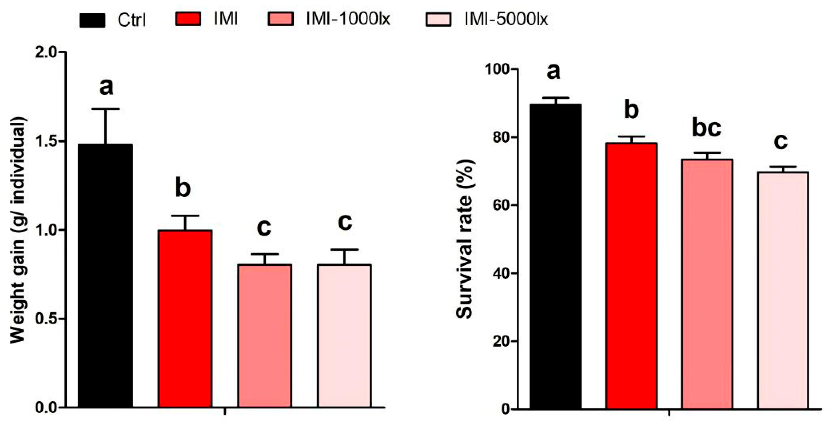
Survival rate and weight gain of P. clarkii were markedly affected by IMI exposure and EPE. As shown in Figure 2, crayfish in the control group exhibited the highest survival rates and weight gain, while IMI exposure alone reduced these parameters. The addition of EPE further exacerbated these effects, with the IMI-5000lx group showing the lowest survival rate (F = 19.17, df = 11, p < 0.001) and weight gain (F = 7.027, df = 11, p < 0.05) among all groups.

3.2. EPE Affects Oxidative Stress in P. clarkii Under IMI Exposure

Oxidative stress markers, including MDA, SOD, and CAT, displayed significant alterations across treatments. IMI exposure slightly induced SOD and CAT activities while significantly increased MDA levels (F = 6.016, df = 11, p < 0.05). These effects were further amplified by extended photoperiod exposure, with the highest SOD (F = 4.883, df = 11, p < 0.05) and CAT (F = 11.86, df = 11, p < 0.001) activities observed in the IMI-5000lx group (Figure 3).

3.3. EPE Affects Immune Response Under IMI Exposure

As shown in Figure 4, immune enzyme activities, including LZM, ACP, and AKP, were suppressed in crayfish exposed to IMI, particularly under extended photoperiod exposure conditions. The IMI-5000lx group showed the lowest LZM (F = 11.07, df = 11, p < 0.001), and AKP (F = 2.259, df = 11, p = 0.158) activities, indicating a substantial reduction in immune function compared to the control group.

3.4. Gene-Expression Analysis

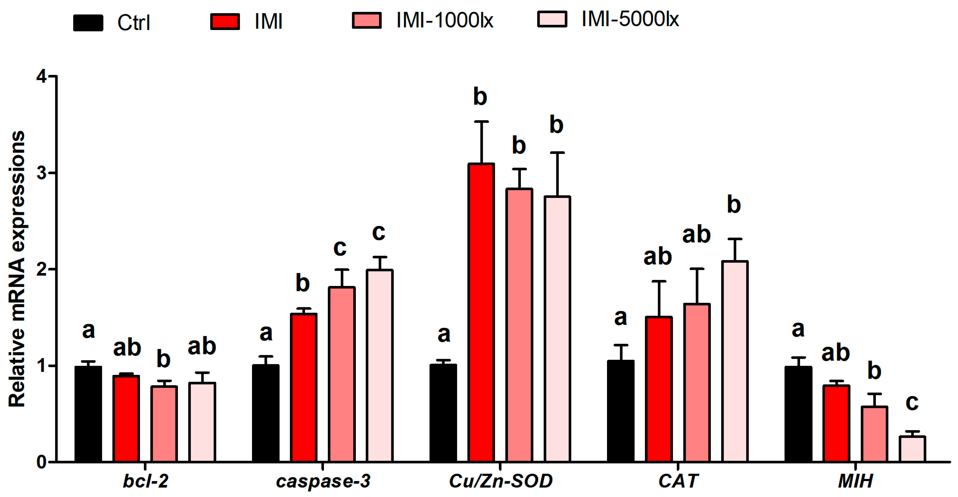
As shown in Figure 5, gene-expression analysis revealed significant dysregulation of oxidative stress-, apoptosis- and growth-related genes in crayfish. The expression of bcl-2 (anti-apoptotic) was significantly downregulated (F = 5.009, df = 11, p < 0.05), while caspase-3 (pro-apoptotic) were upregulated in response to IMI and EPE. The highest level of caspase-3 mRNA was observed in the IMI-5000lx group (F = 35.02, df = 11, p < 0.001), indicating an increased apoptotic induction when treated with extra light illumination. Meanwhile, IMI induced both Cu/Zn SOD (F = 24.56, df = 11, p < 0.001) and CAT expression, which is in line with the biochemical results. Notably, an extended photoperiod aggravated the induction of CAT, with the highest level observed in IMI-5000lx group (F = 6.203, df = 11, p < 0.01). In addition, EPE treatment showed a dramatic reduction in MIH expression, which is significantly lower than any other group (F = 34.19, df = 11, p < 0.001).

3.5. Effects of IMI and EPE on Neuroendocrine Hormone in Eyestalk of P. clarkii

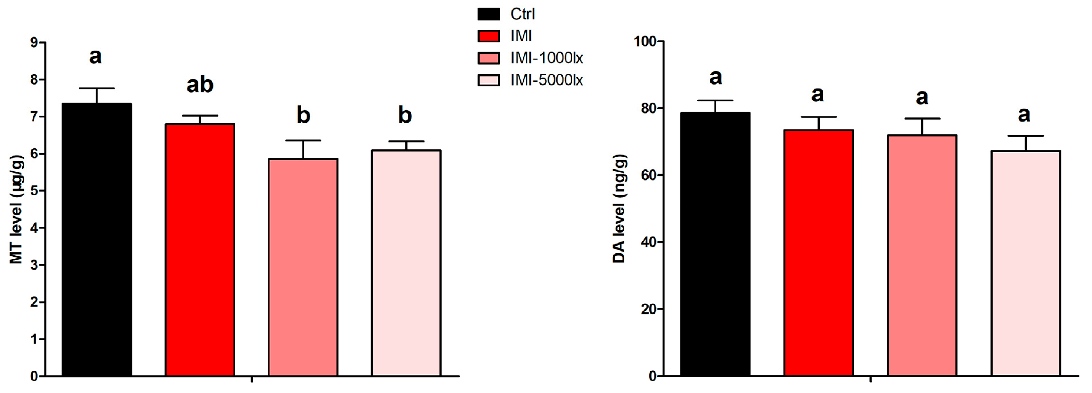
Neuroendocrine hormone levels such as the melatonin (MT) levels were significantly impacted by EPE treatments. MT levels were suppressed in all IMI-treated groups, with the greatest reduction observed under EPE conditions (F = 10.65, df = 11, p < 0.01). Meanwhile, DA levels also showed a decrease in response to IMI exposure and were further suppressed by extended photoperiod exposure, with the lowest levels recorded in the IMI-5000lx group. However, there was no significant difference between control and each treatment (F = 3.440, df = 11, p = 0.072) (Figure 6).

4. Discussion

Extended photoperiod exposure has been shown to amplify the toxicity of chemical pollutants. It disrupts circadian-regulated detoxification pathways and enhances oxidative stress, as observed in organisms exposed to chemicals under EPE conditions [36]. A study on zebrafish reported that the extra light period increased the toxic effects of imidacloprid by disrupting tryptophan metabolism pathways [37]. In the current study, the observed suppression of growth in P. clarkii across all IMI-treated groups, especially those subjected to additional extended photoperiod exposure, reflects the compounded effects of these stressors on energy metabolism and physiological stability. IMI, a potent neonicotinoid, has long been associated with reduced growth in aquatic organisms due to its neurotoxic effects on nicotinic acetylcholine receptors (nAChRs), which impair feeding behavior and energy metabolism. Studies on tiger shrimp P. monodon and zebrafish have similarly reported growth inhibition by IMI exposure due to disrupted feeding efficiency and increased metabolic costs [12,38]. In this study, the weight gain and survival of crayfish were lowest in the IMI-5000lx group, suggesting that EPE further deteriorate the toxic effects of IMI. Extended photoperiod exposure’s interference with circadian rhythms likely mediated IMI-induced growth suppression by disrupting melatonin (MT) production, a hormone essential for regulating antioxidant defenses and energy metabolism [39,40]. Suppressed MT levels force organisms to reallocate energy from growth and reproduction to coping mechanisms, such as antioxidant defense and cellular repair. Dopamine, another critical hormone, plays a central role in regulating motor function, energy balance, and stress responses [41]. In this study, DA levels were slightly reduced in the IMI and EPE-treated groups, particularly under higher light intensities. This reduction is consistent with findings in Drosophila melanogaster, where the IMI-induced disruption of dopaminergic signaling led to impaired behavior and metabolic imbalance [42]. A study involving rats showed that bright light exposure reduces tyrosine hydroxylase (TH)-positive dopamine neurons and dopamine levels by inducing oxidative stress and disrupting circadian-regulated processes such as melatonin production [43]. However, there is no significant difference between treatment and control. Combining this with our results, these findings suggest that extended photoperiod exposure may exacerbate neurodegenerative effects and oxidative damage, mechanisms that could similarly amplify the toxicity of pollutants like imidacloprid in aquatic systems. In particular, the suppression of MT, rather than DA, is more likely to contribute to these amplified physiological effects, since MT plays a pivotal role in maintaining oxidative balance and homeostasis under environmental stress.
The induction of oxidative stress under IMI and EPE is evidenced by elevated MDA levels and increased activities of antioxidant enzymes, such as SOD and CAT. IMI-induced oxidative stress has been extensively documented in aquatic species including fish [11,44], frog [45], and crustaceans [13,32], where it disrupts cellular redox balance and causes lipid peroxidation and DNA damage. On the other hand, however, EPE is reported as having no effect on oxidative status in songbirds [46] and crustacean shredders [47]. Considering the effect of EPE on regulation of neuroendocrine system, the additional oxidative burden imposed by EPE may likely be due to suppressed MT levels and the disrupted circadian regulation of detoxification pathways, forcing crayfish to rely more heavily on enzymatic antioxidant defenses. Prolonged activation of these defenses can lead to cellular exhaustion, further impairing physiological function, which may explain the reduced growth performance of the crayfish after combined exposure of IMI and EPE.
Immune suppression is another critical outcome of this study, as evidenced by reduced LZM, and ACP activities in crayfish exposed to IMI and EPE. These enzymes are integral to innate immunity, and their suppression reflects a weakened defense system, increasing vulnerability to infections and diseases [48]. IMI’s immunosuppressive effects, documented in oyster [49] and frog [45], are primarily mediated through oxidative stress and direct interference with immune cell function. Extended photoperiod exposure amplifies these effects by disrupting MT, which has well-established immunomodulatory properties, including the regulation of cytokines and stimulation of immune cell activity [50,51]. The combined stress of IMI and EPE appears to overwhelm the crayfish’s immune system, leaving it unable to mount an effective response to environmental challenges.
The gene-expression data provide additional insights into the molecular mechanisms underlying these impairments. The downregulation of the anti-apoptotic gene bcl-2 and the upregulation of the pro-apoptotic gene caspase-3 observed in this study reflect a shift toward apoptosis in hepatopancreatic tissues. Apoptotic responses to IMI-induced oxidative stress has been reported in different kinds of animals, including rats [52], carp [53] and shrimp [13], where prolonged oxidative stress triggers mitochondrial dysfunction and activates apoptotic cell death pathways. In addition, the upregulation of antioxidant-related genes, Cu/Zn-SOD and CAT, reflects an adaptive response to oxidative stress in crayfish exposed to IMI and with an extended photoperiod. However, these responses appear insufficient to counteract the heightened oxidative burden, as indicated by increased malondialdehyde (MDA) levels. Similar findings have been reported in honey bees, where IMI-induced oxidative stress led to the overexpression of antioxidant genes, such as SOD, and CAT. A study involving mice showed that the expression of antioxidative genes including Cu/Zn SOD, CAT and the glutathione system genes was relatively higher in light compared to darkness [54]. In this test, a stronger induction of CAT has been observed. Therefore, an extended photoperiod may enhance the oxidative stress induced by IMI and result in higher antioxidative response for redox balance compensation. Moreover, the significant downregulation of the growth-related gene MIH (molt-inhibiting hormone) observed in this study provides a molecular basis for the reduced weight gain in crayfish. MIH is a critical endocrine regulator in crustaceans, responsible for controlling the molting process by inhibiting the secretion of ecdysteroids from the Y-organ, which are hormones that drive molting and growth [55]. MIH ensures proper timing and regulation of molting, which is essential for the survival, growth, and reproduction of crustaceans. Dysregulation of MIH can lead to premature or delayed molting, resulting in stunted growth or increased vulnerability to environmental stressors [56]. Similar results have been found in brown shrimp [57] and honey bees [58] when exposed to IMI, where pesticide-induced endocrine disruption impaired molting, development, and overall fitness. The additional suppression of MIH in extended photoperiod-treated groups suggests that light pollution could increase endocrine disruption under pesticide, like the IMI stress.
While this study successfully demonstrated that an extended photoperiod enhances imidacloprid-induced disruptions in neuroendocrine signaling and oxidative stress pathways, one limitation is the lack of direct behavioral assessments. Behavioral endpoints such as locomotor activity, shelter-seeking, or escape response are highly sensitive to neuroendocrine perturbations and can provide functional evidence of neurotoxicity. Although changes in melatonin levels, as well as apoptosis-related gene expression, strongly suggest neurophysiological impairment, future studies should incorporate quantitative behavioral tracking to validate and extend the current findings. The integration of behavioral data with biochemical and molecular analyses would offer a more comprehensive understanding of the ecological implications of pesticide–light interactions in aquatic organisms.

5. Conclusions

In conclusion, this study highlights the synergistic effects of IMI and EPE on P. clarkii, revealing significant impairments in growth, oxidative balance, immune function, and neuroendocrine regulation. The suppression of MT levels emerges as a central mechanism linking these physiological disruptions, providing insights into how chemical and physical stressors interact to amplify their impacts. These findings underscore the need for integrated management strategies to mitigate the combined effects of pollutants and light pollution on aquatic ecosystems, particularly in regions where both stressors are prevalent. Such approaches are essential for protecting economically and ecologically important species like crayfish and ensuring the sustainability of freshwater environments.

Author Contributions

Methodology, D.Q.; Software, X.L.; Validation, X.H. and Q.H.; Formal analysis, Y.H. and D.Q.; Investigation, X.L. and X.H.; Writing—original draft, Y.H.; Writing—review and editing, Z.H.; Supervision, Z.H.; Project administration, Z.H. All authors have read and agreed to the published version of the manuscript.

Funding

This research was funded by ‘Pandeng’ Project in Xichang University, grant number [117620010] and “Shuanggao” Science Research Program in Xichang University, grant number LGLZ201809. And The APC was funded by ‘Pandeng’ Project in Xichang University.

Institutional Review Board Statement

The animal study protocol was approved by the Animal Bioethics Committee at Xichang University (protocol code: XCC20240704001 and approval date: 2024-07-04).

Informed Consent Statement

Not applicable.

Data Availability Statement

The original contributions presented in this study are included in the article. Further inquiries can be directed to the corresponding author.

Conflicts of Interest

The authors declare no conflicts of interest.

References

  1. Buckingham, S.D.; Lapied, B.; Le Corronc, H.; Grolleau, F.; Sattelle, D.B. Imidacloprid actions on insect neuronal acetylcholine receptors. J. Exp. Biol. 1997, 200, 2685–2692. [Google Scholar] [CrossRef]
  2. Roessink, I.; Merga, L.B.; Zweers, H.J.; Van den Brink, P.J. The neonicotinoid imidacloprid shows high chronic toxicity to mayfly nymphs. Environ. Toxicol. Chem. 2013, 32, 1096–1100. [Google Scholar] [CrossRef] [PubMed]
  3. Anderson, J.C.; Dubetz, C.; Palace, V.P. Neonicotinoids in the Canadian aquatic environment: A literature review on current use products with a focus on fate, exposure, and biological effects. Sci. Total Environ. 2015, 505, 409–422. [Google Scholar] [CrossRef]
  4. Nowell, L.H.; Moran, P.W.; Schmidt, T.S.; Norman, J.E.; Nakagaki, N.; Shoda, M.E.; Mahler, B.J.; Van Metre, P.C.; Stone, W.W.; Sandstrom, M.W.; et al. Complex mixtures of dissolved pesticides show potential aquatic toxicity in a synoptic study of Midwestern U.S. streams. Sci. Total Environ. 2018, 613–614, 1469–1488. [Google Scholar] [CrossRef] [PubMed]
  5. Morrissey, C.A.; Mineau, P.; Devries, J.H.; Sanchez-Bayo, F.; Liess, M.; Cavallaro, M.C.; Liber, K. Neonicotinoid contamination of global surface waters and associated risk to aquatic invertebrates: A review. Environ. Int. 2015, 74, 291–303. [Google Scholar] [CrossRef] [PubMed]
  6. Thunnissen, N.W.; Lautz, L.S.; van Schaik, T.W.G.; Hendriks, A.J. Ecological risks of imidacloprid to aquatic species in the Netherlands: Measured and estimated concentrations compared to species sensitivity distributions. Chemosphere 2020, 254, 126604. [Google Scholar] [CrossRef]
  7. Wu, S.; Wu, L.; Xu, H.; Zhao, X.; Wu, C.; Chen, L.; Zhang, H.; Wang, Q. Study on residue of imidacloprid in rice and field environment. Acta Agric. Zhejiangensis 2004, 16, 274–278. [Google Scholar]
  8. Nugnes, R.; Russo, C.; Orlo, E.; Lavorgna, M.; Isidori, M. Imidacloprid: Comparative toxicity, DNA damage, ROS production and risk assessment for aquatic non-target organisms. Environ. Pollut. 2023, 316, 120682. [Google Scholar] [CrossRef]
  9. Osterberg, J.S.; Darnell, K.M.; Blickley, T.M.; Romano, J.A.; Rittschof, D. Acute toxicity and sub-lethal effects of common pesticides in post-larval and juvenile blue crabs, Callinectes sapidus. J. Exp. Mar. Biol. Ecol. 2012, 424–425, 5–14. [Google Scholar] [CrossRef]
  10. Frew, J.A.; Brown, J.T.; Fitzsimmons, P.N.; Hoffman, A.D.; Sadilek, M.; Grue, C.E.; Nichols, J.W. Toxicokinetics of the neonicotinoid insecticide imidacloprid in rainbow trout (Oncorhynchus mykiss). Comp. Biochem. Physiol. Part C Toxicol. Pharmacol. 2018, 205, 34–42. [Google Scholar] [CrossRef]
  11. Ge, W.; Yan, S.; Wang, J.; Zhu, L.; Chen, A.; Wang, J. Oxidative Stress and DNA Damage Induced by Imidacloprid in Zebrafish (Danio rerio). J. Agric. Food Chem. 2015, 63, 1856–1862. [Google Scholar] [CrossRef]
  12. Butcherine, P.; Kelaher, B.P.; Taylor, M.D.; Barkla, B.J.; Benkendorff, K. Impact of imidacloprid on the nutritional quality of adult black tiger shrimp (Penaeus monodon). Ecotoxicol. Environ. Saf. 2020, 198, 110682. [Google Scholar] [CrossRef]
  13. Fu, Z.; Han, F.; Huang, K.; Zhang, J.; Qin, J.G.; Chen, L.; Li, E. Impact of imidacloprid exposure on the biochemical responses, transcriptome, gut microbiota and growth performance of the Pacific white shrimp Litopenaeus vannamei. J. Hazard. Mater. 2022, 424, 127513. [Google Scholar] [CrossRef] [PubMed]
  14. Souty-Grosset, C.; Anastácio, P.M.; Aquiloni, L.; Banha, F.; Choquer, J.; Chucholl, C.; Tricarico, E. The red swamp crayfish Procambarus clarkii in Europe: Impacts on aquatic ecosystems and human well-being. Limnologica 2016, 58, 78–93. [Google Scholar] [CrossRef]
  15. Wang, Q.; Ding, H.; Tao, Z.; Ma, D. Crayfish (Procambarus clarkii) Cultivation in China: A Decade of Unprecedented Development. In Aquaculture in China; John Wiley & Sons Ltd.: Hoboken, NJ, USA, 2018; pp. 363–377. [Google Scholar]
  16. Hernández, L.; Maeda-Martínez, A.M.; Ruiz-Campos, G.; Rodríguez-Almaraz, G.; Alonzo-Rojo, F.; Sainz, J.C. Geographic expansion of the invasive red crayfish Procambarus clarkii (Girard, 1852) (Crustacea: Decapoda) in Mexico. Biol. Invasions 2008, 10, 977–984. [Google Scholar] [CrossRef]
  17. Gherardi, F. Crayfish invading Europe: The case study of Procambarus clarkii. Mar. Freshw. Behav. Physiol. 2006, 39, 175–191. [Google Scholar] [CrossRef]
  18. Li, Y.; Guo, X.; Cao, X.; Deng, W.; Luo, W.; Wang, W. Population Genetic Structure and Post-Establishment Dispersal Patterns of the Red Swamp Crayfish Procambarus clarkii in China. PLoS ONE 2012, 7, e40652. [Google Scholar] [CrossRef] [PubMed]
  19. Fernández-Cisnal, R.; García-Sevillano, M.A.; García-Barrera, T.; Gómez-Ariza, J.L.; Abril, N. Metabolomic alterations and oxidative stress are associated with environmental pollution in Procambarus clarkii. Aquat. Toxicol. 2018, 205, 76–88. [Google Scholar] [CrossRef]
  20. Suárez-Serrano, A.; Alcaraz, C.; Ibáñez, C.; Trobajo, R.; Barata, C. Procambarus clarkii as a bioindicator of heavy metal pollution sources in the lower Ebro River and Delta. Ecotoxicol. Environ. Saf. 2010, 73, 280–286. [Google Scholar] [CrossRef]
  21. Pastorino, P.; Anselmi, S.; Zanoli, A.; Esposito, G.; Bondavalli, F.; Dondo, A.; Pucci, A.; Pizzul, E.; Faggio, C.; Barceló, D.; et al. The invasive red swamp crayfish (Procambarus clarkii) as a bioindicator of microplastic pollution: Insights from Lake Candia (northwestern Italy). Ecol. Indic. 2023, 150, 110200. [Google Scholar] [CrossRef]
  22. Vioque-Fernández, A.; de Almeida, E.A.; Ballesteros, J.; García-Barrera, T.; Gómez-Ariza, J.L.; López-Barea, J. Doñana National Park survey using crayfish (Procambarus clarkii) as bioindicator: Esterase inhibition and pollutant levels. Toxicol. Lett. 2007, 168, 260–268. [Google Scholar] [CrossRef]
  23. Gaston, K.J.; Sánchez de Miguel, A. Environmental Impacts of Artificial Light at Night. Annu. Rev. Environ. Resour. 2022, 47, 373–398. [Google Scholar] [CrossRef]
  24. Brüning, A.; Hölker, F.; Franke, S.; Kleiner, W.; Kloas, W. Impact of different colours of artificial light at night on melatonin rhythm and gene expression of gonadotropins in European perch. Sci. Total Environ. 2016, 543, 214–222. [Google Scholar] [CrossRef]
  25. Cho, Y.; Ryu, S.-H.; Lee, B.R.; Kim, K.H.; Lee, E.; Choi, J. Effects of artificial light at night on human health: A literature review of observational and experimental studies applied to exposure assessment. Chronobiol. Int. 2015, 32, 1294–1310. [Google Scholar] [CrossRef]
  26. Zhang, M.; Gao, X.; Luo, Q.; Lin, S.; Lyu, M.; Luo, X.; Ke, C.; You, W. Risk assessment of persistent exposure to artificial light at night revealed altered behavior and metabolic patterns of marine nocturnal shellfish. Ecol. Indic. 2024, 160, 111807. [Google Scholar] [CrossRef]
  27. Manríquez, P.H.; Jara, M.E.; González, C.P.; Seguel, M.; Quijón, P.A.; Widdicombe, S.; Pulgar, J.M.; Quintanilla-Ahumada, D.; Anguita, C.; Duarte, C. Effects of artificial light at night and predator cues on foraging and predator avoidance in the keystone inshore mollusc Concholepas concholepas. Environ. Pollut. 2021, 280, 116895. [Google Scholar] [CrossRef]
  28. Jackson, K.M.; Moore, P.A. The intensity and spectrum of artificial light at night alters crayfish interactions. Mar. Freshw. Behav. Physiol. 2019, 52, 131–150. [Google Scholar] [CrossRef]
  29. Nie, X.; Huang, C.; Wei, J.; Wang, Y.; Hong, K.; Mu, X.; Liu, C.; Chu, Z.; Zhu, X.; Yu, L. Effects of Photoperiod on Survival, Growth, Physiological, and Biochemical Indices of Redclaw Crayfish (Cherax quadricarinatus) Juveniles. Animals 2024, 14, 411. [Google Scholar] [CrossRef] [PubMed]
  30. Thomas, J.R.; James, J.; Newman, R.C.; Riley, W.D.; Griffiths, S.W.; Cable, J. The impact of streetlights on an aquatic invasive species: Artificial light at night alters signal crayfish behaviour. Appl. Anim. Behav. Sci. 2016, 176, 143–149. [Google Scholar] [CrossRef]
  31. Liu, Z.; Lv, Y.; Ding, R.; Chen, X.; Pu, G. Light Pollution Changes the Toxicological Effects of Cadmium on Microbial Community Structure and Function Associated with Leaf Litter Decomposition. Int. J. Mol. Sci. 2020, 21, 422. [Google Scholar] [CrossRef]
  32. Huang, Y.; Hong, Y.; Yin, H.; Yan, G.; Huang, Q.; Li, Z.; Huang, Z. Imidacloprid induces locomotion impairment of the freshwater crayfish, Procambarus clarkii via neurotoxicity and oxidative stress in digestive system. Aquat. Toxicol. 2021, 238, 105913. [Google Scholar] [CrossRef] [PubMed]
  33. Bradford, M.M.A. A Rapid and Sensitive Method for Quantitation of Microgram Quantities of Protein Utilizing the Principle of Protein-Dye Binding. Anal. Biochem. 1976, 25, 248–256. [Google Scholar] [CrossRef]
  34. Han, Z.; Li, X.; Li, X.; Xu, W.; Li, Y. Circadian rhythms of melatonin in haemolymph and optic lobes of Chinese mitten crab (Eriocheir sinensis) and Chinese grass shrimp (Palaemonetes sinensis). Biol. Rhythm Res. 2019, 50, 400–407. [Google Scholar] [CrossRef]
  35. Tinikul, Y.; Poljaroen, J.; Kornthong, N.; Chotwiwatthanakun, C.; Anuracpreeda, P.; Poomtong, T.; Hanna, P.J.; Sobhon, P. Distribution and changes of serotonin and dopamine levels in the central nervous system and ovary of the Pacific white shrimp, Litopenaeus vannamei, during ovarian maturation cycle. Cell Tissue Res. 2011, 345, 103–124. [Google Scholar] [CrossRef]
  36. Eberhardt, L.; Binde Doria, H.; Bulut, B.; Feldmeyer, B.; Pfenninger, M. Transcriptomics predicts Artificial Light at Night’s (ALAN) impact on fitness: Nightly illumination alters gene expression pattern and negatively affects fitness components in the midge Chironomus riparius (Diptera: Chironomidae). bioRxiv 2024. [Google Scholar] [CrossRef]
  37. Huang, Y.; Hong, Y.; Wu, S.; Yang, X.; Huang, Q.; Dong, Y.; Xu, D.; Huang, Z. Prolonged darkness attenuates imidacloprid toxicity through the brain-gut-microbiome axis in zebrafish, Danio rerio. Sci. Total Environ. 2023, 881, 163481. [Google Scholar] [CrossRef]
  38. Vignet, C.; Cappello, T.; Fu, Q.; Lajoie, K.; De Marco, G.; Clérandeau, C.; Mottaz, H.; Maisano, M.; Hollender, J.; Schirmer, K.; et al. Imidacloprid induces adverse effects on fish early life stages that are more severe in Japanese medaka (Oryzias latipes) than in zebrafish (Danio rerio). Chemosphere 2019, 225, 470–478. [Google Scholar] [CrossRef]
  39. Touitou, Y.; Reinberg, A.; Touitou, D. Association between light at night, melatonin secretion, sleep deprivation, and the internal clock: Health impacts and mechanisms of circadian disruption. Life Sci. 2017, 173, 94–106. [Google Scholar] [CrossRef] [PubMed]
  40. Liu, H.; Fu, R.; Zhang, Y.; Mao, L.; Zhu, L.; Zhang, L.; Liu, X.; Jiang, H. Integrate transcriptomic and metabolomic analysis reveals the underlying mechanisms of behavioral disorders in zebrafish (Danio rerio) induced by imidacloprid. Sci. Total Environ. 2023, 870, 161541. [Google Scholar] [CrossRef] [PubMed]
  41. Rubí, B.; Maechler, P. Minireview: New Roles for Peripheral Dopamine on Metabolic Control and Tumor Growth: Let’s Seek the Balance. Endocrinology 2010, 151, 5570–5581. [Google Scholar] [CrossRef] [PubMed]
  42. Janner, D.E.; Gomes, N.S.; Poetini, M.R.; Poleto, K.H.; Musachio, E.A.S.; de Almeida, F.P.; de Matos Amador, E.C.; Reginaldo, J.C.; Ramborger, B.P.; Roehrs, R.; et al. Oxidative stress and decreased dopamine levels induced by imidacloprid exposure cause behavioral changes in a neurodevelopmental disorder model in Drosophila melanogaster. NeuroToxicology 2021, 85, 79–89. [Google Scholar] [CrossRef]
  43. Romeo, S.; Viaggi, C.; Di Camillo, D.; Willis, A.W.; Lozzi, L.; Rocchi, C.; Capannolo, M.; Aloisi, G.; Vaglini, F.; Maccarone, R.; et al. Bright light exposure reduces TH-positive dopamine neurons: Implications of light pollution in Parkinson’s disease epidemiology. Sci. Rep. 2013, 3, 1395. [Google Scholar] [CrossRef]
  44. Vieira, C.E.D.; Pérez, M.R.; Acayaba, R.D.A.; Raimundo, C.C.M.; dos Reis Martinez, C.B. DNA damage and oxidative stress induced by imidacloprid exposure in different tissues of the Neotropical fish Prochilodus lineatus. Chemosphere 2018, 195, 125–134. [Google Scholar] [CrossRef]
  45. Rios, F.M.; Wilcoxen, T.E.; Zimmerman, L.M. Effects of imidacloprid on Rana catesbeiana immune and nervous system. Chemosphere 2017, 188, 465–469. [Google Scholar] [CrossRef] [PubMed]
  46. Raap, T.; Casasole, G.; Costantini, D.; AbdElgawad, H.; Asard, H.; Pinxten, R.; Eens, M. Artificial light at night affects body mass but not oxidative status in free-living nestling songbirds: An experimental study. Sci. Rep. 2016, 6, 35626. [Google Scholar] [CrossRef] [PubMed]
  47. Czarnecka, M.; Jermacz, Ł.; Glazińska, P.; Kulasek, M.; Kobak, J. Artificial light at night (ALAN) affects behaviour, but does not change oxidative status in freshwater shredders. Environ. Pollut. 2022, 306, 119476. [Google Scholar] [CrossRef]
  48. Liu, Z.; Yu, P.; Cai, M.; Wu, D.; Zhang, M.; Chen, M.; Zhao, Y. Effects of microplastics on the innate immunity and intestinal microflora of juvenile Eriocheir sinensis. Sci. Total Environ. 2019, 685, 836–846. [Google Scholar] [CrossRef]
  49. Ewere, E.E.; Reichelt-Brushett, A.; Benkendorff, K. The neonicotinoid insecticide imidacloprid, but not salinity, impacts the immune system of Sydney rock oyster, Saccostrea glomerata. Sci. Total Environ. 2020, 742, 140538. [Google Scholar] [CrossRef]
  50. Giannoulia-Karantana, A.; Vlachou, A.; Polychronopoulou, S.; Papassotiriou, I.; Chrousos, G.P. Melatonin and Immunomodulation: Connections and Potential Clinical Applications. Neuroimmunomodulation 2007, 13, 133–144. [Google Scholar] [CrossRef]
  51. Medrano-Campillo, P.; Sarmiento-Soto, H.; Álvarez-Sánchez, N.; Álvarez-Ríos, A.I.; Guerrero, J.M.; Rodríguez-Prieto, I.; Castillo-Palma, M.J.; Lardone, P.J.; Carrillo-Vico, A. Evaluation of the immunomodulatory effect of melatonin on the T-cell response in peripheral blood from systemic lupus erythematosus patients. J. Pineal Res. 2015, 58, 219–226. [Google Scholar] [CrossRef]
  52. Taha, M.A.I.; Badawy, M.E.I.; Abdel-Razik, R.K.; Younis, H.M.; Abo-El-Saad, M.M. Mitochondrial dysfunction and oxidative stress in liver of male albino rats after exposing to sub-chronic intoxication of chlorpyrifos, cypermethrin, and imidacloprid. Pestic. Biochem. Physiol. 2021, 178, 104938. [Google Scholar] [CrossRef]
  53. Miao, Z.; Miao, Z.; Wang, S.; Wu, H.; Xu, S. Exposure to imidacloprid induce oxidative stress, mitochondrial dysfunction, inflammation, apoptosis and mitophagy via NF-kappaB/JNK pathway in grass carp hepatocytes. Fish Shellfish Immunol. 2022, 120, 674–685. [Google Scholar] [CrossRef]
  54. Xu, Y.-Q.; Zhang, D.; Jin, T.; Cai, D.-J.; Wu, Q.; Lu, Y.; Liu, J.; Klaassen, C.D. Diurnal Variation of Hepatic Antioxidant Gene Expression in Mice. PLoS ONE 2012, 7, e44237. [Google Scholar] [CrossRef]
  55. Techa, S.; Chung, J.S. Ecdysteroids Regulate the Levels of Molt-Inhibiting Hormone (MIH) Expression in the Blue Crab, Callinectes sapidus. PLoS ONE 2015, 10, e0117278. [Google Scholar] [CrossRef]
  56. Nakatsuji, T.; Lee, C.-Y.; Watson, R.D. Crustacean molt-inhibiting hormone: Structure, function, and cellular mode of action. Comp. Biochem. Physiol. Part A Mol. Integr. Physiol. 2009, 152, 139–148. [Google Scholar] [CrossRef] [PubMed]
  57. Al-Badran, A.A.; Fujiwara, M.; Mora, M.A. Effects of insecticides, fipronil and imidacloprid, on the growth, survival, and behavior of brown shrimp Farfantepenaeus aztecus. PLoS ONE 2019, 14, e0223641. [Google Scholar] [CrossRef] [PubMed]
  58. Li, Z.; Wang, Y.; Qin, Q.; Chen, L.; Dang, X.; Ma, Z.; Zhou, Z. Imidacloprid disrupts larval molting regulation and nutrient energy metabolism, causing developmental delay in honey bee Apis mellifera. eLife 2024, 12, RP88772. [Google Scholar] [CrossRef] [PubMed]
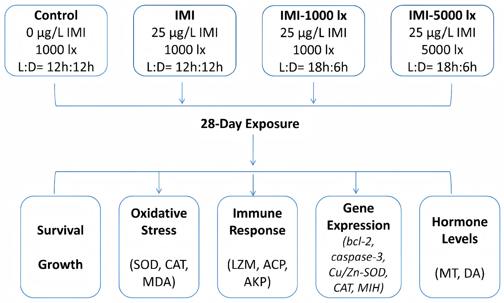
Figure 1. Experimental design showing imidacloprid (IMI) and extended photoperiod exposure (EPE) treatments in juvenile Procambarus clarkii.
Fishes 10 00562 g001
Figure 2. Effects of imidacloprid (IMI) exposure and extended photoperiod on the growth performance and survival of juvenile crayfish (Procambarus clarkii) after 28 days. The experimental groups included (1) control (1000 lx, 12L:12D photoperiod, no IMI); (2) IMI (25 µg/L IMI, 1000 lx, 12L:12D photoperiod); (3) IMI-l000lx (25 µg/L IMI, 1000 lx, 18L:6D photoperiod); and (4) IMI-5000lx (25 µg/L IMI, 5000 lx, 18L:6D photoperiod). The left panel shows mean weight gain; the right panel shows survival rate (%). Bars represent mean ± SD (n = 3). Different lowercase letters above the bars indicate statistically significant differences among groups (p < 0.05; one-way ANOVA with Tukey’s post hoc test).
Fishes 10 00562 g002
Figure 3. Oxidative stress status in hepatopancreas of P. clarkii under IMI and extended photoperiod exposure treatments. (left panel) MDA level; (middle panel) SOD activity; (right panel) CAT activity. The different letters above the columns represent significant differences among groups. N = 3.
Fishes 10 00562 g003
Figure 4. EPE affects immune-related enzyme activities under IMI exposure. (left panel) LZM; (middle panel) ACP; (right panel) AKP. The different letters above the columns represent significant differences among groups. N = 3.
Fishes 10 00562 g004
Figure 5. Expression of apoptotic-, oxidative stress- and growth-related genes in response to IMI and EPE treatment. Different letters above the columns represent significant differences among groups. N = 3.
Fishes 10 00562 g005
Figure 6. Neuroendocrine hormone levels in the eyestalks of P. clarkii under IMI and EPE treatment. (left panel) MT levels; (right panel) DA levels. Different letters above the columns represent significant differences among groups. N = 3.
Fishes 10 00562 g006
Table 1. Oligonucleotide primers used in this study.
Primer NameSequence (5′ to 3′)
bcl-2 FwCTGGGAGGTGTGTGAGGTGTT
bcl-2 RvTCAGAGGTGAGAGTGAGGGAGA
caspase-3 FwTGAAGAATCGCTGAATCTGCTC
caspase-3 RvCATATCCTCCTCCAACCTGCT
Cn/Zn SOD FwAACCAAATCAGTGGCAGGCT
Cu/Zn SOD RvCCTGGAGTCAGCCCATACAC
CAT FwAGTTCAAGAAGAGCCAGAC
CAT RvAGGAATGCGTTCTCTATCAA
MIHAGGTTCTACGAGTTGCTTG
MIHTGCCGTTGTCTGCTGT
β-actin FwTGCCGCCTCATCCTCTTC
β-actin RvCCTCTCGTTGCCAATGGTAATG
Disclaimer/Publisher’s Note: The statements, opinions and data contained in all publications are solely those of the individual author(s) and contributor(s) and not of MDPI and/or the editor(s). MDPI and/or the editor(s) disclaim responsibility for any injury to people or property resulting from any ideas, methods, instructions or products referred to in the content.