Article

Mechanism of Biphasic Activation of NLRP3 Inflammasome in the Fat Greenling (Hexagrammos otakii) Under Hypoxic Stress: From Inflammatory Defense to Pyroptosis Execution

1
Key Laboratory of Applied Biology and Aquaculture of Northern Fishes in Liaoning Province, Dalian Ocean University, Dalian 116023, China
2
College of Fisheries and Life Science, Dalian Ocean University, Dalian 116023, China
3
Key Laboratory of Biotechnology and Bioresources Utilization, Dalian Minzu University, Ministry of Education, Dalian 116600, China
*
Authors to whom correspondence should be addressed.
Fishes 2025, 10(11), 542; https://doi.org/10.3390/fishes10110542 (registering DOI)
Submission received: 19 September 2025 / Revised: 16 October 2025 / Accepted: 22 October 2025 / Published: 24 October 2025
(This article belongs to the Section Physiology and Biochemistry)

Abstract

Hypoxic stress is an important environmental challenge for aquatic organisms, which is detrimental to fish survival and growth. Specifically, the NOD-like receptor thermal protein domain associated protein 3 (NLRP3) has emerged as a pivotal regulator, with accumulating evidence underscoring its central role in inflammatory processes. However, the regulatory functions of NLRP3 during hypoxic stress in fat greenling (Hexagrammos otakii) remain elusive. In this study, we systematically analyzed the molecular features of HoNLRP3 and elucidated its biphasic regulatory mechanism under hypoxic stress in H. otakii using phylogenetic analysis, qRT-PCR, Western blot, and immunofluorescence. Its phylogeny is significantly different from that of mammals and carries FISNA and related motifs specific to bony fishes. Hypoxia induced predominant nlrp3 expression in the brain, peaking at 12–24 h, with strong positive correlation to hif-1α activation. NLRP3-ASC-Caspase1 inflammasomes assembly drove IL-1β maturation, while prolonged hypoxia (48 h) activated Caspase3/GSDME-mediated pyroptosis, accompanied by elevated LDH activity. Reoxygenation partially reversed inflammatory and pyroptosis markers, indicating that NLRP3 balances defense and injury through a biphasic regulatory mechanism. This study provides new insights into the hypoxic adaptation mechanisms in bony fish.
Key Contribution: HoNLRP3 participates in the HIF1-α-mediated immune defense response after acute hypoxic stress in fat greenling (Hexagrammos otakii). HoNLRP3 assembles and interacts with ASC, caspase1 to form inflammatory vesicles. Activation of NLRP3 inflammatory vesicles promotes the production of mature IL-1β. The hypoxia stress triggers the Caspase3/GSDME pyroptosis pathway, signaling the dominant stage of injury.

Graphical Abstract

1. Introduction

Hypoxic stress is one of the major environmental challenges facing aquatic organisms, especially in the context of increasing offshore eutrophication and seasonal fluctuations in dissolved oxygen [1,2,3]. The physiology, metabolism, and immune defense of fish are highly dependent on ambient oxygen partial pressure, and chronic or acute hypoxic stress can lead to energy metabolism disorders, oxidative stress, and immunosuppression, which can lead to large-scale population declines [4,5,6]. Hypoxia signaling triggers adaptive responses through activation of the Hypoxia-Inducible-Factor (HIF) family (especially HIF-1α), such as increased glycolysis, neovascularization, and inflammatory regulation [7,8]. Moreover, hypoxia triggers abnormal accumulation of reactive oxygen species (ROS) and disorders of mitochondrial metabolism, a pathological mechanism that ultimately initiates the organism’s natural immune system response by modulating key signaling pathways [9,10,11].
The NLRP3 inflammasome, as a core component of pattern recognition receptors (PRRs), makes a critical difference in recognizing danger signals (e.g., hypoxia, pathogen invasion) and initiating inflammatory responses [12,13,14,15,16]. In mammals, NLRP3 mediates the maturation and development of the proinflammatory cytokines Interleukin-1β (IL-1β)/Interleukin-18 (IL-18) by recruiting the apoptosis-associated speck-like protein containing a caspase-recruitment domain (ASC) and activating the Cysteine-dependent aspartate-specific proteases 1 (Caspase1) to form inflammasomes, and triggers gasdermin D (GSDMD)-mediated pyroptosis [17], resulting in an “immune defense–cell death” dynamic equilibrium [18,19]. This pathway is indispensable in resisting bacterial infections, clearing damaged cells, and maintaining tissue homeostasis, and its over-activation has been found to be strongly related to a number of chronic inflammatory disorders, including gout and atherosclerosis [20,21]. Notably, the HIF-1α can drive the hyperactivation of inflammasomes and thus exacerbate tissue damage by directly binding to the NLRP3 promoter or modulating ROS levels [22,23].
However, significant gaps remain in understanding NLRP3 inflammasome function within bony fish—a crucial branch of vertebrate evolution. Although inflammasome systems in these species exhibit distinct adaptive features and have been characterized across numerous taxa, current research remains predominantly focused on pathogen infection models. For example, the zebrafish (Danio rerio) NLRP3 homologous gene activates the IL-1β pathway and triggers ACR toxicity [24], whereas carp (Cyprinus carpio) responds to immune responses in bacterial infections through the ASC-Caspase1 axis [25]. Furthermore, the execution mechanism of pyroptosis in fish differs significantly from mammalian dependence on Caspase1/GSDMD, with pyroptosis in some bony fishes being mediated primarily by the Caspase3/gasdermin E (GSDME) pathway [26,27,28,29,30].
As a typical benthic fish that inhabits the intertidal zone and offshore rocky reef areas, the fat greenling is chronically exposed to the environmental pressure of dramatic fluctuations in dissolved oxygen, and its hypoxia tolerance mechanism may have unique evolutionary features [31,32]. Previous studies have conducted preliminary investigations into the HIF-1α/ROS/NLRP3 pathway in the livers. However, compared to the liver, the brain—as the organ with the highest systemic oxygen consumption—is more sensitive to hypoxia [31]. Its unique immune microenvironment is defined by the blood–brain barrier, which strictly modulates the passage of signaling molecules and immune cells [33]. This environment stands in stark contrast to that found in peripheral organs like the liver. Despite the importance of understanding hypoxic adaptation in fish for assessing resilience and aquaculture development, the function of the NLRP3 inflammasome in hypoxia—specifically its regulatory mechanisms in the brain—remains to be elucidated. To understand the various molecular mechanisms by which fish are adapted to hypoxia stress may better determine their ability to cope with this situation, which is of important implications for the growth of the aquaculture industry. Therefore, this study focuses on the temporal response pattern of HoNLRP3, elucidating the molecular basis driving the transition from inflammatory defense to pyroptosis execution. This reveals the central role of brain tissue in hypoxic immunity, offering a new perspective for research on environmental adaptation strategies in fish.

2. Materials and Methods

2.1. Management of Fish

A total of 90 healthy, mixed-sex H. otakii (Length 13.45 ± 0.47 cm, Weight 38.96 ± 0.36 g) from the Key Laboratory of Fish Applied Biology and Aquaculture were used in this study. The fish were housed in three 141-type fiber-reinforced plastic tanks and acclimatized for 2 weeks prior to experimentation. During the experiment, the water temperature was maintained at 12 °C ± 0.5 °C.

2.2. Grouping and Sampling of Experiments

Establish a hypoxic stress model based on previous research [31]. In the hypoxic stress group, dissolved oxygen (DO) was maintained at 2.1 ± 0.3 mg/L, while the control group was kept at 7.7 ± 0.3 mg/L, and it was measured by a DO meter (YSI, Yellow Springs, OH, USA) during the experimental period. At each time point (0, 6, 12, 24, and 48 h), tissues from nine fish were pooled into three independent biological replicates (three fish per pool) to minimize variability arising from individual differences. All sampled fish were anesthetized with tricaine methanesulfonate (MS-222) prior to tissue collection. Multiple tissues, including the brain, gills, heart, liver, spleen, stomach, intestines, and kidneys, were collected. Following the 48h hypoxic exposure, fish were subjected to a 24 h reoxygenation period (R 24 h) to restore normoxic conditions, after which sampling was continued.

2.3. Sequence Analysis of HoNLRP3

Analysis of HoNLRP3 coding sequences included sequence comparison, phylogenetic tree construction, domain analysis, and protein 3D structure prediction. The open reading frame (ORF) for HoNLRP3 was identified using NCBI ORF Finder (https://www.ncbi.nlm.nih.gov/orffinder/, accessed on 1 November 2023), while NLRP3 amino acid sequences from comparative species (Table S1) were sourced from the NCBI database. Multiple sequence alignment was performed using DNAMAN. Conserved motifs within HoNLRP3 were predicted using the online tool MEME (https://web.mit.edu/meme/current/share/doc/meme.htmle/, accessed on 10 November 2024), and its isoelectric point (pI) and molecular weight (Mw) were calculated via the NovoPro online tool (https://www.novopro.cn/tools/, accessed on 10 January 2025). Protein domain architecture was analyzed using NCBI’s Batch CD-Search, and 3D structure modeling was conducted with SWISS-MODEL. Phylogenetic evolutionary relationships were inferred using MEGA 7.0.

2.4. Quantitative Real-Time Polymerase Chain Reaction (qRT-PCR)

We extracted total RNA from the obtained tissues with TRIzol reagent (CWBIO, Suzhou, China) and synthesized CDNA (Takara, Dalian, China), and analyzed the variation in mRNA levels of various genes by FastReal Rapid Fluorescent PCR Premix Reagents (Tiangen, Beijing, China) on a qRT-PCR Analyzer (LifeReal, Hangzhou, China). The primer information used is shown in Table 1 [31]. All sequences used were obtained from the transcriptome data (PRJNA1028859) of H. otakii.

2.5. IL-1β Content Detection

IL-1β levels in the brain were measured using the Nanjing Jiancheng Bioengineering Institute Fish IL-1β ELISA Detection Kit (Nanjing Jiancheng, H002-1-1, Nanjing, China). Procedures followed the manufacturer’s instructions, with IL-1β concentrations (ng/L) calculated based on the standard curve and optical density (OD) values.

2.6. Western Blot

Brain tissue samples from each time point of group were taken, total protein was extracted using RIPA lysis buffer (containing 1% PMSF) (Servicebio, Wuhan, China), using the Bicinchoninic Acid (BCA) assay for protein quantitation (Servicebio, Wuhan, China) and standardized to a homogeneous concentration (4 μg/μL), and fully denatured at 100 °C in a metal bath after adding protein loading buffer (SDS-PAGE sample loading buffer, 5×). Electrophoresis was performed using 15%/10% SDS-PAGE gels (sample volume 20 μg/well) at a constant voltage of 80 V until the separation of the gels, and then adjusted to 120 V until the bromophenol blue reached the bottom end of the gel. After transferring the protein samples to 0.45 μm pore size PVDF membranes using the semi-dry transfer technique, the following treatment process was carried out: Firstly, a 5% skimmed milk solution prepared using TBST buffer was applied to the transfer membrane for 2 h at room temperature to seal it. And then, the membrane was thoroughly rinsed three times using TBST buffer. The treated transfer membrane was incubated with the specific primary antibody solution in a refrigerator at 4 °C for 12–16 h to complete the antigen–antibody binding reaction. After three repeated washes by TBST on the following day, the working solution of secondary antibody coupled to horseradish peroxidase was added, and the reaction was shaken for 2 h at room temperature. The final signal was developed by ECL chemiluminescent substrate chromogenic color development solution, and the experimental results were recorded using high-resolution gel imaging equipment. The optical density analysis of the protein bands was done by ImageJ-win64, and the quantitative comparative analysis was realized by gray scale value conversion. The Antibody information used is shown in Table 2.

2.7. Colocalization of Tissue Immunofluorescence

Brain tissues were fixed with 4% paraformaldehyde for 0–24 h of hypoxia stress, dehydrated and embedded, and cryosections were prepared. Antigen repair was performed for 15 min. Closed for 1 h at room temperature with BSA (Bovine Serum Albumin). Primary antibody ASC was mixed with NLRP3, Caspase1, and incubated with PBS overnight. After washing with PBS, a fluorescent secondary antibody was added and incubated at ambient temperature for 1 h. Nuclei were stained with DAPI, and the slices were sealed with an anti-fluorescence quenching sealer. Fluorescence images were acquired using an inverted fluorescence microscope, and the Pearson correlation coefficient and co-localization rate (Manders’ coefficient) were calculated by ImageJ software to assess the strength of ASC interactions with NLRP3/Caspase1.

2.8. Co-Immunoprecipitation

Brain tissue treated under hypoxic conditions for 24 h was lysed in IP cell lysis buffer (SolarBio, Beijing, China). Following homogenization using a tissue grinder (Sevier, Wuhan, China), the mixture was centrifuged at 12,000× g for 5 min at 4 °C. The supernatant collected served as the total protein sample. Take 50 μL of Protein A/G magnetic Beads (MCE, Shanghai, China), wash three times with pre-chilled IP lysis buffer, then resuspend in 100 μL of buffer for later use. Take 500 μg of total protein from each sample, add 2 μg of antibody to each sample, and incubate overnight at 4 °C with gentle rotation. Add pretreated Protein A/G magnetic beads and incubate at 4 °C for 4 h. After magnetic separation, discard the supernatant. Wash the magnetic beads five times with IP lysis buffer. Add 1× SDS loading buffer and denature at 95 °C for 10 min. After magnetic separation, collect the supernatant for Western blot analysis.

2.9. Measurement of LDH Activity

Brain tissues at each time point of hypoxic stress were taken, and LDH activity was measured by using the Nanjing Jiancheng Institute of Bioengineering LDH assay kit (Nanjing Jiancheng, A020-2-2, Nanjing, China), which was operated according to the instruction manual. Caulmer’s Brilliant Blue was used to determine the protein concentration of the samples, and the LDH activity (U/gprot) was calculated according to the formula. The data were normalized by total protein concentration.

2.10. Histopathological Analysis

Brain tissues from the control (0 h), 24 h hypoxia groups, and R 24 h groups were collected and immediately fixed in 4% paraformaldehyde for 24 h. Following dehydration, the tissues were embedded in paraffin. Sections were cut at a thickness of 5 μm and stained with hematoxylin and eosin (H&E) according to standard protocols. The stained sections were examined under a light microscope for histopathological assessment.

2.11. Statistical Analysis

SPSS 22.0 statistical software was used to complete data processing and analysis in this study. Data were expressed in the form of mean ± standard error (Mean ± SE). In order to assess the differences between groups between time points, one-way analysis of variance (ANOVA) combined with Duncan multiple comparison procedures was chosen for statistical comparison. The results of the experiments are presented graphically, where significant differences are labeled as * p < 0.05, ** p < 0.01, and *** p < 0.001.

3. Results

3.1. Molecular Characteristics and Phylogeny Analysis of HoNLRP3

In this study, we obtained the sequence of NLRP3 from the H. otakii transcriptome database. Sequence analysis showed that the ORF of HoNLRP3 was 1584 bp in length, contained 1055 aa (Figure 1 B), and had a molecular weight of approximately 119.10 kDa and a theoretical isoelectric point of 8.08. The structural domain prediction showed that the HoNLRP3 protein contains six conserved domains, including the FISNA domain (113-184aa), the NACHT domain (194-362aa), the NOD2_WH domain (445-503aa), the NLRC4_HD2 domain (505-636aa), the PPP1R42 superfamily, also known as the LRR domain (703-871aa), and the fish-specific SPRY_PRY_SNTX domain (881-1055aa). The protein structure of HoNLRP3 differs from other species. HoNLRP3 lacks the N-terminal PYRIN and has fewer C-terminal LRRs (Figure 1A).
To further investigate the phylogenetic relationships of NLRP3, we constructed a phylogenetic tree, and the findings suggest that HoNLRP3 is the closest related to Cyclopterus lumpus, followed by Anarrhichthys ocellatus (Figure 1A). Also in this study, several fish-specific Motif sequences were identified by MEME, which are interspersed in various structural domains and may be associated with fish-specific response to hypoxia (Figure 1C).

3.2. Tissue Distribution of NLRP3 and Response to Hypoxic Stress

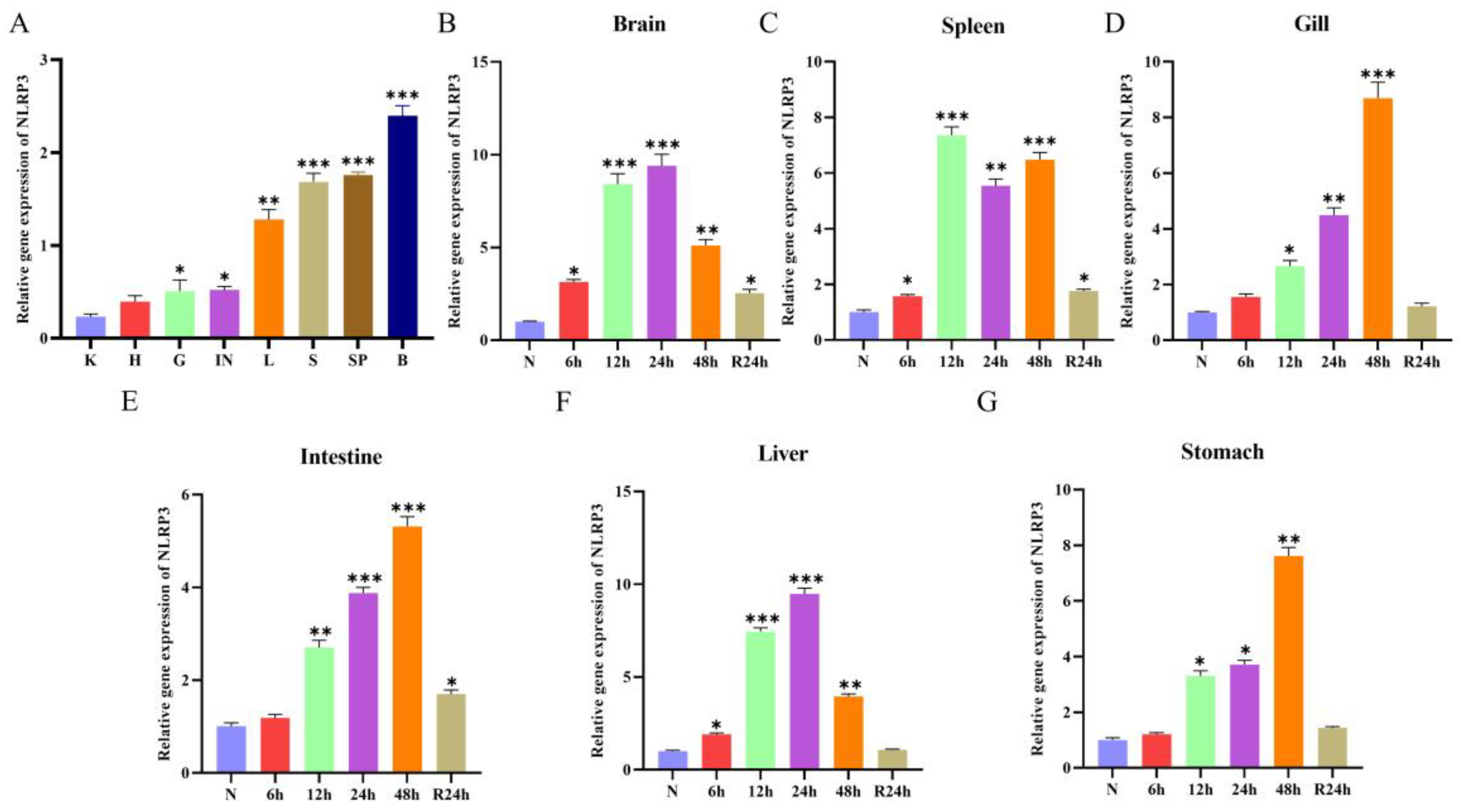
Given its high metabolic rate and sensitivity to hypoxia, we selected the brain as the primary tissue for study. Furthermore, we observed that nlrp3 exhibited the most pronounced transcriptional response in the brain compared to other tissues examined (such as liver, spleen, and gills), and also demonstrated the highest expression levels in healthy fish (Figure 2A). This further validates the rationale for focusing our research on this tissue.
The research further revealed a differential upregulation of nlrp3 across tissues during hypoxic stress, with distinct temporal expression patterns: it peaked at 24 h in the brain (Figure 2B) but not until 48 h in the gills, intestines, and stomach (Figure 2D,E,G). Additionally, the expression of nlrp3 was significantly elevated in the spleen and liver at 24 h post-hypoxia-stress (Figure 2C,F). These results suggest that the nlrp3 may be involved in the immune response of fish under hypoxic stress.

3.3. Dynamics of Gene and Protein Expression of the HIF-1α

The qRT-PCR results showed that the mRNA expression of hif-1α in the brain of H. otakii showed significant time-dependent changes under hypoxic stress (Figure 3A). Under normoxic conditions (0 h), hif-1α mRNA was expressed at basal levels. After 12 h of hypoxia, its expression rapidly increased to the peak (10.34-fold), and then gradually decreased to 8.25-fold at 24 h and 6.05-fold at 48 h. After 24 h of reoxygenation (R 24 h), hif-1α mRNA partially recovered to 2.44-fold. In addition, correlation analysis showed that the changing trend of hif-1α mRNA was highly positively correlated with nlrp3 (R2 = 0.87) (Figure 3D)
The specificity of HIF1-α has been verified by Western blot, and the sizes of the target protein bands are all consistent with the theoretical molecular weight (Figure S1A). Western blot results further verified the changes in protein levels of HIF-1α (Figure 3B). After 24 h of reoxygenation, protein expression partially returned to basal levels (1.89-fold). Notably, the dynamics of HIF-1α protein levels (Figure 3C) were largely consistent with the gene expression, The above results indicate that HIF-1α is rapidly activated in the early stage of hypoxia and is involved in regulating the hypoxic stress response of the H. otakii through transcription and translation, whereas the rapid degradation after reoxygenation suggests that the sensitivity of its dynamic equilibrium to changes in the oxygen environment.

3.4. NLRP3 Inflammasome Activation Promotes Inflammatory Signaling

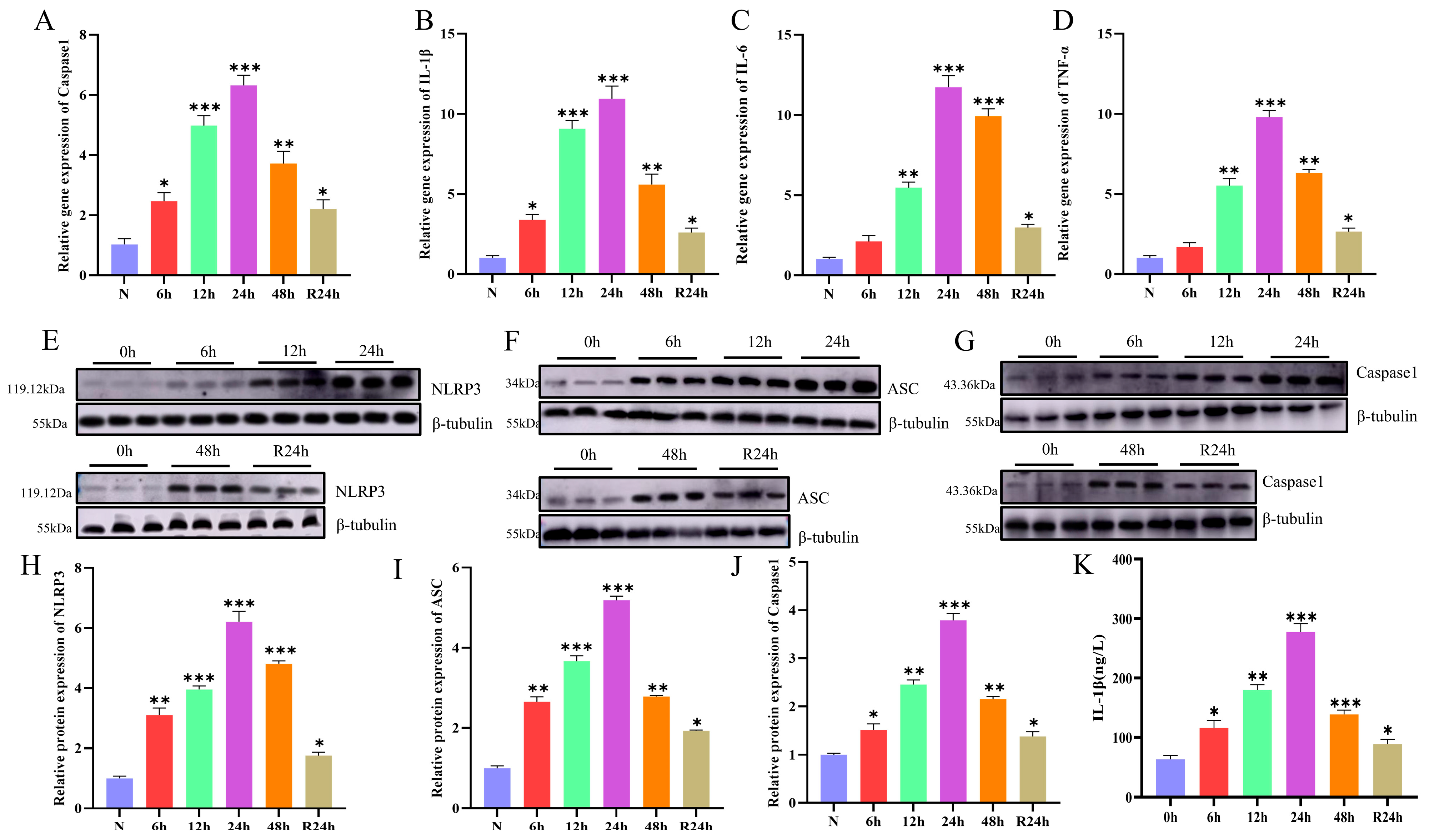
The experimental evidence reveals that hypoxic stress markedly activated the NLRP3 inflammasome pathway, igniting inflammatory cascade responses and the release of pro-inflammatory cytokines. The specificity of the three antibodies has been verified and is consistent with the theoretical molecular weight size (Figure S1B–D). At the early stage of hypoxic stress, NLRP3 was significantly up-regulated in the brain at 12 h (3.95-fold) and peaked at 24 h (6.2-fold) (Figure 4E,H), and its downstream connecting molecule, ASC levels, started to accumulate at 6 h of hypoxia (2.64-fold), peaked at 24 h (5.18-fold), and decreased significantly after reoxygenation (1.92-fold) (Figure 4F,I), and its effector protein, Caspase1, showed a trend of synchronous up-regulation (Figure 4G,J). This suggests that the synergistic activation of ASC and Caspase1 is a critical step in the assembly of inflammasomes.
The content of IL-1β reached the highest level at 24 h (10.93-fold) (Figure 4B,K), whereas il-6 and tnf-α were significantly elevated at 24 h (11.73-fold and 9.79-fold, respectively) (Figure 4C,D), suggesting that NLRP3 inflammasome-driven inflammatory responses predominate in early hypoxia. The down-regulation of inflammatory markers after reoxygenation suggests that the return of oxygen concentration can alleviate the inflammatory response by inhibiting the activity of inflammasomes.
To correlate these molecular changes with tissue-level pathology, we examined brain histology across key time points (Figure S1). While the control group exhibited normal neural architecture, pronounced edema and scattered pyknotic nuclei were evident after 24 h of hypoxia, coinciding with the peak of inflammatory cytokine expression. Notably, following 24 h of reoxygenation (R 24 h), these pathological features were markedly attenuated, indicating a substantial recovery of tissue integrity.

3.5. The Formation of Inflammasomes

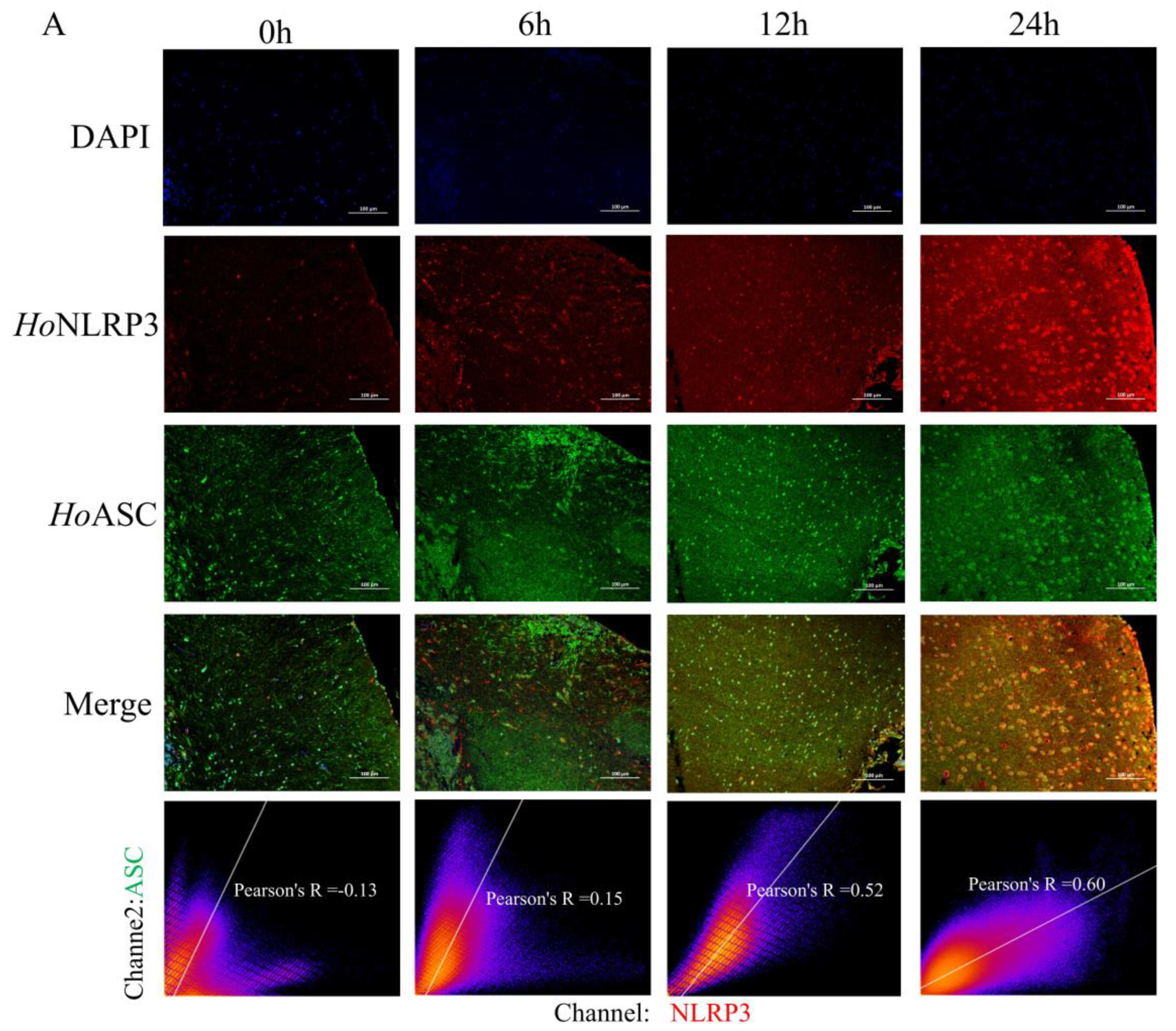
To investigate whether the NLRP3 inflammasome was formed, the interaction of NLRP3, ASC, and Caspase-1 was analyzed by the immunofluorescence co-localization technique in this study. Immunofluorescence co-localization analysis showed that under normoxic conditions (0 h), the Pearson’s R of NLRP3 with ASC was −0.13, indicating that the two were randomly distributed in the brain and did not form a functional complex. After 6 h of hypoxic stress, the Pearson’s R of NLRP3-ASC increased to 0.15, suggesting that the inflammasomes entered the preliminary assembly stage. At 12 h of hypoxia, the co-localization intensity further increased (Pearson’s R of 0.52), and at 24 h, the Pearson’s R of NLRP3-ASC reached the peak (Figure 5A), and the co-localization rate of ASC and Caspase1 reached the peak simultaneously (Figure 5B), indicating that the inflammasomes had completed its maturation and formed the NLRP3 inflammasome.
To confirm that the immunofluorescence-observed colocalization represents genuine physical interactions, we performed immunoprecipitation experiments. Results demonstrated that NLRP3 directly interacts with both ASC and Caspase1 in the brain samples exposed to hypoxia for 24 h (Figure 5C). The negative control (IgG) showed no nonspecific binding, while the Input group confirmed the expression levels of each protein. These findings, together with immunofluorescence colocalization, collectively support the formation of the NLRP3 inflammasome.
The above results provide direct visual evidence for the formation of the NLRP3-ASC-Caspase1 complex, suggesting that hypoxic stress drives inflammasome assembly, which in turn initiates downstream inflammatory signaling cascades.

3.6. Late Hypoxia Triggers Caspase3/GSDME-Mediated Pyroptosis

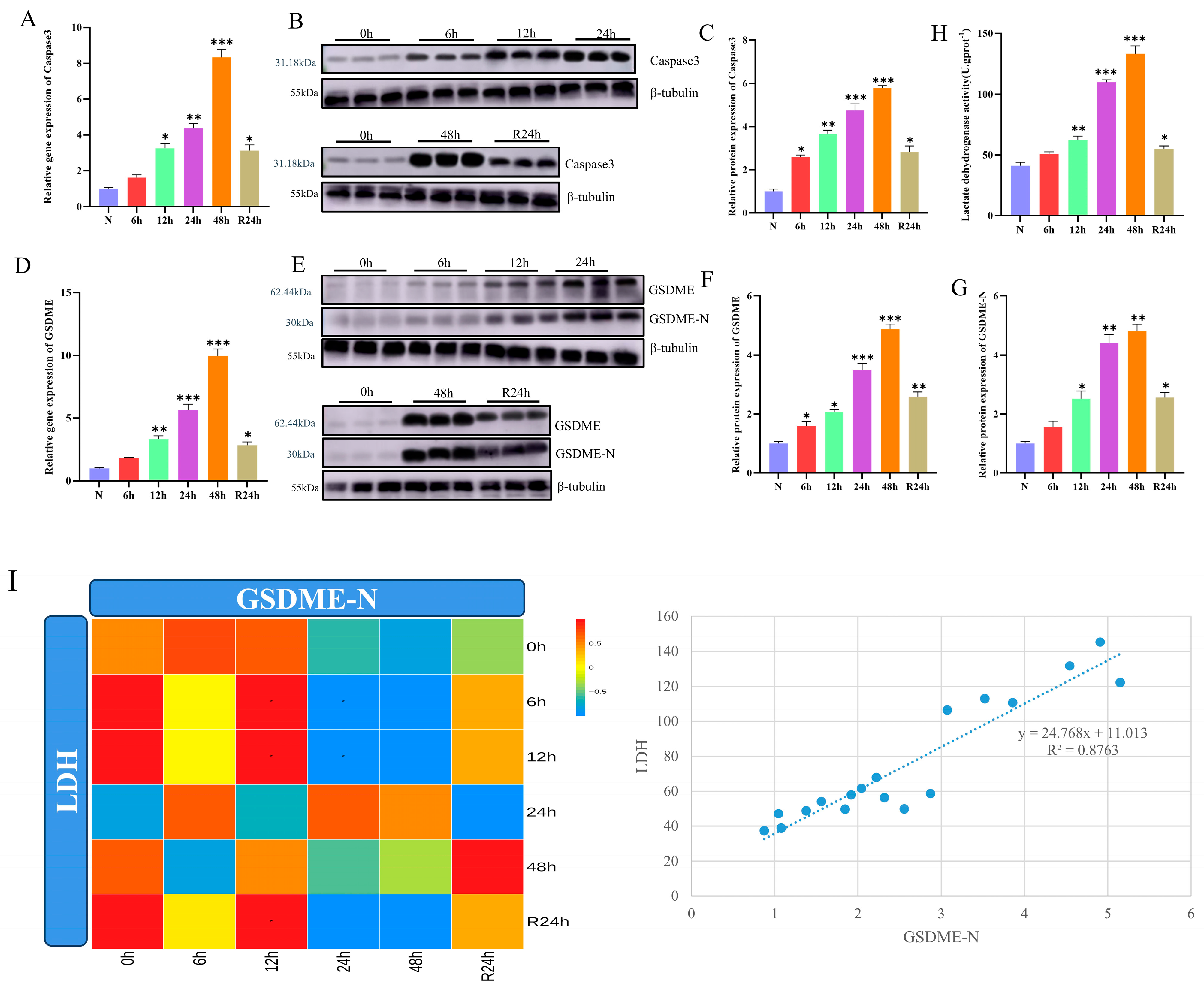
To reveal the global regulatory role of NLRP3 under hypoxic stress, the present study further analyzed the mechanism of its association with late cellular pyroptosis. The qRT-PCR results showed that the mRNA expression of caspase3 and gsdme was noticeably upgraded to peak at 48 h of hypoxia (8.32-fold and 9.96-fold, respectively), and Western blot further verified the accumulation of both proteins as well as GSDME-N-terminus, suggesting the initiation of the executive stage of pyroptosis (Figure 6A–G). The results of antibody specificity validation are shown in Figure S1E,F. Additionally, total LDH activity in the brain peaked after 48 h of hypoxia, exhibiting a dynamic expression pattern highly synchronized with the accumulation of GSDME N-terminus. This strong positive correlation suggests that the dramatic increase in total LDH activity likely reflects the loss of cell membrane integrity mediated by GSDME pore formation, thereby providing crucial indirect evidence for pyroptosis occurrence. (Figure 6H,I). After 24 h of reoxygenation, pyroptosis markers were partially restored, suggesting that the return of oxygen concentration could alleviate the injury by suppressing the downstream signaling of the NLRP3 pathway.

4. Discussion

The immune defense system of bony fishes, as a vertebrate of low evolutionary status, mainly relies on the signal recognition function of PRRs in the innate immune system. NLRP3 belongs to the family of NOD-like receptors (NLRs). Existing studies have clearly elucidated the pivotal role of the NLRP3 inflammasome in pathogen infection processes across teleost fish such as zebrafish, carp, and flounder, establishing it as a key antibacterial defense mechanism [34,35,36,37]. However, this study reveals that under environmental stressors like hypoxia, NLRP3 assumes a more complex, dynamically coordinated role in H. otakii. During early hypoxia (6–24 h), it drives IL-1β-mediated inflammatory responses by assembling NLRP3-ASC-Caspase1 inflammasomes. In contrast, during prolonged hypoxia into the late stage (48 h), it activates the Caspase3/GSDME pathway to induce pyroptosis, thereby eliminating severely damaged cells. This time-dependent “inflammation–pyroptosis” biphasic regulatory pattern significantly expands our understanding of NLRP3 function in fishes—elevating it from a traditional “anti-infective molecular switch” to an adaptive regulatory center that coordinates immune defense and cell death decisions in dynamic environments.
The HoNLRP3 identified in this study, with a coding sequence length of 1584 bp, encodes a protein containing conserved structural domains such as NACHT, LRR, and PYD, which is consistent with the typical features of NLRP3 in mammals and zebrafish, suggesting that it possesses the molecular basis for the assembly of inflammasomes. Phylogenetic analyses showed that HoNLRP3 shares a highly conserved structural domain composition with its mammalian and zebrafish homologs, but there are also significant differences. Sequence alignment showed that HoNLRP3 also contains FISNA structural domains and multiple species-specific motifs (Figure 1), which are unique to fish NLRPs [38]. Notably, there is still a lack of experimental evidence supporting the specific functional mechanisms of these structural units in inflammasome assembly, and, in particular,, the biological functions of the FISNA structural domains and several fish-specific motifs have not yet been systematically resolved after hypoxic or pathogenic stimulation.
The rapid response mechanism of NLRP3 in the brain may involve multiple regulatory networks. In this study, we found that nlrp3 mRNA was significantly up-regulated at 12 h of hypoxia and peaked at 24 h (Figure 2), whereas the early accumulation of hif-1α mRNA (12 h) preceded the peak protein levels of NLRP3 (12–24 h). The two expressions exhibit strong temporal correlation, suggesting a potential regulatory relationship. In mammals, HIF-1α has been demonstrated to directly bind the HRE element within the NLRP3 promoter region. However, future studies are necessary to determine if HIF-1α directly binds to the NLRP3 promoter to drive its expression under hypoxic stress in H. otakii. It has been shown that after thalamic hemorrhagic stroke, HIF-1α was significantly upregulated in the peri-injury region, activated microglia and astrocytes, drove the formation of NLRP3 inflammasome, and facilitated the release of pro-inflammatory cytokines (e.g., IL-1β, TNF-α, IL-6) [39]. Notably, the delayed responses of other tissues (e.g., gills, liver) may reflect their compensatory adaptive strategies (Figure 2), with some studies suggesting that gills may mitigate oxygen deficit by increasing blood flow distribution, whereas the liver prioritizes maintenance of metabolic homeostasis over immune activation [40,41,42,43]. This pattern of tissue-specific regulation may be an evolutionary trade-off designed to ensure preferential protection of critical organs (e.g., brain) under acute hypoxia while reducing energy expenditure in peripheral tissues.
Although the early inflammatory response provides a rapid immune defense against acute hypoxia, sustained activation of the NLRP3 pathway may breach physiological regulatory thresholds, leading to the onset of programmed cell death [44]. The results of qRT-PCR, Western blot, and immunofluorescence showed that activation of the NLRP3 inflammasome drives IL-1β maturation and release, which provides short-term relief of oxygen deficit by recruiting immune cells in the 6–24 h of hypoxia-stress (Figure 4 and Figure 5). However, excessive IL-1β release may activate NF-κB signaling through a positive feedback loop, further upregulating NLRP3 expression and creating an inflammatory self-amplification effect [45,46,47]. This process is consistent with studies in mammals, but may be more tightly regulated in fish to avoid excessive injury. Persistent inflammatory signaling leads to a breakdown in mitochondrial membrane potential, releasing LDH and activating the Caspase8-Caspase3 cascade [48,49], which in turn cleaves the GSDME, triggering pyroptosis at 48 h of hypoxia-stress (Figure 6). The fish execute pyroptosis through Caspase3/GSDME, a difference that may reflect their evolutionary adaptive strategy: the activation threshold of GSDME is higher, requiring stronger or more persistent damage signals to trigger pore formation, thus avoiding premature initiation of cell death under transient hypoxia, which is different from mammals relying on Caspase1/GSDMD. Disruption of cell membrane integrity due to pyroptosis may release intracellular pro-inflammatory signals (e.g., ATP, HMGB1) [50], which may exacerbate tissue injury by activating NLRP3 inflammasome in neighboring cells to create a vicious cycle [51]. Furthermore, the experimental design of this study was primarily focused on the pyroptosis pathway; thus, specific markers for other cell death pathways, such as apoptosis and necroptosis, were not concurrently assessed. While robust GSDME cleavage, significantly elevated LDH activity, and the concomitant inflammatory milieu collectively support pyroptosis as the dominant cell death mechanism, the possibility that other forms of cell death operate in a minor or synergistic capacity within the complex in vivo environment cannot be excluded. Elucidating the potential crosstalk between these distinct cell death pathways under hypoxic stress represents a significant direction for future research.
The results presented of the repair phase of reoxygenation provide new clues for the study of hypoxia tolerance in fish. The expression of NLRP3 and its downstream molecules (e.g., IL-1β, Caspase3) was partially restored after 24 h of reoxygenation, suggesting the involvement of multiple repair pathways (Figure 4 and Figure 6). Primarily, oxygen concentration rebound prompted the degradation of HIF-1α and reduced its transcriptional drive for NLRP3. And then, the anti-inflammatory cytokines (e.g., TGF-β, IL-10) may block inflammasome assembly by inhibiting NF-κB signaling [52,53]. In addition, autophagy activation may reduce ROS generation by removing damaged mitochondria, thereby inhibiting NLRP3 overactivation [54,55]. For example, it is reported that autophagy inhibits NLRP3 inflammasome activation by degrading damaged mitochondria [56], a mechanism that may be equally important in fish. However, this study found that under reoxygenation conditions, proinflammatory cytokine and LDH levels remained elevated above normal. Underlying mechanisms may involve the long-term effects of epigenetic regulation. Research indicates that hypoxic environments may induce alterations in DNA methylation patterns, particularly affecting methylation levels in the promoter regions of proinflammatory genes, thereby promoting their sustained expression [57,58]. Histone modifications, such as increased acetylation levels, may also be activated during hypoxia and maintain pro-inflammatory gene transcriptional activity after returning to normoxia [59]. Furthermore, pyroptosis markers like GSDME-N only partially recover after reoxygenation, suggesting pyroptosis is not fully reversible. This may limit fish’s long-term physiological tolerance to hypoxia. The experimental evidence reveals that the mechanism of the temporal dynamic balance of the hypoxic response through the “HIF-1α/NLRP3/inflammation/Pyroptosis” regulatory axis in H. otakii (Figure 7). Theoretically, this finding enriches our recognition of the system of immunometabolism adaptation in fishes, especially the evolutionary conservation and species-specificity of the cross-regulation of inflammation and cell death. On the application, key nodes of the NLRP3-pyroptosis pathway (e.g., GSDME) may become molecular targets for hypoxia-resistant fish breeding to alleviate tissue damage through gene editing or inhibitor intervention. However, the present study still has limitations: Initially, the direct regulation of NLRP3 by HIF-1α needs to be further verified. Furthermore, the central role of the Caspase3/GSDME pathway in pyroptosis has not been fully clarified. Additionally, it should be noted that this study used mixed-sex fish cohorts. Although our randomized sampling was designed to capture the general response of the species, we cannot exclude potential sex-based differences in hypoxia tolerance or immune regulation as reported in other teleosts [60,61,62]. Ultimately, the mechanism of communication between brain tissues and other organs (e.g., exosomes or cytokines remotely modulated) has not yet been resolved. These unresolved questions point the way forward for future research into the mechanisms of hypoxic adaptation in teleost fish.

5. Conclusions

This study elucidates the molecular characteristics and functional dynamics of NLRP3 inflammasome activation in H. otakii under hypoxic stress, revealing a biphasic regulatory mechanism that transitions from early inflammatory defense to late pyroptosis. The conserved yet evolutionarily distinct features of HoNLRP3, its brain-specific expression, and its correlation with HIF-1α activation highlight its central role in hypoxic adaptation. While the partial recovery of inflammatory and pyroptotic markers upon reoxygenation underscores the plasticity of this pathway, limitations such as unresolved sex-specific responses and inter-organ communication warrant further investigation. These findings advance our understanding of NLRP3-mediated immunometabolism adaptation in teleost and provide a theoretical foundation for hypoxia-resilient aquaculture development.

Supplementary Materials

The following supporting information can be downloaded at https://www.mdpi.com/article/10.3390/fishes10110542/s1, Figure S1. Antibody validation by Western blot analysis. (A) HIF-1α antibody: single band detected (~47 kDa), consistent with theoretical molecular weight (47.31 kDa). (B) Caspase1 antibody: recognizes ~43 kDa band, consistent with theoretical molecular weight (43.36 kDa). (C) NLRP3 antibody: shows clear single band at ~119 kDa (theoretical value 119 kDa). (D) ASC antibody: ~34 kDa target band detected (theoretical value 34 kDa). (E) Caspase3 antibody: recognizes ~31 kDa activation fragment (theoretical value 31.18 kDa). (F) GSDME antibody: specifically recognizes ~62 kDa full-length band (theoretical value 62.44 kDa) and ~35 kDa cut fragment (GSDME-N-terminal). Figure S2. Hypoxia-induced histopathological changes in the brain of H. otakii. H&E-stained sections from the (A) control (0 h) (B) 48 h hypoxia groups. (C) R 24 h groups. N. Normal nerve cell; E. Enlarged nerve cell; P. Pyknotic nuclei; V. Vacuole. Magnification: 400×. Table S1. The GenBank accession numbers of the NLRP3 in different species.

Author Contributions

Y.W.: Data curation, Formal analysis, Investigation, Software, Visualization, Writing—original draft. D.G., L.P. and Y.L.: Validation. X.Z., R.L., L.Z., J.S. and Y.Y.: Investigation, Methodology. W.W. and Z.X.: Conceptualization, Investigation, Resources, Methodology, Project administration, Validation, Writing—review and editing. All authors have read and agreed to the published version of the manuscript.

Funding

This work was supported by the Science and Technology Joint Plan Project of Liaoning Province in 2024 (2024-MSLH-047); Liaoning Province Major Science and Technology Special Project (2024JH1/11700010, for Zhuang Xue and Wei Wang); Basic Research Projects of Higher Education Institutions by Liaoning Provincial Department of Education (JYTZD2023038); Open Fund of Key Laboratory of Biotechnology and Bioresources Utilization (Dalian Minzu University), Ministry of Education (No. KF2024010), China.

Institutional Review Board Statement

All animal experiments were approved by the Ethics Committee of Dalian Ocean University (Approval Code: DLOU20250011; Approval Date: 9 September 2025) and were conducted following ARRIVE (Animal Research: Reporting of In Vivo Experiments) guidelines.

Data Availability Statement

The relevant data (including qRT-PCR, Western blot, immunofluorescence results, and statistical analysis) from this study are included in the paper and its supplementary materials. The rest of the raw data can be obtained from the corresponding author upon reasonable request.

Conflicts of Interest

The authors declare no conflicts of interest.

References

  1. Lima, A.R.; Booms, E.M.; Lopes, A.R.; Martins-Cardoso, S.; Novais, S.C.; Lemos, M.F.; Ribeiro, L.; Castanho, S.; Candeias-Mendes, A.; Pousão-Ferreira, P. Early life stage mechanisms of an active fish species to cope with ocean warming and hypoxia as interacting stressors. Environ. Pollut. 2024, 341, 122989. [Google Scholar] [CrossRef]
  2. Wood, C.M.; Eom, J. The osmorespiratory compromise in the fish gill. Comp. Biochem. Physiol. A Mol. Integr. Physiol. 2021, 254, 110895. [Google Scholar] [CrossRef]
  3. Aaskov, M.L.; Nelson, D.; Lauridsen, H.; Huong, D.T.T.; Ishimatsu, A.; Crossley, D.A., 2nd; Malte, H.; Bayley, M. Do air-breathing fish suffer branchial oxygen loss in hypoxic water? Proc. Biol. Sci. 2023, 290, 20231353. [Google Scholar] [CrossRef]
  4. Johnston, W.; Adil, S.; Cao, C.; Nipu, N.; Mennigen, J.A. Fish models to explore epigenetic determinants of hypoxia-tolerance. Comp. Biochem. Physiol. A Mol. Integr. Physiol. 2025, 302, 111811. [Google Scholar] [CrossRef]
  5. Cerra, M.C.; Filice, M.; Caferro, A.; Mazza, R.; Gattuso, A.; Imbrogno, S. Cardiac hypoxia tolerance in fish: From functional responses to cell signals. Int. J. Mol. Sci. 2023, 24, 1460. [Google Scholar] [CrossRef]
  6. Ma, Q.; Luo, Y.; Zhong, J.; Limbu, S.M.; Li, L.Y.; Chen, L.Q.; Qiao, F.; Zhang, M.L.; Lin, Q.; Du, Z.Y. Hypoxia tolerance in fish depends on catabolic preference between lipids and carbohydrates. Zool. Res. 2023, 44, 954–966. [Google Scholar] [CrossRef]
  7. Eltzschig, H.K.; Bratton, D.L.; Colgan, S.P. Targeting hypoxia signalling for the treatment of ischaemic and inflammatory diseases. Nat. Rev. Drug Discov. 2014, 13, 852–869. [Google Scholar] [CrossRef]
  8. Yuan, X.; Ruan, W.; Bobrow, B.; Carmeliet, P.; Eltzschig, H.K. Targeting hypoxia-inducible factors: Therapeutic opportunities and challenges. Nat. Rev. Drug Discov. 2024, 23, 175–200. [Google Scholar] [CrossRef]
  9. Cyran, A.M.; Zhitkovich, A. HIF1, HSF1, and NRF2: Oxidant-responsive trio raising cellular defenses and engaging immune System. Chem. Res. Toxicol. 2022, 35, 1690–1700. [Google Scholar] [CrossRef]
  10. Zhang, Y.; Yin, K.; Wang, D.; Wang, Y.; Lu, H.; Zhao, H.; Xing, M. Polystyrene microplastics-induced cardiotoxicity in chickens via the ROS-driven NF-κB-NLRP3-GSDMD and AMPK-PGC-1α axes. Sci. Total Environ. 2022, 840, 156727. [Google Scholar] [CrossRef]
  11. Yu, Q.; Shi, H.; Ding, Z.; Wang, Z.; Yao, H.; Lin, R. The E3 ubiquitin ligase TRIM31 attenuates NLRP3 inflammasome activation in Helicobacter pylori-associated gastritis by regulating ROS and autophagy. Cell Commun. Signal 2023, 21, 1. [Google Scholar] [CrossRef]
  12. Li, J.; Lin, Q.; Shao, X.; Li, S.; Zhu, X.; Wu, J.; Mou, S.; Gu, L.; Wang, Q.; Zhang, M.; et al. HIF1α-BNIP3-mediated mitophagy protects against renal fibrosis by decreasing ROS and inhibiting activation of the NLRP3 inflammasome. Cell Death Dis. 2023, 14, 200. [Google Scholar] [CrossRef]
  13. Fu, J.; Wu, H. Structural mechanisms of NLRP3 inflammasome assembly and activation. Annu. Rev. Immunol. 2023, 41, 301–316. [Google Scholar] [CrossRef]
  14. Zheng, S.; Que, X.; Wang, S.; Zhou, Q.; Xing, X.; Chen, L.; Hou, C.; Ma, J.; An, P.; Peng, Y.; et al. ZDHHC5-mediated NLRP3 palmitoylation promotes NLRP3-NEK7 interaction and inflammasomes activation. Mol. Cell 2023, 83, 4570–4585.e7. [Google Scholar] [CrossRef]
  15. Sayaf, K.; Battistella, S.; Russo, F.P. NLRP3 inflammasome in acute and chronic liver diseases. Int. J. Mol. Sci. 2024, 25, 4537. [Google Scholar] [CrossRef]
  16. Wang, H.; Ma, L.; Su, W.; Liu, Y.; Xie, N.; Liu, J. NLRP3 inflammasome in health and disease (Review). Int. J. Mol. Med. 2025, 55, 48. [Google Scholar] [CrossRef]
  17. Vasudevan, S.O.; Behl, B.; Rathinam, V.A. Pyroptosis-induced inflammation and tissue damage. Semin. Immunol. 2023, 69, 101781. [Google Scholar] [CrossRef]
  18. Xu, J.; Núñez, G. The NLRP3 inflammasome: Activation and regulation. Trends Biochem. Sci. 2023, 48, 331–344. [Google Scholar] [CrossRef]
  19. Toldo, S.; Abbate, A. The role of the NLRP3 inflammasome and pyroptosis in cardiovascular diseases. Nat. Rev. Cardiol. 2024, 21, 219–237. [Google Scholar] [CrossRef]
  20. Abderrazak, A.; Syrovets, T.; Couchie, D.; El Hadri, K.; Friguet, B.; Simmet, T.; Rouis, M. NLRP3 inflammasome: From a danger signal sensor to a regulatory node of oxidative stress and inflammatory diseases. Redox Biol. 2015, 4, 296–307. [Google Scholar] [CrossRef]
  21. Tourkochristou, E.; Aggeletopoulou, I.; Konstantakis, C.; Triantos, C. Role of NLRP3 inflammasome in inflammatory bowel diseases. World J. Gastroenterol. 2019, 25, 4796. [Google Scholar] [CrossRef]
  22. Ma, X.; Jiao, J.; Aierken, M.; Sun, H.; Chen, L. Hypoxia Inducible Factor-1α through ROS/NLRP3 pathway regulates the mechanism of acute ischemic stroke microglia scorching mechanism. Biologics 2023, 17, 167–180. [Google Scholar] [CrossRef]
  23. Wu, X.; Gong, L.; Xie, L.; Gu, W.; Wang, X.; Liu, Z.; Li, S. NLRP3 deficiency protects against intermittent hypoxia-induced neuroinflammation and mitochondrial ROS by promoting the PINK1-Parkin pathway of mitophagy in a murine model of sleep apnea. Front. Immunol. 2021, 12, 628168. [Google Scholar] [CrossRef]
  24. Zhang, B.; Zhao, M.; Ji, X.; Xia, Q.; Jiang, L.; Zhao, L. Acrylamide induces neurotoxicity in zebrafish (Danio rerio) via NLRP3-mediated pyroptosis. Sci. Total Environ. 2023, 896, 165208. [Google Scholar] [CrossRef]
  25. Zhao, Y.; Liang, Y.; Chen, Q.; Shan, S.; Yang, G.; Li, H. The function of NLRP3 in anti-infection immunity and inflammasomes assembly of common carp (Cyprinus carpio L.). Fish Shellfish Immunol. 2024, 145, 109367. [Google Scholar] [CrossRef]
  26. Song, Z.; Zou, J.; Wang, M.; Chen, Z.; Wang, Q. A comparative review of pyroptosis in mammals and fish. J. Inflamm. Res. 2022, 15, 2323–2331. [Google Scholar] [CrossRef]
  27. Hao, K.; Yuan, L.; Yu, C.; Xu, H.; Sun, L. Paralichthys olivaceus GSDME-mediated pyroptosis is regulated by multiple caspases in different manners. Fish Shellfish Immunol. 2024, 155, 110002. [Google Scholar] [CrossRef]
  28. Xu, H.; Qin, K.; Hao, K.; Yuan, Z.; Sun, L. Pufferfish gasdermin Ea is a significant player in the defense against bacterial pathogens. Mar. Life Sci. Technol. 2024, 6, 462–474. [Google Scholar] [CrossRef]
  29. Zhao, Y.; Qiao, D.; Zhang, J.; Gao, F.; Pei, C.; Li, C.; Kong, X. Activation mechanism of CcGSDMEb-1/2 and regulation for bacterial clearance in common carp (Cyprinus carpio). J. Immunol. 2023, 211, 658–672. [Google Scholar] [CrossRef]
  30. Zhao, Y.; Zhang, J.; Qiao, D.; Gao, F.; Gu, Y.; Jiang, X.; Zhu, L.; Kong, X. CcGSDMEa functions the pore-formation in cytomembrane and the regulation on the secretion of IL-lβ in common carp (Cyprinus carpio haematopterus). Front. Immunol. 2022, 13, 1110322. [Google Scholar] [CrossRef]
  31. Zhan, Y.; Gao, D.; Peng, L.; Cui, D.; Li, G.; Cao, S.; Chen, Y.; Xue, Z.; Wang, W. Hypoxia induces pyroptosis and inflammation in the liver of fat greenling (Hexagrammos otakii). Comp. Immunol. Rep. 2024, 6, 200146. [Google Scholar] [CrossRef]
  32. Gao, D.; Wu, Y.; Zhan, Y.; Peng, L.; Zhao, L.; Cao, S.; Xue, Z.; Wang, W. Chronic hypoxia drives the occurrence of ferroptosis in liver of fat greening (Hexagrammos otakii) by activating HIF-1α and promoting iron production. Ecotoxicol. Environ. Saf. 2024, 285, 117135. [Google Scholar] [CrossRef]
  33. Yang, Z.; Lin, P.; Chen, B.; Zhang, X.; Xiao, W.; Wu, S.; Huang, C.; Feng, D.; Zhang, W.; Zhang, J. Autophagy alleviates hypoxia-induced blood-brain barrier injury via regulation of CLDN5 (claudin 5). Autophagy 2021, 17, 3048–3067. [Google Scholar] [CrossRef]
  34. Li, J.Y.; Wang, Y.Y.; Shao, T.; Fan, D.D.; Lin, A.F.; Xiang, L.X.; Shao, J.Z. The zebrafish NLRP3 inflammasome has functional roles in ASC-dependent interleukin-1β maturation and gasdermin E-mediated pyroptosis. J. Biol. Chem. 2020, 295, 1120–1141. [Google Scholar] [CrossRef]
  35. Chen, H.; Ding, S.; Tan, J.; Yang, D.; Zhang, Y.; Liu, Q. Characterization of the Japanese flounder NLRP3 inflammasome in restricting Edwardsiella piscicida colonization in vivo. Fish Shellfish Immunol. 2020, 103, 169–180. [Google Scholar] [CrossRef]
  36. Morimoto, N.; Kono, T.; Sakai, M.; Hikima, J.I. Inflammasomes in teleosts: Structures and mechanisms that induce pyroptosis during bacterial infection. Int. J. Mol. Sci. 2021, 22, 4389. [Google Scholar] [CrossRef]
  37. Deng, N.; Zhao, Y.; Xu, J.; Ouyang, H.; Wu, Z.; Lai, W.; Lu, Y.; Lin, H.; Zhang, Y.; Lu, D. Molecular characterization and functional study of the NLRP3 inflammasome genes in Tetraodon nigroviridis. Fish Shellfish Immunol. 2022, 131, 570–581. [Google Scholar] [CrossRef]
  38. Acevedo, W.; Morán-Figueroa, R.; Vargas-Chacoff, L.; Morera, F.J.; Pontigo, J.P. Revealing the salmo salar NLRP3 inflammasome: Insights from structural modeling and transcriptome analysis. Int. J. Mol. Sci. 2023, 24, 4556. [Google Scholar] [CrossRef]
  39. Shi, Z.M.; Jing, J.J.; Xue, Z.J.; Chen, W.J.; Tang, Y.B.; Chen, D.J.; Qi, X.Y.; Huang, L.; Zou, Y.Q.; Wu, X.Z.; et al. Stellate ganglion block ameliorated central post-stroke pain with comorbid anxiety and depression through inhibiting HIF-1α/NLRP3 signaling following thalamic hemorrhagic stroke. J. Neuroinflam. 2023, 20, 82. [Google Scholar] [CrossRef]
  40. Jia, Y.; Gao, Y.; Wan, J.; Gao, Y.; Li, J.; Guan, C. Altered physiological response and gill histology in black rockfish, Sebastes schlegelii, during progressive hypoxia and reoxygenation. Fish Physiol. Biochem. 2021, 47, 1133–1147. [Google Scholar] [CrossRef]
  41. Ding, W.; Cao, L.; Cao, Z.; Bing, X. Gill and brain transcriptomic analysis of mandarin fish (Siniperca chuatsi) reveals hypoxia-induced mitochondrial dysfunction and modulation of metabolism. Comp. Biochem. Physiol. Part. D Genom. Proteom. 2025, 53, 101367. [Google Scholar] [CrossRef]
  42. Liu, Q.; Wang, H.; Ge, J.; Li, L.; Luo, J.; He, K.; Yan, H.; Zhang, X.; Tahir, R.; Luo, W.; et al. Chronic hypoxia and Cu2+ exposure induce gill remodeling of largemouth bass through endoplasmic reticulum stress, mitochondrial damage and apoptosis. Aquat. Toxicol. 2023, 255, 106373. [Google Scholar] [CrossRef]
  43. Wang, Y.; Zhao, Y.; Gong, W.; Hou, Y.; Ren, J.; Duan, C.; Zhang, H.; Nie, X.; Li, J. Aspirin exposure coupled with hypoxia interferes energy metabolism, antioxidant and autophagic processes and causes liver injury in estuarine goby Mugilogobius chulae. J. Hazard. Mater. 2024, 476, 135071. [Google Scholar] [CrossRef]
  44. Zheng, M.; Kanneganti, T.D. The regulation of the ZBP1-NLRP3 inflammasome and its implications in pyroptosis, apoptosis, and necroptosis (PANoptosis). Immunol. Rev. 2020, 297, 26–38. [Google Scholar] [CrossRef]
  45. He, Y.; Li, Z.; Xu, T.; Luo, D.; Chi, Q.; Zhang, Y.; Li, S. Polystyrene nanoplastics deteriorate LPS-modulated duodenal permeability and inflammation in mice via ROS drived-NF-κB/NLRP3 pathway. Chemosphere 2022, 307 Pt 1, 135662. [Google Scholar] [CrossRef]
  46. Shah, B.; Solanki, N. Aegeline attenuates TNBS-induced colitis by suppressing the NF-ƙB-mediated NLRP3 inflammasome pathway in mice. Inflammopharmacology 2024, 32, 2589–2599. [Google Scholar] [CrossRef]
  47. Zheng, X.; Qiu, J.; Gao, N.; Jiang, T.; Li, Z.; Zhang, W.; Gong, Y.; Hong, Z.; Hong, H. Paroxetine attenuates chondrocyte pyroptosis and inhibits osteoclast formation by inhibiting NF-κB pathway activation to delay osteoarthritis progression. Drug Des. Dev. Ther. 2023, 17, 2383–2399. [Google Scholar] [CrossRef]
  48. Huang, Y.; Liu, J.; Lin, C.; Zhu, Q.; Wu, L. Impact of Caspase3/GSDME-mediated pyroptosis on tumor immune microenvironment and clinical prognosis across multiple cancers. Cancer Manag. Res. 2024, 16, 1663–1683. [Google Scholar] [CrossRef]
  49. Tang, J.; Bei, M.; Zhu, J.; Xu, G.; Chen, D.; Jin, X.; Huang, J.; Dong, J.; Shi, L.; Xu, L.; et al. Acute cadmium exposure induces GSDME-mediated pyroptosis in triple-negative breast cancer cells through ROS generation and NLRP3 inflammasome pathway activation. Environ. Toxicol. Pharmacol. 2021, 87, 103686. [Google Scholar] [CrossRef]
  50. Liu, X.; Lieberman, J. Inflammasomes-independent pyroptosis. Curr. Opin. Immunol. 2024, 88, 102432. [Google Scholar] [CrossRef]
  51. Zheng, X.; Wan, J.; Tan, G. The mechanisms of NLRP3 inflammasome/pyroptosis activation and their role in diabetic retinopathy. Front. Immunol. 2023, 14, 1151185. [Google Scholar] [CrossRef]
  52. Shah, B.; Solanki, N. Ameliorative effect of nodakenin in combating TNBS-induced ulcerative colitis by suppressing NF-ƙB-mediated NLRP3 inflammasome pathway. Naunyn Schmiedebergs Arch. Pharmacol. 2025, 398, 673–686. [Google Scholar] [CrossRef]
  53. Drakul, M.; Tomić, S.; Bekić, M.; Mihajlović, D.; Vasiljević, M.; Rakočević, S.; Đokić, J.; Popović, N.; Bokonjić, D.; Čolić, M. Sitagliptin induces tolerogenic human dendritic cells. Int. J. Mol. Sci. 2023, 24, 6829. [Google Scholar] [CrossRef]
  54. Zhou, Y.; Zhang, Y.; Wang, H.; Zhang, X.; Chen, Y.; Chen, G. Microglial pyroptosis in hippocampus mediates sevolfurane-induced cognitive impairment in aged mice via ROS-NLRP3 inflammasome pathway. Int. Immunopharmacol. 2023, 116, 109725. [Google Scholar] [CrossRef]
  55. Han, X.; Xu, T.; Fang, Q.; Zhang, H.; Yue, L.; Hu, G.; Sun, L. Quercetin hinders microglial activation to alleviate neurotoxicity via the interplay between NLRP3 inflammasome and mitophagy. Redox Biol. 2021, 44, 102010. [Google Scholar] [CrossRef]
  56. Qiu, W.Q.; Ai, W.; Zhu, F.D.; Zhang, Y.; Guo, M.S.; Law, B.Y.; Wu, J.M.; Wong, V.K.; Tang, Y.; Yu, L.; et al. Polygala saponins inhibit NLRP3 inflammasome-mediated neuroinflammation via SHP-2-Mediated mitophagy. Free Radic. Biol. Med. 2022, 179, 76–94. [Google Scholar] [CrossRef]
  57. Choudhry, H.; Harris, A.L. Advances in hypoxia-inducible factor biology. Cell Metab. 2018, 27, 281–298. [Google Scholar] [CrossRef]
  58. Qi, R.; Xie, Y.; Zhang, X.; Jiang, S.; Liu, X.; Xie, W.; Jia, X.; Bade, R.; Liu, Y.; Gong, K. Possible involvement of DNA methylation in TSC1 gene expression in neuroprotection induced by hypoxic preconditioning. Oxidative Med. Cell. Longev. 2022, 2022, 9306097. [Google Scholar] [CrossRef]
  59. Kim, J.; Lee, H.; Yi, S.-J.; Kim, K. Gene regulation by histone-modifying enzymes under hypoxic conditions: A focus on histone methylation and acetylation. Exp. Mol. Med. 2022, 54, 878–889. [Google Scholar] [CrossRef]
  60. Lai, K.P.; Tam, N.; Wang, S.Y.; Lin, X.; Chan, T.F.; Au, D.W.T.; Wu, R.S.S.; Kong, R.Y.C. Hypoxia causes sex-specific hepatic toxicity at the transcriptome level in marine medaka (Oryzias melastigma). Aquat. Toxicol. 2020, 224, 105520. [Google Scholar] [CrossRef]
  61. Das, T.; Soren, K.; Yerasi, M.; Kumar, A.; Chakravarty, S. Revealing sex-specific molecular changes in hypoxia-ischemia induced neural damage and subsequent recovery using zebrafish model. Neurosci. Lett. 2019, 712, 134492. [Google Scholar] [CrossRef] [PubMed]
  62. Zhao, C.; Song, Y.; Zhang, G.; Zhang, K.; Yin, S.; Ji, J. Multi-omics analysis identifies sex-specific hepatic protein-metabolite networks in yellow catfish (Pelteobagrus fulvidraco) exposed to chronic hypoxia. Int. J. Biol. Macromol. 2024, 268, 131892. [Google Scholar] [CrossRef] [PubMed]
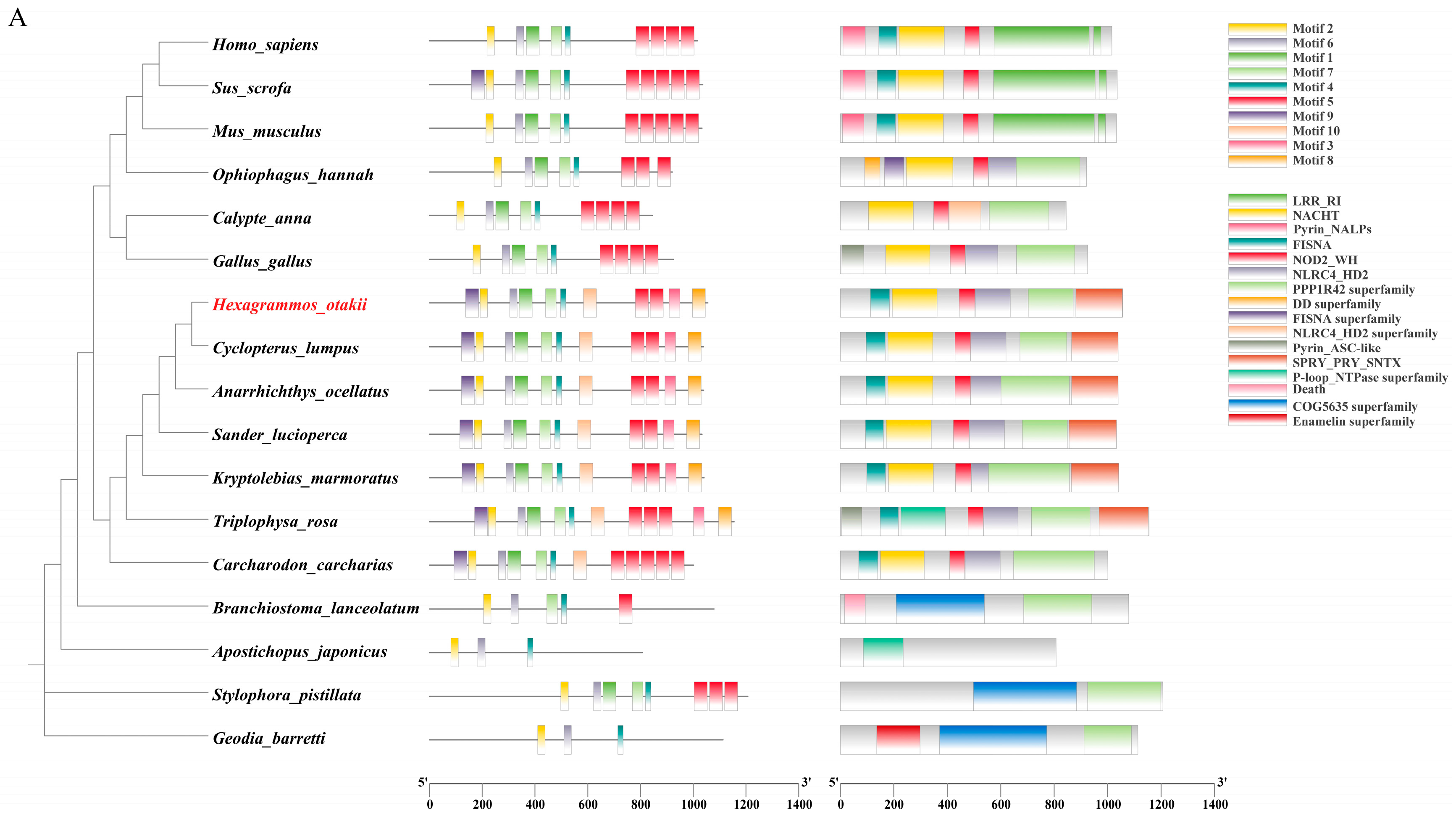
Figure 1. Sequence analysis of HoNLRP3. (A) Phylogenetic, domain, and motif visualization of NLRP3 relationships between fish and other species. Sequence alignment and the phylogenetic tree were constructed in MEGA7.0. The above results were visualized using TBtools-II. H. otakii marked with a special red tag. (B) The results of the HoNLRP3 open reading frame. The yellow region represents its NACHT structural domain; the blue region is the fish-specific FISNA structural domain; purple represents the LRR structural domain, and green and red represent the PYD structural domain. (C) The Motif seqlogo and 3D model of HoNLRP3 protein. A larger value of the bit is more conservative. Red represents Motif10. Pink represents Motif9. Yellow represents Motif3. Blue represents Motif8.
Fishes 10 00542 g001aFishes 10 00542 g001b
Figure 2. The mRNA expression of nlrp3 in various tissues of H. otakii was measured by qPCR. (A) Tissue distribution under normoxic conditions. K-Kidney, H-Heart, G-Gill, L-Liver, S-Stomach, Sp-Spleen, B-Brain; Relative expression of nlrp3 in the brain (B), spleen (C), gills (D), intestine (E), liver (F), stomach (G) at different times of hypoxic stress and after reoxygenation. Data normalized to β-actin. Data are shown as mean ± SE (n = 3 independent biological replicates, each replicate is a pool of tissues from three fish) and were analyzed by Tukey’s HSD test following ANOVA. *** p < 0.001; ** p < 0.01; * p < 0.05.
Fishes 10 00542 g002
Figure 3. The content of HIF-1α in the brain of H. otakii. (A) The relative expression of hif-1α mRNA in hypoxic stress and reoxygenation. Data normalized to β-actin. (B,C) The levels of HIF-1α were measured by Western blot. (D) Intergroup correlation analysis of hif1-α and nlrp3 mRNA, R2 closer to 1 represents higher correlation. Data are shown as mean ± SE (n = 3 independent biological replicates, each replicate is a pool of tissues from three fish) and were analyzed by Tukey’s HSD test following ANOVA. * p < 0.05, ** p < 0.01, *** p < 0.001.
Fishes 10 00542 g003
Figure 4. Effects of hypoxia on gene expression and protein levels associated with inflammatory responses. (AD) Relative expression of caspase1, il-6, tnf-α, and il-1β was measured by qPCR and normalized to β-actin. (EJ) Changes in NLRP3, ASC, and Caspase1 levels were measured by Western blot. (K) IL-1β content detection. Data are shown as mean ± SE (n = 3 independent biological replicates, each replicate is a pool of tissues from three fish) and were analyzed by Tukey’s HSD test following ANOVA. * p < 0.05, ** p < 0.01, *** p < 0.001.
Fishes 10 00542 g004
Figure 5. NLRP3 inflammasome activation in the brain of H. otakii under hypoxic stress. (A) Trends in NLRP3-ASC complex dynamics from 0 to 24 h of hypoxic stress. (200×) DAPI-labeled cell nucleus (blue), HoNLRP3 (red), HoASC (green), co-localization signal (Merge, yellow). (B) Trends in ASC-Caspase1 complex dynamics from 0 to 24 h of hypoxic stress. (200×) DAPI-labeled cell nucleus (blue), HoCaspase1 (red), HoASC (green), co-localization signal (Merge, yellow). Larger Pearson’s R coefficients indicate higher co-localization rates. (C) Immunoprecipitation of NLRP3, ASC, and Caspase1. The immunofluorescence images are representative of the findings observed across all samples in their respective groups.
Fishes 10 00542 g005aFishes 10 00542 g005b
Figure 6. Caspase3/GSDME-mediated pyroptosis and total LDH activity downstream of NLRP3 inflammasome. (A,D) The mRNA expression of caspase3, gsdme was measured by qRT-PCR; the data were normalized to β-actin. (B,C,EG) The levels of Caspase3, GSDME, and GSDME-N-terminal were measured by Western blot. (H) Total LDH activity. (I) Correlation analysis between LDH and GSDME-N. Data are shown as mean ± SE (n = 3 independent biological replicates, each replicate is a pool of tissues from three fish) and were analyzed by Tukey’s HSD test following ANOVA. * p < 0.05, ** p < 0.01, *** p < 0.001.
Fishes 10 00542 g006
Figure 7. Biphasic hypoxic response mechanism of HoNLRP3 inflammasome. Under hypoxic stress, early HIF-1α accumulation is associated with NLRP3 upregulation and the assembly of the NLRP3-ASC-Caspase1 inflammasomes (Pearson’s R > 0.7). This functional inflammasome subsequently drives IL-1β maturation and release, initiating the inflammatory response (6–24 h). Continued hypoxia activates Caspase3 to shear GSDME, GSDME-N is upregulated, resulting in pyroptosis (48 h).
Fishes 10 00542 g007
Table 1. Information of primers used in qRT-PCR.
GenePrimerPrimer Sequence (5′-3′)Product Length (bp)Tm
HIF1-αHIF-1α-FGCTGGGTGACATAAGAGAGATG11255.1
HIF-1α-RTGAAGGCAGCAGAAGTATGG54.8
NLRP3NLRP3-FCCCAGTCCAGAGTGAACTTATG9655.5
NLRP3-RCACCTGGAAGAGAGATACCAATC54.6
Gasdermin-EGasdermin-E-FACAAGTCGTTGAGGGTGAAG12854.8
Gasdermin-E-RCATCCATGTGTCCCGAGTAAA54.4
Caspase3Caspase3-FGTCGATGCTGACCAAAGAGA11454.6
Caspase3-RCCACCTCACACACACATACA54.6
Caspase1Caspase1-FTTTGGCCGCAGGGTAAATA10854.1
Caspase1-RGCTGCAGAGCAACAGATAGA54.7
IL-1βIL-1β-FGAGGAATGCTCGAAGCTGAA11854.9
IL-1β-RGGCACTTCACGGACTCAAA55.4
IL-6IL-6-FGTCTGTATCTGGCCGTGATATG10355.1
IL-6-RATGACCGTTACCTGGAGTTTG54.6
TNF-αTNF-α-FCTTCTACCAGTACGCACATCC12155.3
TNF-α-RAACACTCAGACAGCCATACAC54.6
β-actinβ-actin-FCTGGTCTGGATTGGCTGTGA8957.5
β-actin-RGGAAGGAAGGCTGGAAGAGG58.3
Table 2. Antibody information used in this study.
AntibodyCatalog No.SourceCompany Information
HIF1-αRM7162RabbitBiodragon, Suzhou, China
NLRP368102-1-IgMouseProteintech, Wuhan, China
ASCBY2922RabbitAbways, Shanghai, China
Caspase1BYab-00584RabbitByabscience, Nanjing, China
Caspase3BD-PT0656RabbitBiodragon, Suzhou, China
GSDME13075-1-APRabbitProteintech, Wuhan, China
IgG10284-1-APRabbitProteintech, Wuhan, China
rabbit IgG30000-0-APRabbitProteintech, Wuhan, China
β-tubulinGB15140MouseServicebio, Wuhan, China
Goat Anti-RabbitRGAR001GoatProteintech, Wuhan, China
Goat Anti-MouseRGAM001GoatProteintech, Wuhan, China
Disclaimer/Publisher’s Note: The statements, opinions and data contained in all publications are solely those of the individual author(s) and contributor(s) and not of MDPI and/or the editor(s). MDPI and/or the editor(s) disclaim responsibility for any injury to people or property resulting from any ideas, methods, instructions or products referred to in the content.