Article

Assessment of Hepatic Enzyme Biomarkers in Northern Pike (Esox lucius) from Lotic and Lentic Freshwater Habitats: Implications for Monitoring Metal Pollution and Ecological Stress in Aquatic Ecosystems

by
Katarina Jovičić
1,
Vesna Đikanović
1,
Srđan Subotić
2,
Milena Dimitrijević
3,
Snežana Kovačević
3,
Branko Miljanović
4 and
Jelena S. Vranković
1,*
1
Department of Hydroecology and Water Protection, Institute for Biological Research “Siniša Stanković”—National Institute of Republic of Serbia, University of Belgrade, Bulevar Despota Stefana 142, 11008 Belgrade, Serbia
2
Faculty of Biology, University of Belgrade, Studentski Trg 16, 11000 Belgrade, Serbia
3
Life Sciences Department, Institute for Multidisciplinary Research—National Institute of Republic of Serbia, University of Belgrade, Kneza Višeslava 1, 11000 Belgrade, Serbia
4
Department of Biology and Ecology, Faculty of Sciences, University of Novi Sad, Dositeja Obradovića Square 2, 21000 Novi Sad, Serbia
*
Author to whom correspondence should be addressed.
Fishes 2025, 10(11), 541; https://doi.org/10.3390/fishes10110541 (registering DOI)
Submission received: 18 September 2025 / Revised: 16 October 2025 / Accepted: 23 October 2025 / Published: 24 October 2025
(This article belongs to the Section Physiology and Biochemistry)

Abstract

Rapid urbanization and increased anthropogenic activities have led to the release of an increasing number of pollutants, including metals, into freshwater ecosystems, posing a significant threat to aquatic life. In this study, the bioaccumulation of metals and hepatic enzyme activities in northern pike (Esox lucius) from two contrasting freshwater ecosystems in Serbia, the lotic Tisza River and the lentic Bela Crkva Reservoirs, were investigated. A total of 22 specimens (11 per site) were sampled in autumn 2024. The liver tissue was analyzed for the concentrations of 11 metals (As, Cd, Cr, Co, Cu, Pb, Li, Fe, Zn, Mn, Ni) and the activities of the liver enzymes (ALT, AST, AP, GGT). The results showed ecosystem-specific patterns of metal accumulation, with the northern pike in the rivers showing significantly higher Cu and Fe levels, while the fish in the reservoirs showed increased Zn concentrations. Enzymatic biomarkers showed different responses between ecosystems, with river fish showing increased ALT and AST activities, indicating hepatocellular stress, while reservoir fish showed increased GGT, indicating enhanced detoxification processes. The body condition factor was negatively correlated with liver Mn and Zn concentrations, emphasizing its utility as an integrative bioindicator of metal-induced stress. No significant sex-specific differences in metal or enzyme levels were found. These results suggest the suitability of northern pike as a sentinel species for environmental monitoring in freshwaters and highlight the different physiological adaptations to local metal stress in lentic and lotic habitats.
Key Contribution: This study demonstrates that the northern pike (Esox lucius) exhibits ecosystem-specific hepatic metal bioaccumulation and enzyme biomarker responses in lentic versus lotic freshwater habitats. The highlight of this research is the suitability of this fish as a sentinel species for effective monitoring of metal pollution and ecological stress in different aquatic ecosystems.

1. Introduction

Due to rapid urbanization and advances in industrial and agricultural production, more and more pollutants are being discharged into river systems [1]. The bioaccumulation of metals and trace elements in water and sediment and their long residence time in environment, non-biodegradability and toxicity make them more hazardous for aquatic life forms [2]. As a result, the river environment (lotic ecosystem) is under severe threat, with heavy metals being one of the most common pollutants in rivers worldwide [3]. In Serbia, the Tisza River basin is heavily influenced by human activities, with the main impacts coming from industrial activities, municipal wastewater discharges and agricultural practices [4].
On the other side, lentic water bodies, like lakes and ponds, are often overloaded by pollution from domestic and industrial sources and by excessive exploitation of their resources [5]. Artificial reservoirs act as sediment sinks that reduce downstream sediment transport and accumulate pollutants bound to particles (e.g., metals) [6]. Metals accumulated in reservoirs can be later released from the bottom sediments to the water column and have a toxic effect on aquatic organisms [7]. The greatest pressure on the Bela Crkva reservoirs (Serbia) is environmental degradation due to excessive urbanization and tourism-related pollution, including plastic waste, illegal fishing and unregulated construction of cottages and facilities that disturb the natural ecosystem [8].
To protect aquatic biota, it is necessary to determine trace element contamination through chemical biomonitoring and the assessment of biomarkers, which are early indicators of biological impacts [9]. Fish are excellent models for biochemical and comparative physiological studies, as they live in different environments and have to adapt to different conditions and stressors. They often serve as early indicators of the risks posed by emerging chemicals and potential environmental pollutants [10]. The accumulation of metals in fish organs depends on abiotic (i.e., pH, water temperature, hardness, etc.) and biotic (species, size, age, sex, feeding type and position in the trophic chain) factors [11]. Metal distribution between different fish tissues depends on the type of contact, i.e., dietary and/or aqueous contact [12,13].
The northern pike (Esox lucius) is an apex predator that is heavily dependent on overgrown river areas for spawning and recruitment [14]. It is considered a valuable bioindicator due to its wide distribution in temperate freshwaters, its trophic position as an apex predator, its sedentary and territorial behavior with limited migrations, and its importance to commercial, recreational and sport fisheries. These characteristics make the northern pike a reliable species for assessing the health of aquatic ecosystems [15]. Top predators (i.e., piscivorous fish) and species with high lipid contents have been shown to be the most sensitive indicators of environmental contamination [16,17].
Various fish organs are used in the biomonitoring of environmental pollution, with the liver being a crucial organ for the accumulation of heavy metals in fish metabolism. The fish liver as an active organ tends to accumulate a large number of metals and its biochemical parameters are sensitive to detect possible harmful effects of metal accumulation [13]. The activity levels of certain enzymes in fish liver serve as sensitive biochemical indicators of toxic stress before visible damage or death occurs, making them valuable tools for analyzing water quality and the presence of toxins in water bodies. Certain enzymes such as transaminases (e.g., ALT, AST) and alkaline phosphatase (AP) are frequently monitored as they are related to the function of vital organs such as the liver and kidneys and may show altered activity in response to environmental stressors [18]. As has already been shown, gamma (γ) glutamyl transferase (GGT) activity can also serve as a reliable biomarker for the assessment of metal toxicity in aquatic organisms. The GGT is often determined together with other liver enzymes (ALT, AST, AP) as part of panels that measure physiological disorders in fish due to environmental metals [19,20]. The enzymes AST, ALT and AP are involved in the metabolism of amino acids, and their alterations allow the identification of tissue damage in organs such as the liver and kidney [21,22].
Given the critical role of the mentioned enzymes (ALT, AST, AP and GGT), this study aims to evaluate the impact of anthropogenic stressors on the liver physiology of northern pike living in the Tisza River (lotic system) and its associated reservoirs (lentic system). For this purpose, the concentrations of metals and trace elements as well as enzyme activities will be analyzed to investigate the suitability of northern pike as a bioindicator species in different freshwater environments.

2. Materials and Methods

2.1. Sampling of Fish

For the study, northern pike specimens, 11 specimens per locality, were collected from the Bela Crkva reservoirs and Tisza River in autumn of 2024 (November). The Bela Crkva reservoirs are located southwest of the town of Bela Crkva. They are artificial reservoirs, created by the exploitation of gravel for industrial purposes from the bottom of the Pannonian Basin. They consist of a series of artificial reservoirs, all originating from the same geological formation and characterized by uniform bottom composition. Due to their shared origin, similar hydrological features and consistent sediment structure, the reservoirs were treated as a single locality for the purposes of this study. The Tisa River is the largest tributary of the Danube River, and its length through Serbia is 164 km. The anthropogenic activity causes the permanent pollution of Tisza River from communal, industrial and agricultural activities. At this locality, sampling was performed at the 31st river kilometer (Adorjan village) (Figure 1). At both sampling localities, basic physico-chemical parameters were measured.
Sampling was carried out by electrofishing, using standard equipment for reversible stunning, electric generator type ELT62II GI HONDA GCV160, 230/400 V, power 11.9/7.4A DC and frequency 360 Hz (Honda Motor Co., Ltd., Tokyo, Japan). The fish were euthanized with an overdose of 2-phenoxyethanol and immediately frozen in liquid nitrogen after capture on site and transported to the laboratory on the same day of capture, where they were frozen stored at −80 °C until biochemical determinations were carried out. Basic measurements, including wet weight (W), were obtained using an electric scale, and total length (TL) was measured using a metal ruler for each specimen. The sex of the fish was determined by macroscopic examination of the gonads, while age was determined by scale analyses. Water samples at each locality were collected for the analysis of metals and metalloids.

2.2. Elemental Analysis

The frozen fish were allowed to thaw and were rinsed with milli-Q water prior to dissection. Analytical portions of an approximately 0.5 g liver sample were accurately weighed and then processed in a microwave digestion unit. Samples were mineralized by adding 6 mL of 65% HNO3 and 4 mL of 30% H2O2 (Merck, Darmstadt, Germany). Microwave-assisted digestion was performed in the ETHOS EASY Advanced Microwave Digestion System 230 V/50 Hz, Milestone, Italy. After cooling, the digested samples were diluted with distilled water to a total volume of 15 mL.
The concentrations of elements, expressed as μg g−1 dry weight (dw), were measured by inductively coupled plasma optical emission spectrometry (ICP-OES, Avio 200, PerkinElmer, Shelton, CT, USA). We have determined the concentrations of 11 selected chemical element. The elements tested include both essential trace elements and toxic elements. The wavelength lines (λ, nm) of the measured elements were as follows: As (193.696), Cd (228.802), Cr (267.716), Co (228.616), Cu (327.393), Pb (220.353), Li (670.784), Fe (238.204), Zn (206.200), Mn (257.610), and Ni (231.604). In order to determine contamination by the reagents used in this study, analytical blanks (four in total) with no tissue were run in the same way as the samples. The assessment of the analytical process quality control was performed using the IAEA-336 lichen (AQCS, International Atomic Energy Agency, Vienna, Austria) and BCR-185R bovine liver (European Commission Joint Research Center, Karlsruhe, Germany) reference materials. For all elements, the concentrations found were within 90–115% of the certified values. The values equal to half of the ICP-OES sensitivity for certain elements were used when its concentration was below the detection limits.

2.3. Hepatic Enzymes

Fish livers were extracted from each individual and homogenized in 10 volumes (w/v) of 0.25 M sucrose buffer, pH 7.4, and then centrifuged at 3000× g for 20 min in a refrigerated centrifuge at 4 °C to remove cell debris, and clear cell-free extracts were used as enzyme source, according to David et al. [23]. The supernatant was taken in Teflon tubes for the analysis of AST, ALT, AP, and GGT levels. The enzyme activities in the liver homogenate were measured using appropriate diagnostic commercial kits (Bioscience Medical SL, Madrid, Spain) according to the method of Bergmeyer et al. [24]. Briefly, 400 µL of R1 reagent was taken in the test tube, and then 100 µL of R2 reagent was added to the solution. These two solutions were mixed together and kept for incubation at 37 °C. Then, 50 µL of sample was added and immediately reading was taken from the autoanalyzer for three minutes. All the assays were run in triplicate to avoid error as much as possible. Enzymatic activities data of homogenate livers were expressed as UL−1.

2.4. Statistical Analysis

Normality of data was tested using Shapiro–Wilk test. If the data followed normal distribution, the Independent Samples t test was performed. In cases of non-normal distribution, the Mann–Whitney U Test was used. The correlation analysis between hepatic element concentrations and enzyme activities in lentic ecosystem and the lotic ecosystem between female and males subsamples was conducted using Pearson Correlation. Since there was significant difference in body condition (Fulton’s condition factor—FCF) between northern pike sampled from accumulations and those sampled from the river, linear regression analysis between hepatic element/enzyme concentrations and body condition was conducted for both subsamples. Females and males did not significantly differ in body condition; hence, linear regression was conducted on the whole sample. Statistical analysis was performed in IBM SPSS 22 Statistics version 22.0.
Fulton’s condition factor was calculated with the following equation [25]:
FCF = 100 × W/L3
where W—total body weight of fish (g), and L—total length of fish (cm).

3. Results

3.1. Physico-Chemical Parameters in Water

The physico-chemical characteristics of the water at the studied localities are presented in Table 1 (O2, temperature, pH, conductivity and transmittance). The obtained values are within the established standard limits and varied in narrow ranges.

3.2. Fish Length, Weight and Fulton’s Condition Factor

Fish length (TL), weight (W) and Fulton’s Condition Factor (FCF) were measured in northern pike. The mean values of TL, W and the calculated FCF of the investigated fish are shown in Table 2. Fulton’s condition factor is a non-invasive biomarker that reflects the overall physiological condition of the fish and serves as an indicator of nutritional status, health and response to environmental stress. Northern pike from the Tisza River had a significantly higher average TL, W and FCF compared to fish from the Bela Crkva reservoirs. There were no significant differences in these parameters between male and female fish.

3.3. Element Concentrations in Water

In Bela Crkva reservoirs, all analyzed elements in water were below detection limit. Most of the elements in the water samples from the Tisza were below the detection limit, with the exception of Li (0.01 ± 0.001 mg L−1) and Zn (0.04 ± 0.020 mg L−1).

3.4. Differences in Hepatic Element Concentrations and Enzyme Activities in All Fish Specimens (Males and Females) Between Reservoirs and River Ecosystem

Northern pike samples from the Tisza River had significantly higher concentrations of Cu and Fe (p < 0.05), while the samples from the reservoirs had significantly higher concentrations of Zn). Also, ALT and AST concentrations were significantly higher in the liver of fish from the river (p < 0.05), while GGT concentrations were significantly higher in samples from reservoirs (Table 3).

3.5. Differences in Hepatic Element Concentrations and Enzymes Between Females and Males

Although the majority of the elements analyzed were higher in females, significant differences were only found for Li and Mn. There were no significant differences in enzyme concentrations between females and males (p > 0.05) (Table 4).

3.6. Correlation Analysis of Metal and Enzyme Concentrations in Northern Pike Liver

In the northern pike samples, there were a similar number of strong significant correlations (p < 0.01) between element concentration and enzyme activities in fish liver from reservoirs and rivers. In both samples, the positive correlations were more numerous than the negative ones. At both sampling sites, only AST was significantly correlated, negatively with Cr in the reservoirs and positively with Zn in the Tisza River (Supplementary Tables S1 and S2). There were more significant correlations (p < 0.01) between element concentrations and liver enzyme activities in males than in females. The positive correlations were far more numerous in both sexes. In both sexes, AST correlated with Cu and in females also with Fe. In males, a positive correlation was found between GGT and Mn, while ALT and Zn were negatively correlated (Supplementary Tables S3 and S4).

3.7. Regression of Hepatic Element Concentration and Enzyme Activities with Fish Condition Factor

Our approach to statistical data processing ensures that the first regression analysis captures the full variability of the dataset by including all fish samples from both localities. Using the entire sample size (n = 22) increases the statistical power and reliability of the results and allows a more comprehensive assessment of the relationships between metal concentrations or enzyme activities and their potential effects under different environmental conditions. It also avoids biases that could occur if individual sites were analyzed separately with smaller sample sizes.
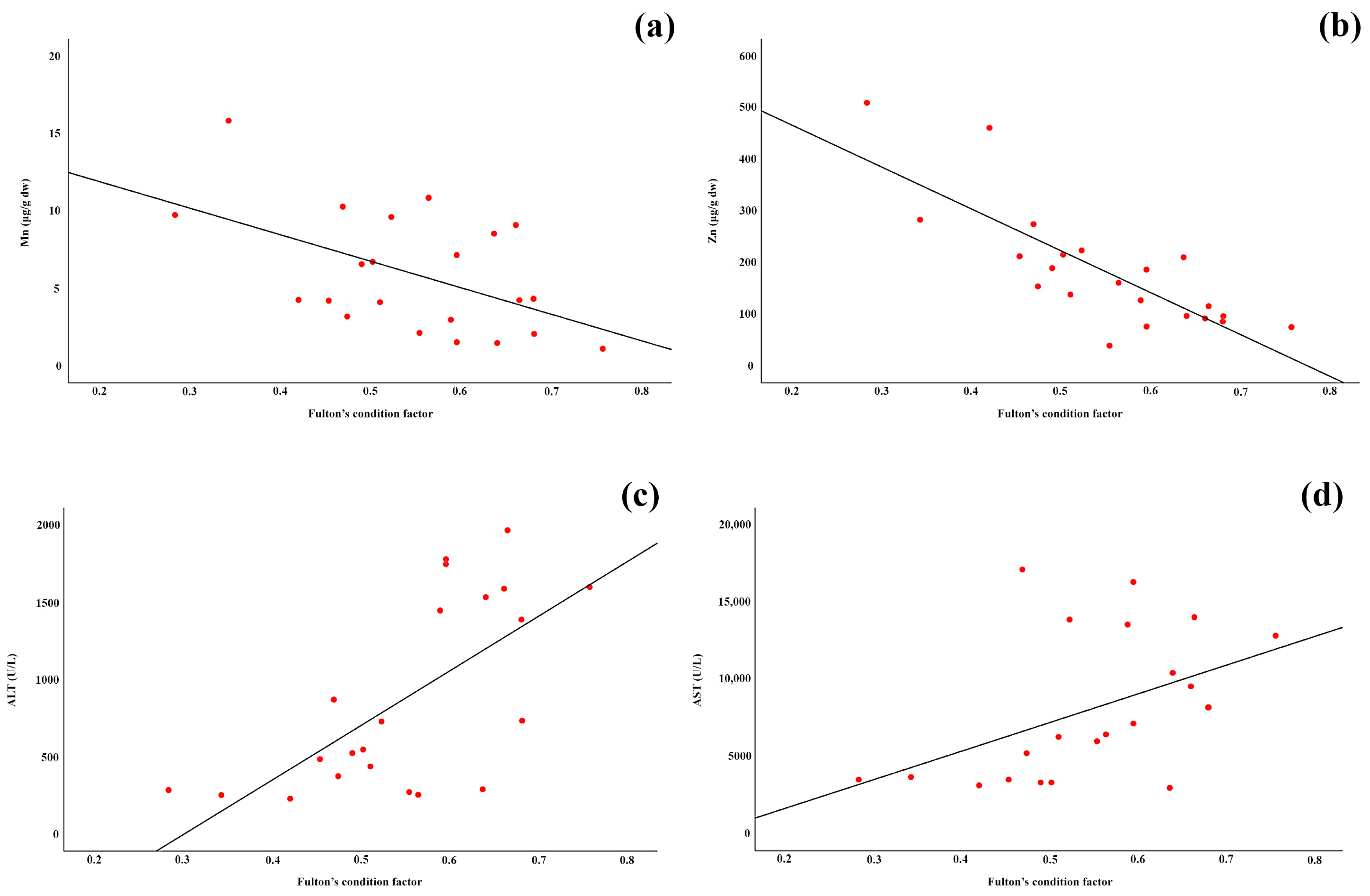
Regression analysis, for all the samples, showed a negative relationship between the hepatic concentrations of Mn (R2adj = 0.231, F(20) = 7.319, p = 0.014) and Zn (R2adj = 0.617, F(20) = 34.798, p < 0.001) with FCF, as well as positive relationships in the hepatic concentrations of ALT (R2adj = 0.421, F(20) = 16.293, p = 0.001) and AST (R2adj =0.175, F(20) = 5.457, p = 0.030) with FCF (Figure 2a–d).
Every significant regression, in both lentic and lotic ecosystem, was negative. In lentic habitat, significant linear regression with body condition was observed for concentrations of Zn (R2adj =0.483, F(9) = 10.352, p = 0.011) and AP (R2adj =0.357, F(9) = 6.559, p = 0.031) (Figure 3a,b).
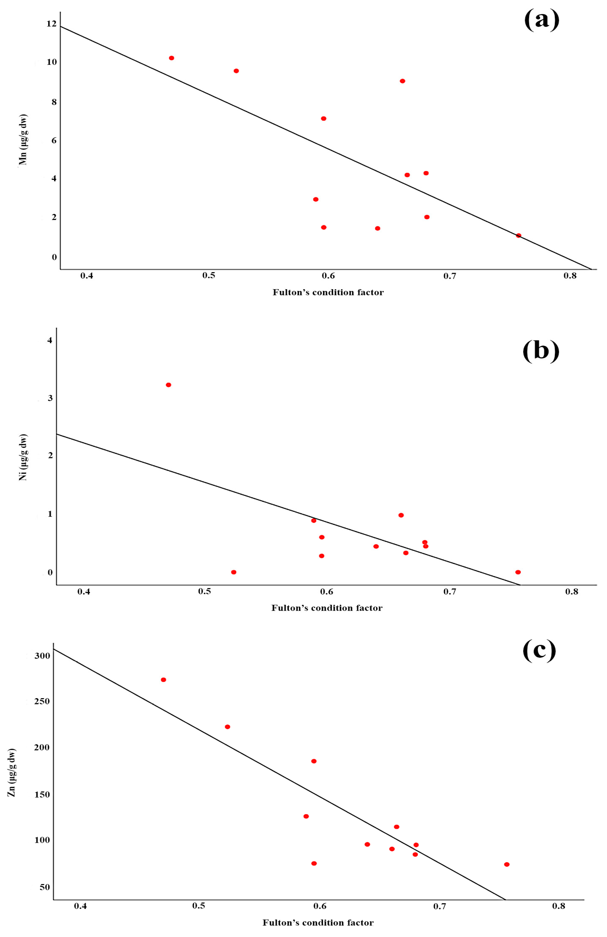
In lotic habitat, significant linear regression with body condition was observed for concentrations of Mn (R2adj = 0.359, F(9) = 6.594, p = 0.030), Ni (R2adj = 0.306, F(9) = 5.412, p = 0.045), and Zn (R2adj = 0.708, F(9) = 25.301, p = 0.001) (Figure 4a–c).

4. Discussion

In this study, we analyzed the liver health of northern pike (Esox lucius) from two different aquatic ecosystems to establish reference values for the main liver enzyme activities of this predatory fish species to serve as a basis for future ecological assessments of different freshwater environments. The Tisza River is a lotic ecosystem that is continuously polluted by various sources such as chemical and petrochemical industries, municipal wastewater, mining [26], water management infrastructure, sewerage network [27,28] and agricultural runoff [4]. On the other hand, the Belocrkvan Lakes, as a lentic ecosystem, represent a valuable natural and tourist resource with high ecological value and high-water quality, according to a report on the 2024 surface water measurement [29,30].
Comparative analysis of hepatic element concentrations revealed ecosystem-specific bioaccumulation patterns in northern pike specimens. Northern pike from the lotic ecosystem exhibited significantly higher hepatic concentrations of Cu and iron Fe compared to reservoir specimens. This finding could be the result of sediment profile and element accumulation in Tisza River, as was shown in the work of Nguyen et al. [31], where sediment layers of the Tisza River, with varying depths, were burdened with both Cu and Fe, among other elements. Fish specimens collected from lentic environments had statistically higher Zn concentration, which could be an indication of a higher environmental load of this metal in the analyzed reservoirs. Some studies have shown the impact of the vehicle-related and road-related sources on the environmental Zn load [32,33,34]. Furthermore, Erdoğan et al. [35] showed that Cu, Fe and Zn concentrations in pike liver are significantly, and positively, correlated with concentrations of these elements in sediments and water.
In addition, Zn concentrations are linked to natural sources, with the main source of Zn enrichment in water being the accumulation of Zn in sediments [36]. The measured Li concentrations in the Tisza may originate from combustion processes, vehicle emissions (from tire abrasion and fuel additives) and possible secondary contamination from mining/smelter waste, thus making a small contribution. Lithium in river water may originate from the natural weathering of lithium-bearing minerals in the surrounding geology and sediments, which gradually leach into groundwater and surface water [37]. As most of the analyzed elements in the water were below detection limits, the bioaccumulation of metals in the northern pike liver can be attributed to diet. Several studies [15,35,38] also showed different bioaccumulation patterns in the liver of northern pike, suggesting that this species is a good indicator of elemental pollution in ecosystems.
The liver serves as the primary site for the accumulation of Cu [39] and is responsible for the homeostasis and excretion of this element [40] in both fish and other vertebrates [41,42]. In our study, northern pike from the Tisza River exhibited approximately 5.4-fold significantly higher Cu concentrations (~42 µg/g dry weight) compared to specimens from the reservoirs., This finding is consistent with the results reported by Štrbac et al. [43], who documented elevated Cu levels in the liver of fish species from similar freshwater environments in the region. Although copper is an essential trace element, concentrations above certain thresholds are known to cause physiological impairment and toxicity in aquatic organisms. The ~42 µg/g dry weight copper concentration measured in Tisza pike liver falls within the range of tissue levels associated with sublethal toxic effects, as demonstrated in several previous studies. For example, Paris-Palacios et al. [44] observed hepatic copper accumulation inducing biochemical and ultrastructural perturbations in zebrafish liver at comparable concentrations. Similarly, Malhotra et al. [45] reviewed copper toxicity in fish and reported that copper accumulation in liver tissue within this concentration range is linked to oxidative stress and adverse physiological effects in species like Oreochromis niloticus and rainbow trout. Furthermore, Jin et al. [46] found that juvenile fish exposed to elevated copper concentrations accumulated liver copper at levels around 40 µg/g dry weight, which correlated with histopathological liver damage. These findings suggest that the elevated hepatic copper levels in northern pike from the Tisza River may reflect environmentally relevant exposure with potential implications for fish health and ecosystem quality. The liver and kidneys serve as the primary sites for Fe storage and regulation in fish, playing a crucial role in maintaining iron homeostasis [47]. The elevated Fe concentrations observed in the liver of northern pike from the Tisza River likely reflect the physiological accumulation of this essential element, which is necessary for vital functions such as oxygen transport, enzymatic reactions, and cellular metabolism. These results are consistent with previous findings that Fe concentration in the liver tissue of the studied fish was the highest among the metals analyzed. This may be due to Fe being one of the main components in liver tissue [48,49]. The Tisza River, subject to various anthropogenic disturbances, can introduce higher Fe loads into the aquatic environment through sediment disturbance and runoff, potentially increasing iron bioavailability and accumulation in fish tissues. This environmental pressure may explain the significantly higher hepatic Fe levels in northern pike from the lotic ecosystem compared to lentic reservoirs. The liver plays a central role in the detoxification, storage and metabolism of metals and is therefore the main target organ for the bioaccumulation of Zn in fish. Several studies show that Zn accumulates significantly more in the liver than in other tissues such as the gills, muscles, skin and intestines [50,51,52]. In our study, only the Zn concentrations in fish from the Belocrkvan reservoirs showed statistically significant differences. The results agree with those of Sujing et al. [53] and Nyeste et al. [54], which state that predators accumulate more zinc than benthivores, i.e., concentration Zn increased with trophic level. The analyzed individuals from the Tisza represent a wide range of age classes, so it is not possible to clearly define the relationship between age and the degree of bioaccumulation, especially as the number of individuals is also a limiting factor.
Liver enzymes such as ALT, AST, AP and GGT are important biomarkers for assessing the health and function of the liver. ALT and AST are mainly found in liver cells. Elevated levels of these enzymes in the blood often indicate damage or inflammation of the liver cells. While ALT is mainly found in the liver, AST is also found in other tissues such as the heart and muscles, so an elevated level may also indicate damage outside the liver. AP is an enzyme found mainly in the liver and its elevated level may indicate liver disease. GGT is mainly found in the liver and bile ducts. Elevated GGT levels often indicate liver inflammation or damage, especially in conjunction with conditions such as fatty liver, hepatitis or alcohol abuse. Together, these enzymes provide valuable insight into various aspects of liver function and aid in the diagnosis of various liver and biliary diseases [55].
In this study, enzymatic biomarker analysis showed significant differences in hepatic enzyme activities between lentic and lotic pike populations. ALT and AST activities were significantly elevated in the liver tissue of the river fish. Elevated ALT and AST levels in river fish may indicate direct hepatocellular damage, primarily due to higher concentrations of pro-oxidant metals, especially Fe and Cu, present in Tisza sediment [56] and bioaccumulated in fish livers. Both Fe and Cu catalyze the generation of reactive oxygen species through Fenton and Haber–Weiss reactions [57]. These oxidative reactions can damage hepatocyte membranes and organelles, affecting transaminases such as ALT and AST, which are hallmarks of hepatocellular injury [58]. Alkaline phosphatase activity showed no significant differences between northern pike samples from two different localities, suggesting that this enzymatic biomarker may be less sensitive to ecosystem-specific environmental conditions. The preservation of AP activity indicates that membrane transport functions are relatively intact, suggesting that liver damage is primarily hepatocellular and not cholestatic [59].
In contrast, the reservoir system shows elevated hepatic Zn and high GGT activity, suggesting a more chronic detoxification response rather than acute cellular damage. Zinc, though essential, can be toxic in excess and often accumulates due to sediment resuspension and anthropogenic inputs [60]. Chronic exposure to Zn and mixed, lower-level stressors may induce adaptive upregulation of glutathione-related detoxification pathways, for which GGT is a key enzyme. GGT elevation indicates increased demand for glutathione recycling, crucial for neutralizing metal-induced oxidative stress and facilitating excretion of metal conjugates [61]. In this study, chronic metal exposure in lentic habitats can result in metabolic adaptation rather than acute injury, as reflected by these biochemical changes. Thus, the Tisza River’s metal profile—dominated by pro-oxidant metals like Fe and Cu—results in direct hepatocellular oxidative damage, while the reservoirs, with a higher chronic burden of Zn and possibly mixed pollutants, induce an upregulated detoxification response. The different GGT activity patterns in the different ecosystem types indicate that northern pike populations are exposed to varying degrees of environmental stress and detoxification requirements. Elevated AP and GGT activities have been documented in several fish species following Pb exposure, including Oreochromis niloticus [19,62], Cyprinus carpio [63,64], Clarias gariepinus [65] and Mystus species [66]. A simultaneous increase in AP and GGT levels is characteristic of impaired liver function as a result of prolonged Pb intoxication. While increased AP activity usually indicates biliary obstruction, increased GGT activity confirms the hepatic origin of the AP elevation [67].
The elevated liver enzymes in riverine fish from this investigation suggest some level of hepatotoxicity. Alanine aminotransferase activity shows a 3.9-fold increase, while AST demonstrates a 2.8-fold increase in river specimens compared to reservoir specimens. These aminotransferases are classic “leakage enzymes” that escape into circulation when hepatocytes are damaged or undergo necrosis [68]. AST correlated with metals in liver of fish from both aquatic environments (with Cr in reservoirs and with Zn in the Tisza River), suggesting the link between these parameters and potential as biomarker for pollution assessment. Research demonstrates that elevated ALT and AST levels directly indicate liver cell damage, which could be result of excessive heavy metal exposure.
Given that the analysis of liver elements and enzyme activities revealed minimal significant differences between the sexes of northern pike, neither sex can be identified as a definitive indicator. It is important to consider that metal accumulation is primarily influenced by hormonal activity, while factors such as growth rate, diet, and environment also play significant roles [50,69]. In our study, only Li and Mn were significantly higher in females. The results are in accordance with the study of Varol and Kaçar [70], where authors also reported higher levels of Mn accumulation in female liver of Capoeta tinca. In the study by Wu et al. [71], the authors reported higher concentrations of Fe, Zn and Cu in the livers of females, whereas in males, Mn, Pb and Cr concentrations were higher in the marine fish species Gnathodentex aureolineatus. The authors attribute the observed differences in metal accumulation to the potential combination of metals with steroid hormones, which are then stored in the ovary or sperm, resulting in varying metal concentrations between genders. The spawning period of northern pike is between February and June [14], so the fact that specimens were caught in the post-spawning period (autumn) may be a reason why there were no significant variations in the levels of metals and enzymes between male and female fish.
The condition factor for the investigated northern pike corresponded, albeit slightly lower, to the results of several authors [15,72,73].
Several significant correlations were observed in our study, unlike previous findings [15] that reported no significant relationship between element concentrations in the liver and fish condition. This discrepancy may result from the different age classes analyzed, as the earlier work focused on younger animals (including age class 0+), while our study examined older specimens.
The significant negative correlation observed between the concentrations of Mn and Zn and the FCF in the northern pike studied indicates that fish with better FCF values tend to accumulate less Mn in their tissues. This pattern suggests that healthier fish may have better detoxification abilities or lower exposure pathways for this essential trace element, which becomes toxic at elevated concentrations. Research by Vieira et al. [74] found that Mn exposure causes oxidative stress in fish, with better detoxification abilities observed in fish in better physiological condition. The same study found that bioaccumulation of Mn in fish tissues occurs in the order liver > gills > gut > muscle, confirming tissue-specific accumulation patterns. Łuczyńska et al. [75] investigated the bioaccumulation of trace metals in various freshwater fish and found that fish with higher condition factors tended to have lower tissue Mn, Zn and Cu concentrations, which is consistent with our regression results, and suggests that healthier fish may regulate metal uptake or detoxify more efficiently. Varol et al. [76] confirmed a tissue-specific accumulation of Mn in fish, with the highest concentrations found in the liver, followed by the gills, gut and muscle, reflecting the central role of the liver in detoxification and metal storage.
In the case of Mn, the negative correlation with FCF could also be due to the diet of the fish, as Musharraf and Khan [77] showed a negative correlation between fish condition and dietary Mn. No such correlation with diet was found for Zn [78]. In studies on gudgeon (Gobio gobio), Bervoets and Blust [79] found no direct correlation between metal levels in the environment and the condition factor and surmised that other factors could influence the low levels.
Research by Naz & Chatha [80] and Kasimoglu [81] found that condition factor values can be significantly affected by heavy metal exposure, with differences observed according to species, age and environmental exposure, suggesting that the condition factor may serve as an effective predictor of exposure to metal bioaccumulation. Kumar et al. [82] found that Zn loading in female Mystus vittatus resulted in a decrease in FCF, gonadosomatic index and hepatosomatic index. In particular, this study showed that elevated tissue Zn concentrations were associated with impaired reproductive function and the poorer overall condition of the fish. No statistically significant trends in Zn accumulation as a function of fish size (length and weight) were found in most of the fish species studied (Cyprinus carpio and Tinca tinca) [83]. However, species-specific differences were found in the study, with C. carpio having higher liver Zn concentrations than T. tinca. Other studies show that the effects of zinc on the condition of fish are dose-dependent: optimal concentrations (30–40 mg kg−1 Zn in the diet) increase growth performance, improve condition factors and maintain healthy liver function, while excessive concentrations (>50 mg kg−1) lead to toxicity, reduced growth rates, impaired liver health, and finally deficiency, which leads to poor growth and skeletal deformities [84,85].
In contrast to Mn and Zn concentrations, AP and AST show a positive correlation with FCF. Increased AP activity in fish with better conditions may indicate improved metabolic capacity and cell function. The enzyme serves as a sensitive biomarker for environmental contamination and the assessment of liver function. Previous studies on rainbow trout have shown that there is a significant positive correlation between total weight and AP activity under controlled conditions. This study showed that AP activity increases with improved fish condition, indicating improved metabolic capacity in healthier fish [86].
Although transaminase activity is established as a reliable biomarker of metabolic status and liver function in fish [68], the increased liver metabolic activity observed in larger or older individuals from the Tisza River likely could reflect biological variation associated with fish size and health status, independent of pollutant exposure. This aligns with the idea that optimal liver function in healthy fish (fish with higher condition factor) maintains elevated enzyme activities [87]. Given the relatively small sample size (n = 22) and pronounced inter-site variation in fish size within this study, the observed positive correlation between FCF and ALT/AST may represent a statistical artifact or collinearity rather than an independent effect of environmental factors.
While the limited sample size may reduce statistical power, several results demonstrated large effect sizes and strong linear relationships that are unlikely to arise by chance. The clear ecological contrasts between sites, and the consistency of patterns across sexes and habitats support the reliability of the findings. Moreover, the observed enzyme and metal responses align with known physiological mechanisms in fish exposed to metal stress, reinforcing their biological plausibility. Future studies based on larger sample sets will help confirm these trends and further elucidate the mechanisms linking hepatic metal accumulation, enzyme activity and body condition in northern pike.

5. Conclusions

The results show that the populations of northern pike from the two types of ecosystems studied differ significantly in terms of element concentrations and enzyme activities in the liver. These results show that the northern pike could be used as an effective bioindicator species to assess ecosystem-specific pollution patterns and environmental stress responses. The differential bioaccumulation of trace elements and patterns of enzyme activities between lentic and lotic ecosystems could provide valuable insights into habitat-specific exposure scenarios and physiological adaptation mechanisms in freshwater fish populations. The observed variations in hepatic element concentrations and enzymatic activities reflect the complex interactions between environmental geochemistry, hydrodynamic conditions and biological responses in freshwater ecosystems. Obtained results can serve as a good basis for further studies regarding environmental monitoring strategies and ecological risk assessments for freshwaters.

Supplementary Materials

The following supporting information can be downloaded at https://www.mdpi.com/article/10.3390/fishes10110541/s1, Table S1: Correlation of hepatic elements and enzymes in northern pike sampled from reservoirs; Table S2: Correlation of hepatic elements and enzymes in northern pike sampled from river; Table S3: Correlation of hepatic elements and enzymes in female northern pike; Table S4: Correlation of hepatic elements and enzymes in male northern pike.

Author Contributions

Conceptualization, K.J. and V.Đ.; data collection, B.M., K.J. and V.Đ.; laboratory analysis, K.J., J.S.V., M.D. and S.K.; data analysis, S.S. and J.S.V.; writing—original draft preparation K.J., V.Đ.; writing—review and editing, J.S.V. All authors have read and agreed to the published version of the manuscript.

Funding

This research was funded by the Ministry of Science, Technological Development and Innovation of the Republic of Serbia, Contract Nos. 451-03-136/2025-03/200007, 451-03-136/2025-03/200053, 451-03-136/2025-03/200125 and 451-03-136/2025-03/200178.

Institutional Review Board Statement

The animal study protocol was approved by the Ministry of Science, Technological Development and Innovation of the Republic of Serbia (protocol code 000410410 2024 14850 004 003 501 082 and approval date: 19 March 2024).
Informed: Consent Statement Not applicable.

Data Availability Statement

The datasets and analyses for this study are available from the corresponding author upon request.

Acknowledgments

This research was conducted in collaboration with Rezervati prirode, Zrenjanin, as part of their ongoing monitoring of fish populations. The content of this study is in line with following United Nations (UN) Sustainable Development Goals, under the frame of the UN Agenda for Sustainable Development (Transforming our World: The 2030 Agenda for Sustainable Development—UN, 2015): Goal 6: Clean water and sanitation (6.6) and Goal 12: Ensure sustainable consumption and production patterns (12.4).

Conflicts of Interest

The authors declare no conflicts of interest.

References

  1. Luo, W.; Su, L.; Craig, N.J.; Du, F.; Wu, C.; Shi, H. Comparison of microplastic pollution in different water bodies from urban creeks to coastal waters. Environ. Pollut. 2019, 246, 174–182. [Google Scholar] [CrossRef]
  2. Pandey, S.; Kumari, N. Impact assessment of heavy metal pollution in surface water bodies. In Metals in Water: Global Sources, Significance, and Treatment; Shukla, S.K., Kumar, S., Madhav, S., Mishra, P.K., Eds.; Elsevier: Amsterdam, The Netherlands, 2023; pp. 129–154. [Google Scholar]
  3. Saravanan, P.; Saravanan, V.; Rajeshkannan, R.; Arnica, G.; Rajasimman, M.; Gurunathan, B.; Pugazhendhi, A. Comprehensive review on toxic heavy metals in the aquatic system: Sources, identification, treatment strategies, and health risk assessment. Environ. Res. 2024, 249, 119440. [Google Scholar] [CrossRef] [PubMed]
  4. Štrbac, S.; Kašanin Grubin, M.; Vasić, N. Importance of background values in assessing the impact of heavy metals in river ecosystems: Case study of Tisza River, Serbia. Environ. Geochem. Health 2018, 40, 1247–1263. [Google Scholar] [CrossRef] [PubMed]
  5. Zhao, G.; Ye, S.; Yuan, H.; Ding, X.; Wang, J. Surface sediment properties and heavy metal pollution assessment in the Pearl River Estuary, China. Environ. Sci. Pollut. Res. 2017, 24, 2966–2979. [Google Scholar] [CrossRef] [PubMed]
  6. García-Ordiales, E.; Esbrí, J.M.; Covelli, S.; López-Berdonces, M.A.; Higueras, P.L.; Loredo, J. Heavy metal contamination in sediments of an artificial reservoir impacted by long-term mining activity in the Almadén mercury district (Spain). Environ. Sci. Pollut. Res. 2016, 23, 6024–6038. [Google Scholar] [CrossRef]
  7. Sojka, M.; Jaskuła, J.; Barabach, J.; Ptak, M.; Zhu, S. Heavy metals in lake surface sediments in protected areas in Poland: Concentration, pollution, ecological risk, sources, and spatial distribution. Sci. Rep. 2022, 12, 15006. [Google Scholar] [CrossRef]
  8. Boškov, J. Impact of tourism on spatial transformation: A case study of the Bela Crkva municipality (Serbia). Eur. J. Econ. Stud. 2015, 3, 131–138. [Google Scholar] [CrossRef]
  9. Annabi, A.; Said, K.; Messaoudi, I. Cadmium: Bioaccumulation, histopathology, and detoxifying mechanisms in fish. Am. J. Res. Commun. 2013, 1, 60–79. [Google Scholar]
  10. Wannas, A.K.; Mohammed, S.H. The role of environmental stress in fish health: A review. GSC Biol. Pharm. Sci. 2025, 32, 17–27. [Google Scholar] [CrossRef]
  11. Jezierska, B.; Witeska, M. The metal uptake and accumulation in fish living in polluted waters. In Soil and Water Pollution Monitoring, Protection and Remediation; Springer: Dordrecht, The Netherlands, 2006; pp. 107–114. [Google Scholar]
  12. Avigliano, E.; Monferrán, M.V.; Sánchez, S.; Wunderlin, D.A.; Gastaminza, J.; Volpedo, A.V. Distribution and bioaccumulation of 12 trace elements in water, sediment, and tissues of the main fishery from different environments of the La Plata basin (South America): Risk assessment for human consumption. Chemosphere 2019, 236, 124394. [Google Scholar] [CrossRef]
  13. Zaghloul, G.Y.; Eissa, H.A.; Zaghloul, A.Y.; Kelany, M.S.; Hamed, M.A.; Moselhy, K.M.E. Impact of some heavy metal accumulation in different organs on fish quality from Bardawil Lake and human health risks assessment. Geochem. Trans. 2024, 25, 1. [Google Scholar] [CrossRef]
  14. Kottelat, M.; Freyhof, J. Handbook of European Freshwater Fishes; Publications Kottelat, Cornol and Freyhof: Berlin, Germany, 2007; p. 646. [Google Scholar]
  15. Nikolić, D.; Skorić, S.; Janković, S.; Hegediš, A.; Djikanović, V. Age-specific accumulation of toxic metal(loid)s in northern pike (Esox lucius) juveniles. Environ. Monit. Assess. 2021, 193, 229. [Google Scholar] [CrossRef]
  16. Dusek, L.; Svobodova, Z.; Janouskova, D.; Vykusova, B.; Jarkovsky, J.; Smid, R.; Pavlis, P. Bioaccumulation of mercury in muscle tissue of fish in the Elbe River (Czech Republic): Multispecies monitoring study 1991–1996. Ecotoxicol. Environ. Saf. 2005, 61, 256–267. [Google Scholar] [CrossRef]
  17. de Pinho, A.P.; Guimarães, J.R.D.; Martins, A.S.; Costa, P.A.S.; Olavo, G.; Valentin, J. Total mercury in muscle tissue of five shark species from Brazilian offshore waters: Effects of feeding habit, sex, and length. Environ. Res. 2002, 89, 250–258. [Google Scholar] [CrossRef]
  18. Bavia, L.; da Silva, A.P.; Carneiro, M.C.; Kmecick, M.; Pozzan, R.; Esquivel-Muelbert, J.; Isaac, L.; Prodocimo, M.M. Health status biomarkers and hemato-biochemical indices in Nile tilapia. Comp. Immunol. Microbiol. Infect. Dis. 2024, 7, 200168. [Google Scholar] [CrossRef]
  19. Khalefa, H.S.; AbuBakr, H.O.; Aljuaydi, S.H.; Kotp, Y.H.; Al-Mokaddem, A.K.; Abdel-Moneam, D.A. Aquatic assessment of the chelating ability of silica-stabilized magnetite nanocomposite to lead nitrate toxicity with emphasis on their impact on hepatorenal, oxidative stress, genotoxicity, histopathological, and bioaccumulation parameters in Oreochromis niloticus and Clarias gariepinus. BMC Vet. Res. 2024, 20, 262. [Google Scholar]
  20. Shahsavani, D.; Baghshani, H.; Nourian, K. Impact of betaine and allicin against circulating oxidative and biochemical alterations induced by cadmium intoxication in common carp. Aquac. Res. 2022, 53, 3637–3646. [Google Scholar] [CrossRef]
  21. Begun, G. In vivo biochemical changes in liver and gill of Clarias batrachus during cypermethrin exposure and following cessation of exposure. Pestic. Biochem. Physiol. 2005, 82, 185–196. [Google Scholar] [CrossRef]
  22. Borges, A.; Scotti, L.; Siqueira, D.; Zanini, R.; do Amaral, F.; Jurinitz, D.; Wassermann, G. Changes in hematological and serum biochemical values in jundiá (Rhamdia quelen) due to sub-lethal toxicity of cypermethrin. Chemosphere 2007, 69, 920–926. [Google Scholar] [CrossRef]
  23. David, M.; Mushigeri, S.B.; Shivakumar, R.; Philip, G.H. Response of Cyprinus carpio (Linn) to sublethal concentration of cypermethrin: Alterations in protein metabolic profiles. Chemosphere 2004, 56, 347–352. [Google Scholar] [CrossRef]
  24. Bergmeyer, H.U.; Bowers, G.N., Jr.; Horder, M.; Moss, D.W. Provisional recommendations on IFCC methods for the measurement of catalytic concentrations of enzymes. Part 2. IFCC method for aspartate aminotransferase. Clin. Chim. Acta 1976, 70, F19–F29. [Google Scholar] [CrossRef]
  25. Ricker, W.E. Computation and interpretation of the biological statistics of fish populations. Bull. Fish. Res. Board Can. 1975, 191, 10019384629. [Google Scholar]
  26. Sakan, S.; Gržetić, I.; Đorđević, D. Distribution and fractionation of heavy metals in the Tisa (Tisza) River sediments. Environ. Sci. Pollut. Res. Int. 2007, 14, 229–236. [Google Scholar] [CrossRef]
  27. Milanović, A.; Milijašević, D.; Brankov, J. Assessment of polluting effects and surface water quality using water pollution index: A case study of Hydro-system Danube-Tisa-Danube, Serbia. Carpath. J. Earth Environ. Sci. 2011, 6, 269–277. [Google Scholar]
  28. Stojanović, Ž. Agriculture in Serbia. In The Geography of Serbia; Manić, E., Nikitović, V., Djurović, P., Eds.; Springer: Cham, Switzerland, 2022; pp. 269–288. [Google Scholar]
  29. Mitić, D.-L.; Živković, M.; Teofilović, V. Tragovi teških metala u vodama belocrkvanskih jezera (in Serbian). In Proceedings of the 27th Conference on Biotechnology with International Participation, Čačak, Srbija, 25–26 March 2022; pp. 431–436. [Google Scholar]
  30. Report on the Measurement of Surface Waters of the “Belocrkvan Lakes” on 8th August 2024. Available online: https://www.jezera.rs/obavestenja/52-a-8-8-2024 (accessed on 3 September 2025). (In Serbian).
  31. Nguyen, H.L.; Braun, M.; Szaloki, I.; Baeyens, W.; Grieken, R.V.; Leermakers, M. Tracing the metal pollution history of the Tisza River through the analysis of a sediment depth profile. Water Air Soil Pollut. 2009, 200, 119–132. [Google Scholar] [CrossRef]
  32. Apeagyei, E.; Bank, M.S.; Spengler, J.D. Distribution of heavy metals in road dust along an urban-rural gradient in Massachusetts. Atmos. Environ. 2011, 45, 2310–2323. [Google Scholar] [CrossRef]
  33. Yu, H.; Ni, S.J.; He, Z.W.; Zhang, C.J.; Nan, X.; Kong, B.; Weng, Z.Y. Analysis of the spatial relationship between heavy metals in soil and human activities based on landscape geochemical interpretation. J. Geochem. Explor. 2014, 146, 136–148. [Google Scholar] [CrossRef]
  34. Thapalia, A.; Borrok, D.M.; Van Metre, P.C.; Wilson, J. Zinc isotopic signatures in eight lake sediment cores from across the United States. Environ. Sci. Technol. 2015, 49, 132–140. [Google Scholar] [CrossRef]
  35. Erdoğan, K.; Kandemir, Ş.; Doğru, M.I.; Doğru, A.; Şimşek, I.; Yılmaz, S.; Örün, G.; Altaş, L.; Yazıcıoğlu, A.; Korkmaz, N.; et al. The effects of seasonal heavy-metal pollution of Ladik Lake on pike fish (Esox lucius). Biol. Rhythm Res. 2021, 52, 821–845. [Google Scholar] [CrossRef]
  36. Algül, F.; Beyhan, M. Concentrations and sources of heavy metals in shallow sediments in Lake Bafa, Turkey. Sci. Rep. 2020, 10, 11782. [Google Scholar] [CrossRef]
  37. Adeel, M.; Zain, M.; Shakoor, N.; Ahmad, M.A.; Azeem, I.; Aziz, M.A.; Tulcan, R.X.S.; Rathore, A.; Tahir, M.; Horton, R.; et al. Global navigation of lithium in water bodies and emerging human health crisis. npj Clean Water 2023, 6, 33. [Google Scholar] [CrossRef]
  38. Salamat, N.; Movahedinia, A.; Etemadi-Deylami, E.; Mohammadi, Y. Pike (Esox lucius) bio-indicator of heavy metal pollution in Anzali Wetland. Water Qual. Expos. Health 2015, 7, 251–254. [Google Scholar] [CrossRef]
  39. Kamunde, C.; Wood, C.M. The influence of ration size on copper homeostasis during sublethal dietary copper exposure in juvenile rainbow trout, Oncorhynchus mykiss. Aquat. Toxicol. 2003, 62, 235–254. [Google Scholar] [CrossRef]
  40. Tao, T.Y.; Gitlin, J.D. Hepatic copper metabolism: Insights from genetic disease. Hepatology 2003, 37, 1241–1247. [Google Scholar] [CrossRef]
  41. Grosell, M.; McGeer, J.C.; Wood, C.M. Plasma copper clearance and biliary copper excretion are stimulated in copper-acclimated trout. Am. J. Physiol. Regul. Integr. Comp. Physiol. 2001, 280, R796–R806. [Google Scholar] [CrossRef]
  42. Grosell, M.; McDonald, M.D.; Walsh, P.J.; Wood, C.M. Effects of prolonged copper exposure in the marine gulf toadfish (Opsanus beta) II: Copper accumulation, drinking rate and Na+/K+-ATPase activity in osmoregulatory tissues. Aquat. Toxicol. 2004, 68, 263–275. [Google Scholar] [CrossRef]
  43. Štrbac, S.; Kašanin-Grubin, M.; Jovančićević, B.; Simonović, P. Bioaccumulation of heavy metals and microelements in silver bream (Brama brama L.), northern pike (Esox lucius L.), sterlet (Acipenser ruthenus L.), and common carp (Cyprinus carpio L.) from Tisza River, Serbia. J. Toxicol. Environ. Health A 2015, 78, 663–665. [Google Scholar] [CrossRef]
  44. Paris-Palacios, S.; Biagianti-Risbourg, S.; Vernet, G. Biochemical and (ultra)structural hepatic perturbations of Brachydanio rerio (Teleostei, Cyprinidae) exposed to two sublethal concentrations of copper sulfate. Aquat. Toxicol. 2000, 50, 109–124. [Google Scholar] [CrossRef]
  45. Malhotra, N.; Ger, T.R.; Uapipatanakul, B.; Huang, J.C.; Chen, K.H.; Hsiao, C.D. Review of Copper and Copper Nanoparticle Toxicity in Fish. Nanomaterials 2020, 10, 1126. [Google Scholar] [CrossRef]
  46. Jin, Z.; Xu, Y.; Zhou, H.; Cui, A.; Jiang, Y.; Wang, B.; Zhang, W. Effects of copper exposure and recovery in juvenile yellowtail kingfish (Seriola lalandi): Histological, physiological and molecular responses. Aquac. Rep. 2023, 31, 101669. [Google Scholar] [CrossRef]
  47. Szarek-Gwiazda, E.; Amirowicz, A. Bioaccumulation of trace elements in roach, silver bream, Rudd, and perch living in an inundated opencast sulphur mine. Aquat. Ecol. 2006, 40, 221–236. [Google Scholar] [CrossRef]
  48. Alshkarchy, S.S.; Raesen, A.K.; Najim, S.M. Effect of heavy metals on physiological and histological status in liver of common carp Cyprinus carpio, reared in cages and wild in the Euphrates River, Babil/Iraq. IOP Conf. Ser. Earth Environ. Sci. 2021, 779, 012066. [Google Scholar] [CrossRef]
  49. Ben Salem, Z.; Capelli, N.; Laffray, X.; Elise, G.; Ayadi, H.; Aleya, L. Seasonal variation of heavy metals in water, sediment and roach tissues in a landfill draining system pond (Etueffont, France). Ecol. Eng. 2014, 69, 25–37. [Google Scholar] [CrossRef]
  50. Al-Yousuf, M.H.; El-Shahawi, M.S.; Al-Ghais, S.M. Trace metals in liver, skin and muscle of Lethrinus lentjan fish species in relation to body length and sex. Sci. Total Environ. 2000, 256, 87–94. [Google Scholar] [CrossRef]
  51. Rajkowska, M.; Protasowicki, M. Distribution of metals (Fe, Mn, Zn, Cu) in fish tissues in two lakes of different trophy in Northwestern Poland. Environ. Monit. Assess. 2013, 185, 3493–3502. [Google Scholar] [CrossRef]
  52. Milošković, A.; Dojčinović, B.; Simić, S.; Pavlović, M.; Simić, V. Heavy metal and trace element bioaccumulation in target tissues of three edible predatory fish species from Bovan Reservoir (Serbia). Fresenius Environ. Bull. 2014, 23, 1884–1891. [Google Scholar]
  53. Wang, S.; Zheng, N.; Sun, S.; An, Q.; Li, P.; Li, X.; Li, Z.; Zhang, W. Trends and health risk of trace metals in fishes in Liaodong Bay, China, from 2015 to 2020. Front. Mar. Sci. 2022, 8, 2021. [Google Scholar] [CrossRef]
  54. Nyeste, K.; Zulkipli, N.; Uzochukwu, I.E.; Somogyi, D.; Nagy, L.; Czeglédi, I.; Harangi, S.; Baranyai, E.; Simon, E.; Nagy, S.A.; et al. Assessment of trace and macroelement accumulation in cyprinid juveniles as bioindicators of aquatic pollution: Effects of diets and habitat preferences. Sci. Rep. 2024, 14, 11288. [Google Scholar] [CrossRef]
  55. Lee, T.H.; Kim, W.R.; Poterucha, J.J. Evaluation of elevated liver enzymes. Clin. Liver Dis. 2012, 16, 183–198. [Google Scholar] [CrossRef]
  56. Sakan, M.S.; Đorđević, D.S.; Manojlović, D.D.; Polić, S.P. Assessment of heavy metal pollutants accumulation in the Tisza river sediments. J. Environ. Manag. 2009, 90, 3337–3343. [Google Scholar] [CrossRef]
  57. Ansari, W.A.; Srivastava, K.; Nasibullah, M.; Khan, M.F. Reactive oxygen species (ROS): Sources, generation, disease pathophysiology, and antioxidants. Discov. Chem. 2025, 2, 191. [Google Scholar] [CrossRef]
  58. Allameh, A.; Niayesh-Mehr, R.; Aliarab, A.; Sebastiani, G.; Pantopoulos, K. Oxidative Stress in Liver Pathophysiology and Disease. Antioxidants 2023, 12, 1653. [Google Scholar] [CrossRef] [PubMed]
  59. Levitt, M.D. Alkaline phosphatase pathophysiology with emphasis on its role in the homeostasis of the enzyme. Int. J. Mol. Sci. 2022, 23, 3144. [Google Scholar]
  60. Huseen, H.M.; Mohammed, A.J. Heavy Metals Causing Toxicity in Fishes. J. Phys. Conf. Ser. 2019, 1294, 062028. [Google Scholar] [CrossRef]
  61. Koenig, G.; Seneff, S. Gamma-Glutamyltransferase: A Predictive Biomarker of Cellular Antioxidant Inadequacy and Disease Risk. Dis. Markers 2015, 2015, 818570. [Google Scholar] [CrossRef]
  62. Elgaml, S.A.; Khalil, R.; Hashish, E.A.; El-Murr, A. Protective effects of selenium and alpha-tocopherol against lead-induced hepatic and renal toxicity in Oreochromis niloticus. J. Aquac. Res. Dev. 2015, 6, 1. [Google Scholar]
  63. Mohiseni, M.; Asayesh, S.; ShafieeBazarnoie, S.; Mohseni, F.; Moradi, N.; Matouri, M.; Mirzaee, N. Biochemical alteration induced by cadmium and lead in common carp via an experimental food chain. Iran J. Toxicol. 2016, 10, 4. [Google Scholar] [CrossRef]
  64. Nourian, K.; Baghshani, H.; Shahsavani, D. The effect of vitamin C on lead-induced plasma biochemical alterations in fish, Cyprinus carpio. Iran J. Toxicol. 2019, 2, 25–29. [Google Scholar] [CrossRef]
  65. Osman, A.G.; AbouelFadl, K.Y.; Abd El Baset, M.; Mahmoud, U.M.; Kloas, W.; Moustafa, M.A. Blood biomarkers in Nile tilapia Oreochromis niloticus and African catfish Clarias gariepinus to evaluate water quality of the River Nile. J. Fish Sci. Com. 2018, 12, 1–15. [Google Scholar] [CrossRef]
  66. Tabrez, S.; Zughaibi, T.A.; Javed, M. Bioaccumulation of heavy metals and their toxicity assessment in Mystus species. Saudi J. Biol. Sci. 2021, 28, 1459–1464. [Google Scholar] [CrossRef]
  67. Tang, P.; Liao, Q.; Tang, Y.; Yao, X.; Du, C.; Wang, Y.; Song, F.; Deng, S.; Wang, Y.; Qiu, X.; et al. Independent and combined associations of urinary metals exposure with markers of liver injury: Results from the NHANES 2013–2016. Chemosphere 2023, 338, 139455. [Google Scholar] [CrossRef]
  68. Bojarski, B.; Witeska, M.; Kondera, E. Blood biochemical biomarkers in fish toxicology—A review. Animals 2025, 15, 965. [Google Scholar] [CrossRef]
  69. Oguz, A.R.; Yeltekin, A. Metal levels in the liver, muscle, gill, intestine, and gonad of Lake Van fish (Chalcalburnus tarichi) with abnormal gonad. Biol. Trace Elem. Res. 2014, 159, 219–223. [Google Scholar] [CrossRef] [PubMed]
  70. Varol, M.; Kaçar, E.; Karadede Akın, H. Accumulation of trace elements in muscle, gill and liver of fish species (Capoeta umbla and Luciobarbus mystaceus) in the Tigris River (Turkey), and health risk assessment. Environ. Res. 2020, 186, 109570. [Google Scholar] [CrossRef]
  71. Wu, Z.; Xu, Y.; Cai, M.; Cheng, S.Y.; Chen, H.; Huang, D.; Chen, K.; Lin, Y.; Li, T.; Liu, M.; et al. Metals in fishes from Yongshu Island, southern South China Sea: Human health risk assessment. J. Toxicol. 2017, 2017, 2458293. [Google Scholar] [CrossRef]
  72. Salam, A.; Davies, P.M. Body composition of northern pike (Esox lucius L.) in relation to body size and condition factor. Fish. Res. 1994, 19, 193–204. [Google Scholar] [CrossRef]
  73. Yazicioğlu, O.; Yilmaz, S.; Polat, N. The growth feature of pike, Esox lucius L., 1758 inhabiting Lake Ladik. Acta Biol. Turc. 2024, 37, J4:1–J4:8. [Google Scholar]
  74. Vieira, M.C.; Torronteras, R.; Córdoba, F.; Canalejo, A. Acute toxicity of manganese in goldfish Carassius auratus is associated with oxidative stress and organ specific antioxidant responses. Ecotoxicol. Environ. Saf. 2012, 78, 212–217. [Google Scholar] [CrossRef] [PubMed]
  75. Łuczyńska, J.; Tońska, E.; Paszczyk, B.; Łuczyński, M. The relationship between biotic factors and the content of chosen heavy metals (Zn, Fe, Cu and Mn) in six wild freshwater fish species collected from two lakes (Łańskie and Pluszne) located in northeastern Poland. Iran J. Fish Sci. 2020, 19, 421–442. [Google Scholar]
  76. Varol, M.; Kaçar, E. Bioaccumulation of metals in various tissues of fish species in relation to fish size and gender and health risk assessment. Curr. Pollut. Rep. 2023, 9, 327–337. [Google Scholar] [CrossRef]
  77. Musharraf, M.; Khan, M. Dietary manganese requirement of fingerling Indian major carp, Labeo rohita (Hamilton) estimated by growth, tissue manganese concentration and hepatic manganese-superoxide dismutase activity. Aquaculture 2021, 736, 734–742. [Google Scholar] [CrossRef]
  78. Zhou, C.; Lin, H.; Huang, Z.; Wang, J.; Wang, Y.; Yu, W. Effects of dietary zinc levels on growth performance, digestive enzyme activities, plasma physiological response, hepatic antioxidant responses and metallothionein gene expression in juvenile spotted sea bass (Lateolabrax maculatus). Aquac. Nutr. 2021, 27, 1421–1432. [Google Scholar] [CrossRef]
  79. Bervoets, L.; Blust, R. Metal concentrations in water, sediment and gudgeon (Gobio gobio) from a pollution gradient: Relationship with fish condition factor. Environ. Pollut. 2003, 126, 9–19. [Google Scholar] [CrossRef]
  80. Naz, S.; Chatha, A.M.M. Metals mixture effects on growth performance and their bioaccumulation in fish. Iran. J. Fish. Sci. 2022, 21, 601–614. [Google Scholar]
  81. Kasimoglu, C. The Effect of Fish Size, Age and Condition Factor on the Contents of Seven Essential Elements in Anguilla nguilla from Tersakan Stream Mugla (Turkey). J. Pollut. Eff. Cont. 2014, 2, 123. [Google Scholar] [CrossRef]
  82. Kumar, S.; Das, D.; Sarbajna, A.; Chakraborty, S.B. Zinc modulates hypothalamo-pituitary-gonadal-liver axis to impair reproduction in female Mystus vittatus (Bloch, 1794). Environ. Sci. Pollut. Res. 2024, 31, 42212–42229. [Google Scholar] [CrossRef] [PubMed]
  83. Tharwat, M.; Almundarij, T.I.; Marzok, M. Diseases and disorders of trace elements deficiency in farm animals: An illustrated review. Int. J. Vet. Sci. 2025, 14, 624–640. [Google Scholar]
  84. Sayed, J.A.; Haque, A.; Akter, N.; Akter, S.; Satter, A.; Sarker, P.K.; Marshall, D.J.; Paray, B.A.; Hossain, M.B. Effects of dietary supplementation of Zn-nanoparticles on the growth performance and nutritional quality of Asian catfish, Clarias batrachus. Front. Sustain. Food Syst. 2024, 8, 1410557. [Google Scholar] [CrossRef]
  85. Tahir, M.; Khan, N.; Fatima, M.; Haque, N.U.; Davies, S.J. Bioavailability of zinc from different organic zinc chelates and their effect on the growth, whole body, tissue zinc content, enzymes activity and proximate composition of Labeo rohita. PLoS ONE 2025, 20, e0314146. [Google Scholar] [CrossRef]
  86. Pastorino, P.; Bergagna, S.; Dezzutto, D.; Barbero, R.; Righetti, M.; Pagliasso, G.; Gasco, L.; Gennero, M.S.; Pizzul, E.; Dondo, A.; et al. Long-term assessment of baseline blood biochemistry parameters in rainbow trout (Oncorhynchus mykiss) maintained under controlled conditions. Animals 2020, 10, 1466. [Google Scholar] [CrossRef]
  87. Sun, S.; Wu, Y.; Yu, H.; Su, Y.; Ren, M.; Zhu, J.; Ge, X. Serum biochemistry, liver histology and transcriptome profiling of bighead carp Aristichthys nobilis following different dietary protein levels. Fish Shellfish Immunol. 2019, 86, 832–839. [Google Scholar] [CrossRef] [PubMed]
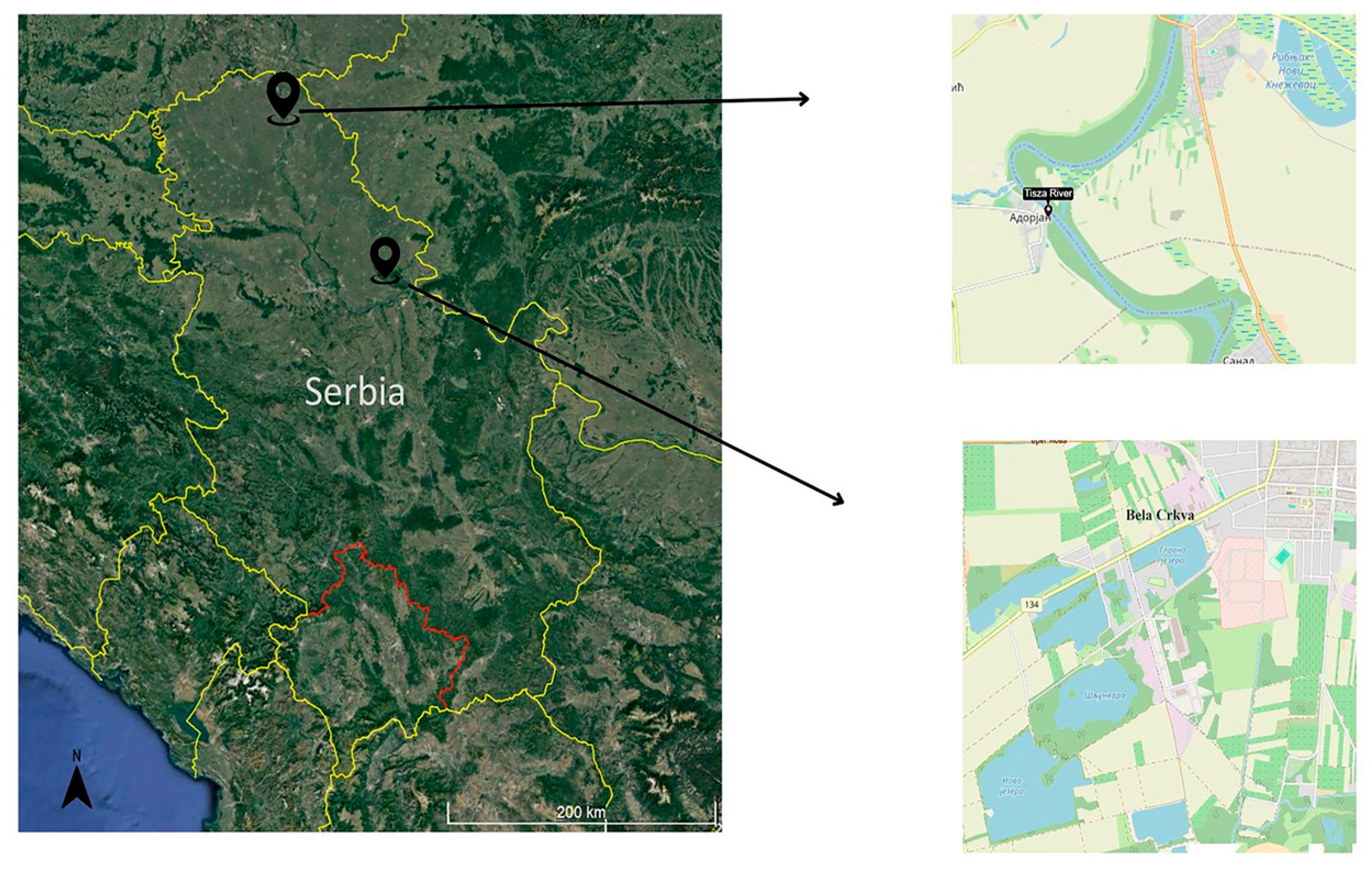
Figure 1. Map of the two sampling localities: Tisza River and Bela Crkva reservoirs.
Fishes 10 00541 g001
Figure 2. Regression analysis for all samples (n = 22) in the study. (a) Linear regression between hepatic Mn concentration and Fulton’s condition factor; (b) linear regression between hepatic Zn concentration and Fulton’s condition factor; (c) linear regression between hepatic ALT concentration and Fulton’s condition factor; (d) linear regression between hepatic AST concentration and Fulton’s condition factor.
Fishes 10 00541 g002
Figure 3. Lotic ecosystem: (a) linear regression between hepatic Zn concentration and Fulton’s condition factor (n = 11); (b) linear regression between hepatic AP concentration and Fulton’s condition factor (n = 11).
Fishes 10 00541 g003
Figure 4. Lentic ecosystem: (a) linear regression between hepatic Mn concentration and Fulton’s condition factor (n = 11); (b) linear regression between hepatic Ni concentration and Fulton’s condition factor (n = 11); (c) linear regression between hepatic Zn concentration and Fulton’s condition factor (n = 11).
Fishes 10 00541 g004
Table 1. Physico-chemical parameters in water on both analyzed localities.
ParametersBela Crkva Reservoirs
Values (Min–Max)
Tisza River
Values
O2 (mg/L)7.35–9.027.7
O2 (%)72.4–85.872.9
Temperature (°C)12.6–13.112.9
pH7.68–7.97.49
Conductivity (µS/cm−1)398–483588
Transmittance (cm)125–170100
Table 2. Mean values (mean ± SD) of total length (TL), body weight (W) and Fulton’s Condition Factor (FCF) of the northern pike analyzed.
TL (cm)W (g)AgeFCF
Reservoirs30.23 ± 8.0 *163.85 ± 183.1 *1+–2+0.48 ± 0.1 *
Tisza River42.81 ± 15.0 *693.16 ± 911.1 *1+–5+0.62 ± 0.1 *
Females37.17 ± 17.5552.08 ± 1032.91+–5+0.56 ± 0.1
Males36.07 ± 10.4342.96 ± 342.81+–3+0.55 ± 0.1
* statistically significant differences (p < 0.05).
Table 3. Hepatic element concentrations (mean ± SD; μg g−1 dry weight, dw) and enzyme activity (UL−1) in reservoirs and Tisza River northern pike.
LiverReservoirsTisza Riverp Value
ElementAs0.47 ± 0.50.22 ± 0.4ns
Cr0.35 ± 0.20.38 ± 0.2ns
Cu7.78 ± 3.1 *41.92 ± 35.8 *0.000
Fe151.59 ± 153.3 *862.73 ± 516.5 *0.001
Li0.09 ± 0.10.08 ± 0.1ns
Ni1.78 ± 5.10.70 ± 0.9ns
Mn6.91 ± 4.14.88 ± 3.5ns
Pb1.11 ± 0.61.24 ± 0.5ns
Zn233.59 ± 138.8 *131.17 ± 66.9 *0.034
EnzymeALT359.20 ± 119.5 *1397.91 ± 430.3 *0.000
AST4302.84 ± 1376.3 *11,923.59 ± 3413.1 *0.000
AP990.68 ± 324.9988.22 ± 287.2ns
GGT5.20 ± 1.9 *3.46 ± 0.8 *0.016
* Significant differences (p < 0.05); ns—not significant (p > 0.05).
Table 4. Hepatic element concentration (mean ± SD; μg g−1 dry weight, dw) and enzyme activities of ALT, AST, AP and GGT (UL−1) in female and male northern pike.
LiverFemalesMalesp Value
Element
(μg g−1 dw)
As0.36 ± 0.50.33 ± 0.5ns
Cr0.43 ± 0.3 0.32 ± 0.2ns
Cu31.71 ± 44.320.10 ± 15.5ns
Fe532.99 ± 571.6489.28 ± 504.9ns
Li0.14 ± 0.1 *0.05 ± 0.1 *0.011
Ni0.62 ± 1.01.67 ± 4.7ns
Mn7.87 ± 2.7 *4.53 ± 4.0 *0.030
Pb1.02 ± 0.51.28 ± 0.5ns
Zn176.41 ± 71.7186.51 ± 145.3ns
Enzyme
(UL−1)
ALT757.86 ± 557.6962.11 ± 659.5ns
AST8762.01 ± 5692.37664.05 ± 3971.0ns
AP908.55 ± 271.81045.46 ± 314.9ns
GGT4.39 ± 1.74.28 ± 1.7ns
* Significant differences (p < 0.05); ns—not significant (p > 0.05).
Disclaimer/Publisher’s Note: The statements, opinions and data contained in all publications are solely those of the individual author(s) and contributor(s) and not of MDPI and/or the editor(s). MDPI and/or the editor(s) disclaim responsibility for any injury to people or property resulting from any ideas, methods, instructions or products referred to in the content.