Article

Distribution and Comparative Analysis of Genomic Microsatellites in Nine Species of Family Sillaginidae

1
Fishery College, Zhejiang Ocean University, Zhoushan 316022, China
2
Fishery College, Guangdong Ocean University, Zhanjiang 524088, China
3
Key Laboratory of Tropical and Subtropical Fishery Resources Application and Cultivation, Ministry of Agriculture and Rural Affairs, Pearl River Fisheries Research Institute, Chinese Academy of Fishery Sciences, Guangzhou 510380, China
*
Authors to whom correspondence should be addressed.
Fishes 2025, 10(11), 536; https://doi.org/10.3390/fishes10110536
Submission received: 30 August 2025 / Revised: 15 October 2025 / Accepted: 20 October 2025 / Published: 22 October 2025
(This article belongs to the Section Genetics and Biotechnology)

Abstract

We conducted a comparative analysis of the identified microsatellite sequences across the genomes of nine sillaginids. We examined the microsatellites with motifs ranging from 1 to 6 bp in length and analyzed their distribution and frequency across different genomic regions. Microsatellite occurrence differed significantly with the degree of coverage ranging from 1.47 to 3.21%. The number and proportion of each repeat type were consistent across the nine species, with di-nucleotide repeats being the most abundant, followed by mono-nucleotide repeats, and gradually decreasing as the number of repeat units increased. The mono-nucleotide repeat motifs were dominated by A/T, while di-nucleotide repeat motifs were mainly AC/GT, and tri-nucleotide repeat motifs were primarily AGG/CCT. Regarding the number of repeats, microsatellites in Sillaginidae were generally concentrated between 5 and 18 repeat units, with peaks observed at 6 and 10 repetitions. The abundance of microsatellite loci consistently decreased as the number of repetitions increased beyond 10. These findings provide valuable insights into genome evolution and microsatellite DNA dynamics, supporting future investigations into their structural and functional characteristics, compositional patterns, and applications in molecular marker development for these species.
Key Contribution: This study presents the first genome-wide comparative analysis of microsatellites in nine species of the family Sillaginidae, revealing consistent patterns in abundance, motif preference, and repeat length, enabling future molecular marker development for this economically important fish family.

1. Introduction

Sillaginidae, or whitings, a family within the Order Eupercaria incertae sedis (Osteichthyes: Teleostei), are widely distributed in the tropical, subtropical, and temperate waters of the Indian Ocean and the western Pacific Ocean (including the coast of China). Sillaginid species are commonly found in shallow coastal waters, often forming shoals in sandy areas and estuaries, with some species entering freshwater habitats [1,2,3,4]. Known for their burrowing behavior, sillaginid species are prized for their delicious and nutritious meat, making them of high economic importance as a target for offshore fisheries and a popular food source [5,6,7,8,9].
Molecular markers are indispensable tools for investigating genetic diversity, population structure, and evolutionary history, which are crucial for the sustainable management and conservation of fishery resources. Among these markers, microsatellites, or simple sequence repeats (SSRs), are particularly powerful due to their high polymorphism, co-dominant inheritance, and genome-wide distribution [10,11,12]. These characteristics have made SSRs widely applicable in studies of genetic diversity [13], genetic relationship analysis [14], and genetic map construction [15,16]. The advent of next-generation sequencing technologies has revolutionized the development of molecular markers by enabling efficient and large-scale screening of SSRs directly from whole-genome sequences [17,18,19,20]. This approach has been successfully applied to characterize microsatellites and develop markers in various fish species, such as darkbarbel catfish (Pelteobagrus vachelli) [21], grass carp (Ctenopharyngodon idella) [22], channel catfish (Ictalurus punctatus) [23], groupers (Epinephelus spp.) [24], silver carp (Hypophthalmichthys molitrix), and bighead carp (Hypophthalmichthys nobilis) [25], demonstrating its utility for non-model organisms.
A robust phylogenetic framework is crucial for interpreting comparative genomic data. Within the family Sillaginidae, complex evolutionary relationships, particularly in the genus Sillago, have been revealed by recent molecular studies, which indicate that the genus is not monophyletic, with several species groups demonstrating paraphyly or polyphyly [26,27]. For instance, the species formerly considered a single widespread taxon, Sillago sihama, is actually a complex containing multiple cryptic species. This has been confirmed through DNA barcoding and detailed morphological comparisons, leading to the description of several new species such as S. sinica [28], S. shaoi [29], and S. parasihama [30]. Importantly, a recent phylogenomic study by Liu et al. [31], which shared genomic data and provided a high-resolution phylogenetic tree for the entire family based on genome-wide markers, further clarified intra-generic and interspecific evolutionary relationships. The established phylogenetic context offers a solid evolutionary foundation for the current comparative analysis of microsatellite distribution and evolution across the nine targeted species of Sillaginidae. These studies not only provided valuable genetic tools for population genetics and germplasm assessment in those species but also demonstrated the efficiency and reliability of genome-wide SSR development. We applied a similar approach to systematically identify and characterize microsatellites in the genomes of nine sillaginid species. In this study, we used the microsatellite screening software MISA to screen and statistically analyze the microsatellites in the whole genomes of nine sillaginid species (Sillago sinica, Sillago sihama, Sillago japonica, Sillago ingenuua, Sillago asiatica, Sillago maculata, Sillago aeolus, Sillago nigrofasciata, and Sillaginopsis panijus). Our work focuses on systematically identifying and characterizing these microsatellites, including analyzing the types, lengths, and distribution patterns of their repeat motifs, as well as exploring the evolutionary dynamics of microsatellites across the sillaginid genome with the goal of filling the current gap in microsatellite research for this family. By preliminarily clarifying the distribution characteristics of microsatellites in the sillaginid genome, this study lays a robust foundation for future research on the genetic structure, genetic diversity, and molecular marker development of particular sillaginid species.

2. Materials and Methods

2.1. Genomic Sequences

The whole-genome sequences of the nine sillaginid species (Sillago sinica, S. sihama, S. japonica, S. ingenuua, S. asiatica, S. maculata, S. aeolus, S. nigrofasciata, Sillaginopsis panijus) analyzed in this study were obtained from the recent whole-genome sequencing study by Liu et al. [31]. The raw sequencing data and draft genome assemblies were generated following standardized protocols as described in their work. The genome sizes ranged from 511.71 Mb to 578.27 Mb, with scaffold N50 values varying between 5.27 kb and 21,469.63 kb. These metrics indicate that the genomic data provide a solid foundation for comparative microsatellite analysis across these species.

2.2. Microsatellite Identification

The microsatellite search software MISA v2.1 (http://pgrc.ipk-gatersleben.de/misa/, accessed on 20 October 2025) was used to identify and characterize microsatellite sequences in the genomes of Sillaginidae. We employed its perfect search model (detecting uninterrupted, mismatch-free repeat units) and set the minimum repeat numbers as follows: mono-nucleotide ≥10, di-nucleotide ≥6, tri-nucleotide ≥5, and tetra-/penta-/hexa-nucleotide ≥4. These correspond to the minimum lengths defined in this study (mono- ≥ 10 bp, di- ≥ 12 bp, tri- ≥ 15 bp, tetra- ≥ 20 bp, penta- ≥ 25 bp, hexa- ≥ 30 bp) [32]. We mainly examined the distribution of perfect repeats ≥10 bp, as microsatellites are often disrupted by single-base substitutions [33].

3. Results

3.1. The Number of SSRs

We examined the number, relative frequency (microsatellite numbers per Mb of the sequence), average distance, and coverage degree (percentage of the total microsatellite length in the sequence) of microsatellites with motif lengths of 1–6 nucleotides in the nine fish genomes (Table 1 and Supplementary Materials). The total length of the assembled genome ranges from 522 (S. sihama) to 652 Mb (S. panijus). The total number of microsatellites, ranging from 409,623 (S. ingenuua) to 723,688 (S. nigrofasciata), differed between species, and the degree of coverage varied from 1.65% (S. ingenuua) to 3.27% (S. sinica) (Table 1). The lowest relative frequency was found in S. maculata (673.52 loci/Mb). The highest relative frequency was found in S. sinica (1174.36 loci/Mb). The average distance between SSRs was found to range from 852 bp (S. sinica) to 1485 bp (S. maculata). The number of compound microsatellites ranged from 55,836 (S. maculata) to 172,203 (S. nigrofasciata).

3.2. The Frequency of SSRs

Based on the analysis of microsatellite repeat sequences in the genomes of nine species of Sillaginidae, we found an abundant variety of nucleotide repeat motifs, with 200–300 types identified across genomes. The numbers and proportions of each repeat type were consistent across all nine species. In terms of the variety of repeat motifs, di-nucleotide repeats had the highest number of distinct types, followed by mono-nucleotide repeats, while the variety decreased as the number of repeat units increased (Table 2).
Through the statistical analysis of the repeat types and repeat numbers of microsatellite loci in the genomes of nine Sillaginidae, the following patterns were observed. The mono-nucleotide repeat motifs were predominantly A/T, while the dominant motifs for di- and tri-nucleotide repeats were AC/GT and AGG/CCT, respectively. Preference in tetra-nucleotide repeats, however, varied among the nine species: AGGG/CCCT was favored by S. sinica, S. sihama, S. japonica, and S. asiatica, whereas ACAG/CTGT was dominant in S. ingenuua, S. maculata, S. aeolus, S. nigrofasciata, and S. panijus. The penta-nucleotide and hexa-nucleotide repeats exhibited greater diversity, with the predominant motifs differing across species (Table 3).

3.3. The Number of Repetitions of Microsatellite Loci

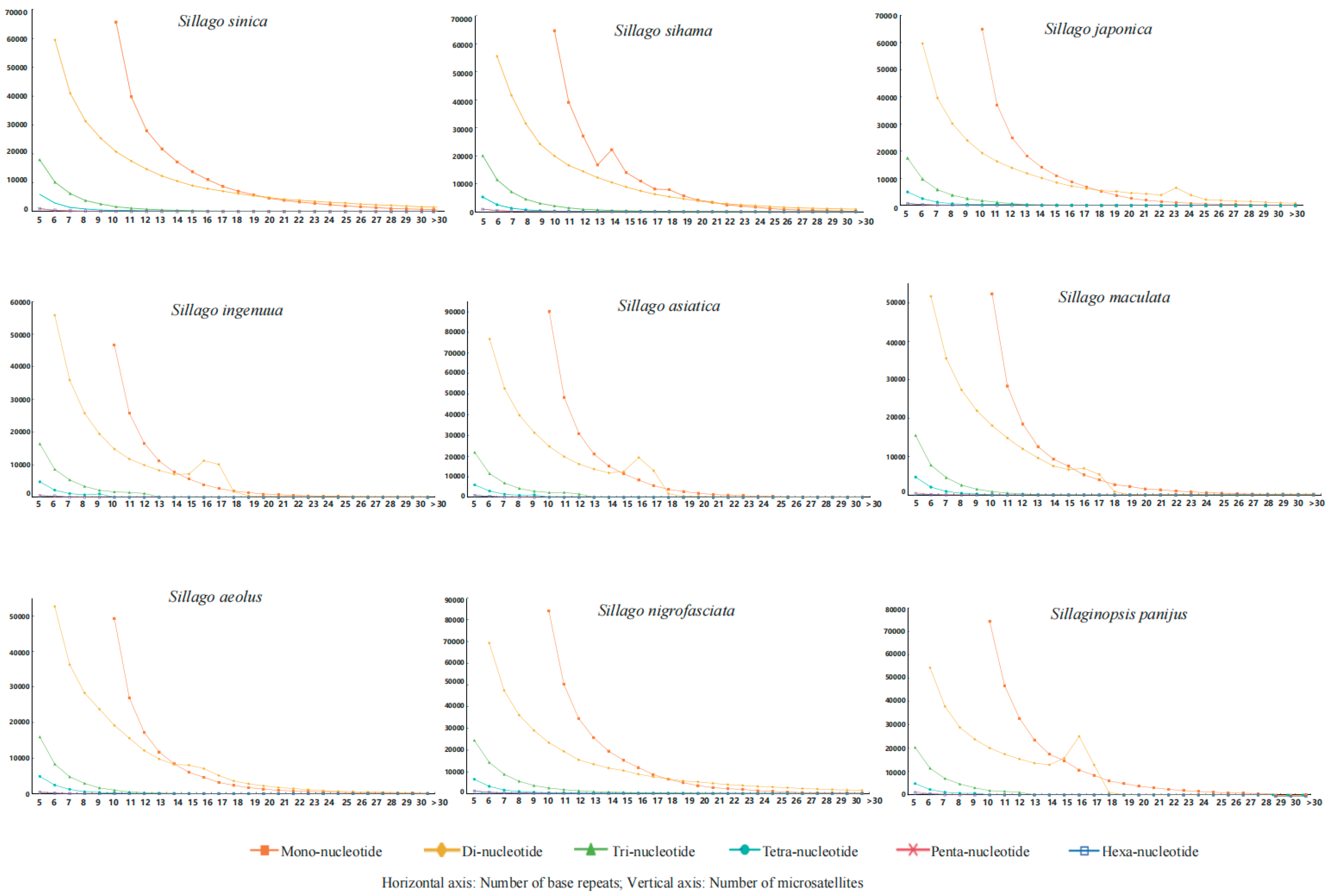
Sillaginid species generally exhibited consistent numbers of nucleotide repeats within arrays, primarily concentrated between 5 and 18. The distribution of the number of motifs repeated within arrays peaked at 6 and 10 repeats, and the total number of corresponding sites decreased as repeat counts exceeded 10 (Figure 1). The peak distributions varied by nucleotide type: mono-nucleotide repeats peaked at 10, di-nucleotide repeats at 6, and tri-nucleotide repeats at 5. Repeats for tetra-nucleotide, penta-nucleotide, and hexa-nucleotide motifs were mainly concentrated between 5 and 10 (Figure 2).

3.4. Microsatellite Sequence Length and Variation Analysis

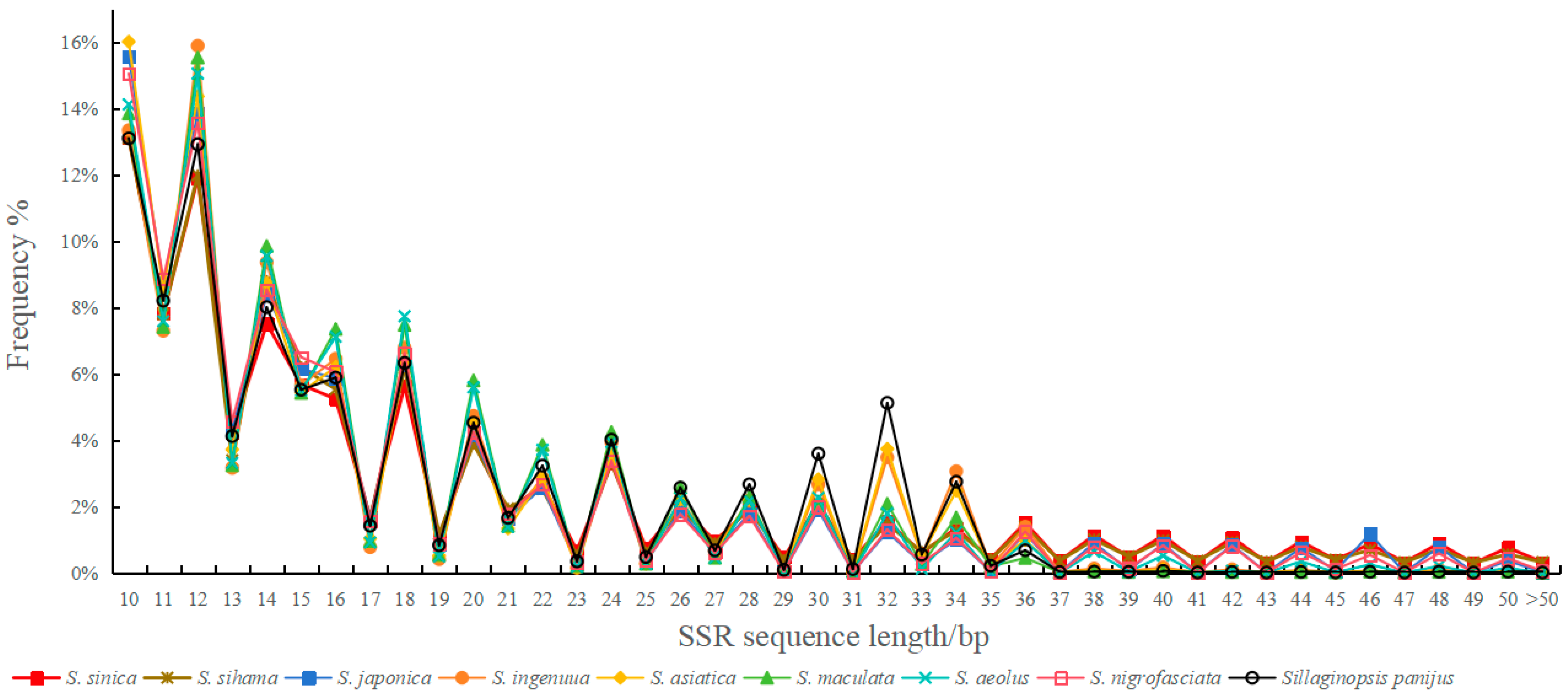
The overall microsatellite sequence length in nine species of Sillaginidae ranged from 10 to 300 bp. The longest average length is 19.89 bp (S. nigrofasciata), while the shortest is 17.24 bp (S. asiatica). Microsatellite sequence lengths were polymorphic across different repeat unit types, with longer sequences occurring less frequently (Figure 3). When considering the total count of microsatellite sequences, mono-nucleotide repeats were the most frequently occurring class, followed by di-nucleotide repeats, while other nucleotide repeats were less common. Among the microsatellites in Sillago, sequences with a length of 12 bp were the most frequent, irrespective of repeat type, followed by 10 bp sequences. Generally, the frequency of microsatellites decreased as the number of repeating units increased across all six base types.

4. Discussion

We examined microsatellites composed of motifs 1–6 bp long in the genomes of nine species of the family Sillaginidae and analyzed their distribution and frequency in genomic regions. Microsatellite occurrence varied significantly, with coverage ranging from 1.47% to 3.21%. A comparison with data from other species, including humans (3%) [33], primates (0.83–0.88%) [32,34,35], birds (0.13–0.49%) [36], and plants and fungi (0.04–0.15%) [37,38,39,40,41], suggests that microsatellite abundance differs widely across species and may represent a general phenomenon among taxa [42]. Our findings also suggested that microsatellite density is not strictly positively correlated with genome size. For instance, S. sinica exhibits a lower microsatellite density than S. sihama, yet possesses a larger genome. Similarly, the microsatellite density of S. japonica is lower than that of S. ingenuua, despite its larger genome size. Although a general positive correlation between microsatellite density and genome size has been widely reported [43,44,45], our results were consistent with those of several previous studies [32,36,46,47]. Consequently, the comparative analysis of microsatellites indicated that there was great variation in microsatellite content across nine species of Sillago.
The variation remained evident even after accounting for the varying degrees of completeness among the underlying draft genomes. Assembly continuity can affect the absolute number of microsatellites detected. However, the consistent and distinct patterns we observed across species, including the relative abundances of different repeat types and the predominant motifs, strongly indicate that these trends represent robust biological signals. Moreover, the genome-wide scope of our analysis further minimizes the potential impact of localized assembly gaps.
Our findings on microsatellite composition gain significant evolutionary context when viewed alongside the recently established phylogenetic framework of the family Sillaginidae [31]. For instance, the divergence in the dominant tetra-nucleotide motif aligns with major phylogenetic lineages. The preference for AGGG/CCCT was observed in a group comprising S. sinica, S. sihama, S. japonica, and S. asiatica, which represent a more derived clade. In contrast, the motif ACAG/CTGT was predominant in a group containing S. ingenuua, S. maculata, S. aeolus, S. nigrofasciata, and S. panijus, a grouping that includes the phylogenetically basal species S. ingenuua and the distinct genus Sillaginopsis. This correlation suggests that evolutionary shifts in dominant microsatellite motifs may track the deeper evolutionary splits within the family. This result might be indicative that differential selective constraints may play an important role in microsatellite evolution and result in the accumulated preference for different microsatellite types. Furthermore, the species that displayed unique microsatellite characteristics in our analyses, such as S. sihama, S. ingenuua, S. asiatica, and S. panijus, are precisely those that occupy distinct phylogenetic positions or have experienced unique evolutionary histories, as revealed by Liu et al. [31]. For example, the basal position of S. ingenuua is mirrored in its distinct microsatellite profile, which may reflect an ancestral genomic state. Similarly, the unique patterns in S. panijus underscore the considerable evolutionary divergence from the Sillago genus. Therefore, these departures from general trends are not simple outliers but likely hold evolutionary significance, potentially linked to historical population dynamics, differential selective pressures, or genetic drift specific to these lineages.
Generally, microsatellite instability of di-nucleotide repeats is higher than tri-nucleotide, tetra-nucleotide, and penta-nucleotide repeats [48]. In conclusion, the mutation rate of microsatellites depends on the repeated unit length and is biased from the trend of gradual decrease. This could explain the high numbers of mono−/di-nucleotide-motif microsatellites and the low numbers of penta−/hexa-nucleotide-motif microsatellites in the genomes. It should be noted that in all nine genomes, di-nucleotide has the most repeats, followed by mono-nucleotide, and decreases with the increase in the number of repeat units. For example, di-nucleotide was the dominant SSR type in black pepper (Piper nigrum) [49], common carp (Cyprinus carpio) [50], Siamese mud carp (Henicorhynchus siamensis) [51], water mold (Phytophthora) [52], and whiteleg shrimp (Penaeus vannamei) [53], respectively.
(AC)n was the most frequently identified di-nucleotide repeat motif across the nine Sillaginidae genomes, followed by (AG)n, (AT)n, and the considerably less common (CG)n. Our findings on microsatellite abundance and distribution in Sillaginidae align with broader patterns observed in other fish lineages and beyond. This hierarchy of motif prevalence is consistent with patterns reported in diverse taxonomic groups, including humans and mosquitoes [33,54], as well as in elephantfish (Callorhinchus milii), Odobenus rosmarus [55], and P. vannamei [53]. Specifically, a recent genome-wide survey of fishes by Lei et al. [56] similarly reported (AC)n and (AG)n as dominant di-nucleotide motifs across 10 species from different genera. However, a key distinction emerges in tri-nucleotide repeat content: unlike some species in Lei et al.’s study, which showed high proportions, Sillaginidae species generally exhibited lower levels. The consistent underrepresentation of the (CG)n motif, a phenomenon observed across most eukaryotic genomes [44], can be attributed to the general A/T-rich genomic background of many species, coupled with the higher thermodynamic stability of the C≡G base pair compared to A=T, which makes strand separation and polymerase progression more difficult during replication [57]. These differences may reflect lineage-specific evolutionary histories, selective pressures, or genomic architecture.
The prevalence of A/T-rich mono-nucleotide and AC/GT-rich di-nucleotide repeats we observed is consistent with the overall AT-biased base composition reported in many teleost genomes [50]. This compositional bias not only influences the baseline mutation spectrum but also the potential for microsatellites to form specific DNA secondary structures, which can have implications for gene regulation and genome architecture. The concentration of microsatellites in the 5–18 repeat unit range, with distribution peaks at specific lengths (e.g., 6 and 10 repeats), likely represents a balance between the high mutability and potential functional cost of longer repeats, which are prone to contraction or disruption, and the neutral or nearly neutral evolution of shorter repeats, allowing for their persistence and accumulation over time. This dynamic may contribute to the raw material for evolutionary adaptation while being shaped by the evolutionary history of each lineage, as evidenced by the phylogenetic patterns in motif usage.
According to the microsatellite sequence length of Sillago, the number of microsatellite loci with a sequence length of 12 bp was the largest, regardless of the type of primitive microsatellite repeating motif. This was followed by sites 10 bp in length. The general trend for the six base types was that the number of microsatellites decreased gradually with the increase in duplicate number. This phenomenon has been observed in yellow catfish (Pelteobagrus fulvidraco) [58], filefish (Thammaconus modestus) [59], spotted sea bass (Lateolabrax maculatus) [13], and tiger puffer (Takifugu rubripes) [60]. The main reason for this rule might be, on the one hand, that the sequence length of microsatellites is negatively correlated with their stability, while the mutation rate of microsatellites is positively correlated with their duplicate copy number. That is, the longer the sequence of microsatellites, the lower the stability, the higher the mutation frequency, and the lower the number [61]. On the other hand, single-base and two-base screening start from ≥12 and ≥10 repeats, respectively, so peaks are formed at these two points, and different screening principles lead to different statistical results for different species.

5. Conclusions

The distribution and frequency of microsatellite tracts in Sillaginidae were reported in this study, which provided valuable insights into genome evolution and the evolutionary dynamics of microsatellite DNA, supporting further exploration of their structure, function, composition patterns, and the development of molecular markers in these species.

Supplementary Materials

The following supporting information can be downloaded at: https://www.mdpi.com/article/10.3390/fishes10110536/s1, the complete sequence for all microsatellite markers.

Author Contributions

Y.Q.: writing—original draft, methodology, investigation, data curation. C.Q.: writing—original draft, methodology. C.T.: data curation, writing—review. T.G.: conceptualization, data curation, funding acquisition, project administration, supervision, validation. Y.Z.: writing—review, editing. All authors have read and agreed to the published version of the manuscript.

Funding

The work was supported by the National Natural Science Foundation of China (41976083).

Institutional Review Board Statement

Not applicable.

Data Availability Statement

The complete sequences of all microsatellite markers are available in the Supplementary Materials.

Conflicts of Interest

The authors declare no conflicts of interest.

References

  1. Mckay, R.J. Silaginid fishes of the world (family Sillaginidae): An annotated and illustrated catalogue of the Sillago, smelt or Indo-Pacific whiting species known to date. In FAO Fish Synop; Food and Agriculture Organization of the United Nations: Rome, Italy, 1992; Volume 14, pp. 1–82. [Google Scholar]
  2. Tetsuji, N. Fish Retrieval from Japan; Tokai University Press Association: Tokyo, Japan, 2013; pp. 974–975. [Google Scholar]
  3. Nelson, J.S.; Grande, T.; Wilson, M.V.H. Fishes of the World; John Wiley and Sons, Inc.: Hoboken, NJ, USA, 2016; p. 707. [Google Scholar]
  4. Wu, H.L.; Zhong, J.S. Marine and Estuarine Fish System Retrieval in China; China Agriculture Press: Beijing, China, 2021; pp. 680–682. [Google Scholar]
  5. Tatsuya, K. Phylogenetic systematics of the family Sillaginidae (Percomorpha: Order Perciformes). Zootaxa 2013, 3642, 1–105. [Google Scholar] [CrossRef]
  6. Ye, M.; Kong, L.; Jian, Z.; Qiu, Z.; Lin, X.; Zhang, Y.; Huang, Y.; Li, G.; Tian, C. Genetic insights into hypoxia tolerance in silver sillago (Sillago sihama) through QTL mapping and SNP association analysis. Aquaculture 2024, 592, 741174. [Google Scholar] [CrossRef]
  7. Cheng, Q.T.; Zheng, B.S. Systematic Synopsis of Chinese Fishes; Science Press: Beijing, China, 1987. [Google Scholar]
  8. Konishi, H.; Nakabo, T. Color Guide to the Japanese Fishes for SportFishermen; Enterbrain, Inc.: Tokyo, Japan, 2007; p. 400. [Google Scholar]
  9. Yu, J.W.; Wang, Y.R.; Lin, X.H.; Shen, Y.J.; Li, G.L.; Huang, Y.; Zhu, C.H.; Tian, C.X. Effects of morphological traits on body weight in 12-month-old silver sillago (Sillago sihama). J. Guangdong Ocean Univ. 2022, 42, 137–143. (In Chinese) [Google Scholar]
  10. Tautz, D. Hypervariability of simple sequences as a general source for polymorphic DNA markers. Nucl. Acids. Res. 1989, 17, 6463–6471. [Google Scholar] [CrossRef] [PubMed]
  11. Litt, M.; Luty, J.A. A hypervariable microsatellite revealed by in vitro amplification of a dinucleotide repeat within the cardiac muscle actin gene. Am. J. Hum. Genet. 1989, 44, 397–401. [Google Scholar] [PubMed]
  12. Madesis, P.; Ganopoulos, I.; Tsaftaris, A. Microsatellites: Evolution and contribution. Methods Mol. Biol. 2013, 1006, 1–13. [Google Scholar]
  13. Fan, S.G.; Huang, H.; Liu, Y.; Wang, P.F.; Zhao, C.; Yan, L.L.; Qiao, X.T.; Qiu, L.H. Genome-wide identification of microsatellite and development of polymorphic SSR markers for spotted sea bass (Lateolabrax maculatus). Aquacult. Rep. 2021, 20, 100677. [Google Scholar]
  14. Serbezov, D.; Bernatchze, L.; Olsen, E.M.; Vøllestad, L.A. Mating patterns and determinants of individual reproductive success in brown trout (Salmo trutta) revealed by parentage analysis of an entire stream living population. Conserv. Genet. Resour. 2010, 19, 3193–3205. [Google Scholar] [CrossRef]
  15. Shen, X.Y.; Yang, G.P.; Liu, Y.J.; Liao, M.J.; Wang, X.C.; Zhu, M.Z.; Song, W.B.; Zou, G.W.; Wei, Q.W.; Wang, D.Q.; et al. Construction of genetic linkage maps of guppy (Poecilia reticulata) based on AFLP and microsatellite DNA markers. Aquaculture 2007, 271, 178–187. [Google Scholar] [CrossRef]
  16. Xia, J.H.; Liu, F.; Zhu, Z.Y.; Fu, J.J.; Feng, J.B.; Li, J.L.; Yue, G.H. A consensus linkage map of the grass carp (Ctenopharyngodon idella) based on microsatellites and SNPs. BMC Genom. 2010, 11, 135–151. [Google Scholar] [CrossRef]
  17. Kuo, J.; Chen, C.Y.; Han, C.C.; Ju, Y.M.; Tew, K.S. Analyses of diet preference of larval orange-spotted grouper (Epinephelus coioides) grown under inorganic fertilization method using next-generation sequencing. Aquaculture 2021, 531, 735916. [Google Scholar] [CrossRef]
  18. Wang, H.Y.; Fu, Y.; Gu, P.; Zhang, Y.Y.; Tu, W.L.; Chao, Z.; Wu, H.L.; Cao, J.G.; Zhou, X.; Liu, B.; et al. Genome-wide characterization and comparative analyses of simple sequence repeats among four miniature pig breeds. Animals 2020, 10, 1792. [Google Scholar] [CrossRef] [PubMed]
  19. Liu, W.C.; Xu, Y.T.; Li, Z.K.; Fan, J.; Yang, Y. Genome-wide mining of microsatellites in king cobra (Ophiophagus hannah) and cross-species development of tetranucleotide SSR markers in Chinese cobra (Naja atra). Mol. Biol. Rep. 2019, 46, 6087–6098. [Google Scholar] [CrossRef] [PubMed]
  20. Bastías, A.; Correa, F.; Rojas, P.; Martin, C.; Pérez-Diaz, J.; Yáñez, C.; Cuevas, M.; Verdugo, R.; Sagredo, B. Draft genome sequence data of maqui (Aristotelia chilensis) and identification of SSR markers. Data Brief. 2019, 27, 104545. [Google Scholar] [CrossRef]
  21. Peng, Y.; Li, J.; Wang, T.; Zhang, K.; Ning, X.H.; Ji, J.; Yin, S.W. Preliminary study on distribution characteristics and positioning of microsatellites in whole genome of Pelteobagrus vachelli. South China Fish. Sci. 2022, 18, 90–98. (In Chinese) [Google Scholar]
  22. Fu, J.J.; Shen, Y.B.; Xu, X.Y.; Chen, Y.; Li, D.; Li, J.L. Multiplex microsatellite PCR sets for parentage assignment of grass carp (Ctenopharyngodon idella). Aquacult. Int. 2013, 21, 1195–1207. [Google Scholar] [CrossRef]
  23. Zhong, L.Q.; Zhang, S.Y.; Jiang, H.C.; Wang, M.H.; Chen, X.H. Bioinformatics analysis of perfect microsatellite in the whole genome of channel catfish, Ictalurus punctatus. Genom. Appl. Biol. 2022, 41, 504–513. (In Chinese) [Google Scholar]
  24. Wang, X.L.; Gao, J.; Qi, X.; Wang, Y.B.; Chen, F.X.; Liu, J.Y.; Fu, S.Y. Screening and characteristics analysis of microsatellites in whole genome of five groupers. Prog. Fish. Sci. 2023, 45, 149–158. (In Chinese) [Google Scholar]
  25. Jian, J.B.; Yang, L.D.; Gan, X.N.; Wu, B.; Gao, L.; Zeng, H.H.; Wang, X.Z.; Liang, Z.Q.; Wang, Y.; Fang, L.H.; et al. Whole genome sequencing of silver carp (Hypophthalmichthys molitrix) and bighead carp (Hypophthalmichthys nobilis) provide novel insights into their evolution and speciation. Mol. Ecol. Resour. 2021, 21, 912–923. [Google Scholar] [CrossRef]
  26. Xiao, J.G.; Song, N.; Gao, T.X.; McKay, R.J. Redescription and DNA barcoding of Sillago indica (Perciformes: Sillaginidae) from the Coast of Pakistan. Pak. J. Zool. 2016, 48, 317–323. [Google Scholar]
  27. Bae, S.E.; Kwun, H.J.; Kim, J.K.; Kweon, S.M.; Kang, C.B. New record of Sillago sinica (Pisces: Sillaginidae) in Korean waters, and re-identification of Sillago parvisquamis previously reported from Korea as S. sinica. Anim. Syst. Evol. Divers. 2013, 29, 288–293. [Google Scholar] [CrossRef]
  28. Gao, T.X.; Ji, D.P.; Xiao, Y.S.; Xue, T.Q.; Yanagimoto, T.; Setoguma, T. Description and DNA barcoding of a new Sillago species, Sillago sinica (Perciformes: Sillaginidae) from coastal waters of China. Zool. Stud. 2011, 50, 254–263. [Google Scholar]
  29. Xiao, J.G.; Song, N.; Han, Z.Q.; Gao, T.X. Description and DNA barcoding of a new Sillago species, Sillago shaoi (Perciformes: Sillaginidae), in the Taiwan Strait. Zool. Stud. 2016, 55, e47. [Google Scholar]
  30. Yu, Z.S.; Guo, T.; Xiao, J.G.; Song, N.; Gao, T.X. Identification and DNA barcoding of a new Sillago species in Beihai and Zhanjiang, China, with a key to related species. J. Ocean Univ. China 2022, 21, 1334–1342. [Google Scholar] [CrossRef]
  31. Liu, Q.; Mao, W.H.; Wang, Y.T.; Xiao, J.G.; Saha, S.; Gao, T.X.; Liu, F. Whole genome sequencing and phylogenetic analyses of the Sillaginidae family fish. Mol. Phylogenet. Evol. 2025, 207, 108340. [Google Scholar] [CrossRef]
  32. Liu, S.X.; Hou, W.; Sun, T.L.; Xu, Y.T.; Li, P.; Yue, B.S.; Fan, Z.X.; Li, J. Genome-wide mining and comparative analysis of microsatellites in three macaque species. Mol. Gene. Genom. 2017, 292, 537–550. [Google Scholar] [CrossRef]
  33. Subramanian, S.; Mishra, R.K.; Singh, L. Genome-wide analysis of microsatellite repeats in humans: Their abundance and density in specific genomic regions. Genom. Biol. 2003, 4, R13. [Google Scholar] [CrossRef]
  34. Xu, Y.T.; Li, W.J.; Hu, Z.X.; Zeng, T.; Shen, Y.M.; Liu, S.X.; Zhang, X.Y.; Li, J.; Yue, B.S. Genome-wide mining of perfect microsatellites and tetranucleotide orthologous microsatellites estimates in six primate species. Gene 2018, 643, 124–132. [Google Scholar] [CrossRef]
  35. Xu, Y.T.; Hu, Z.X.; Wang, C.; Zhang, X.Y.; Li, J.; Yue, B.S. Characterization of perfect microsatellite based on genome-wide and chromosome level in Rhesus monkey (Macaca mulatta). Gene 2016, 592, 269–275. [Google Scholar] [CrossRef]
  36. Huang, J.; Li, W.J.; Jian, Z.Y.; Yue, B.S.; Yan, Y.F. Genome-wide distribution and organization of microsatellites in six species of birds. Biochem. Syst. Ecol. 2016, 67, 95–102. [Google Scholar] [CrossRef]
  37. Karaoglu, H.; Lee, C.M.Y.; Meyer, W. Survey of simple sequence repeats in completed fungal genomes. Mol. Biol. Evol. 2005, 22, 639–649. [Google Scholar] [CrossRef]
  38. Li, C.Y.; Liu, L.; Yang, J.; Li, J.B.; Su, Y.; Zhang, Y.; Wang, Y.Y.; Zhu, Y.Y. Genome-wide analysis of microsatellite sequence in seven filamentous fungi. Interdiscip. Sci. Comput. Life Sci. 2009, 1, 141–150. [Google Scholar] [CrossRef]
  39. Qian, J.; Xu, H.B.; Song, J.Y.; Xu, J.; Zhu, Y.J.; Chen, S.L. Genome-wide analysis of simple sequence repeats in the model medicinal mushroom Ganoderma lucidum. Gene 2013, 512, 331–336. [Google Scholar] [CrossRef]
  40. Cai, G.; Leadbetter, C.W.; Muehlbauer, M.F.; Molnar, T.J.; Hillman, B.I. Genome-wide microsatellite identification in the fungus Anisogramma anomala using Illumina sequencing and genome assembly. PLoS ONE 2013, 8, e82408. [Google Scholar] [CrossRef]
  41. Wang, Y.; Chen, M.J.; Wang, H.; Wang, J.F.; Bao, D.P. Microsatellites in the genome of the edible mushroom, Volvariella volvacea. BioMed Res. Int. 2014, 2014, 281912. [Google Scholar] [CrossRef]
  42. Ellegren, H. Microsatellites: Simple sequences with complex evolution. Nat. Rev. Genet. 2004, 5, 435–445. [Google Scholar] [CrossRef]
  43. Katti, M.V.; Ranjekar, P.K.; Gupta, V.S. Differential distribution of simple sequence repeats in eukaryotic genome sequences. Mol. Biol. Evol. 2001, 18, 1161–1167. [Google Scholar] [CrossRef]
  44. Tóth, G.; Gáspári, Z.; Jurka, J. Microsatellites in different eukaryotic genomes: Survey and analysis. Genome Res. 2000, 10, 967–981. [Google Scholar] [CrossRef] [PubMed]
  45. Hancock, J.M. Simple sequences in a ‘minimal’ genome. Nat. Genet. 1996, 14, 14–15. [Google Scholar] [CrossRef] [PubMed]
  46. Burranboina, K.; Abraham, S.; Murugan, K. Genome wide identification and analysis of microsatellite repeats in the largest DNA viruses (Poxviridae Family): An insilico approach. Annu. Res. Rev. Biol. 2018, 22, 1–11. [Google Scholar] [CrossRef]
  47. Qi, W.H.; Jiang, X.M.; Du, L.M.; Xiao, G.S.; Hu, T.Z.; Yue, B.S.; Quan, Q.M. Genome-wide survey and analysis of microsatellite sequences in bovid species. PLoS ONE 2015, 10, e0133667. [Google Scholar] [CrossRef]
  48. Perinchery, G.; Nojima, D.; Goharderakhshan, R.; Tanaka, Y.; Alonzo, J.; Dahiya, R. Microsatellite instability of dinucleotide tandem repeat sequences is higher than trinucleotide, tetranucleotide and pentanucleotide repeat sequences in prostate cancer. Int. J. Oncol. 2000, 16, 1203–1212. [Google Scholar] [CrossRef] [PubMed]
  49. Kumari, R.; Wankhede, D.P.; Bajpai, A.; Maurya, A.; Prasad, K.; Gautam, D.; Rangan, P.; Latha, M.; Joseph, J.K.; Suma, A.; et al. Genome wide identification and characterization of microsatellite markers in black pepper (Piper nigrum): A valuable resource for boosting genomics applications. PLoS ONE 2019, 14, e0226002. [Google Scholar] [CrossRef] [PubMed]
  50. Ji, P.F.; Zhang, Y.; Li, C.; Zhao, Z.; Wang, J.; Li, J.T.; Xu, P.; Sun, X.W. High throughput mining and characterization of microsatellites from common carp genome. Int. J. Mol. Sci. 2012, 13, 9798–9807. [Google Scholar] [CrossRef]
  51. Iranawati, F.; Jung, H.; Chand, V.; Hurwood, D.A.; Mather, P.B. Analysis of genome survey sequences and SSR marker development for Siamese mud carp, Henicorhynchus siamensis, using 454 pyrosequencing. Int. J. Mol. Sci. 2012, 13, 10807–10827. [Google Scholar] [CrossRef]
  52. Mathew, D.; Anju, P.S.; Tom, A.; Johnson, N.; George, M.L.; Davis, S.P.; Ravisankar, V.; Asha, K.N. Genome-wide microsatellites and species specific markers in genus Phytophthora revealed through whole genome analysis. 3 Biotech 2020, 10, 442. [Google Scholar] [CrossRef]
  53. Zhang, X.J.; Yuan, J.B.; Sun, Y.M.; Li, S.H.; Gao, Y.; Yu, Y.; Liu, C.Z.; Wang, Q.C.; Lv, X.J.; Zhang, X.X.; et al. Penaeid shrimp genome provides insights into benthic adaptation and frequent molting. Nat. Commun. 2019, 10, 356. [Google Scholar] [CrossRef] [PubMed]
  54. Wang, X.T.; Zhang, Y.J.; Qiao, L.; Chen, B. Comparative analyses of simple sequence repeats (SSRs) in 23 mosquito species genomes: Identification, characterization and distribution (Diptera: Culicidae). Insect Sci. 2019, 26, 607–619. [Google Scholar] [CrossRef]
  55. Jiang, Q.; Li, Q.; Yu, H.; Kong, L.F. Genome-wide analysis of simple sequence repeats in marine animals—A comparative approach. Mar. Biotechnol. 2014, 16, 604–619. [Google Scholar] [CrossRef]
  56. Lei, Y.; Zhou, Y.; Price, M.; Song, Z.B. Genome-wide characterization of microsatellite DNA in fishes: Survey and analysis of their abundance and frequency in genome-specific regions. BMC Genom. 2021, 22, 421. [Google Scholar] [CrossRef]
  57. Zhao, X.Y.; Tan, Z.Y.; Feng, H.P.; Yang, R.H.; Li, M.F.; Jiang, J.H.; Shen, G.L.; Yu, R.Q. Microsatellites in different Potyvirus genomes: Survey and analysis. Gene 2011, 488, 52–56. [Google Scholar] [CrossRef]
  58. Shan, Y.J.; Lu, C.Y.; Li, C.; Cheng, L.; Sun, X.W. Isolation and characterization of 57 novel polynucleotide microsatellites from yellow catfish (Pelteobagrus fulvidraco) genome for genetic analysis. Conser. Genet. Resour. 2014, 6, 73–77. [Google Scholar] [CrossRef]
  59. Wang, J.L.; Li, H.L.; Yin, S.; Hu, Z.H.; Ye, M.; Zhang, P.P. Distribution characteristics of whole genome microsatellite of Thammaconus modestus. J. Yantai Univ. 2022, 35, 285–293. (In Chinese) [Google Scholar]
  60. Takagi, M.; Sato, J.; Monbayashi, C.; Aoki, K.; Tsuji, T.; Hatanaka, H.; Takahashi, H.; Sakai, H. Evaluation of microsatellites identified in the tiger puffer Takifugu rubripes DNA database. Fish. Sci. 2003, 69, 1085–1095. [Google Scholar] [CrossRef]
  61. Wierdl, M.; Dominska, M.; Petes, T.D. Microsatellite instability in yeast: Dependence on the length of the microsatellite. Genetics 1997, 146, 769–779. [Google Scholar] [CrossRef] [PubMed]
Figure 1. The relationship between microsatellite repeats and frequencies in the 9 Sillago species genomes.
Fishes 10 00536 g001
Figure 2. Distribution of number of base repetitions of SSR types in the 9 Sillago species’ genomes.
Fishes 10 00536 g002
Figure 3. Frequency of different microsatellite sequence lengths in the 9 Sillago species genomes.
Fishes 10 00536 g003
Table 1. Genomic SSR loci information of the 9 Sillago species.
ItemsGenome Size/MbMCNSPCFTLM/bpPGSADS/bpRA/(loci/Mb)
S. sinica534627,460166,29417,156,7573.21%8521174.36
S. sihama522599,804153,80816,005,5723.07%8691149.84
S.japonica562560,704137,89614,600,8752.58%1009990.78
S. ingenuua554409,62363,2389,139,6781.65%1353739.14
S. asiatica701657,299113,40514,552,1362.07%1067937.11
S. maculata630424,02355,8369,293,6561.47%1485673.52
S. aeolus524426,08577,7099,867,9441.88%1230812.57
S. nigrofasciata633723,688172,20319,649,6873.11%8741160.56
S. panijus652619,95892,13114,491,3742.22%1051951.57
Note: MC indicates microsatellite count; NSPCF indicates number of SSRs present in compound formation; TLM indicates total length of microsatellites; PGS indicates proportion of genome sequence; ADS indicates average distance between SSRs; RA indicates relative abundance.
Table 2. Statistical analysis of repeat types and repeat numbers for microsatellite loci in the 9 Sillago species genomes.
ItemsNPMNPDNPTRNPTENPPNPH
S. sinica247,032
(39.37)
316,071
(50.37)
46,514
(7.41)
13,640
(2.17)
1805
(0.29)
2398
(0.38)
S. sihama235,100
(39.20)
294,271
(49.06)
53,701
(8.95)
12,318
(2.05)
2205
(0.37)
2209
(0.37)
S.japonica205,275
(36.61)
294,781
(52.57)
44,627
(7.96)
12,711
(2.27)
1673
(0.30)
1637
(0.29)
S. ingenuua129,886
(31.71)
226,368
(55.26)
40,872
(9.98)
10,171
(2.48)
1320
(0.32)
1006
(0.25)
S. asiatica247,906
(37.72)
338,625
(51.52)
54,205
(8.25)
13,027
(1.98)
2027
(0.31)
1509
(0.23)
S. maculata152,822
(36.04)
227,791
(53.72)
33,905
(8.00)
8462
(2.00)
771
(0.18)
272
(0.06)
S. aeolus136,660
(32.07)
242,913
(57.01)
35,527
(8.34)
9701
(2.28)
823
(0.19)
461
(0.11)
S. nigrofasciata278,656
(38.50)
355,441
(49.12)
67,478
(9.23)
16,280
(2.25)
2947
(0.41)
2886
(0.40)
S. panijus262,679
(42.37)
291,764
(47.06)
51,903
(8.37)
9954
(1.61)
2405
(0.39)
1253
(0.20)
Note: NPM indicates number and proportion of mono-nucleotide; NPD indicates number and proportion of di-nucleotide; NPTR indicates number and proportion of tri-nucleotide; NPTE indicates number and proportion of tetra-nucleotide; NPP indicates number and proportion of penta-nucleotide; NPH indicates number and proportion of hexa-nucleotide.
Table 3. Dominant duplicate copy categories for each repeat type in sillaginid species.
ItemsMono-
Nucleotide
Di-
Nucleotide
Tri-
Nucleotide
Tetra-
Nucleotide
Penta-
Nucleotide
Hexa-
Nucleotide
S. sinicaA/T
(63.01)
AC/GT
(79.62)
AGG/CCT
(39.28)
AGGG/CCCT
(13.50)
AAGCT
/AGCTT
(12.80)
ACACGC
/CGTGTG
(27.27)
S. sihamaA/T
(72.32)
AC/GT
(79.77)
AGG/CCT
(39.37)
AGGG/CCCT
(18.87)
AGCCG
/CGGCT
(11.88)
AACCCT
/AGGGTT
(37.89)
S.japonicaA/T
(76.42)
AC/GT
(82.19)
AGG/CCT
(36.39)
AGGG/CCCT
(14.48)
AGAGG
/CCTCT
(11.36)
ACCAGG
/CCTGGT(20.04)
S. ingenuuaA/T
(79.97)
AC/GT
(77.21)
AGG/CCT
(41.03)
ACAG/CTGT
(19.88)
AAGCT/AGCTT
(15.76)
ACCAGG
/CCTGGT
(26.54)
S. asiaticaA/T
(75.82)
AC/GT
(81.99)
AGG/CCT
(43.09)
AGGG/CCCT
(14.98)
AGCCG
/CGGCT
(11.30)
ACCAGG
/CCTGGT
(18.75)
S. maculataA/T
(76.53)
AC/GT
(81.05)
AGG/CCT
(41.65)
ACAG/CTGT
(18.19)
AGAGG
/CCTCT
(12.58)
ACCAGG
/CCTGGT
(17.65)
S. aeolusA/T
(82.47)
AC/GT
(81.80)
AGG/CCT
(43.02)
ACAG/CTGT
(22.67)
AGAGG
/CCTCT
(12.52)
ACACGC
/CGTGTG
(16.70)
S. nigrofasciataA/T
(74.30)
AC/GT
(80.56)
AGG/CCT
(38.74)
ACAG/CTGT
(16.63)
AGCCG
/CGGCT
(13.27)
AACCCT
/AGGGTT
(25.95)
S. panijusA/T
(75.30)
AC/GT
(81.04)
AGG/CCT
(41.94)
ACAG/CTGT
(15.16)
AATCT
/AGATT
(25.32)
ACCAGG
/CCTGGT
(12.21)
Disclaimer/Publisher’s Note: The statements, opinions and data contained in all publications are solely those of the individual author(s) and contributor(s) and not of MDPI and/or the editor(s). MDPI and/or the editor(s) disclaim responsibility for any injury to people or property resulting from any ideas, methods, instructions or products referred to in the content.