Article

Report on Intersex and Abnormal Mature Aquacultured Walleye Pollock, Gadus chalcogrammus

Aquaculture Industry Research Division, East Sea Fisheries Institute, National Institute of Fisheries Science, Gangneung 25435, Republic of Korea
*
Author to whom correspondence should be addressed.
Fishes 2025, 10(1), 35; https://doi.org/10.3390/fishes10010035
Submission received: 4 November 2024 / Revised: 14 January 2025 / Accepted: 16 January 2025 / Published: 18 January 2025

Abstract

:
Walleye pollock (Gadus chalcogrammus) is a commercially important species widely distributed in cold-water regions. We have been culturing this species artificially since 2015. The average embryo diameter was 1.43 ± 0.056 mm, and hatching occurred at 5 °C approximately 339 h post-fertilization. Gonadal development became visibly apparent at a body size of 10–15 cm after ~180 days, initially distinguishing ovaries with the development of germ cells, whereas testes were observable after further maturation. We discovered two intersexes from F1 and F2 generations, and one abnormal mature individual from F2. Morphologically, intersex walleye pollock exhibited distinct characteristics of ovary and testes, with male gonads visibly connected to the end of ovaries. In intersex walleye pollock aged 3–6 years, the gonads developed normally, and oocyte resorption was restricted to the area near the connections between testicular and ovarian tissues, with numerous atretic oocytes observed in the resorption zone. Primordial germ cells were found together in individuals who had not undergone spawning, indicating an abnormal maturation pattern. Although no significant differences in the gonadosomatic index were observed between intersex and normal individuals, further research on intersexuality is necessary to understand the reproductive development of this species and the health of offspring spawned by intersex individuals, which are rare.
Key Contribution: This study reports the discovery of intersex and the abnormal development of artificially produced walleye pollock; Gadus chalcogrammus.

1. Introduction

The development of gonads in fish begins with sex determination. Sex determination acts as the master switch for the bipotential gonadal primordium, activating the differentiation pathway. Depending on the species, sex determination can be genetic (GSD), environmental (ESD), or a combination of both [1]. Once the fate of the bipotential gonadal primordium is determined in the testes or ovaries, sex differentiation takes over the process of gonadal development. In teleost fish, sex differentiation is labile and influenced by various factors, such as genes, hormones, and external factors, throughout gonadal development [2]. Sex differentiation results from a competitive relationship between genes involved in the development and maturation of testes and ovaries [3,4]. Environmental factors influencing sex differentiation include temperature [5], pH [6], population density [7], oxygen concentration [8], and social status [9]; during gonadal differentiation, these play a significant role in maintaining the activation and suppression of various male and female pathways depending on sexual fate.
Fish are a group of vertebrates known to exhibit hermaphroditism, with two main types identified. The first major type is sequential hermaphroditism, in which individuals initially mature as females or males, reverse their sex, and subsequently keep that specific sex for the remainder of their lives. The second major type is simultaneous hermaphroditism, in which both male and female reproductive cells are produced simultaneously or within a very short period by the same individual [10]. Approximately 2% of all existing teleost species are known to exhibit functional hermaphroditism, spanning 27 teleost families across seven orders [11].
Gadiformes fish, including walleye pollock (Gadus chalcogrammus), are assumed to exhibit gonochorism [12,13] and, usually, sex reversal does not occur in adult fish in which sex differentiation has been completed. However, even in fish that do not exhibit sex reversal, there are rare instances of the presence of both ovary and testis (i.e., intersex) [14,15,16]. In a previous report, intersex was reported in walleye pollock by Zhukova and Privalikhin [13], with 15 instances recorded between 1987 and 2014 in the Bering and Okhotsk seas.
The walleye pollock is a cold-water fish species widely distributed from the east coast of Korea and the Bering Sea to the coastal waters of central California, North America [17]. These fish constitute the major seafood imported from Korea, being economically significant in Korea and Japan as a food source [18,19,20]. However, their harvest in Korea has declined since 2000 [21], primarily attributed to rising water temperatures owing to climate change and overfishing [22]. The East Fisheries Research Institute in Korea developed and implemented a technology for the artificial aquaculture of walleye pollock in 2015. In aquaculture, walleye pollock maintain their sex throughout their lifetime, either as males or females [23]. Although the level of intersex in this species and the consequent effect on spawning are unclear, we observed intersex in this species during our aquaculture.
Atlantic cod is an economically important cold-water marine species widely distributed in the North Atlantic Ocean. Cod farming has attracted interest owing to the decline in many cod stocks; however, production is hampered by precocious early sexual maturation, particularly in males [4,24,25]. Accordingly, we herein investigated the development of gonads across growth stages in walleye pollock to identify the rare occurrence and factors associated with intersex in artificially reared individuals. Moreover, abnormal maturation was found to have occurred in artificially produced walleye pollock, and precocious maturity in farmed fish has been reported in previous studies in Atlantic salmon and tilapia [26,27]. Further research is thus needed to determine whether abnormal maturation represents a stage of precocious maturation.
In the present study, we also aimed to understand maturation in this species as well as the growth and characteristics of intersex and abnormal maturation in artificially produced individuals.

2. Materials and Methods

2.1. Parental Broodstock and Spawning

The walleye pollock broodstock, comprising one female and three males (56.7 ± 10.4 cm, 1183 ± 1042 g), was sourced from the East Sea near Korea in March 2014 and transported alive to the laboratory. Eggs were collected in February 2015 from wild adults captured in March 2014. Wild walleye pollock were acclimated in indoor tanks by supplying lugworms and oysters, and the water temperature was maintained at approximately 5 °C. After approximately 6 months of indoor acclimation, they laid eggs naturally. Floating eggs were collected and hatched into the F1 generation within 14 days while maintaining the water temperature at 5–7 °C and providing adequate aeration. The F1 generation was a group with mixed full-siblings or half-siblings. After hatching, the fish were sequentially fed enriched rotifers (Brachionus plicatilis) from 4 days post-hatching (dph) to 70 dph, enriched Artemia (Artemia nauplius) from 60 to 120 dph, and copepods from 100 to 150 dph. The resulting fish were raised as parental broodstock at the East Sea Fisheries Research Institute, Korea. Approximately 100 walleye pollock broodstock (8 years old) were housed in a flow-through tank with a diameter of 4 m and a depth of 1 m, and they were exposed to a 12:12 h light:dark photoperiod. Filtered seawater was continuously supplied, maintaining water temperature, dissolved oxygen (DO), salinity, and pH at 6.5 ± 0.5 °C, 7–8 mg/L, 30–33 psu, and 7.5 ± 0.2, respectively. The rearing conditions, including temperature, DO, salinity, and pH, were recorded daily using a YSI ProDSS (YSI Incorporated, Yellow spring, OH, USA), with a water exchange rate of 0.5 L/s. Since 2017, these broodstocks have spawned naturally in tanks between October and February of the following year, with floating eggs collected daily from the surface outflow of the tank using a 1.2 mm mesh net.

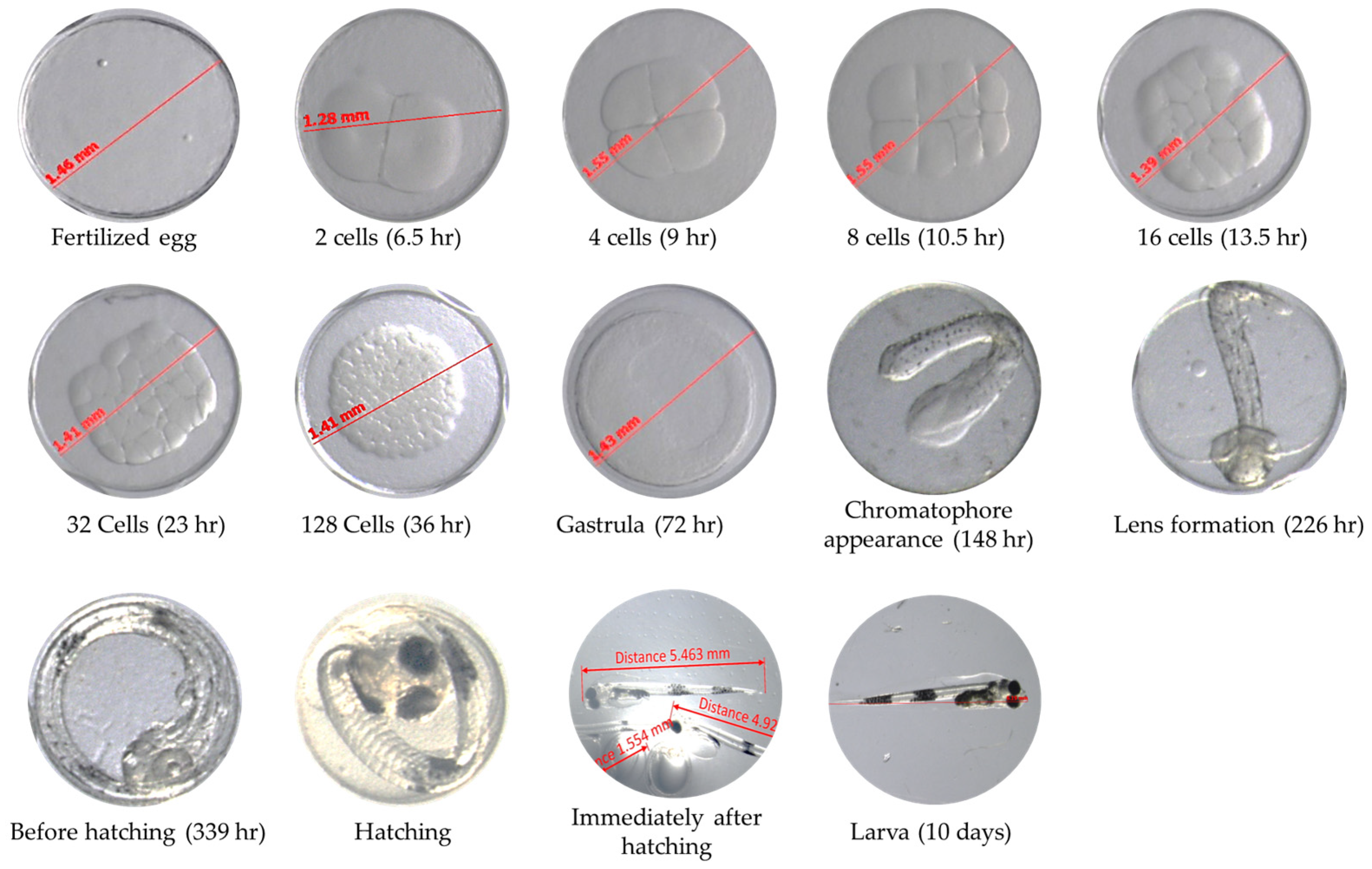
2.2. The Embryos and F1 Generation Measurement

The developmental stage of the embryos was measured daily using a stereomicroscope (SteREO Discovery V8; Carl Zeiss GmbH, Oberkochen, Germany) at 40×, and eggs in tank were maintained at 5–6 °C, with adequate aeration. A total of 100 embryos were measured daily during the spawning period. In addition, the growth of the walleye pollock (F1) hatched in 2015 was measured each period: immediately after hatching (100 larvae), 100 days (100 fish), 180 days (100 fish), 360 days (50 fish) after hatching, and every year thereafter (50 fish). Fish were anesthetized using MS-222 (A5040, Sigma, St louis, MO, USA) at a concentration of 500 ppm and then measured for growth.

2.3. Fish and Sampling

The walleye pollocks used in this study were of two generations: the F1 generation, hatched in 2015 from wild parents, and the F2 generation, hatched in 2017 and 2022 from the F1 generation as parents. Fish hatched in 2015 spawned approximately seven times after 2017, whereas those hatched in 2017 spawned approximately four times after their initial spawning in 2019.
Total length was measured using a fish scale plate (FS-1000), and body weight was measured to the nearest gram using a weighing balance. Each individual used in the investigation was randomly selected. The F1 generation hatched in 2015 comprised 80 fish, the F2 generation hatched in 2017 comprised 50 fish, and the F2 generation hatched in 2022 comprised 50 fish. Gonadal development in walleye pollock was monitored through morphological and histological observations. Gonadal development typically became apparent at a body length of approximately 10–15 cm. As they grew further, the gonads developed into either male or female, and could only be distinguished through dissection, i.e., it was difficult to distinguish between the sexes with the naked eye. During the study, fish were anesthetized using MS-222 (A5040, Sigma) at a concentration of 100 ppm and then dissected for collection of gonads.
The gonadosomatic index (GSI) of walleye pollock was calculated using the following formula [28]:
GSI (%) = (Gonadal mass/(observed wet body mass − Gonad mass)) × 100

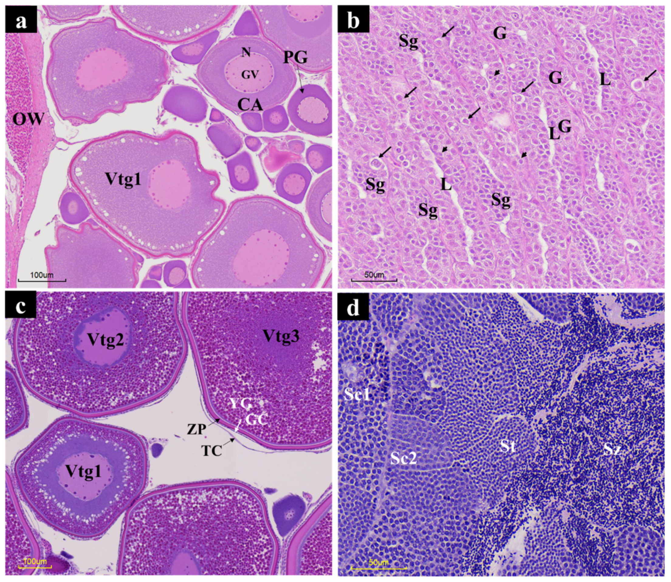
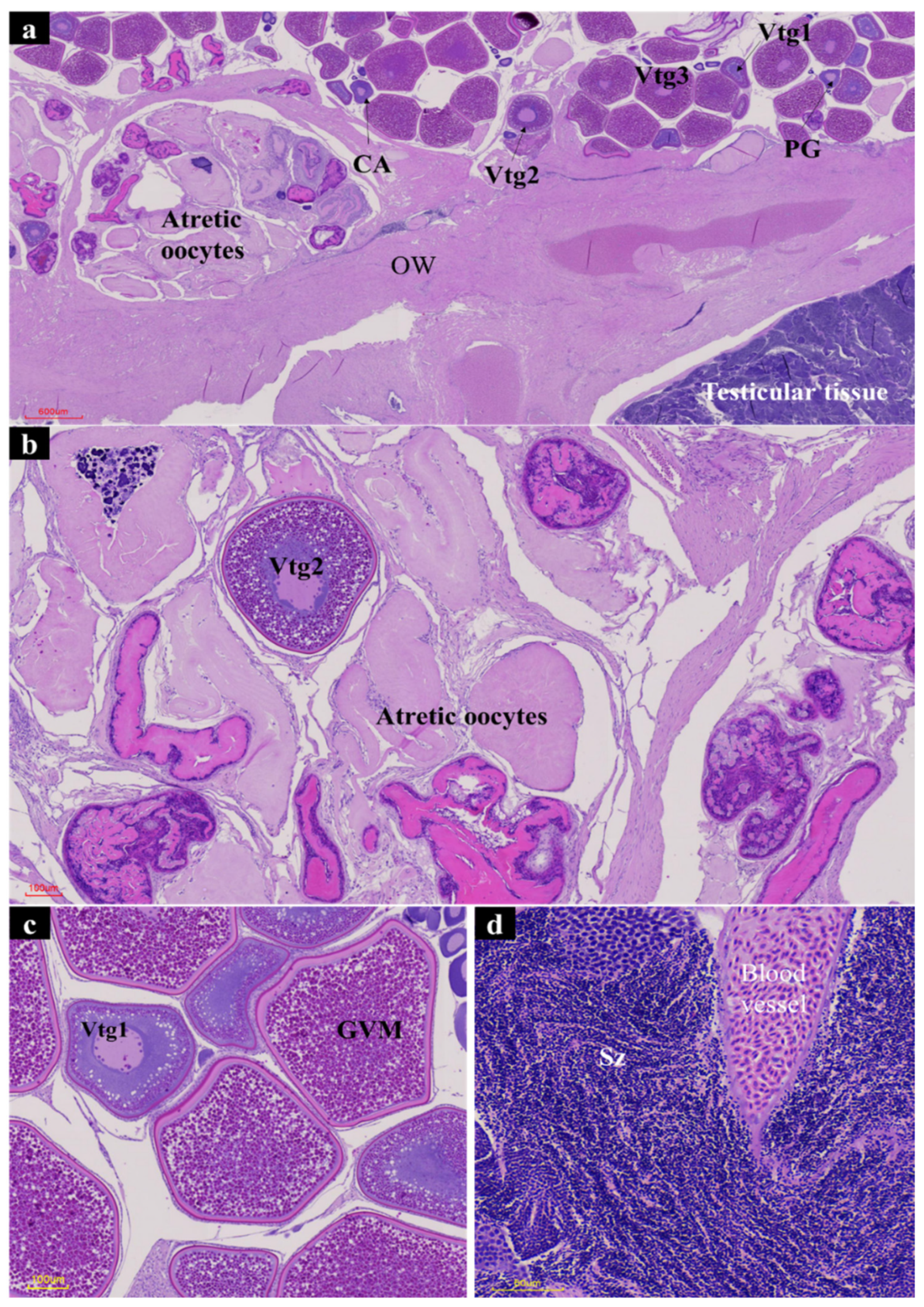
2.4. Histological Characterization

Five female and five male gonads, as well as two intersex gonads, were fixed in 10% neutral buffered formalin for 48 h, dehydrated in a graded alcohol series, embedded in paraffin wax, and sectioned using a rotary microtome to produce approximately 4 to 5 μm thick sections (Leica, YD-355AT, Wetzlar, Hessen, Germany). These sections were then stained with Harris hematoxylin and eosin (H&E). Tissue slides were observed using a slide scanner (Motic EASYSCAN PRO6; Motic, Universal city, CA, USA). The developmental stages of the gonads are shown with reference to previous studies [29].

2.5. Statistical Analyses

Statistical analyses were performed using SPSS version 27 (SPSS Inc., Chicago, IL, USA). Before statistical analysis, Levene’s test was used to examine normality and homogeneity of variances between distinct data. Differences between females and males were determined using t-tests (p < 0.05). All data are presented as mean ± standard deviation.

3. Results

The development of the embryos of the walleye pollock is shown in Figure 1. The average diameter of 3000 embryos was 1.43 ± 0.056 mm. Embryos went through the cleavage stage for up to 52 h post-fertilization (hpf) and reached the gastrula stage at 70.5 hpf. Chromophores were observed at 148 hpf, lenses formed at 226.5 hpf, and the pre-hatching stage was reached at 339 hpf. After hatching, the fish fed on egg yolk for 3–4 days and then began to consume rotifers, B. plicatilis.
The walleye pollock hatched in 2015 showed an average total length of 0.49 ± 0.007 cm immediately after hatching. At 100 days after hatching, the total length was 1.76 ± 0.015 cm and weight was 0.047 ± 0.006 g; they grew to a length of 10.83 ± 2.1 cm and weight of 11.2 ± 2.9 g after about 180 days. Thereafter, their length and weight were 19.7 ± 1.1 cm and 53.2 ± 17.4 g at 1 year old; 37.8 ± 6.2 cm and 402.7 ± 236.7 g at 2 years old; 46.5 ± 4.2 cm and 882.7 ± 283.2 g at 3 years old; 51.3 ± 4.2 cm and 1340.8 ± 468.3 g at 4 years old; 59.7 ± 3.7 cm and 2211.7 ± 453.6 g at 5 years old; and 62.2 ± 4.4 cm and 2189.1 ± 599.4 g at 6 years old, respectively (Table 1). Thus, there was no dramatic difference in the growth between the fifth and sixth years.
The information on the different generations of fish used as samples is shown in Table 2.
Mature walleye pollock gonads are displayed in Figure 2a,b.
Notably, we identified intersex individuals with both female and male gonads while monitoring sexual maturation. Two intersex specimens, one in the F1 generation in 2015 and one in the F2 generation in 2017, were directly observed (Figure 2c,d). They had lengths of 68.0 and 51.5 cm, weights of 1970.4 and 1567.3 g, and gonad weights of 376.8 and 196.8 g, respectively (Table 2). Their GSIs were 23.6% and 14.4%, respectively. The average GSI of normal ovaries of fish hatched in 2015 was 26.5% ± 3.3%, and the average GSI of normal ovaries of fish hatched in 2017 was 14.2% ± 4.1%, i.e., there was no difference in the GSI between normal and intersex individuals. Intersex gonads had distinct ovaries and testes, displaying an asymmetrical arrangement with unevenly shaped ovaries and testes at both ends. Two testes were observed (Figure 2e,f).
H&E staining results for gonadal tissue from normally developed walleye pollock females and males are presented in Figure 3. Oocytes of walleye pollock hatched in 2022 displayed early developmental stages, including primary growth (PG) and cortical alveolar (CA; Figure 3a) stages, whereas the testes exhibited primary spermatogonia (long arrows), secondary spermatogonia (Sg), and undifferentiated spermatogonia (G; Figure 3b) stages. Oocytes hatched in 2015 exhibited various stages, including PG, primary (Vtg1), secondary (Vtg2), and tertiary (Vtg3) vitellogenesis (Figure 3c). Testes displayed a developing stage, having Sg, primary spermatocytes (Sc1), secondary spermatocytes (Sc2), spermatids (St), and spermatozoa (Sz; Figure 3d).
Figure 4 illustrates H&E staining results of intersex walleye pollock gonads. These intersex gonads were divided into oocytes and testes by the ovarian wall (OW), which featured the development of various oocytes. The testes developed normally, and oocyte resorption was limited to the area near the testicular and ovarian tissue connections, with numerous atretic oocytes in the resorption zone.
Figure 5 shows that walleye pollock hatched in January 2022 showed morphological maturity, but the GSI was low at 1.93% ± 1.4%, and the gonad weight was 2.74–10.8 g, being smaller than that of the F1 and F2 walleye pollock generations hatched in 2017. The individual that showed abnormal maturation had a length of 31.5 cm, weighed 258 g, and gonad weight of 10.8 g, with a GSI of 4.4%. Eggs were discernible and opaque when observed with the naked eye. In addition, H&E staining showed primordial germ cells (PGC) and early and late developing vitellogenic oocyte stages, which is the sexual maturity stage.

4. Discussion

This study focused on the growth, characteristics of intersex, and the abnormal maturation of artificially produced walleye pollock. The gonadal development of walleye pollock and the distinction between female and male gonads became evident after the fish reached a size of approximately 10–15 cm after about 180 days. The most commonly reported intersex condition involves the presence of single or multiple oocytes within the testes of subadult or adult males. Numerous studies have reported intersex as a consequence of endocrine disruptor contamination [30,31,32]. Due to limitations in the selection of reference sites for comparison with polluted environments, it is difficult to separate background levels of natural intersex from those reported in most studies. Few studies have investigated levels of intersex at reference sites that have been documented as being free of environmental influences [33]. One intersex was reported after sampling 2660 Chinook salmon (Oncorhynchus tshawytscha) over 3 years in New Zealand and over 3000 coho salmon (O. kisutch) in Chile [34]. Zhukova and Privalikhim [13] consistently observed intersex characteristics in wild-caught walleye pollock, and each individual possessed a distinct sex based on the appearance and classification of the gonads. Assuming normal gonadal development, they may have participated in spawning a number of times, with the exception of the ovarian region adjacent to the testis, where the oocyte maturation process had ceased. In our observation, intersex individuals had ovaries with testes connected to the ends, consistent with a previous report. Except for the area where the ovaries and testes are connected [13], the remaining oocytes and sperms were developing normally. A previous study reported the occurrence of total and partial oocyte atresia in normally developing walleye pollock females from the Bering and Okhotsk seas [35]. However, in this study, in intersex individuals, there was no atresia in other parts of the ovary and more distally, suggesting that the proximity of the testes may have had a hormonal effect on oocytes. Based on our observations, the GSI in intersex individuals was 23.6% and 14.4%, respectively. The average GSI of normal ovaries of the fish hatched in 2015 was 26.5% ± 3.3%, and that for those hatched in 2017 was 14.2% ± 4.1. Therefore, the GSI did not appear to differ between normal and intersex individuals. Herein, unlike intersex individuals discovered in the wild, the age, parent generation, and offspring could be accurately traced along with the development of the gonads for intersex individuals discovered in artificial breeding; moreover, the gonads of such intersex individuals undergo sexual maturity.
Abnormal maturation was observed in approximately 16-month-old F2 generation walleye pollock hatched in 2022 that had not yet spawned. For the growth of the F1 generation, they started spawning after 2 years, and at the time, their length was 39.3 ± 5.3 cm, and weight was 535.4 ± 175.1 g. Therefore, individuals with abnormal maturation hatched in 2022 had a length of 31.5 cm and weight of 258 g, suggesting that they had not yet reached the laying length and/or weight. It is generally known that in the wild, walleye pollock spawns prior to the winter and spring seasons, but not multiple times throughout the year [36]. In our observations, most walleye pollock gonads showed similar maturation, independent of whether immature, developing, or pre-spawning. However, this individual showed pre-spawning involving PGC, and both early and late developing vitellogenic oocyte stages. Normally, after fertilization of the egg and during the early stage of embryogenesis, a small number of non-dividing PGCs are produced in teleost. During egg development, including the formation of PGCs, the transformation of PGCs into oocytes, and then into primary oocytes, initiates meiosis. This is followed by the huge growth of the oocyte during vitellogenesis, accumulating the nutrients necessary for embryonic development. Maturation is characterized by the reduction in or cessation of endocytosis, resumption of meiosis, germ vesicle breakdown (GVBD), formation of a monolayer of cortical alveoli under the oolemma, lysis of yolk platelets, and hydration of the lipophilic oocytes [37]. Therefore, it is rare to observe PGC and vitellogenic stages co-occurring during sexual development and maturation in the teleost. Stahl and Kruse [38] performed a histological characterization of the gonads of 173 female walleye pollocks caught in the Bering Sea in 2003 and showed five stages of maturation in ovaries. In our observations, the oocytes presented pre-spawning stages with yellow and orange gonads and opaque and translucent eggs. The ovary generally occupies 2/3 of the body in the pre-spawning period, but the GSI of this individual hatched in 2022 was still too low for pre-spawning. Precocious maturation occurs in several cultured species, including Nile tilapia (Oreochromis niloticus) and Atlantic salmon (Salmo salar), which tend to become sexually mature and reproduce before they reach a suitable body size for harvest [26,27]. This precocious maturation results in slower growth as energy is diverted to reproduction and large differences in the size of the product at harvest. Female Atlantic salmon often divert energy (e.g., lipids) to reproductive processes, resulting in reduced quality as they reach sexual maturity [27]. Although this pattern has not been reported for this species in the wild and thus cannot be compared, it is assumed that this phenomenon is due to factors, such as precocious maturation or stress, which occur in a controlled environment.
Therefore, the results of this study suggest that intersex individuals do not differ in terms of maturity and growth from individuals with normal development, except for differences in the area near the testicular and ovarian tissue connections. Further, this species shows abnormal ovary development in individuals who have not yet reached maturity, and PGCs are found along with precocious maturation, suggesting that further studies should be considered to understand the maturation of this species.

5. Conclusions

Intersex walleye pollock were observed in both F1 and F2 generations in aquaculture, with male testes observed at the end of female gonads. Except for the area where the ovaries and testes are connected, the remaining oocytes and sperm were developing normally. In intersex cases, there was no atresia in other parts of the ovary and those located more distally, suggesting that the proximity of the testes may have had a hormonal effect on oocytes. Therefore, the results of this study suggest that intersex individuals do not differ in terms of maturity and growth from individuals with normal development. PGC and early and late developing vitellogenic oocyte stages were observed in individuals hatched in 2022. It can be assumed that this phenomenon can be attributed to factors, such as precocious maturation or stress, which occur in a controlled environment.

Author Contributions

Writing—original draft, H.-K.Y. and S.-S.K.; methodology, H.-K.Y., S.-S.K., K.-W.L. and K.-D.K., investigation, S.-J.W. and M.-S.J.; project administration, J.-J.P.; supervision, S.-S.K. All authors have read and agreed to the published version of the manuscript.

Funding

This study was funded by the National Institute of Fisheries Science, Ministry of Oceans and Fisheries, Korea (grant number: R2025028).

Institutional Review Board Statement

The animal protocol used in this study was reviewed and approved by the National Institute of Fisheries Science–Institutional Animal Care and Use Committee in accordance with the ethical procedures and scientific care guidelines (approval number 2022-NIFS-IACUC-4).

Informed Consent Statement

Not applicable.

Data Availability Statement

All data used in this study are available upon request from the corresponding author.

Conflicts of Interest

The authors declare no conflicts of interest.

References

  1. Todd, E.V.; Liu, H.; Muncaster, S.; Gemmell, N.J. Bending genders: The biology of natural sex change in fish. Sex. Dev. 2016, 10, 223–241. [Google Scholar] [CrossRef] [PubMed]
  2. Lau, E.S.-W.; Zhang, Z.; Qin, M.; Ge, W. Knockout of zebrafish ovarian aromatase gene (cyp19a1a) by TALEN and CRISPR/Cas9 leads to all-male offspring due to failed ovarian differentiation. Sci. Rep. 2016, 6, 37357. [Google Scholar] [CrossRef] [PubMed]
  3. Graves, J.A.M.; Peichel, C.L. Are homologies in vertebrate sex determination due to shared ancestry or to limited options? Genome Biol. 2010, 11, 205. [Google Scholar]
  4. McClelland, K.; Bowles, J.; Koopman, P. Male sex determination: Insights into molecular mechanisms. Asian J. Androl. 2012, 14, 164. [Google Scholar] [CrossRef] [PubMed]
  5. Hara, S.; Furukawa, F.; Mukai, K.; Yazawa, T.; Kitano, T. Peroxisome proliferator-activated receptor alpha is involved in the temperature-induced sex differentiation of a vertebrate. Sci. Rep. 2020, 10, 11672. [Google Scholar] [CrossRef]
  6. Römer, U.; Beisenherz, W. Environmental determination of sex in Apistogramma (Cichlidae) and two other freshwater fishes (Teleost). J. Fish Biol. 1996, 48, 714–725. [Google Scholar]
  7. Ramee, S.W.; Lipscomb, T.N.; DiMaggio, M.A. Evaluation of the effect of larval stocking density, salinity, and temperature on stress response and sex differentiation in the Dwarf Gourami and Rosy Barb. Aquac. Rep. 2020, 16, 100287. [Google Scholar] [CrossRef]
  8. Robertson, C.E.; Wright, P.A.; Köblitz, L.; Bernier, N.J. Hypoxia-inducible factor-1 mediates adaptive developmental plasticity of hypoxia tolerance in zebrafish, Danio rerio. Proc. R. Soc. B Biol. Sci. 2014, 281, 20140637. [Google Scholar] [CrossRef]
  9. Pfennig, F.; Kurth, T.; Meißner, S.; Standke, A.; Hoppe, M.; Zieschang, F.; Reitmayer, C.; Göbel, A.; Kretzschmar, G.; Gutzeit, H.O. The social status of the male Nile tilapia (Oreochromis niloticus) influences testis structure and gene expression. Reproduction 2012, 143, 71–84. [Google Scholar] [CrossRef] [PubMed]
  10. Atz, J.W. Intersexuality in fishes. In Intersexuality in Vertebrates Including Man; Armstrong, C.N., Marshall, A.J., Eds.; Academic Press: London, UK, 1964; pp. 145–232. [Google Scholar]
  11. Pauly, D. Darwin’s Fishes: An Encyclopedia of Ichthyology, Ecology, and Evolution; Cambridge University Press: Cambridge, UK, 2004. [Google Scholar]
  12. Sadovy, M.Y.; Liu, M. Functional hermaphroditism in fishes. Copeia 2008, 1, 136–156. [Google Scholar]
  13. Zhukova, K.; Privalikhin, A.M. First record of intersexuality in walleye pollock Theragra chalcogramma (Pallas, 1814). Mar. Biodivers. Rec. 2015, 8, 89–91. [Google Scholar] [CrossRef]
  14. Caprioli, R.; Costa, C.; Cataldi, E.; Boglione, C.; Cataudella, S. First record of female germ cells in the testis of a wild Mediterranean northern bluefin tuna Thunnus thynnus L. 1758. J. Fish Biol. 2007, 71, 620–622. [Google Scholar] [CrossRef]
  15. Macías, D.; Saber, S.; Osuna, A.M.; Cruz-Castán, R.M.; Gómez-Vives, M.J.; Báez, J.C. First record of intersexuality in Euthynnus alletteratus in the Mediterranean Sea: Histological description. Marine Biodiv. Rec. 2014, 7, e3. [Google Scholar] [CrossRef]
  16. Sawada, Y.; Seoka, M.; Okada, T.; Miyashita, S.; Murata, O.; Kumai, H. Hermaphroditism in a captive-raised Pacific bluefin tuna. J. Fish. Biol. 2022, 60, 263–265. [Google Scholar]
  17. Hiatt, T.; Dalton, M.; Felthoven, R.; Fissel, B.; Garber-Yonts, B.; Haynie, A.; Kasperski, S.; Lew, D.; Package, C.; Sepez, J.; et al. Economic status of the ground fish fisheries off Alaska. In Stock Assessment and Fishery Evaluation Report for the Ground Fish Fisheries of the Gulf of Alaska and Bering Sea/Aleutian Islands Area; North Pacific Fishery Management Council: Anchorage, AK, USA, 2010; Volume 2009, p. 254. [Google Scholar]
  18. Livingston, P.A. Importance of predation by groundfish, marine mammals and birds on walleye Pollock Theragra chalcogramma and Pacific herring Clupea pallasi in the eastern Bering Sea. Mar. Ecol. Prog. Ser. 1993, 102, 205–215. [Google Scholar] [CrossRef]
  19. Napp, J.M.; Kendall, A.W., Jr.; Schumacher, J.D. A synthesis of biological and physical processes affecting the feeding environment of larval walleye pollock (Theragra chalcogramma) in the eastern Bering Sea. Fish. Oceanogr. 2000, 9, 147–162. [Google Scholar] [CrossRef]
  20. Sinclair, E.H.; Vlietstra, L.S.; Johnson, D.S.; Zeppelin, T.K.; Byrd, G.V.; Springer, A.M.; Ream, R.R.; Hunt, G.L. Patterns in prey use among fur seals and seabirds in the Pribilof Islands. Deep Sea Res. II 2008, 55, 1897–1918. [Google Scholar] [CrossRef]
  21. FAO. Global Fishery and Aquaculture Production. 2022. Available online: http://www.fao.org/fishery/statistics/en (accessed on 10 April 2024).
  22. Hinckley, S. The reproductive biology of walleye pollock, Theragra chalcogramma, in the Bering Sea with reference to spawning stock structure. Fish. Bull. 1987, 85, 481–498. [Google Scholar]
  23. Kim, S.S.; Lee, C.J.; Yoo, H.K.; Choi, J.; Byun, S.G.; Kim, W.J.; Lim, H.J.; Park, J.S. Effect of water temperature on walleye pollock (Gadus chalcogrammus) embryos, larvae and juveniles: Survival, HSP70 expression, and physiological responses. Aquaculture 2022, 554, 738136. [Google Scholar] [CrossRef]
  24. Haugen, T.; Andersson, E.; Norberg, B.; Taranger, G.L. The production of hermaphrodites of Atlantic cod (Gadus morhua) bymasculinization with orally administered 17-a-methyltestosterone, and subsequent production of all-female cod populations. Aquaculture 2010, 31, 248–254. [Google Scholar]
  25. Kirubakaran, T.G.; Andersen, Ø.; Rosa, M.C.; Andersstuen, T.; Hallan, K.; Peter Kent, M.P.; Lien, S. Characterization of a male specific region containing a candidate sex determining gene in Atlantic cod. Sci. Rep. 2019, 9, 116. [Google Scholar] [CrossRef] [PubMed]
  26. Mires, D. Production of aquatic animals. In The Tilapias; Nash, C.E., Novotony, A.J., Eds.; Elsevier: New York, NY, USA, 1995; pp. 133–152. [Google Scholar]
  27. Piferrer, F.; Beaumont, A.; Falguière, J.-C.; Flajšhans, M.; Haffray, P.; Colombo, L. Polyploid fish and shellfish: Production, biology and applications to aquaculture for performance improvement and genetic containment. Aquaculture 2009, 293, 125–156. [Google Scholar] [CrossRef]
  28. Gunderson, D.R.; Dygert, P.H. Reproductive effort as a predictor of natural mortality rate. J. Cons. Int. Explor. Mer. 1988, 44, 200–209. [Google Scholar] [CrossRef]
  29. Browen-Peterson, N.J.; Wyanski, D.M.; Saborido-Rey, F.; Macewicz, B.; Lowerre-Barbieri, S.K. A Standardized termonology for describing reproductive development in fishes. Mar. Coast. Fish. 2011, 3, 52–70. [Google Scholar] [CrossRef]
  30. Nolan, M.; Jobling, S.; Brighty, G.; Sumpter, J.P.; Tyler, C.R. A histological description of intersexuality in the roach. J. Fish Biol. 2001, 58, 160–176. [Google Scholar] [CrossRef]
  31. Metcalfe, C.D.; Kidd, K.A.; Sumpter, J.P. Chemically induced alterations to gonadal differentiation in fish, fish diseases and disorders. In Non-Infectious Disorders, 2nd ed.; CABI: Wallingford, UK, 2010; Volume 2, pp. 144–165. [Google Scholar]
  32. Schmitt, C.J.; Hinck, J.E.; Blazer, V.S.; Denslow, N.D.; Dethloff, G.M.; Bartish, T.M.; Coyle, J.J.; Tillitt, D.E. Environmental contaminants and biomarker responses in fish from the Rio Grande and its U.S. tributaries: Spatial and temporal trends. Sci. Total Environ. 2005, 350, 161–193. [Google Scholar] [CrossRef] [PubMed]
  33. Bahamonde, P.A.; Munkittrick, K.R.; Martyniuk, C.J. Intersex in teleost fish: Are we distinguishing endocrine disruption from natural phenomena? Gen. Comp. Endocrinol. 2013, 192, 25–35. [Google Scholar] [CrossRef]
  34. Kinnison, M.T.; Unwin, M.J.; Jara, F. Macroscopic intersexuality in salmonid fishes. N. Z. J. Mar. Freshwat. Res. 2000, 34, 125–134. [Google Scholar] [CrossRef]
  35. Privalikhin, A.M. Resorption of developing oocytes as a mechanism of formation of individual and population fecundity in walleye pollock Theragra chalcogramma (Gadidae). J. Ichthyol. 2003, 43, 454–463. [Google Scholar]
  36. Baird, T.A.; Olla, B.L. Social and reproductive behavior of a captive group of walleye pollock, Theragra chalcogramma. Environ. Biol. Fish. 1991, 30, 295–301. [Google Scholar] [CrossRef]
  37. Lubzens, E.; Young, G.; Bobe, J.; Cerdà, J. Oogenesis in teleosts: How fish eggs are formed. Gen. Comp. Endocrinol. 2010, 165, 367–389. [Google Scholar] [CrossRef] [PubMed]
  38. Stahl, J.P.; Kruse, G.H. Classification of ovarian stages of Walleye Pollock Theragra chalcogramma. In Resiliency of Gadid Stocks to Fishing and Climate Change; Alaska Sea Grant College Program AK-SG-08-01; Alaska Sea Grant: Anchorage, AK, USA, 2008; pp. 1–20. [Google Scholar]
Figure 1. The developmental stages of embryos of walleye pollock over 0–339 hpf at 5 °C.
Fishes 10 00035 g001
Figure 2. Gonads of walleye pollock. (a) Ovary of a female hatched in January 2015. (b) Testis of a male hatched in January 2015. Gonads of intersexes hatched in (c) January 2015 and (d) January 2017. Morphological characteristics of intersex gonad with ovary and testis. Gonads of intersexes hatched in (e) January 2015 and (f) January 2017.
Fishes 10 00035 g002
Figure 3. Histological characterization of normal gonadal tissues from immature and mature individuals. (a) Oocytes of female walleye pollock hatched in January 2022. (b) Testicular tissue of male walleye pollock hatched in January 2022. (c) Oocytes of female walleye pollock hatched in January 2015. (d) Testicular tissue of female walleye pollock hatched in January 2015. PG, primary growth oocyte; CA, cortical alveolar oocytes; nu, nucleus; ne, nucleolus; GV, germinal vesicles; GC, granulosa cells; TC, theca cells; ZP, zona pellucida; YG, York globules; Vtg1, primary vitellogenic oocyte; Vtg2, secondary vitellogenic oocyte; Vtg3, tertiary vitellogenic oocyte; OW, ovarian wall in the ovary of females; long arrows, primary spermatogonia; short arrows, Sertoli cells; Sg, secondary spermatogonia; G, differentiated spermatogonia (germ cells); L, lumen of lobule; Sc1, primary spermatocyte; Sc2, secondary spermatocyte; St, spermatid; Sz, spermatozoa in the testis of males.
Fishes 10 00035 g003
Figure 4. Histological characterization of intersex gonadal tissues from mature individuals. (a) Ovary and testis junction area. (b) Oocyte resorption area between ovarian and testicular tissue junctions. (c) Ovary in intersex gonadal tissues. (d) Testis in intersex gonadal tissues. PG, primary growth oocyte; CA, cortical alveolar oocytes; Vtg1, primary vitellogenic oocyte; Vtg2, secondary vitellogenic oocyte; Vtg3, tertiary vitellogenic oocyte; GVM, germinal vesicle migration, OW, ovarian wall; Sz, spermatozoa in the testes of males.
Fishes 10 00035 g004
Figure 5. Morphological characteristics and histological characterization of walleye pollock with abnormal ovary. (a) Morphological characteristics of gonads of walleye pollock hatched in January 2022. (b) Abnormal mature ovary. (c) Histological characterization of gonadal tissues from abnormal immature walleye pollock hatched in January 2022. (d) Primordial germ cells (PGC) can be observed in the dotted box in panel (c). PG indicates primary growth oocyte, CA indicates cortical alveolar oocytes, Vtg2 indicates secondary vitellogenic oocyte, and Vtg3 indicates tertiary vitellogenic oocyte.
Fishes 10 00035 g005
Table 1. Measurement of F1 generation walleye pollock according to time.
AgeLength (cm)Weight (g)
1 day0.49 ± 0.007-
100 days1.76 ± 0.0150.047 ± 0.006
180 days10.83 ± 2.111.2 ± 2.9
360 days19.7 ± 1.153.2 ± 17.4
2 years39.3 ± 5.3535.4 ± 175.1
3 years46.5 ± 4.2882.7 ± 283.2
4 years51.3 ± 4.21340.8 ± 468.3
5 years59.7 ± 3.72211.7 ± 453.6
6 years62.2 ± 4.42189.1 ± 599.4
Table 2. Measurement information of generational walleye pollock.
Hatched
(Generation)/Age
Length (cm)Weight (g)GSI (%)
FemaleMalep ValueFemaleMalep ValueOvaryIntersex
* 2015 (F1)/660.4 ± 4.957.6 ± 4.20.1252226.4 ± 649.51903.5 ± 483.50.00926.5 ± 3.323.6
* 2017 (F2)/451.9 ± 2.647.3 ± 2.80.6991163.3 ± 153.4961.2 ± 203.50.31914.2 ± 4.114.4
** 2022 (F2)/1.428.4 ± 2.826.5 ± 3.60.316184.8 ±59.3161.0 ± 36.50.7191.93 ± 1.4-
* One intersex individual was found; ** these individuals have not yet experienced spawning.
Disclaimer/Publisher’s Note: The statements, opinions and data contained in all publications are solely those of the individual author(s) and contributor(s) and not of MDPI and/or the editor(s). MDPI and/or the editor(s) disclaim responsibility for any injury to people or property resulting from any ideas, methods, instructions or products referred to in the content.

