Standardized Workflow for the Generation of Patient-Derived Glioblastoma Spheroids

 , , , ,
  and
and 
Abstract
1. Introduction
2. Experimental Design
2.1. Materials
- 1X Dulbecco’s Phosphate-Buffered Saline (DPBS) w/o Ca2+ and Mg2+ (Dutscher, Bernolsheim, France, Cat. no.: L0615);
- Phosphate-Buffered Saline (PBS) (Gibco—Themo Fischer Scientific, Waltham, MA, USA, Cat. no.: 18912-014);
- 1X Dulbecco’s Modified Eagle Medium (DMEM) (Corning, Corning, NY, USA, Cat. no.: 10-013-CV);
- Euromed-N Medium (Euroclone, Pero (MI), Italy, Cat. no.: ECM0883L);
- Nutrient Mixture F-12 Ham (Gibco, Cat. no.:21765-029);
- Heat-inactivated Fetal Bovine Serum (FBS) (Sigma-Aldrich, St. Louis, MO, USA, Cat. no.: F7524);
- L-Glutamine 2 mM (Corning, Cat. no.: 25-005-CI);
- Penicillin 100 U/mL (Lonza Bioscience, Walkersville, MD, USA, Cat. no.: DE17-602E);
- Streptomycin 100 μg/mL (Lonza Bioscience, Cat. no.: DE17-602E);
- Antibiotic-antimycotic (100X) (Gibco, Cat. no.: 15240062);
- Trypsin/EDTA Solution 100X (Gibco, Cat. no.: R001100);
- Type II collagenase 250 U/mL (Gibco, Cat. no.: 17101015);
- MEM Non-Essential Amino Acids Solution (100X) (Gibco, Cat. no.: 11140050);
- Agarose powder (Sigma-Aldrich, Cat. no.: A5093);
- 4% paraformaldehyde (PFA) (w/v) in 1X PBS (Thermo Fisher Chemicals—Themo Fischer Scientific, Cat. no.: 416785000);
- 3% BSA (w/v) in 1X PBS, i.e., blocking solution (Dutscher, Cat. no.: P6154);
- Sodium Citrate Buffer, i.e., unmasking solution (10 mM trisodium citrate, 0.05% Tween 20, pH 6) (Sigma Aldrich, Cat. no.: S4641);
- Primary antibodies: mouse anti-PCNA, used at 1:50 for IF (Santa Cruz Biotechnology, Santa Cruz, California, USA, Cat# sc-25280, RRID:AB_628109); rabbit anti-AIF, used at 1:50 for IF (Santa Cruz Biotechnology Cat# sc-5586, RRID:AB_2224668);
- Fluorochrome-conjugated secondary antibodies diluted 1:100 in 1X PBS: anti-rabbit IgG Atto 647N antibody produced in goat, 40839, Sigma-Aldrich, St. Louis, MO, USA; anti-mouse IgG-Atto 488 antibody produced in goat, 62197, Sigma-Aldrich, St. Louis, MO, USA; anti-rabbit IgG (whole molecule)-FITC antibody produced in goat, F0382, Sigma-Aldrich, St. Louis, MO, USA; anti-mouse IgG (whole molecule)-TRITC antibody produced in goat, T5393, Sigma-Aldrich, St. Louis, MO, USA;
- DAPI (4′,6-diamidino-2-phenylindole) (Sigma-Aldrich, Cat. no.: D9542);
- Quick-hardening mounting media (e.g., Eukitt®) (Sigma-Aldrich, Cat. no.: 03989).
2.2. Equipment
- Biological safety cabinet;
- Chemical hood;
- 60 mm Nunc™ EasYDish™ plates (Thermo Scientific—Thermo Fischer Scientific, TM: 150462);
- 100 mm Nunc™ EasYDish™ plates (Thermo Scientific TM: 150466);
- 25 cm2 Nunc™ EasYFlask™ flasks (Thermo Scientific Cat. no.: 156367);
- Incubator humidified at 5% CO2 and 37 °C;
- Scalpel;
- BD Falcon™ 70 μm Cell Strainer (Product Number: 352350);
- 15 mL Polypropylene conical tubes (Falcon—Corning; Cat. no.: 352096);
- 50 mL Polypropylene conical tubes (Falcon; Cat. no.: 352070);
- 100–1000 μL and 20–200 μL pipettes and corresponding tips;
- Polyethylene Pasteur pipettes;
- 1.5 mL Microcentrifuge tubes;
- Benchtop centrifuge;
- Orbital shaker;
- Burker chamber;
- Microwave oven;
- Thermoblock;
- Tissue culture plate, 96-well, flat bottom;
- Hydrophobic pap pen;
- Glass microscope slides and coverslips;
- Confocal microscope (Leica Confocal Microscope TCS SP8; Leica Microsystems, Heidelberg, Germany);
- Inverted phase-contrast microscope.
3. Procedure
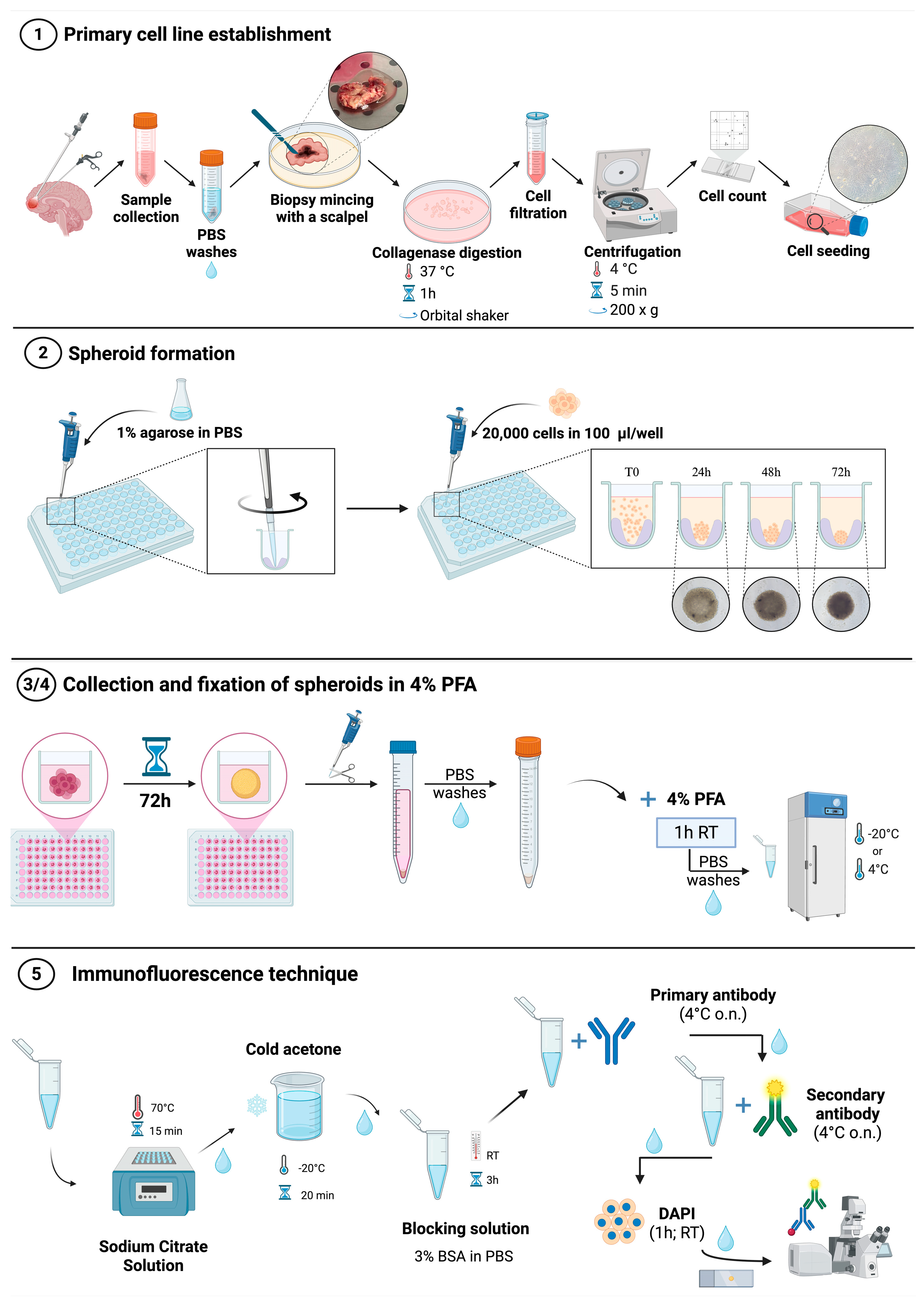
3.1. Primary Cell Line Establishment (~2 h)
- Collect the bioptic sample inside an appropriate medium (1X DMEM supplemented with 100 U/mL penicillin, 100 μg/mL streptomycin and 1X antibiotic-antimycotic 100X);
- In a biological safety cabinet, wash the biopsy, using 1X DPBS w/o Ca2+ and Mg2+, to eliminate any traces of blood;
- Cut the biopsy with a scalpel into small pieces (~1–5 mm) on a 100 mm plate;
- Incubate the obtained small pieces in an orbital shaker at 37 °C for 1 h on a 60 mm plate containing 5 mL of DMEM supplemented with 2 mM L-Glutamine, 100 U/mL penicillin, 100 μg/mL streptomycin, and 250 U/mL collagenase type II;
- Filter the cell suspension with a 70 μm cell strainer into a 50 mL conical tube to collect cells;
- Centrifuge the tube containing the cell suspension at 200× g for 5′ at 4 °C;
- Resuspend the cell pellet with the appropriate medium: 50% Euromed-N Medium, 50% 1X DMEM:F-12, 10% FBS, 1% L-Glutamine, 1% penicillin/streptomycin, 1% Non-Essential Amino Acids;
- Seed the cells into T25 flasks (~5 × 10⁵ cells/flask) and place in a humidified atmosphere of 5% CO2 at 37 °C;
- Monitor GBM primary cells using an optical phase-contrast microscope (Nikon ECLIPSE Ti2, Amstelveen, The Netherlands, EU);
- Passage GBM primary cells of use for downstream experiments when they reach an 80% of confluency.

- CRITICAL STEP: The biopsy cut should be performed as rapidly as possible, while continuously keeping the biopsy moist, in order to prevent necrotic cell death. Be sure to remove all blood from the biopsy to improve isolation yield. The efficiency of primary GBM cell isolation depends primarily on the cellularity and viability of the starting biopsy. In our experience, highly necrotic regions—often identifiable by a dark-red appearance—show reduced isolation yield and lower proliferative potential. For this reason, during the mechanical dissection step, visibly necrotic areas are carefully removed prior to enzymatic digestion to enrich for viable tumor cells. This pre-selection step improves reproducibility and consistency in primary culture establishment and subsequent spheroid formation.NOTE: The serum-containing culture conditions described in this protocol (50% Euromed-N Medium, 50% DMEM:F-12 supplemented with 10% FBS) are intended to support the expansion of differentiated bulk GBM cells and do not selectively enrich for glioblastoma stem-like cells (GSCs). The isolation and propagation of GSCs typically require serum-free defined media supplemented with specific growth factors, which promote spontaneous neurosphere formation.

3.2. 3D Cell Culture (Spheroid) Establishment (at Least 3 Days)
- Prepare a 1% agarose solution by dissolving the agarose powder in 1X sterile PBS and heating in a microwave oven until totally dissolved (usually when the solution is boiling at about 100 °C). For instance, prepare a total of 10 mL of 1% agarose solution for each 96-well plate.
- Using a 200 µL pipette, dispense 50 µL of agarose solution into each well of a 96-well plate.
- Immediately after dispensing, gently perform circular movements to allow the formation of a low-attachment surface as illustrated in Figure 1.
- Place the 96-well plate into an incubator humidified at 5% CO2 and 37 °C for at least 20 min.
- Meanwhile, enzymatically detach the cells using 1X Trypsin/EDTA Solution in 1X DPBS w/o Ca2+ and Mg2+ for 5 min at 37 °C. Inactivate the trypsin solution by adding an equal amount of complete medium, collect and centrifuge for 5 min at 200× g. Resuspend the cell pellet in an appropriate amount of complete medium and count the GBM primary cells.
- Seed 2 × 10⁴ cells into previous agarose-coated wells in the appropriate culture medium (100 μL/well).
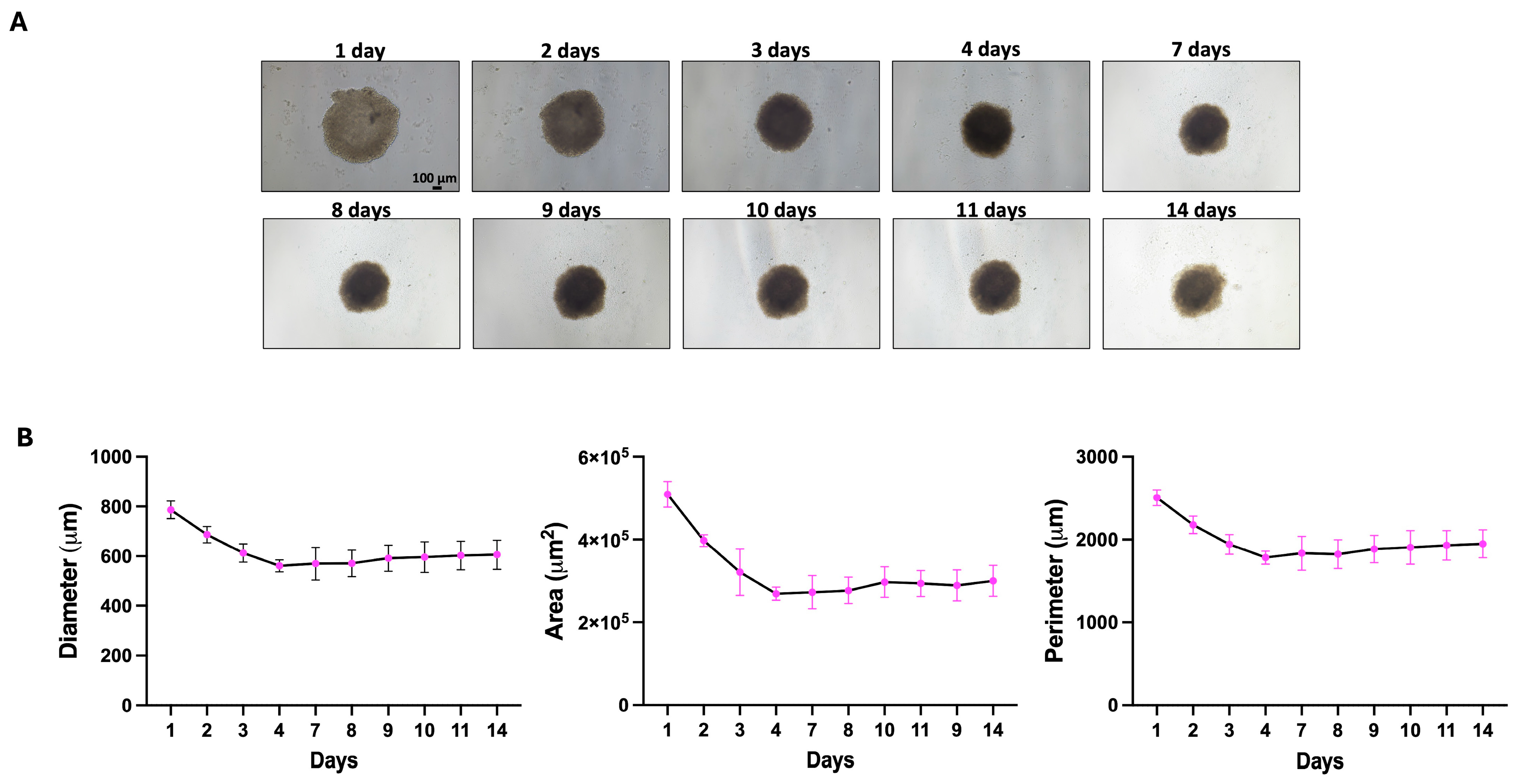
- Monitor the growth and formation of spheroids using an optical phase-contrast microscope.
- OPTIONAL STEP: Upon cell seeding, a short initial centrifugation of the 96-well plate may facilitate cell aggregation. During the spheroid maintenance, add fresh culture medium if necessary.
3.3. Spheroid Collection and Washing (~10′–30′, Depending on the Number of Spheroids)
- After at least 72 h of culturing, collect the 3D spheroids in a 15 mL conical tube using a 200 μL pipette in a biological safety cabinet;
- Allow the 3D spheroids to settle spontaneously at the bottom of the 15 mL conical tube;
- Carefully remove the supernatant with an appropriate pipette (100–1000 μL or 20–200 μL);
- Add 1 mL of 1X DPBS w/o Ca2+ and Mg2+ and wash the spheroids by gently pipetting up and down;
- Repeat the previous step until the supernatant appears clear.

- CRITICAL STEP: During collection, the 3D spheroids may be damaged by mechanical insults. To avoid this issue, cut off the end of the 200 μL tip, gently pipette, ensuring each spheroid is collected from the well, and release carefully at the bottom of the 15 mL tube, avoiding bubble formation.OPTIONAL STEP: If the spheroids do not settle spontaneously at the bottom of the tube after collection and each wash step, you can centrifuge them at 115× g for 5 min and carefully remove the supernatant, without disturbing the pellet.
3.4. Spheroid Fixation in 4% PFA (~1.5 h)
- After the last wash, ensure that the 3D spheroids have pelleted at the bottom of the tube and carefully remove the supernatant;
- Working under a chemical hood, resuspend the pellet in 1 mL of 4% PFA and incubate for 1 h at room temperature (RT);
- After fixation, ensure that the spheroids have pelleted at the bottom of the tube and carefully remove and discard the 4% PFA completely;
- Wash the pellet twice in 1 mL of sterile 1X PBS;
- After the last wash, transfer the spheroids to a new 1.5 mL conical tube in 1 mL of sterile 1X PBS;
- Proceed with the immunofluorescent staining.
- OPTIONAL STEP: If the spheroids do not settle spontaneously at the bottom of the tube after fixation and each wash step, you can centrifuge them at 115× g for 5 min and carefully remove the supernatant, without disturbing the pellet.

- PAUSE STEP: Fixed samples can be stored at 4 °C for up to four months, or directly at −20 °C for a longer period.

3.5. Single or Double Immunofluorescence Analysis (3–5 Days)
- Ensure that the spheroids are at the bottom of the tube, and then carefully remove the supernatant.
- Gently add 0.5–1 mL of the unmasking solution, and incubate for 15 min at 70 °C. You can place the eppendorf tube containing the spheroids resuspended in the unmasking solution in a thermoblock at 70 °C.
- Ensure that the spheroids are at the bottom of the tube, and then carefully remove the supernatant and rinse 2–5 times with 0.5–1 mL of 1X PBS.
- Ensure that the spheroids are at the bottom of the tube, and then carefully remove the supernatant and gently add 0.5–1 mL of cold acetone for 20 min at −20 °C.
- Ensure that the spheroids are at the bottom of the tube, and then carefully remove the supernatant and rinse 2–5 times with 0.5–1 mL of 1X PBS.
- Ensure that the spheroids are at the bottom of the tube, and then carefully remove the supernatant, add 0.5 mL of blocking solution and incubate for 3 h at RT.
- After the blocking, transfer the spheroids to different eppendorf tubes (always using a cut pipette tip) based on the number of primary antibodies to be incubated and considering one eppendorf tube for the negative control. Next, make sure that the spheroids are at the bottom of the tube, and then carefully remove the supernatant.
- Add the primary antibody diluted in 1X PBS according to the manufacturer’s instructions and incubate overnight (o.n.) at 4 °C. In the negative control, incubate o.n. with 1X PBS only.
- The next day, ensure that the spheroids are at the bottom of the tube, and then carefully remove the supernatant and rinse 1–5 times with 0.5–1 mL of 1X PBS.
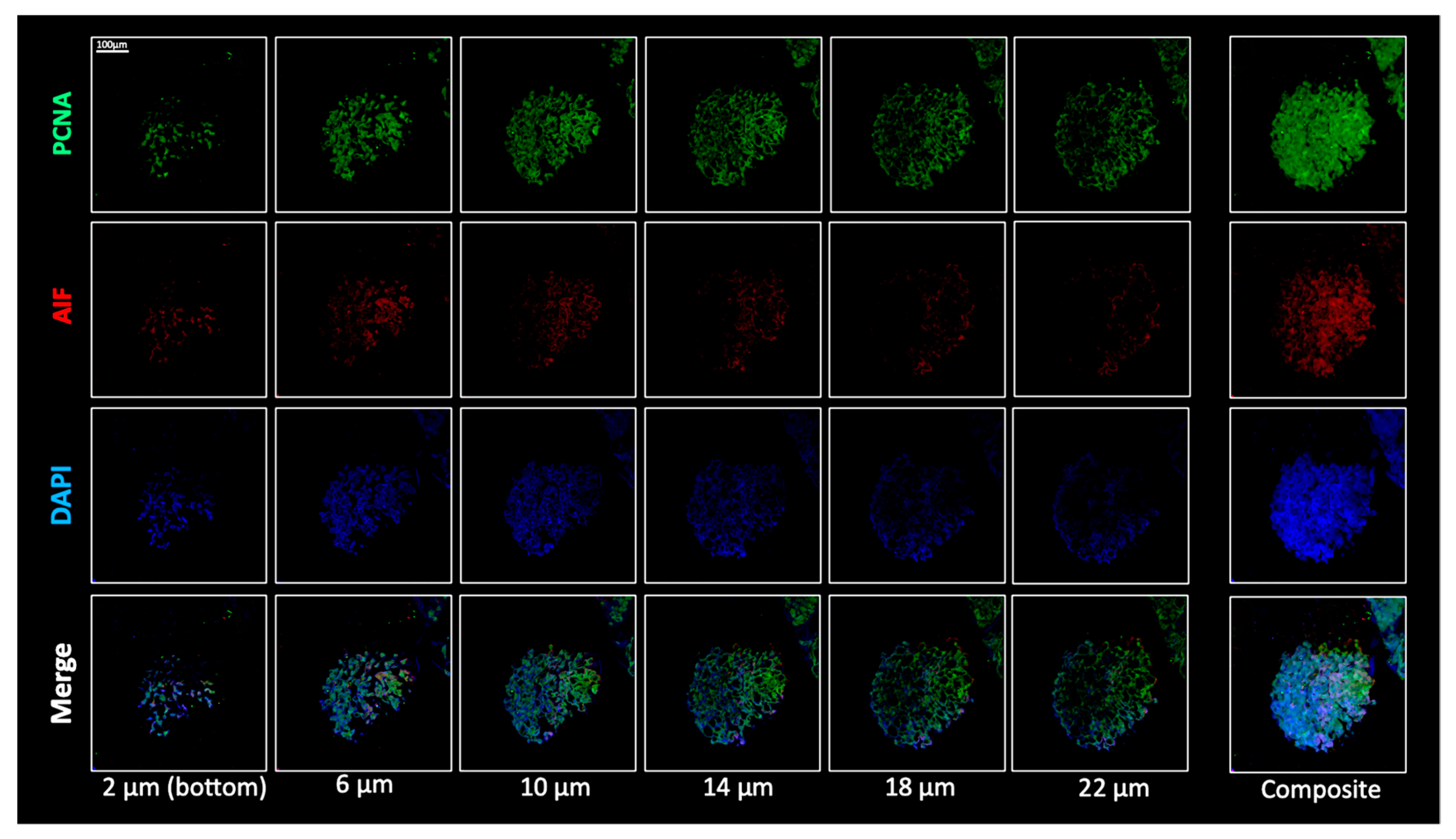
- Ensure that the spheroids are at the bottom of the tube, and then carefully remove the supernatant and incubate o.n. at 4 °C with an appropriate secondary fluorochrome-conjugated antibody diluted in 1X PBS according to the manufacturer’s instructions.Remember to add the secondary antibody to both the sample(s) and the negative control.If you perform a double immunofluorescence, carefully remove the supernatant and rinse 2–5 times with 0.5–1 mL of 1X PBS before repeating the previous three steps with a second primary antibody and a corresponding appropriate secondary fluorochrome-conjugated antibody.
- On the last day, ensure that the spheroids are at the bottom of the tube, and then carefully remove the supernatant and rinse 2–5 times with 0.5–1 mL of sterile 1X PBS.
- Add DAPI diluted 1:1000 in 1X PBS for 1 h at RT to counterstain the nuclei.
- Ensure that the spheroids are at the bottom of the tube, and then carefully remove the supernatant and rinse 2–5 times with 0.5–1 mL of 1X PBS.
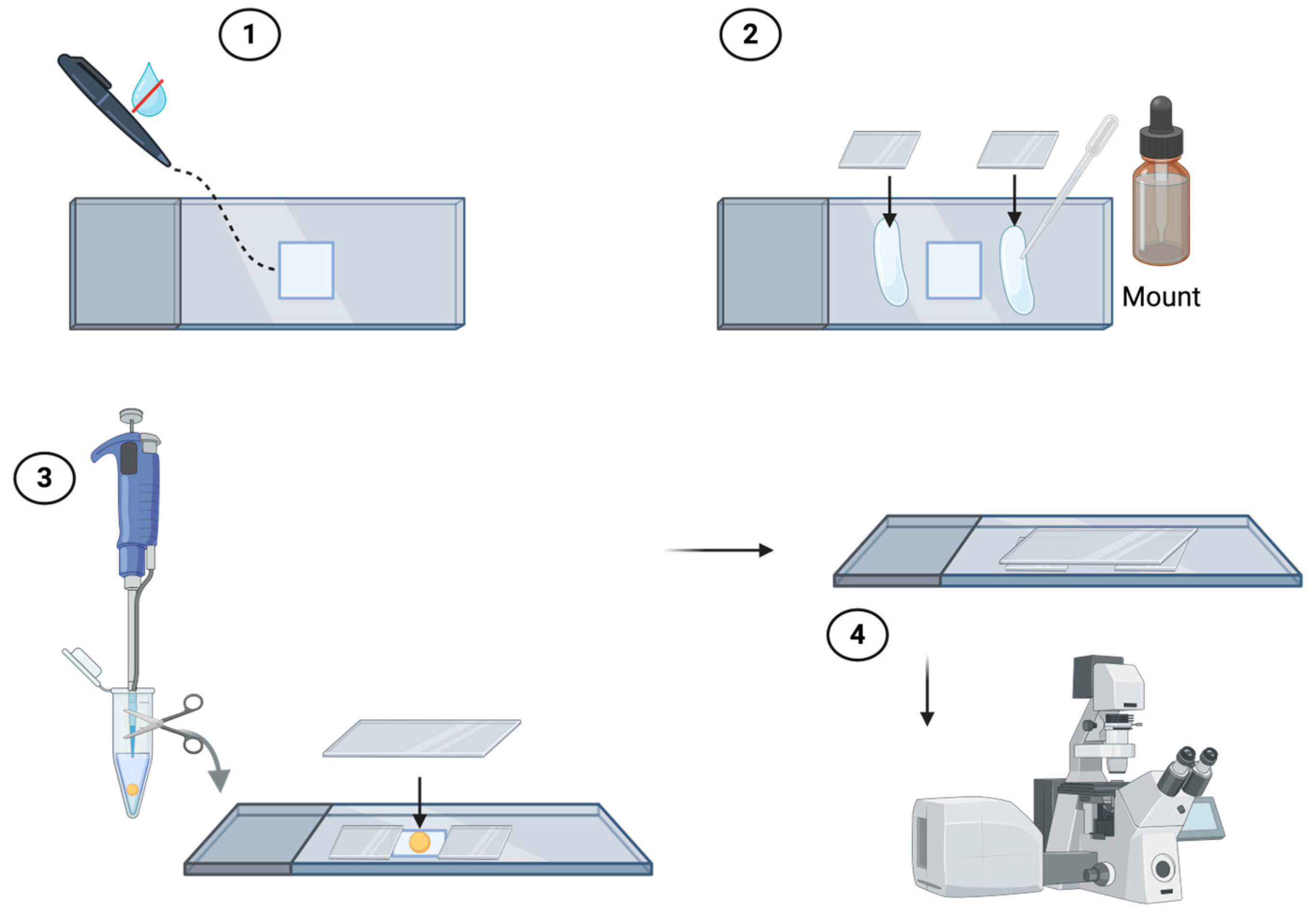
- Prepare a chamber consisting of a slide and two (or more) coverslips to ensure that it is the same thickness as the spheroids to be analyzed. Construct the chamber using a hydrophobic pap pen and aqueous mount (e.g., Eukitt) as illustrated in Figure 2. Resuspend the spheroids in 1X PBS and spot them into the constructed chamber. Finally, close the chamber with a coverslip on top. The samples are now ready for observation under a confocal microscope.

- CRITICAL STEP: During the different steps, the spheroids may be damaged or lost. To minimize this risk, remove and add all volumes gently, avoiding bubble formation. Both the volumes and the number of washing steps can be adjusted as required.OPTIONAL STEP: If the spheroids do not settle spontaneously at the bottom of the tube at each step, you can centrifuge them at 115× g for 5 min and carefully remove the supernatant, without disturbing the pellet.
4. Expected Results
Supplementary Materials
Author Contributions
Funding
Institutional Review Board Statement
Informed Consent Statement
Data Availability Statement
Acknowledgments
Conflicts of Interest
Abbreviations
| 2D | Two-dimensional |
| 3D | Three-dimensional |
| BSA | Bovine Serum Albumin |
| DAPI | 4′,6′-diamidino-2-phenylindole |
| DMEM | Dulbecco’s Modified Eagle Medium |
| DPBS | Dulbecco’s Phosphate-Buffered Saline |
| FBS | Fetal Bovine Serum |
| GBM | Glioblastoma |
| PBS | Phosphate-Buffered Saline |
| PFA | Paraformaldehyde |
| RT | Room temperature |
References
- Verdugo, E.; Puerto, I.; Medina, M.Á. An update on the molecular biology of glioblastoma, with clinical implications and progress in its treatment. Cancer Commun. 2022, 42, 1083–1111. [Google Scholar] [CrossRef] [PubMed]
- Louis, D.N.; Perry, A.; Wesseling, P.; Brat, D.J.; Cree, I.A.; Figarella-Branger, D.; Hawkins, C.; Ng, H.K.; Pfister, S.M.; Reifenberger, G.; et al. The 2021 WHO Classification of Tumors of the Central Nervous System: A summary. Neuro Oncol. 2021, 23, 1231–1251. [Google Scholar] [CrossRef]
- Read, R.D.; Tapp, Z.M.; Rajappa, P.; Hambardzumyan, D. Glioblastoma microenvironment—From biology to therapy. Genes Dev. 2024, 38, 360–379. [Google Scholar] [CrossRef] [PubMed]
- Mathur, R.; Wang, Q.; Schupp, P.G.; Nikolic, A.; Hilz, S.; Hong, C.; Grishanina, N.R.; Kwok, D.; Stevers, N.O.; Jin, Q.; et al. Glioblastoma evolution and heterogeneity from a 3D whole-tumor perspective. Cell 2024, 187, 446–463.e16. [Google Scholar] [CrossRef]
- Lin, H.; Liu, C.; Hu, A.; Zhang, D.; Yang, H.; Mao, Y. Understanding the immunosuppressive microenvironment of glioma: Mechanistic insights and clinical perspectives. J. Hematol. Oncol. 2024, 17, 31. [Google Scholar] [CrossRef]
- Erices, J.I.; Bizama, C.; Niechi, I.; Uribe, D.; Rosales, A.; Fabres, K.; Navarro-Martínez, G.; Torres, Á.; San Martín, R.; Roa, J.C.; et al. Glioblastoma Microenvironment and Invasiveness: New Insights and Therapeutic Targets. Int. J. Mol. Sci. 2023, 24, 7047. [Google Scholar] [CrossRef]
- Sharma, P.; Aaroe, A.; Liang, J.; Puduvalli, V.K. Tumor microenvironment in glioblastoma: Current and emerging concepts. Neurooncol. Adv. 2023, 5, vdad009. [Google Scholar] [CrossRef]
- Graziano, F.; Iacopino, D.G.; Cammarata, G.; Scalia, G.; Campanella, C.; Giannone, A.G.; Porcasi, R.; Florena, A.M.; Conway de Macario, E.; Macario, A.J.L.; et al. The Triad Hsp60-miRNAs-Extracellular Vesicles in Brain Tumors: Assessing Its Components for Understanding Tumorigenesis and Monitoring Patients. Appl. Sci. 2021, 11, 2867. [Google Scholar] [CrossRef]
- Burko, P.; D’Amico, G.; Miltykh, I.; Scalia, F.; Conway de Macario, E.; Macario, A.J.L.; Giglia, G.; Cappello, F.; Caruso Bavisotto, C. Molecular Pathways Implicated in Radioresistance of Glioblastoma Multiforme: What Is the Role of Extracellular Vesicles? Int. J. Mol. Sci. 2023, 24, 4883. [Google Scholar] [CrossRef]
- Caruso Bavisotto, C.; Marino Gammazza, A.; Rappa, F.; Fucarino, A.; Pitruzzella, A.; David, S.; Campanella, C. Exosomes: Can Doctors Still Ignore Their Existence? Euromediterr. Biomed. J. 2013, 8, 136–139. [Google Scholar] [CrossRef]
- Liu, P.; Griffiths, S.; Veljanoski, D.; Vaughn-Beaucaire, P.; Speirs, V.; Brüning-Richardson, A. Preclinical models of glioblastoma: Limitations of current models and the promise of new developments. Expert Rev. Mol. Med. 2021, 23, e20. [Google Scholar] [CrossRef]
- Joseph, V.J.; Blaavand, M.S.; Daubon, T.; Kruyt, F.A.; Thomsen, M.K. Three-dimensional culture models to study glioblastoma—Current trends and future perspectives. Curr. Opin. Pharmacol. 2021, 61, 91–97. [Google Scholar] [CrossRef]
- Li, A.; Walling, J.; Kotliarov, Y.; Center, A.; Steed, M.E.; Ahn, S.J.; Rosenblum, M.; Mikkelsen, T.; Zenklusen, J.C.; Fine, H.A. Genomic changes and gene expression profiles reveal that established glioma cell lines are poorly representative of primary human gliomas. Mol. Cancer Res. 2008, 6, 21–30. [Google Scholar] [CrossRef]
- Soubéran, A.; Tchoghandjian, A. Practical Review on Preclinical Human 3D Glioblastoma Models: Advances and Challenges for Clinical Translation. Cancers 2020, 12, 2347. [Google Scholar] [CrossRef] [PubMed]
- Rae, C.; Amato, F.; Braconi, C. Patient-Derived Organoids as a Model for Cancer Drug Discovery. Int. J. Mol. Sci. 2021, 22, 3483. [Google Scholar] [CrossRef]
- Jo, H.; Lee, S.; Kim, M.H.; Park, S.; Lee, S.Y. Recapitulating Glioma Stem Cell Niches Using 3D Spheroid Models for Glioblastoma Research. Biosensors 2024, 14, 539. [Google Scholar] [CrossRef] [PubMed]
- Bruns, J.; Egan, T.; Mercier, P.; Zustiak, S.P. Glioblastoma spheroid growth and chemotherapeutic responses in single and dual-stiffness hydrogels. Acta Biomater. 2023, 163, 400–414. [Google Scholar] [CrossRef]
- Gerigk, M.; Bulstrode, H.; Shi, H.H.; Tönisen, F.; Cerutti, C.; Morrison, G.; Rowitch, D.; Huang, Y.Y.S. On-chip perivascular niche supporting stemness of patient-derived glioma cells in a serum-free, flowable culture. Lab Chip 2021, 21, 2343–2358. [Google Scholar] [CrossRef]
- Bruns, J.; Zustiak, S.P. Hydrogel-Based Spheroid Models of Glioblastoma for Drug Screening Applications. Mo. Med. 2021, 118, 346–351. [Google Scholar]
- Jacob, F.; Salinas, R.D.; Zhang, D.Y.; Nguyen, P.T.T.; Schnoll, J.G.; Wong, S.Z.H.; Thokala, R.; Sheikh, S.; Saxena, D.; Prokop, S.; et al. A Patient-Derived Glioblastoma Organoid Model and Biobank Recapitulates Inter- and Intra-tumoral Heterogeneity. Cell 2020, 180, 188–204.e22. [Google Scholar] [CrossRef] [PubMed]
- Vtale, A.M.; D’Amico, G.; Santonocito, R.; Spinnato, G.; Di Marco, M.; Scalia, F.; Campanella, C.; Tringali, G.; Giusti, I.; Dolo, V.; et al. An overview of glioblastoma multiforme in vitro experimental models. J. Biol. Res. 2024, 97, 11920. [Google Scholar] [CrossRef]
- Ma, J.; Li, N.; Wang, Y.; Wang, L.; Wei, W.; Shen, L.; Sun, Y.; Jiao, Y.; Chen, W.; Liu, J. Engineered 3D tumour model for study of glioblastoma aggressiveness and drug evaluation on a detachably assembled microfluidic device. Biomed. Microdevices 2018, 20, 80. [Google Scholar] [CrossRef] [PubMed]
- Marino, A.; Battaglini, M.; Carmignani, A.; Pignatelli, F.; De Pasquale, D.; Tricinci, O.; Ciofani, G. Magnetic self-assembly of 3D multicellular microscaffolds: A biomimetic brain tumor-on-a-chip for drug delivery and selectivity testing. APL Bioeng. 2023, 7, 036103. [Google Scholar] [CrossRef]
- Pun, S.; Prakash, A.; Demaree, D.; Krummel, D.P.; Sciumè, G.; Sengupta, S.; Barrile, R. Rapid Biofabrication of an Advanced Microphysiological System Mimicking Phenotypical Heterogeneity and Drug Resistance in Glioblastoma. Adv. Healthc. Mater. 2024, 13, e2401876. [Google Scholar] [CrossRef]
- Xiao, W.; Sohrabi, A.; Seidlits, S.K. Integrating the glioblastoma microenvironment into engineered experimental models. Future Sci. OA 2017, 3, FSO189. [Google Scholar] [CrossRef]
- Bach, C.; Glasow, A.; Baran-Schmidt, R.; Oppermann, H.; Bach, C.; Meixensberger, J.; Güresir, E.; Gaunitz, F. Rapid and reproducible generation of glioblastoma spheroids for high-throughput drug screening. Front. Bioeng. Biotechnol. 2024, 12, 1471012. [Google Scholar] [CrossRef]
- Trautmann, R.K.; Dennison, N.; McCortney, K.; Klier, S.; Cosacak, M.I.; Werner, C.; Akyoldas, G.; Horbinski, C.M.; Freudenberg, U.; Kizil, C. High-Throughput 3D Glioblastoma Model in Glycosaminoglycan Hydrogels for Personalized Therapeutic Screening. Macromol. Biosci. 2026, 26, e00394. [Google Scholar] [CrossRef]
- Gamboa, C.M.; Jara, K.; Pamarthy, S.; Liu, L.; Aiken, R.; Xiong, Z.; Danish, S.; Sabaawy, H.E. Generation of glioblastoma patient-derived organoids and mouse brain orthotopic xenografts for drug screening. STAR Protoc. 2021, 2, 100345. [Google Scholar] [CrossRef]
- Mirab, F.; Kang, Y.J.; Majd, S. Preparation and characterization of size-controlled glioma spheroids using agarose hydrogel microwells. PLoS ONE 2019, 14, e0211078. [Google Scholar] [CrossRef]
- Unnikrishnan, B.S.; Preethi, G.U.; Sreelekha, T.T. A comprehensive study on 2D, 3D and solid tumor environment to explore a multifunctional biogenic nanoconjugate. Sci. Rep. 2021, 11, 8721. [Google Scholar] [CrossRef] [PubMed]


Disclaimer/Publisher’s Note: The statements, opinions and data contained in all publications are solely those of the individual author(s) and contributor(s) and not of MDPI and/or the editor(s). MDPI and/or the editor(s) disclaim responsibility for any injury to people or property resulting from any ideas, methods, instructions or products referred to in the content. |
© 2026 by the authors. Licensee MDPI, Basel, Switzerland. This article is an open access article distributed under the terms and conditions of the Creative Commons Attribution (CC BY) license.
Share and Cite
D’Amico, G.; Vitale, A.M.; Marco, M.D.; Lo Giudice, A.; Cecala, F.C.; Cappello, F.; Caruso Bavisotto, C. Standardized Workflow for the Generation of Patient-Derived Glioblastoma Spheroids. Methods Protoc. 2026, 9, 61. https://doi.org/10.3390/mps9020061
D’Amico G, Vitale AM, Marco MD, Lo Giudice A, Cecala FC, Cappello F, Caruso Bavisotto C. Standardized Workflow for the Generation of Patient-Derived Glioblastoma Spheroids. Methods and Protocols. 2026; 9(2):61. https://doi.org/10.3390/mps9020061
Chicago/Turabian StyleD’Amico, Giuseppa, Alessandra Maria Vitale, Martina Di Marco, Alessandro Lo Giudice, Francesca Chiara Cecala, Francesco Cappello, and Celeste Caruso Bavisotto. 2026. "Standardized Workflow for the Generation of Patient-Derived Glioblastoma Spheroids" Methods and Protocols 9, no. 2: 61. https://doi.org/10.3390/mps9020061
APA StyleD’Amico, G., Vitale, A. M., Marco, M. D., Lo Giudice, A., Cecala, F. C., Cappello, F., & Caruso Bavisotto, C. (2026). Standardized Workflow for the Generation of Patient-Derived Glioblastoma Spheroids. Methods and Protocols, 9(2), 61. https://doi.org/10.3390/mps9020061