A Protocol for Regulating Protein Liquid–Liquid Phase Separation Using NMR-Guided Mutagenesis


Abstract
1. Introduction
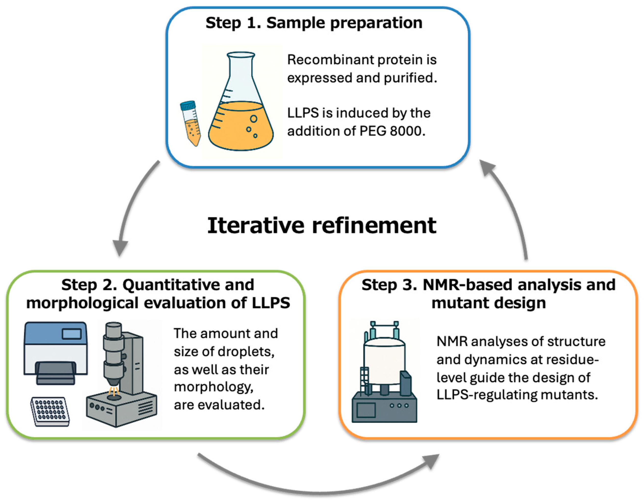
2. Experimental Design
2.1. Materials
- Plasmids and Vectors: pCold-GST expression vector (3372; Takara Bio, Kusatsu, Japan) containing the VAPB MSP domain insert.
- Bacterial Strain: Escherichia coli BL21 Rosetta (DE3) (70954; Novagen/Merck, Darmstadt, Germany).
- Culture Media: Luria–Bertani (LB) broth; M9 minimal medium.
- Affinity Resin: Glutathione Sepharose 4B (GE17-0756-01; GE Healthcare, Chicago, IL, USA).
- Protease: Human rhinovirus (HRV) 3C protease (7360; Takara Bio).
- Gel filtration Chromatography Column: Superdex 75 26/60 (GE28-9893-34; GE Healthcare).
- Mutagenesis Kit: QuikChange Site-Directed Mutagenesis Kit (200518; Agilent Technologies, Santa Clara, CA, USA)
- Chemicals: PEG 8000 (202452; Sigma-Aldrich, St. Louis, MO, USA), DTT (040-29224; Wako, Osaka, Japan), KCl (163-03545; Wako), potassium phosphate buffer reagents (164-04245, 169-04245; Wako).
- Polystyrene 96-well microplate, flat bottom (1-6776-13; AS ONE, Osaka, Japan)
- Silica glass beads: Spherical silica particles (43-00-701, 43-00-202, 43-00-402, 43-00-502, 43-00-802, 43-00-103, 43-00-153, 43-00-303, for 70 nm, 200 nm, 400 nm, 500 nm, 800 nm, 1000 nm, 1500 nm, 3000 nm, respectively; micromod Partikeltechnologie GmbH, Rostock, Germany)
- Water: Ultrapure Milli-Q water (Millipore/Merck, Darmstadt, Germany)
- Lysis buffer (Buffer A): 50 mM Tris–HCl (pH 8.0), 300 mM KCl, 0.1 mM EDTA, 1 mM DTT.
- Elution buffer (Buffer B): Buffer A supplemented with 50 mM reduced glutathione.
- Gel filtration buffer (Buffer C): 50 mM potassium phosphate (pH 6.8), 100 mM KCl, 1 mM DTT.
- PEG 8000 (crowding agent): 30% (w/v) stock; final concentration 15%. Prepare one day in advance, dissolve at room temperature.
- Potassium acetate buffer (pH 4.7): 50 mM with 100 mM KCl and 1 mM DTT.
- D2O: Purity > 99.9%, 0.75 mL (31270356, Merck, Darmstadt, Germany)
- NMR tube: 4 mmφ Symmetrical Micro Sample Tubes for Aqueous Solutions for Bruker NMR equipment (BMS-004B, Shigemi, Tokyo, Japan)
2.2. Equipment
- Incubator Shaker: For bacterial culture at 37 °C (BR-43FL; TAITEC, Tokyo, Japan).
- Ultrasonic Disruptor: For cell disruption during protein extraction (UD-211; TOMY Seiko, Tokyo, Japan).
- Centrifuges: High-speed refrigerated centrifuge for cell harvest and clarification; ultracentrifuge (Optima™ TLX; Beckman Coulter, Brea, CA, USA) for protein fractionation.
- Chromatography System: ÄKTA purifier for affinity and gel-filtration chromatography (Cytiva/Danaher, Washington, DC, USA).
- UV–Vis Microplate Reader: Capable of absorbance measurements from 330 to 800 nm for LLPS assays (Varioskan Flash 2.4; Thermo Fisher Scientific, Waltham, MA, USA).
- Phase-Contrast Microscope: For visualization of LLPS droplets and fusion dynamics (DMI3000B; Leica Microsystems, Wetzlar, Germany) equipped with a CCD camera (DFC series; Leica Microsystems).
- Spectrophotometer: For measuring protein concentration at 280 nm (NanoDrop One; Thermo Fisher Scientific).
- NMR Spectrometer: Bruker Avance III HD 600 MHz equipped with a cryogenic probe (Bruker, Billerica, MA, USA).

- An equivalent NMR spectrometer with a frequency of 500 MHz or higher (Bruker or JEOL, Tokyo, Japan) can be used.

- Ultrasonic Cleaner: For solution mixing and degassing prior to absorbance measurements (ASU-10; AS ONE).
- pH Meter: For buffer preparation and condition control (LAQUA F-71; Horiba, Kyoto, Japan).
- Degassing Devices: Dry vacuum pump (0.095 MPa; GM-20D), vacuum filtration bell jar (3929-0001), and silicone stopper (3923-40/28) (AS ONE).
3. Procedure
3.1. Step 1. Sample Preparation
3.1.1. Procedure 1-1. Protein Expression and Purification
- Clone the gene encoding the VAPB MSP domain into the pCold-GST expression vector.
- Generate site-directed mutants using the QuikChange mutagenesis kit (Agilent Technologies) according to the manufacturer’s instructions.
- Transform the plasmid into Escherichia coli BL21 Rosetta (DE3).
- Inoculate a single colony into 10 mL LB medium and incubate overnight at 37 °C with shaking.
- Inoculate the preculture into 2 L LB medium and grow cells at 37 °C until OD600 reaches 0.4–0.6.
- Cool the culture by shaking in an ice–water bath for approximately 5 min.
- Induce protein expression by adding IPTG to a final concentration of 1 mM.
- Incubate the culture at 15 °C for 16–24 h with shaking.
- Harvest cells by centrifugation at 3300× g for 15 min at 4 °C.
- Wash the cell pellet with physiological saline and centrifuge again at 3000× g for 20 min at 4 °C.
- Store the cell pellets at −80 °C until use.
- Thaw cell pellets on ice and resuspend them in Buffer A.
- Lyse cells by sonication on ice for 15 min (power 6.0, duty 20%).
- Clarify the lysate by ultracentrifugation (50,000 rpm, 15 min, 4 °C).
- Load the supernatant onto a glutathione Sepharose 4B column equilibrated with Buffer A.
- Elute GST-fusion proteins with Buffer B.
- Add HRV 3C protease (1 μL per 1 mg protein) and incubate at 4 °C overnight to remove the GST tag.
- Confirm tag cleavage by SDS–PAGE.
- Purify the cleaved protein by gel filtration chromatography using a Superdex 75 column (GE Healthcare) equilibrated with Buffer C.
- Collect fractions corresponding to monomeric VAPB MSP domain.
- Concentrate protein fractions using centrifugal filter units (10 kDa cutoff).
- Store purified proteins at 4 °C for short-term use.
3.1.2. Procedure 1-2. Induction of LLPS
- Prepare a 30% (w/v) PEG 8000 stock solution at least one day before the assay.
- Equilibrate protein and PEG solutions at the target temperature for at least 15 min.
- Dispense purified VAPB MSP domain solution into wells of a 96-well microplate.
- Gently add PEG solution to each well without direct pipette mixing.
- Avoid introducing air bubbles during sample preparation.
- Incubate plates at 10 °C or 37 °C for 24 h to allow LLPS formation.
- For kinetic measurements, prepare samples within 10 min prior to data acquisition.
3.2. Step 2. Quantitative and Morphological Evaluation of LLPS
3.2.1. Procedure 2-1. Microplate Reader Measurements
- Use flat-bottom 96-well polystyrene plates with a final volume of 200 μL per well.
- Measure absorbance spectra from 330 to 800 nm at 10 nm intervals.
- Control the measurement temperature between 10 °C and 37 °C as required.
- Record blank spectra (buffer + PEG) and subtract them from sample spectra.
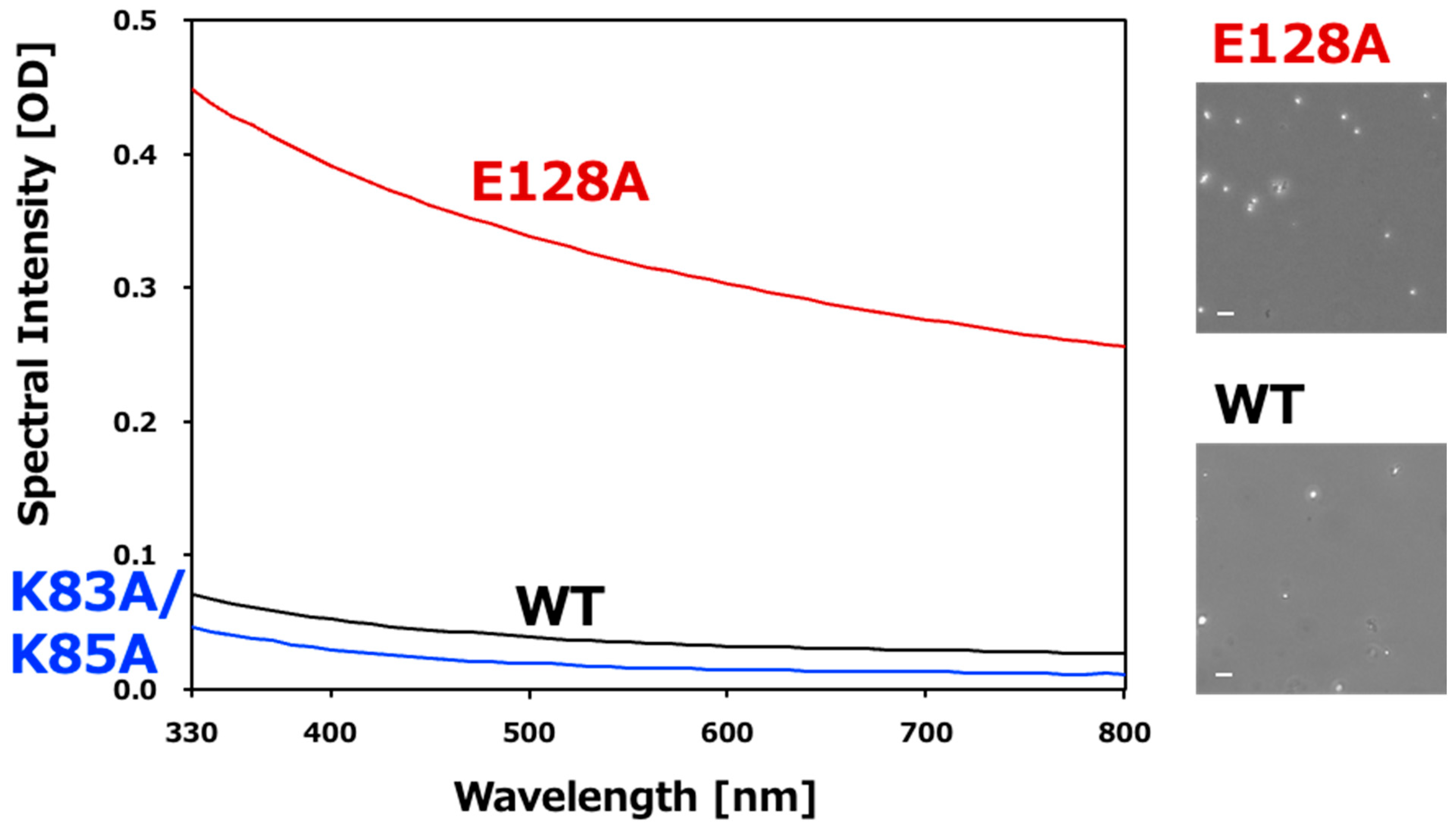
- Normalize spectra and calculate OD660/OD330 ratios.

- The OD660/OD330 ratio is used as an indicator of particle size. Larger values indicate larger particles.
- Perform endpoint measurements after 24 h incubation or kinetic measurements at defined time intervals.
- Measure silica glass beads (70–3000 nm) under identical conditions to obtain reference spectra.
- Fit experimental spectra to reference spectra using RMSD analysis to generate droplet size heatmaps.
- Perform at least three technical replicates for each condition.
3.2.2. Procedure 2-2. Microscopic Observation
- Collect 3 μL of sample from each well immediately after microplate measurements.
- Place the sample on a glass slide and cover with a coverslip.
- Observe samples using phase-contrast microscopy at 10×–40× magnification.
- Record images and videos to monitor droplet morphology and fusion events.
- Analyze images using ImageJ (version 1.54 or higher) to quantify droplet size and circularity.

- Use microscopy to distinguish LLPS droplets from aggregates; turbidity alone cannot make this distinction. Classical LLPS droplets are typically spherical and dynamic.
3.3. Step 3. NMR-Based Structural Analysis and Mutant Design
3.3.1. Procedure 3-1. NMR Measurement
- Express 15N-labeled VAPB MSP domain (WT or mutants) in E. coli grown in M9 minimal medium supplemented with 15NH4Cl [8], and purify the protein as described in Procedure 1-1.
- Prepare purified protein at a final concentration of 0.1–0.4 mM in Buffer C (50 mM potassium phosphate, pH 6.8, 100 mM KCl, and 1 mM DTT).
- Add 150 µL of the sample containing 5% D2O to the outer tube of a Shigemi 4 mm NMR tube, degas the sample using a vacuum pump, and seal the tube with the inner tube, taking care to avoid the introduction of air bubbles.

- When using a Shigemi 5 mm NMR tube, add 250 µL of the sample instead of 150 µL.
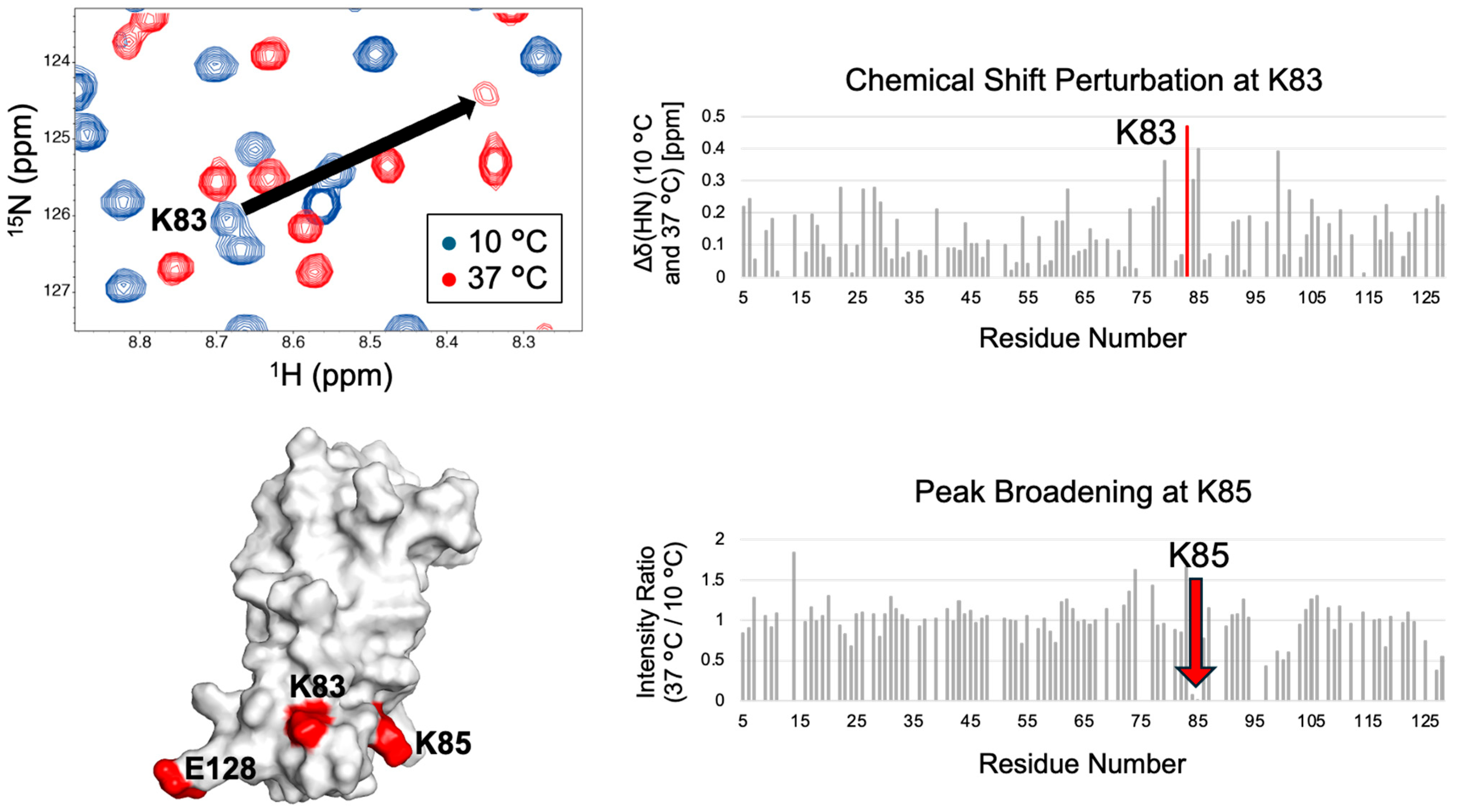
- Record two-dimensional 1H–15N HSQC spectra and 1H–15N heteronuclear NOE spectra at 10 °C to evaluate the soluble state and at 37 °C to assess conditions prone to phase separation, following the procedure below.
- Set the NMR temperature to 10 °C or 37 °C.
- Insert the sample into the NMR spectrometer (Bruker).
- Control the spectrometer and perform measurements using TopSpin software (version 3.6 or higher).
- Lock the spectrometer on H2O/D2O.
- Perform probe tuning and matching.
- Optimize shim values using the TopShim module of TopSpin software.
- Determine the 1H 90° pulse length and power.
- For 1H–15N HSQC, load the parameter set “FHSQCGPPH”; for 1H–15N heteronuclear NOE, load “HSQCNOEF3GPSI3D”.
- Enter “getprosol 1H” (determined 1H 90° pulse length) (determined 1H 90° pulse power in dB) to set the pulse parameters corrected for the determined 90° pulse.
- Set the number of scans (NS) according to the sample concentration.
- Determine the receiver gain (RG).
- Start acquisition.
- Process the acquired spectra using NMRPipe. First, convert the raw data into the NMRPipe format. For Bruker datasets, launch the conversion GUI using the “bruker” command, which automatically generates and executes the conversion script. Next, perform Fourier transformation. In NMRDraw GUI of NMRPipe, the Macro Edit interface can be used to create, save, and execute shell scripts for spectrum processing.
- Convert the NMRPipe data into the UCSF format for Poky software using the pipe2ucsf program.
- Display each spectrum in Poky, perform peak picking, and annotate resonance assignments.

- If the resonance assignments are unavailable, perform a series of NMR experiments with a [13C, 15N]-labeled sample to obtain them. If large chemical shift perturbations are observed between 10 °C and 37 °C, record additional 1H–15N HSQC spectra at intermediate temperatures and trace the signals. For the VAPB MSP domain, resonance assignments were performed at 30 °C [13]. Thus, 1H–15N HSQC spectra were recorded at 10 °C, 20 °C, and 37 °C to observe temperature dependence.
- Using the Poky “lt” command, save a list of peak positions and intensities for each residue.
- Calculate residue-specific 1H–15N HSQC chemical shift perturbations (CSPs) and peak intensity ratios at the two temperatures.
- Determine the heteronuclear NOE for each residue as the ratio of peak intensities measured with and without 1H saturation.
- Evaluate temperature-dependent changes in residue-specific heteronuclear NOE values.
3.3.2. Procedure 3-2. Mutant Design
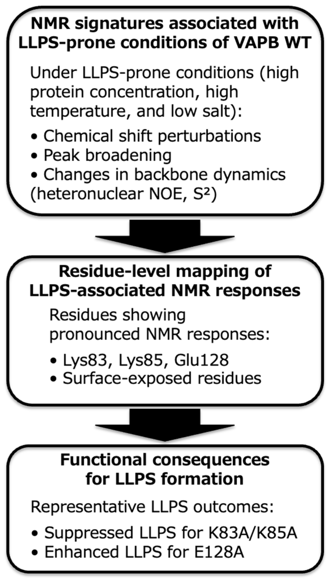
- Identify candidate residues for mutagenesis from those showing large temperature-dependent changes in chemical shifts, peak intensities, and/or 1H–15N heteronuclear NOE values. Prioritize residues that are solvent-exposed, likely to participate in intermolecular interactions (e.g., charged residues), and evolutionarily conserved. The CSPs primarily reflect local structural changes, while changes in peak intensities and 1H–15N heteronuclear NOE values mainly reflect alterations in the dynamics of the corresponding residues.

- Changes in NMR features that depend on temperature can identify candidate residues that regulate LLPS. This approach applies to proteins whose condensate formation is promoted at either lower or higher temperatures. Currently, however, there is no method to unambiguously design mutants at these residues to promote or suppress LLPS formation.
- Introduce mutations by site-directed mutagenesis using the QuikChange method. Replace candidate residues with alanine residues as the initial mutation, as recommended in biochemical studies, because it often provides a straightforward interpretation of side-chain contribution. For a deeper mechanistic understanding, more refined designs are necessary (e.g., charge-conserving substitutions, such as Lys → Arg).
- Express and purify the newly designed mutants, and perform LLPS assays under the same conditions as those used for the wild-type protein.
4. Expected Results
Author Contributions
Funding
Institutional Review Board Statement
Informed Consent Statement
Data Availability Statement
Acknowledgments
Conflicts of Interest
Abbreviations
| HSQC | Heteronuclear single quantum coherence |
| LLPS | Liquid–liquid phase separation |
| MSP | Major sperm protein |
| NMR | Nuclear magnetic resonance |
| NOE | Nuclear Overhauser effect |
| PEG | Polyethylene glycol |
| S2 | Generalized order parameter |
| VAPB | VAMP-associated protein B |
References
- Banani, S.F.; Lee, H.O.; Hyman, A.A.; Rosen, M.K. Biomolecular condensates: Organizers of cellular biochemistry. Nat. Rev. Mol. Cell Biol. 2017, 18, 285–298. [Google Scholar] [CrossRef] [PubMed]
- Brangwynne, C.; Tompa, P.; Pappu, R. Polymer physics of intracellular phase transitions. Nat. Phys. 2015, 11, 899–904. [Google Scholar] [CrossRef]
- Lin, Y.; Protter, D.S.; Rosen, M.K.; Parker, R. Formation and Maturation of Phase-Separated Liquid Droplets by RNA-Binding Proteins. Mol. Cell 2015, 60, 208–219. [Google Scholar] [CrossRef] [PubMed]
- Wang, J.; Choi, J.-M.; Holehouse, A.S.; Lee, H.O.; Zhang, X.; Jahnel, M.; Maharana, S.; Lemaitre, R.; Pozniakovsky, A.; Drechsel, D.; et al. A Molecular Grammar Governing the Driving Forces for Phase Separation of Prion-like RNA Binding Proteins. Cell 2018, 174, 688–699.e16. [Google Scholar] [CrossRef] [PubMed]
- Choi, J.M.; Holehouse, A.S.; Pappu, R.V. Physical Principles Underlying the Complex Biology of Intracellular Phase Transitions. Annu. Rev. Biophys. 2020, 49, 107–133. [Google Scholar] [CrossRef]
- Alberti, S.; Gladfelter, A.; Mittag, T. Considerations and Challenges in Studying Liquid-Liquid Phase Separation and Biomolecular Condensates. Cell 2019, 176, 419–434. [Google Scholar] [CrossRef] [PubMed]
- Qing, G.; Ma, L.-C.; Khorchid, A.; Swapna, G.V.; Mal, T.K.; Takayama, M.M.; Xia, B.; Phadtare, S.; Ke, H.; Acton, T.; et al. Cold-shock induced high-yield protein production in Escherichia coli. Nat. Biotechnol. 2004, 22, 877–882. [Google Scholar] [CrossRef] [PubMed]
- Hayashi, K.; Kojima, C. pCold-GST Vector: A Novel Cold-Shock Vector Containing GST Tag for Soluble Protein Production. Protein Expr. Purif. 2008, 62, 120–127. [Google Scholar] [CrossRef] [PubMed]
- Patel, A.; Lee, H.O.; Jawerth, L.; Maharana, S.; Jahnel, M.; Hein, M.Y.; Stoynov, S.; Mahamid, J.; Saha, S.; Franzmann, T.M.; et al. Liquid-to-Solid Phase Transition of the ALS Protein FUS Accelerated by Disease Mutation. Cell 2015, 162, 1066–1077. [Google Scholar] [PubMed]
- Riback, J.A.; Zhu, L.; Ferrolino, M.C.; Tolbert, M.; Mitrea, D.M.; Sanders, D.W.; Wei, M.-T.; Kriwacki, R.W.; Brangwynne, C.P. Composition-dependent thermodynamics of intracellular phase separation. Nature 2020, 581, 209–214. [Google Scholar] [CrossRef] [PubMed]
- Enomoto-Kusano, M.; Kodama, T.S.; Anzawa, S.; Furuita, K.; Kobayashi, R.; Sekiyama, N.; Togawa, W.; Fujiwara, T.; Miyanoiri, Y.; Tochio, H.; et al. LLPS droplet size estimation via UV-Vis spectroscopy using a microplate reader. Sci. Rep. 2026, 16, 3533. [Google Scholar] [CrossRef]
- McSwiggen, D.T.; Mir, M.; Darzacq, X.; Tjian, R. Evaluating phase separation in live cells: Diagnosis, caveats, and functional consequences. Genes Dev. 2019, 33, 1619–1634. [Google Scholar] [CrossRef]
- Furuita, K.; Enomoto-Kusano, M.; Togawa, W.; Komuro, R.; Nakayama, K.; Kobayashi, N.; Kodama, T.S.; Miyanoiri, Y.; Fujiwara, T.; Kojima, C. Structural basis of condensate formation of VAPB protein. Sci. Rep. 2026, under revision.
- Murthy, A.C.; Fawzi, N.L. The (un) structural biology of biomolecular liquid-liquid phase separation using NMR spectroscopy. J. Biol. Chem. 2020, 295, 2375–2384. [Google Scholar] [CrossRef] [PubMed]
- Delaglio, F.; Grzesiek, S.; Vuister, G.; Zhu, G.; Pfeifer, J.; Bax, A. NMRPipe: A multidimensional spectral processing system based on UNIX pipes. J. Biomol. NMR 1995, 6, 277–293. [Google Scholar] [CrossRef]
- Lee, W.; Rahimi, M.; Lee, Y.; Chiu, A. POKY: A software suite for multidimensional NMR and 3D structure calculation of biomolecules. Bioinformatics 2021, 37, 3041–3042. [Google Scholar] [CrossRef]


Disclaimer/Publisher’s Note: The statements, opinions and data contained in all publications are solely those of the individual author(s) and contributor(s) and not of MDPI and/or the editor(s). MDPI and/or the editor(s) disclaim responsibility for any injury to people or property resulting from any ideas, methods, instructions or products referred to in the content. |
© 2026 by the authors. Licensee MDPI, Basel, Switzerland. This article is an open access article distributed under the terms and conditions of the Creative Commons Attribution (CC BY) license.
Share and Cite
Enomoto-Kusano, M.; Furuita, K.; Kodama, T.S.; Kojima, C. A Protocol for Regulating Protein Liquid–Liquid Phase Separation Using NMR-Guided Mutagenesis. Methods Protoc. 2026, 9, 26. https://doi.org/10.3390/mps9010026
Enomoto-Kusano M, Furuita K, Kodama TS, Kojima C. A Protocol for Regulating Protein Liquid–Liquid Phase Separation Using NMR-Guided Mutagenesis. Methods and Protocols. 2026; 9(1):26. https://doi.org/10.3390/mps9010026
Chicago/Turabian StyleEnomoto-Kusano, Mayu, Kyoko Furuita, Takashi S. Kodama, and Chojiro Kojima. 2026. "A Protocol for Regulating Protein Liquid–Liquid Phase Separation Using NMR-Guided Mutagenesis" Methods and Protocols 9, no. 1: 26. https://doi.org/10.3390/mps9010026
APA StyleEnomoto-Kusano, M., Furuita, K., Kodama, T. S., & Kojima, C. (2026). A Protocol for Regulating Protein Liquid–Liquid Phase Separation Using NMR-Guided Mutagenesis. Methods and Protocols, 9(1), 26. https://doi.org/10.3390/mps9010026