Protocol

Optimization of Nile Tilapia Artificial Breeding Using Human Chorionic Gonadotropin (hCG) Hormone

by
Golam Rbbani
1,
Prabhugouda Siriyappagouder
1,
Riaz Murshed
1,
Rajesh Joshi
2,
Artem Nedoluzhko
3,
Jorge Galindo-Villegas
1 and
Jorge M. O. Fernandes
1,4,*
1
Faculty of Fisheries and Bioscience, Nord University, 8026 Bodø, Norway
2
GenoMar Genetics, 0252 Oslo, Norway
3
Paleogenomics Laboratory, European University at Saint Petersburg, St. Petersburg 191187, Russia
4
Institute of Marine Sciences (ICM-CSIC), 08003 Barcelona, Spain
*
Author to whom correspondence should be addressed.
Methods Protoc. 2025, 8(3), 57; https://doi.org/10.3390/mps8030057
Submission received: 22 November 2024 / Revised: 21 May 2025 / Accepted: 26 May 2025 / Published: 2 June 2025
(This article belongs to the Section Biomedical Sciences and Physiology)

Abstract

Nile tilapia (Oreochromis niloticus) is the most widely farmed tilapia species globally, making it one of the most important aquaculture species. To meet increasing demand, hatcheries occasionally use artificial breeding techniques such as hormonal induction to synchronize breeding. Despite the common use of human chorionic gonadotropin (hCG) in fish breeding, no detailed protocol has been established specifically for Nile tilapia. The objective of this study is to establish an effective hCG-induced artificial breeding protocol for gene editing and aquaculture production, optimizing fertilization, hatching, and survival rates. We employed a single intramuscular injection of 2 IU/g hCG to induce ovulation. The protocol achieved an average fertilization rate of 88.3% and a larval survival rate of 90.5%, demonstrating its potential for obtaining high-quality embryos for functional studies and enhancing reproductive performance on a commercial scale.

1. Introduction

Nile tilapia (Oreochromis niloticus) is one of the most important freshwater fish species cultured globally and contributes significantly to the global aquaculture industry. This species alone accounts for approximately 5 million metric tons of annual production, and with increasing demand, global production is projected to reach 7.3 million tons by 2030 [1]. Several factors contribute to the choice of Nile tilapia in aquaculture, including their large size, high growth rates, short lifecycle, prolific breeding attributes, low protein requirements, and omnivorous feeding. The relatively low cost of production compared to other aquaculture species makes Nile tilapia a cost-effective option for fish farmers. In addition to commercial production, Nile tilapia also serves as a valuable model for studying growth and reproductive biology due to its rapid growth and shorter time to reach adulthood compared to many other species [2,3,4]. They can thrive in a wide range of environmental conditions, including varying temperatures, osmotic pressures, alkalinity levels, and low dissolved oxygen concentrations, making them highly adaptable to diverse farming environments [5,6].
The reproduction of Nile tilapia in captivity can be controlled through environmental manipulations, such as adjustments to photoperiod, temperature, or spawning substrate [2,7,8]. Under suitable conditions, Nile tilapia can reach sexual maturity as early as 3 to 4 months after hatching, enabling rapid population growth. Female Nile tilapia are capable of producing between 75 and 1000 offspring every 14 to 22 days of the spawning cycle [9,10]. Achieving successful captive production of Nile tilapia requires proficiency in advanced breeding techniques, ensuring high-quality larvae in sufficient quantities to meet aquaculture demands. Producers with expertise in Nile tilapia breeding methods are increasingly adopting advanced systems to maximize fry production. In certain cases, exogenous hormones are employed to stimulate ovulation in hatchery fish, providing better control of fertilization and enhancing the overall efficiency of the breeding process [2,11].
Ovulation and spermiation in fish can be induced by several varieties of natural and synthetic hormones that are commercially available. For example, human chorionic gonadotropin (hCG), pituitary homogenate, Ovaprim (analogue of salmon gonadotropin-releasing hormone (GnRH) and domperidone as a dopamine receptor antagonist) and Ovopel (mammalian GnRH analogue and metoclopramide as a dopamine receptor antagonist) are commonly used for artificial breeding [12,13,14,15]. Hormonal induction protocols are widely utilized in fish breeding to facilitate the collection of gametes for chromosome manipulation, reproductive biology research, and selective breeding programs. The use of hormonal induction is also very important for the breeding and management of fishes that migrate during their spawning season, such as Neotropical reofilic fish, which normally do not spawn naturally in captivity. Injection with carp pituitary extract has been successfully used to promote spawning in five such species of commercial importance (Piaractus mesopotamicus, Brycon orbignyanus, Prochilodus lineatus, Salminus brasiliensis, and Leporinus macrocephalus) [16], as well as in Astyanax altiparanae, which is proposed as a model fish for the Neotropical region [17].
The effectiveness of hormonal treatments depends on factors such as dosage, the targeted species, and the maturation stage of the fish. Among the commonly used hormones, hCG is one of the most popular and effective solutions for stimulating ovulation in female broodfish. Despite the effectiveness of hCG in stimulating ovulation, only three studies have specifically documented its use in Nile tilapia breeding, focusing primarily on its efficacy rather than its use in commercial aquaculture settings [12,13,18]. Developing a detailed protocol could significantly aid fish farms in utilizing hCG effectively on a commercial scale while minimizing its residual hormonal effects. To address this gap, we present a detailed protocol of the method that we used in a recent study for synchronizing final oocyte maturation using hCG in the artificial reproduction of Nile tilapia [3]. In addition to its relevance for functional studies in the laboratory (e.g., for gene editing), hCG-induced breeding methods may find application in the aquaculture industry, improving breeding efficiency and production outcomes.

2. Experimental Design

Materials

  • Mature male and mature female Nile tilapia (Oreochromis niloticus);
  • Human chorionic gonadotropin (Merck, Darmstadt, Germany);
  • NaCl (sodium chloride 98.80%) (Maldon, Essex, UK);
  • Nitrate-Nitrite (0–40 mg/L NO3-N, 0.015–0.60 mg/L NO2-N) and ammonia test kit (0–2.4 mg/L NH3-N) (product numbers: 1408100 and 2428700, Hach, Loveland, CO, USA);
  • 100 L plastic buckets;
  • Heater (EHEIM thermocontrol 150 and 300 Watt, cat-4011708011713 and 4011708011744, Eheim GmbH & Co., Deizisau, Germany);
  • Aerator (TetraTec APS 120-400, 400 L/h 4.5 watts, Tetra GmbH, Melle, Germany);
  • Syringe (0.3 mL, Beckton-Dickinson, Oslo, Norway);
  • Needle (Gauge 22, WVR, Radnor, PA, USA);
  • Borosilicate glass beaker (500 mL);
  • Aquarium tanks (500 L);
  • Fine mesh net (7.5 cm)
  • Lamp (Unilite SLR-3000, Worcestershire, UK);
  • Timer (Bachmann, Bremen, Germany);
  • Egg rocker (5 L capacity) (item number 1407, FIAP GmbH, Ursensollen, Germany);
  • Air pump (Eheim Compacton 600 Pump, Deizisau, Germany);
  • EBI temperature probe (item no. 620-1860P, WVR, Radnor, PA, USA);
  • pH meter (item no. 665-0499P, WVR, Radnor, PA, USA);
  • Oxygen meter (H05: Polaris C, Oxyguard, Farum, Denmark);
  • Leica MZ16 FA dissecting stereomicroscope (Leica Microsystems GmbH, Wetzlar, Germany).

3. Procedure

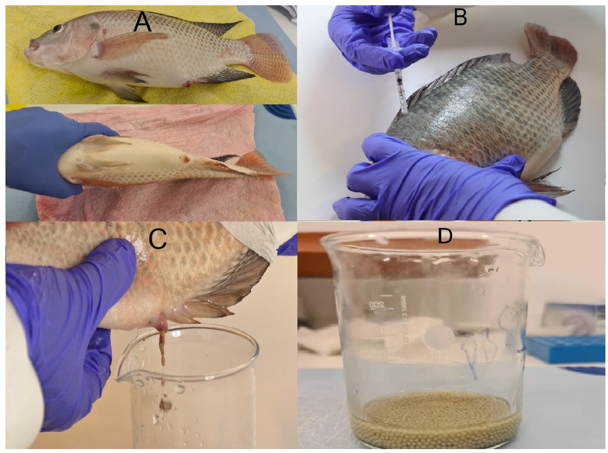
3.1. Selection of Female Tilapia

Select one mature female Nile tilapia by looking for the following secondary sexual characteristics: reddish genital papilla, pinkish fin color, and a swollen abdomen (as shown in Figure 1A).

3.2. Hormone Induction

Administer an intramuscular injection of hCG below the dorsal fin to the selected female at a dosage of 2 IU/g body weight, based on our preliminary trials (see Figure 1B).

3.3. Post-Injection Care

Place the injected female in a 100 L bucket, 2/3 full of water; with continuous aeration, maintain the fish there for 24 h. Keep the water temperature stable at 28 °C using an EHEIM thermocontrol 150 heater (cat-4011708011713, Eheim GmbH & Co., Deizisau, Germany).

3.4. Selection of Male Tilapia

Collect male tilapia that are ready to release sperm by identifying the following secondary sexual characteristics: pointed dorsal and anal fins and a noticeable reddish coloration on the body.

3.5. Egg Collection

After 24 h post-injection, gently massage the female’s abdomen to collect the eggs into a clean beaker (Figure 1C).

3.6. Sperm Collection and Fertilization

Immediately strip the male and collect the sperm directly into the beaker containing eggs. Add water (approximately 2 mL) to activate the sperm, and gently shake (by hand or using a feather) the beaker continuously for 8–10 min to ensure the mixing of sperm and eggs (Figure 1D).

3.7. Washing Fertilized Eggs
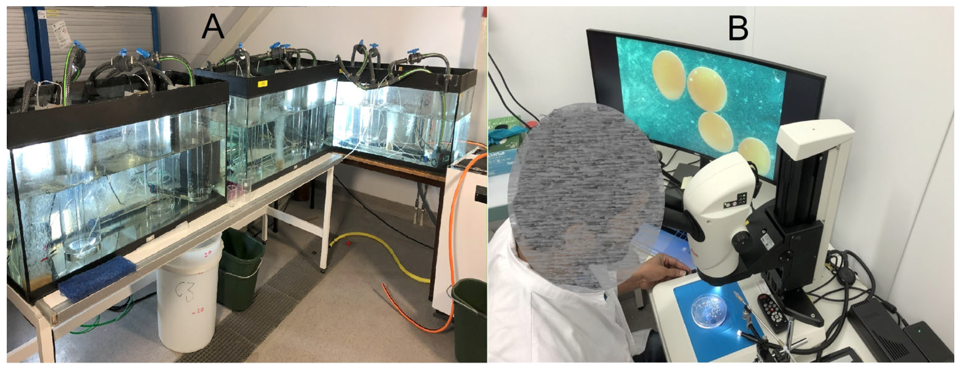
After fertilization, rinse the fertilized eggs 5–6 times with system water (100 mL) to remove debris and redundant sperm.

3.8. Incubation

Transfer the fertilized eggs to three different tanks (500 L) containing three egg rockers (5 L) each. Each egg rocker contained 230 fertilized eggs, ensuring appropriate temperature and aeration conditions were maintained (see Figure 2A). Tank temperature (24 °C, 28 °C and 32 °C) was maintained using the EHEIM thermocontrol 300 (cat-4011708011744, Eheim GmbH & co., Deizisau, Germany).

3.9. Water Quality Parameters

Dissolved oxygen was determined with a Polaris C meter (Oxyguard, Farum, Denmark), and pH, ammonia, nitrite, and nitrate were periodically monitored during the entire trial using appropriate measuring kits (Supplementary Table S1).

3.10. Photoperiod

Maintain fertilized embryos under an artificial photoperiod of 13 h light and 11 h dark (13 h:11 h) using lamps (3000 lm, Unilite SLR-3000, Redditch, UK) controlled by a timer.

3.11. Monitoring the Fertilized Eggs

Monitor the fertilized eggs every 6 h for 16 days until the larvae absorb the yolk sac (Figure 2B).

3.12. Data Collection

Calculate fertilization rate, hatching rate, and larval survival rate as development progresses. Fertilization rate was determined as the percentage of embryos relative to the total number of eggs, multiplied by 100. The hatching rate was calculated as the percentage of embryos that successfully hatched at 96, 128, and 200 h post-fertilization at 32, 28, and 24 °C, respectively. The survival rate was based on the number of larvae that survived 3 days after hatching. These metrics were used to assess the effectiveness of the incubation process and the overall health and viability of the developing embryos.

4. Results and Discussion

In this study, fertilized eggs were incubated in a large tank using egg rockers and replicated in triplicate at each temperature until yolk sac absorption [3]. Continuous water flow was maintained in the tanks using a pump to ensure that the eggs remained in constant motion, preventing them from settling at the bottom (Figure 2A). This setup closely mimics the natural mouth incubation process in Nile tilapia.
Table 1 shows the fertilization, hatching, and survival rates in the breeding trials. The fertilization rates were assessed 1 day post-fertilization, and hatching rates were recorded 3 days post-fertilization. Across all temperature groups, the fertilization rates were consistently high, ranging from approximately 82.6% to 91.3%. The hatching rates showed some variation across different temperatures. The highest hatching rate was observed at 24 °C in Replicate 3 (80.5%). Finally, the survival rate of larvae post-hatching remained relatively high across all temperature conditions. The highest survival rate was observed at 32 °C in Replicate 2 (94.6%), while the lowest was at 24 °C in Replicate 3 (88.9%).
These results are in line with those of previous studies examining the effects of hormonal treatments on Nile tilapia reproduction. For instance, Fernandes et al. [13], using a large number of fish, achieved a spawning percentage ranging from 53% to 82%. Importantly, the untreated group produced no larvae at the end of the study, underscoring the effectiveness of hormonal treatments. Similarly, El-Gamal and El-Greisy reported comparable fertilization rates but used a hormone dose 25 times higher than the dose applied in this study [19]. These findings highlight the efficiency and practicality of this protocol, particularly in achieving high reproductive outcomes with a significantly lower hormonal dosage.
Egg spawning of Nile tilapia in farms often poses challenges due to asynchronous influences of various factors, including temperature, photoperiod, oxygen availability, and rainfall [20,21,22]. Temperature proves particularly influential, as Nile tilapia experience a wide range of temperatures due to strong seasonal thermal variations, with natural ranges spanning 22 °C to 34 °C [23,24]. These variations are encountered in diverse aquatic ecosystems, such as rivers, lakes with consistently cooler temperatures, and hydrothermal hot springs. The selected incubation temperatures in this study (24 °C, 28 °C, and 32 °C) were chosen to reflect this natural thermal adaptability and to assess the potential impacts of environmental temperature variations on reproductive success. Comparative analysis of the results across temperatures and developmental stages—fertilization, hatching, and larval survival—indicates that 28 °C offers the most balanced outcomes. This underscores the importance of maintaining optimal thermal conditions to improve reproductive efficiency and aquaculture practices.
Furthermore, the hormonal induction protocol facilitates the collection of fully mature eggs, thus ensuring efficient fertilization, which is crucial for the success of gene-editing techniques such as CRISPR/Cas9. The resulting embryos provide a reliable biological platform for microinjecting genetic material, enabling enhanced precision and consistency in genetic manipulations [25,26,27]. In contrast, traditional methods of egg collection, which rely on observing females and harvesting eggs during spawning in aquariums, often result in variability due to asynchronous oocyte maturation and inconsistent developmental stages of embryos for gene editing. Such inconsistencies can lead to lower fertilization rates, impede gene-editing efficiency, and reduce the reproducibility of experimental outcomes. By addressing these limitations, this protocol enables the collection of high-quality eggs with uniform maturity, thereby enhancing the success of gene-editing experiments and promoting the broader application of genetic tools in aquaculture and scientific research.
To achieve these results, it is important to focus on several key points. These are as follows:
  • Breeders should be maintained in a controlled wet lab environment for proper evaluation, ensuring the selection of superior females. Housing selected males and females in separate aquarium systems facilitates female maturation for spawning and allows more effective monitoring and management.
  • Throughout the incubation process, we carefully monitored the eggs, removing any dead embryos to prevent deterioration of water quality.
  • Sperm was stripped from the male and directly added to the eggs to ensure its viability. This immediate mixing helps maximize the chances of successful fertilization.
  • It is essential to keep the eggs in motion at the bottom of the egg rockers to ensure successful development.
  • The water in the tanks was changed once daily and replaced (75%) with freshwater manually using a bucket.
  • When calculating the concentration of hormones used during breeding, particular attention should be given, as even slight variations in hormone dosage can have a significant impact on fertilization rates and larval survival. Proper dosing is crucial to ensuring successful reproduction and optimal outcomes in the breeding process.

Supplementary Materials

The following supporting information can be downloaded at: https://www.mdpi.com/article/10.3390/mps8030057/s1, Table S1: Water quality parameters (dissolved oxygen, pH, salinity, nitrates, nitrites and ammonia) throughout the experimental period.

Author Contributions

Conceptualization, J.M.O.F., A.N. and G.R.; methodology, G.R., P.S.; formal analysis, G.R., R.M.; investigation, G.R., P.S. and R.M.; data curation, G.R. and R.M.; writing—original draft preparation, G.R.; writing—review and editing, G.R., P.S., R.J., A.N., J.G.-V. and J.M.O.F.; visualization, G.R. and R.M.; supervision, A.N., J.G.-V. and J.M.O.F.; project administration, J.M.O.F.; funding acquisition, J.M.O.F. and J.G.-V. All authors have read and agreed to the published version of the manuscript.

Funding

The authors declare that financial support was received for the research, authorship, and/or publication of this article. This work was supported by the European Research Council (ERC) under the European Union’s Horizon 2020 research and innovation programme (grant agreements no. 683210, 2016 and 101081935, 2023) and by the Research Council of Norway (grant agreements no. 250548, 2016 and 310103, 2021), with additional support from the grant ‘Severo Ochoa Centre of Excellence’ accreditation (CEX2019-000928-S) funded by AEI 10.13039/501100011033. This study also received co-funding from the EU H2020 ERANET BlueBio Cofund project, grant agreement 817992 (BESTBROOD), with further support from the Research Council of Norway under project number 311799.

Institutional Review Board Statement

The animal study protocol was approved by the Institutional Review Board of the Norwegian Animal Research Authority, Norway (FOTS ID 15362, 4 July 2019).

Informed Consent Statement

Not applicable.

Conflicts of Interest

Author Rajesh Joshi was employed by the company GenoMar Genetics, Norway. The remaining authors declare that the research was conducted in the absence of any commercial or financial relationships that could be construed as a potential conflict of interest.

References

  1. FAO. The State of World Fisheries and Aquaculture 2020; Sustainability in Action; FAO: Rome, Italy, 2020. [Google Scholar]
  2. Li, M.; Sun, L.; Zhou, L.; Wang, D. Tilapia, a good model for studying reproductive endocrinology. Gen. Comp. Endocrinol. 2024, 345, 114395. [Google Scholar] [CrossRef] [PubMed]
  3. Rbbani, G.; Murshed, R.; Siriyappagouder, P.; Sharko, F.; Nedoluzhko, A.; Joshi, R.; Galindo-Villegas, J.; Raeymaekers, J.A.; Fernandes, J.M. Embryonic temperature has long-term effects on muscle circRNA expression and somatic growth in Nile tilapia. Front. Cell Dev. Biol. 2024, 12, 1369758. [Google Scholar] [CrossRef]
  4. Rbbani, G.; Nedoluzhko, A.; Siriyappagouder, P.; Sharko, F.; Galindo-Villegas, J.; Raeymaekers, J.A.M.; Joshi, R.; Fernandes, J.M.O. The novel circular RNA CircMef2c is positively associated with muscle growth in Nile tilapia. Genomics 2023, 115, 110598. [Google Scholar] [CrossRef] [PubMed]
  5. Abd El-Hack, M.E.; El-Saadony, M.T.; Nader, M.M.; Salem, H.M.; El-Tahan, A.M.; Soliman, S.M.; Khafaga, A.F. Effect of environmental factors on growth performance of Nile tilapia (Oreochromis niloticus). Int. J. Biometeorol. 2022, 66, 2183–2194. [Google Scholar] [CrossRef] [PubMed]
  6. El-Sayed, A.F.M.; Fitzsimmons, K. From Africa to the world—The journey of Nile tilapia. Rev. Aquac. 2023, 15, 6–21. [Google Scholar] [CrossRef]
  7. Abaho, I.; Masembe, C.; Akoll, P.; Jones, C.L. The use of plant extracts to control tilapia reproduction: Current status and future perspectives. J. World Aquac. Soc. 2022, 53, 593–619. [Google Scholar] [CrossRef]
  8. Biswas, A.K.; Morita, T.; Yoshizaki, G.; Maita, M.; Takeuchi, T. Control of reproduction in Nile tilapia Oreochromis niloticus (L.) by photoperiod manipulation. Aquaculture 2005, 243, 229–239. [Google Scholar] [CrossRef]
  9. Arumugam, M.; Jayaraman, S.; Sridhar, A.; Venkatasamy, V.; Brown, P.B.; Abdul Kari, Z.; Tellez-Isaias, G.; Ramasamy, T. Recent advances in tilapia production for sustainable developments in Indian aquaculture and its economic benefits. Fishes 2023, 8, 176. [Google Scholar] [CrossRef]
  10. Akian, D.D.; Quenum, C.L.; Koua, D.N.Z.; Houra, J.A.K.; Clota, F.; Bégout, M.L.; Yao, K. Comparative study of larvae production by the Nile tilapia (Oreochromis niloticus, Linné, 1758) Bouaké strain between earthen ponds and hapas. Aquac. Res. 2022, 53, 6049–6055. [Google Scholar] [CrossRef]
  11. Mylonas, C.C.; Fostier, A.; Zanuy, S. Broodstock management and hormonal manipulations of fish reproduction. Gen. Comp. Endocrinol. 2010, 165, 516–534. [Google Scholar] [CrossRef]
  12. Azevedo, R.O.; de Alvarenga, É.R.; Fernandes, A.F.A.; da Silva, M.A.; Alves, G.F.d.O.; Menezes, W.F.; Turra, E.M. Use of hCG hormone in the natural and artificial reproduction of Nile tilapia (Oreochromis niloticus). Aquac. Res. 2021, 52, 6380–6388. [Google Scholar] [CrossRef]
  13. Fernandes, A.; Alvarenga, É.; Oliveira, D.; Aleixo, C.; Prado, S.; Luz, R.; Sarmento, N.; Teixeira, E.; Luz, M.; Turra, E. Production of oocytes of Nile tilapia (Oreochromis niloticus) for in vitro fertilization via hormonal treatments. Reprod. Domest. Anim. 2013, 48, 1049–1055. [Google Scholar] [CrossRef] [PubMed]
  14. Peter, R.; Lin, H.; Kraak, G.v.d.; Little, M. Releasing Hormones, Dopamine Antagonists and Induced Spawning. In Recent Advances in Aquaculture; Blackwell Scientific: Oxford, UK, 1993. [Google Scholar]
  15. Acharjee, A.; Chaube, R.; Joy, K. Ovaprim, a commercial spawning inducer, stimulates gonadotropin subunit gene transcriptional activity: A study correlated with plasma steroid profile, ovulation and fertilization in the catfish Heteropneustes fossilis. Gen. Comp. Endocrinol. 2017, 251, 66–73. [Google Scholar] [CrossRef] [PubMed]
  16. Reynalte-Tataje, D.; Lopes, C.; Ávila-Simas, S.; Garcia, J.; Zaniboni-Filho, E. Artificial reproduction of neotropical fish: Extrusion or natural spawning? Nat. Sci. 2013, 5, 1–6. [Google Scholar] [CrossRef]
  17. Yasui, G.S.; Ferreira do Nascimento, N.; Pereira-Santos, M.; Santos Silva, A.P.d.; Coelho, G.C.Z.; Visintin, J.A.; Porto-Foresti, F.; Okada Nakaghi, L.S.; Vianna, N.C.; Carvalho, G.B. Establishing a model fish for the Neotropical region: The case of the yellowtail tetra Astyanax altiparanae in advanced biotechnology. Front. Genet. 2022, 13, 903990. [Google Scholar] [CrossRef]
  18. Tuzine, T.A.R.; Freitas, R.T.d.F.d.; Gaya, L.d.G.; Paula, D.A.d.J.; Tuzine, M.S.; Felizardo, V.d.O. Hormonal induction and synchronization in the reproduction of nile tilapia (Oreochromis Niloticus). Bol. De Indústria Anim. 2022, 79, 1–14. [Google Scholar] [CrossRef]
  19. El-Gamal, A.; El-Greisy, Z.A. Effect of photoperiod, temperature and HCG on ovarian recrudescence and ability of spawning in Nile tilapia Oreochromis niloticus (Teleostei, Cichlidae). Egypt. J. Aquat. Res. 2005, 31, 419–431. [Google Scholar]
  20. Campos-Mendoza, A.; McAndrew, B.; Coward, K.; Bromage, N. Reproductive response of Nile tilapia (Oreochromis niloticus) to photoperiodic manipulation; effects on spawning periodicity, fecundity and egg size. Aquaculture 2004, 231, 299–314. [Google Scholar] [CrossRef]
  21. Piamsomboon, P.; Mehl, N.S.; Sirivaidyapong, S.; Wongtavatchai, J. Assisted reproduction in Nile tilapia Oreochromis niloticus: Milt preservation, spawning induction and artificial fertilization. Aquaculture 2019, 507, 139–143. [Google Scholar] [CrossRef]
  22. Yoshida, G.M.; Oliveira, C.A.L.d.; Kunita, N.M.; Rizzato, G.S.; Ribeiro, R.P. Reproduction performance of female Nile tilapia under different environments and age classes. Acta Scientiarum. Anim. Sci. 2015, 37, 221–226. [Google Scholar] [CrossRef]
  23. Bezault, E.; Clota, F.; Derivaz, M.; Chevassus, B.; Baroiller, J.-F. Sex determination and temperature-induced sex differentiation in three natural populations of Nile tilapia (Oreochromis niloticus) adapted to extreme temperature conditions. Aquaculture 2007, 272, S3–S16. [Google Scholar] [CrossRef]
  24. Nivelle, R.; Gennotte, V.; Kalala, E.J.K.; Ngoc, N.B.; Muller, M.; Mélard, C.; Rougeot, C. Temperature preference of Nile tilapia (Oreochromis niloticus) juveniles induces spontaneous sex reversal. PLoS ONE 2019, 14, e0214689. [Google Scholar]
  25. Li, M.; Yang, H.; Zhao, J.; Fang, L.; Shi, H.; Li, M.; Sun, Y.; Zhang, X.; Jiang, D.; Zhou, L. Efficient and heritable gene targeting in tilapia by CRISPR/Cas9. Genetics 2014, 197, 591–599. [Google Scholar] [CrossRef] [PubMed]
  26. Li, M.; Liu, X.; Dai, S.; Xiao, H.; Wang, D. High efficiency targeting of non-coding sequences using CRISPR/Cas9 system in Tilapia. G3 Genes Genomes Genet. 2019, 9, 287–295. [Google Scholar] [CrossRef]
  27. Segev-Hadar, A.; Slosman, T.; Rozen, A.; Sherman, A.; Cnaani, A.; Biran, J. Genome editing using the CRISPR-Cas9 system to generate a solid-red germline of Nile tilapia (Oreochromis niloticus). CRISPR J. 2021, 4, 583–594. [Google Scholar] [CrossRef]
Figure 1. Illustration of the hormone-induced spawning protocol in Nile tilapia (Oreochromis niloticus). (A) Sexually mature female with prominent reddish genital papilla and pinkish fin color. (B) Intramuscular injection of hCG below the dorsal fin. (C) Manual egg extrusion was performed by applying gentle pressure along the abdominal cavity toward the genital pore. (D) Eggs exhibiting optimal golden-yellow coloration and uniform size distribution.
Mps 08 00057 g001
Figure 2. Incubation setup (A) and fertilized egg monitoring (B). Fertilized eggs are incubated in egg rockers placed in tanks with continuous water flow, maintained using a water pump. Regular monitoring of the developing embryos is performed using a Leica MZ16 FA dissecting stereomicroscope (Leica Microsystems GmbH, Wetzlar, Germany), precisely mounting them on a Petri dish and securing them in place using 3% methylcellulose (Sigma-Aldrich, St. Louis, MO, USA). Monitoring helps in assessing the viability of the embryos during the incubation period.
Mps 08 00057 g002
Table 1. Breeding data collected throughout the trial.
GroupReplicateTotal
Eggs
Dead EggsFertilized EggsFertilization Rate (%)Hatch-Out LarvaeHatching Rate (%)Total Surviving LarvaeSurvival Rate (%)
32 °CRep 12302620488.713566.211383.7
Rep 22302420689.612962.612294.6
Rep 32302021091.312760.511691.3
28 °CRep 12302220890.414067.312891.4
Rep 22303219886.114573.213190.3
Rep 32302520589.114872.213389.9
24 °CRep 12302220890.414770.713793.2
Rep 22303119986.515175.913891.4
Rep 32304019082.615380.513688.9
Disclaimer/Publisher’s Note: The statements, opinions and data contained in all publications are solely those of the individual author(s) and contributor(s) and not of MDPI and/or the editor(s). MDPI and/or the editor(s) disclaim responsibility for any injury to people or property resulting from any ideas, methods, instructions or products referred to in the content.

