Review

Thirty-Year Lessons from the Newborn Screening for Congenital Adrenal Hyperplasia (CAH) in Japan

1
Department of Systems BioMedicine, National Research Institute for Child Health and Development, Tokyo 157-8535, Japan
2
Department of Pediatrics and Developmental Biology, Tokyo Medical and Dental University (TMDU), Tokyo 113-8510, Japan
*
Author to whom correspondence should be addressed.
Int. J. Neonatal Screen. 2021, 7(3), 36; https://doi.org/10.3390/ijns7030036
Submission received: 27 May 2021 / Revised: 24 June 2021 / Accepted: 25 June 2021 / Published: 29 June 2021
(This article belongs to the Collection Newborn Screening in Japan)

Abstract

:
Congenital adrenal hyperplasia (CAH) is an inherited disorder caused by the absence or severely impaired activity of steroidogenic enzymes involved in cortisol biosynthesis. More than 90% of cases result from 21-hydroxylase deficiency (21OHD). To prevent life-threatening adrenal crisis and to help perform appropriate sex assignments for affected female patients, newborn screening (NBS) programs for the classical form of CAH have been introduced in numerous countries. In Japan, the NBS for CAH was introduced in 1989, following the screenings for phenylketonuria and congenital hypothyroidism. In this review, we aim to summarize the experience of the past 30 years of the NBS for CAH in Japan, composed of four parts, 1: screening system in Japan, 2: the clinical outcomes for the patients with CAH, 3: various factors that would impact the NBS system, including timeline, false positive, and LC-MS/MS, 4: Database composition and improvement of the screening program.

1. Introduction

Congenital adrenal hyperplasia (CAH) is an inherited disorder caused by the loss or severely impaired activity of steroidogenic enzymes involved in cortisol biosynthesis (Figure 1A,B) [1,2]. More than 90 percent of cases result from 21-hydroxylase deficiency (21OHD) caused by mutations in CYP21A2. The prevalence of 21OHD is estimated to be 1:15,000–16,000 in the USA and Europe [3] and slightly lower in Japan (1:18,000) [4,5,6]. The clinical spectrum of the disease ranges from the most severe to mild forms, depending upon the degree of enzyme deficiency [2].
The disease is mainly classified into two forms: classical and nonclassical. The classical form is associated with two major problems: life-threatening adrenal crisis in both sexes and virilization of the external genitalia in 46,XX patients. The classical form is further subdivided into two subtypes, the severest, salt wasting (SW) form, and simple virilizing (SV) form. The SW form is associated with cortisol and aldosterone deficiencies, in which neonates are likely to develop life-threatening adrenal crises with severe hyponatremia and hyperkalemia. Virilization of the external genitalia in newborn females and precocious puberty due to overproduction of androgens by the adrenal cortex are the other major clinical manifestations of the SW and SV forms. However, the clinical phenotypes of the SW type and the SV forms may overlap, and attempts to differentiate them based on endocrinological evaluation without genetic analysis are sometimes inconclusive [2].
To prevent a life-threatening adrenal crisis and help perform appropriate sex assignments for affected female patients, newborn screening (NBS) programs for the classical form of CAH have been introduced in numerous countries [7]. In Japan, the NBS for CAH was introduced in 1989, following that for PKU and congenital hypothyroidism [4].
In contrast to the classical form, the nonclassical form has a milder phenotype in which clinical problems are not obvious during the neonatal period or childhood, generally developing during adolescence or adulthood [1,2]. The prevalence of nonclassical form in Japan is estimated much lower than that in western countries [8,9,10]. Although some of them are screened by the NBS, the screening program is not designed to detect all the newborns with the nonclassical form.
The aim of this review is to summarize the experience of the past 30 years of the NBS for CAH in Japan, comprising four parts: 1, screening system in Japan; 2, clinical outcomes for patients with CAH; 3, factors that would impact the NBS system, including timeline, false positive, and LC-MS/MS; and 4, database composition and improvement of the screening program.

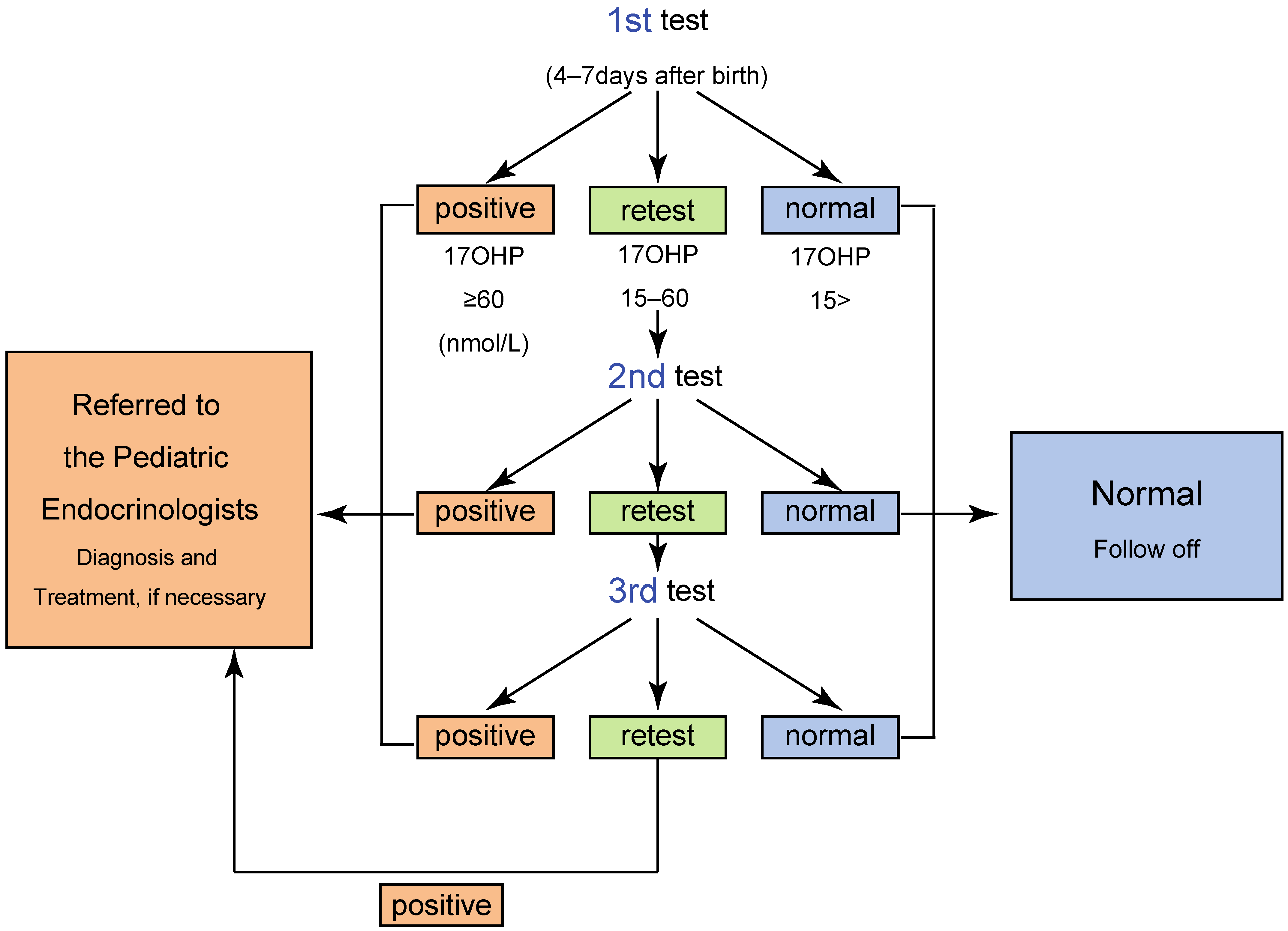
2. Screening System in Japan

The NBS in Japan was introduced individually into the prefectural administration according to a government notification by the Ministry of Health and Welfare in 1977. The basis of the NBS system, such as the timeline, and the screening panel are identical in all local governments. After informed consent is obtained from a legal guardian, blood samples are collected by a heel prick blotted on a filter paper from neonates at 4–7 days from birth, and the filter paper samples are immediately sent to a laboratory allocated by the prefectural government.
The details of the screening system are different among laboratories, and as a representative, the screening algorithm in Tokyo was shown in Figure 2. In Tokyo, the 1st screening is divided into two procedures. The level of 17-hydroxyprogesterone (17αOHP) is initially determined by enzyme-linked immunosorbent assay (ELISA) without steroid extraction. We select blood samples in the 97th percentile or higher for 17αOHP values for subjecting the second-tier test, which is carried out after steroid extraction [4,5,11,12]. The cutoff criteria for the second-tier test are shown in Table 1.
The NBS has two different cutoff values: for “screening positive” and for “retest”. When the 17αOHP level is higher than the screening positive cutoff value, the neonate is directly referred to a pediatric endocrinologist for further endocrinological evaluation. Neonates with 17αOHP levels more than the retest cutoff value are retested. When the 17αOHP levels are higher than the retest cutoff value two–three times, the screening is considered positive (Figure 2) [4,5,11,12,13].
In some female patients, blood sampling for the screening is performed ahead of schedule due to atypical genitalia, which is one of the major clinical symptoms in female neonates with 21OHD and is frequently recognized at birth.
To reduce the number of false-positive results in preterm newborns, one of the most serious issues in the screening for 21OHD, some laboratories, including that of Tokyo, employ cutoff values based on gestational age and/or birth weight. The cutoff values were determined according to a pilot study of serum 17αOHP levels in full-term and preterm infants. As a representative screening system in Japan, the algorithm and criteria for the screening in Tokyo are shown in Table 1 and Figure 2 [5], respectively.
For the quality control of the screening, most screening laboratories perform follow-up surveys of the patients who were referred to hospitals. In the surveys, clinical information of the patients, including confirmed diagnosis, is collected from the pediatric endocrinologists at the hospitals [13].

3. Clinical Outcomes of the Newborn Screening for CAH in Japan

3.1. The Effects of the Screening

The clinical profiles of 21OHD before the introduction of the screening differ remarkably from the current profiles [14]. Before the introduction of the screening, Suwa S et al. conducted a nationwide survey and reported the clinical profiles of 21OHD in Japan. According to the survey, the estimated prevalence of 21OHD was 1/43,764, and the average age in days when the patients firstly visited the hospitals was 1102. In the SW type, the average age of the first hospital visit was 55 days (male: 63 days (range, 1 days to 3 years), female: 47 days (range: 0 days to 3.9 years)), and in the SV form, the average age at first visit was 6.4 years (male: 5.9 years (range: 14 days to 34 years), female: 6.5 years (range: 0 days to 44 years)) (Table 2) [15]. The ratio of male to female was 1:1.5, and the number of male patients was significantly lower than that of females, implying that a substantial number of male patients were missed, i.e., the SV form remained undiagnosed or the fatal cases with the SW form in the neonatal-infantile period. Consistently, the survey revealed that the mortality rate was 10.6% in neonates with the SW form, which is consistent with the reports from other countries [15]. In 46,XX cases, 12.9% were firstly assigned as male because of atypical genitalia and corrected to female sex after the diagnosis of 21OHD [15].
After the introduction of the screening, the clinical outcomes of 21OHD during the neonatal/infantile period were remarkably improved. The average ages at the first visit were 8.2 and 7.6 days (male: 9.2 days, female: 6.0 days) in Sapporo and Tokyo, respectively (Table 2) [6,13]. To date, no fatal cases have been identified.
Although the follow-up surveys and the screening systems are not designed for detecting false-negative cases, based on a survey for the literature and the annual reports from NBS programs, no false-negative cases have been reported since the introduction of the screening [5,6,13]. We presume that the sex of all 46,XX cases was correctly assigned.

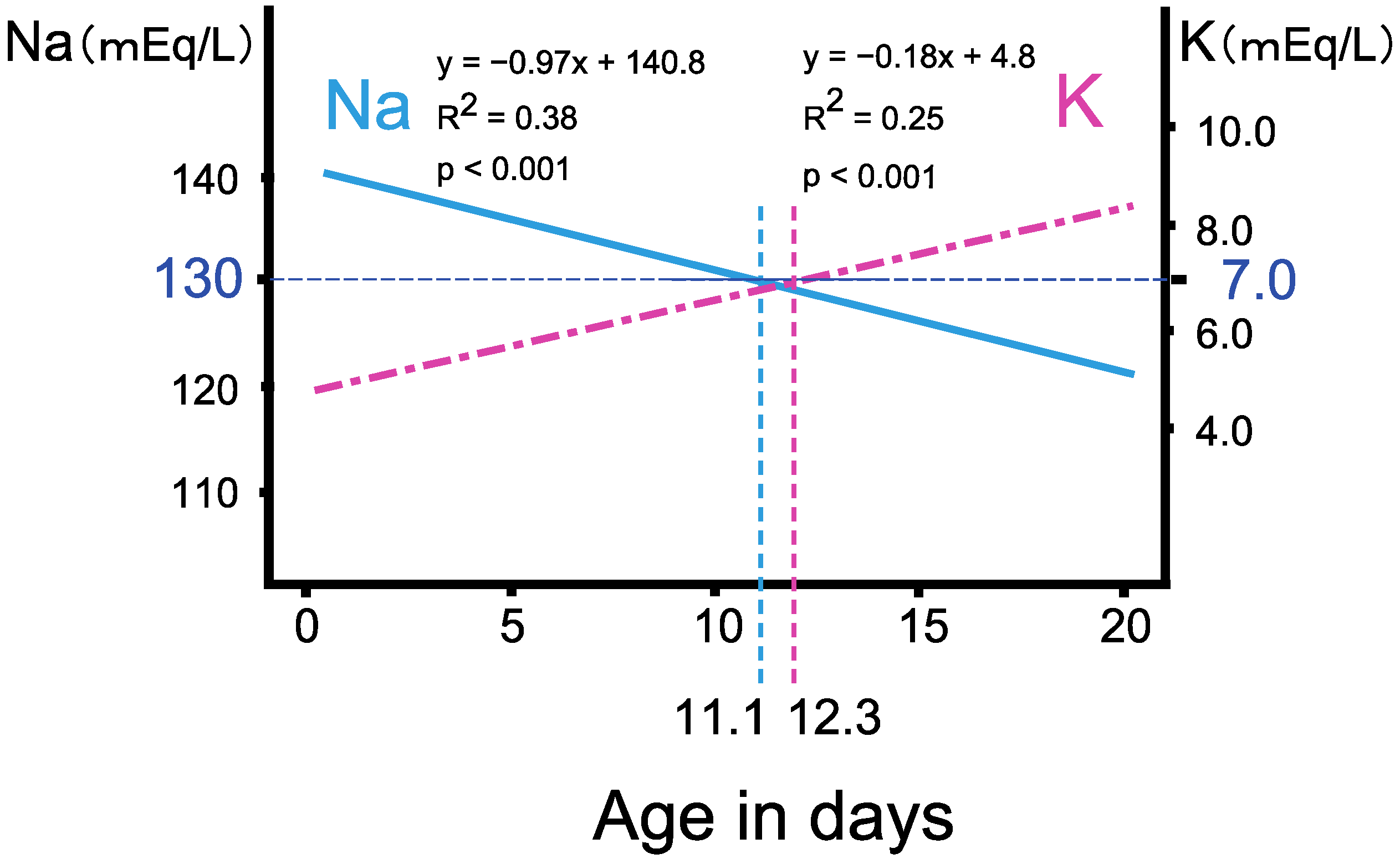
3.2. The Progression of Salt Wasting during the First Two Weeks of Life

Adrenal crisis is a life-threatening medical emergency, and eradicating the lethal cases of 21OHD is one of the major goals of newborn screening [16]. Although the fact that there were no reported fatal cases suggests the primary goal of the screening has been accomplished, 37.4% of 21OHD neonates already developed severe salt wasting, which is defined by Na < 130 mEq/L, K > 7 mEq/L, on arrival at medical hospitals in Tokyo screening [13]. Furthermore, some of the 21OHD neonates exhibited life-threatening salt wasting, such as more than 10 mEq/L of serum K [13].
Severe adrenal crisis during the neonatal to early infantile period would cause neurological comorbidities. According to the nationwide survey before the introduction of NBS in Japan, a substantial number of the 21OHD patients revealed to have neurological comorbidities including intellectual disability and epilepsy. The prevalence associated with the SW form was higher, 18.5%, than with the SV form, 9.4%, suggesting that delayed diagnosis of adrenal crisis causes intellectual disability [17]. Consistently, in the retrospective study from the U.K., where NBS for 21OHD is not introduced, more than 20% of the SW-type 21OHD patients developed learning difficulties [14]. Those suggest that just eradication of lethal cases would not be sufficient for the goal of the 21OHD screening, and avoiding severe adrenal crisis should be considered.
Retrospective analysis of the follow-up survey of the NBS in Tokyo revealed that, in classical 21OHD patients, the serum Na and K levels linearly deteriorated with age in days, and the age when the regression lines reached Na < 130 mEq/L, K > 7 mEq/L approximately coincided at 11.1 and 12.3 days, respectively [13] (Figure 3). The risk of developing severe salt wasting increases during the second week of life without a threshold, and, therefore, an early intervention, ideally during the first week of life, is desirable [13,18,19].

3.3. Triage of the Neonates with Salt Wasting by Body Weight Change

The follow-up survey in Tokyo revealed that from the second week of life, changes in body weight provide a useful index in the evaluation of neonates with positive CAH screening results [13]. Neonates with decreasing body weight from the birth weight are likely to have classical 21OHD, and neonates with increasing body weight after birth are more likely to be false positives [13]. Furthermore, even in cases of 21OHD, the possibility of developing severe salt wasting, such as hyponatremia (<130 mEq/L) or hyperkalemia (>7 mEq/L), is extremely low without loss of body weight (Figure 4). Contrary to body weight change, the relevance of predicting severe salt wasting based on the 17αOHP level is extremely low because the 17αOHP level is not associated with Na or K levels [13].
Although the findings of body weight change in patients cannot be the direct criteria for the CAH screening protocol, they may assist in some individual cases, e.g., for triaging a neonate with a positive result who is living in a region with limited access to a pediatric endocrinologist or in which there is no CAH screening.

4. Potential Issues of Testing Practices in the Newborn Screening for CAH in Japan

4.1. The Timeline of the Newborn Screening for 21OHD

The timeline of the NBSs is becoming earlier worldwide because newly added inborn errors to the screening panel require early intervention immediately after birth. In the U.S., SIMD (Society for Inherited Metabolic Disorders) defines the critical condition as a condition in which serious symptoms may present acutely in the first weeks of life with a short pre-symptomatic window and require immediate treatment to mitigate morbidity and mortality [20]. More than 10 inborn errors of organic acid disorders and fatty acid oxidation disorders are involved in the list, and the SIMD recommends considering the list as an important starting point for discussion between clinicians and laboratories [20]. Accordingly, blood samples for screening are collected 48 h after birth in the U.S., and the recommended age in days when the first results are obtained should be seven [21]. Indeed, in 2018, 64% of the first results were available within 5 days after birth (Table 3) [22]. The situation is similar in the EU, and, in most countries, blood sampling starts 72 h after birth (Table 3) [23].
In the NBS for 21OHD, several factors should be considered in terms of timing for the blood sampling. Especially, given the rate of 37.4% neonates with severe salt wasting in Japan, earlier sampling can be discussed for the prevention of life-threatening salt wasting. However, an increase in serum 17αOHP level has been observed in unaffected neonates during the first 1–2 days of life, and there is evidence of false negatives associated with the early collection of specimens in the U.S. [24]. Further, the timeline is determined by various factors of other diseases in the screening panels, which are different among countries (Table 3). For optimizing the timeline of the screening, we need careful discussion continuously.

4.2. High Rate of False Positive

For the 17αOHP measurement, immunoassays have been used because of their sensitivity, cost, and simplicity. However, immunoassays lead to high rates of false positives, seriously affecting the screening efficiency [5,18,26].
One of the major reasons is the cross-reactivity with steroids, such as 17-hydroxypregnenolone sulfate and 15β-hydroxylated compounds, which is high in preterm infants, and the ratio of false positives is extremely high in preterm infants. To minimize false positives, cutoff points stratified by gestational age and/or birth weight have been used in some screening systems. Although the stratified cutoff improves positive predictive value (PPV) to some extent, its efficiency is limited [5,27,28,29,30]. In the Tokyo system, gestational age and birth weight cutoff points have been used since the introduction of the NBS (Table 1). While the average PPV in Japan was reported as 6.6%, the Tokyo screening program achieved 25.8% (Figure 5). On the other hand, the PPV in preterm infants with a gestational age of ≤37 weeks was only 2% [5].
Another cause for the high false-positive rate is the nature of 17αOHP itself. Historically, 17αOHP was originally considered as the pathogenic androgen in cases of 21OHD rather than as a diagnostic marker, and it has several shortcomings as a diagnostic for 21OHD [31]. The level of 17αOHP is high in cord blood during the first 1–2 days of life, and stress from other illnesses may result in the 17αOHP remaining high in unaffected neonates. Furthermore, in other forms of CAH, including 11-hydroxylase deficiency (11OHD), 3β-hydroxysteroid dehydrogenase deficiency (3βHSDD), and P450 oxidoreductase deficiency (PORD), 17αOHP may be elevated to almost the same level as that of 21OHD [32]. For further improving PPV in 21OHD screening, measuring other biomarkers with high specificity for 21OHD would be required.

4.3. LC-MS/MS Analysis of 17αOHP as a Second-Tier Test and Diagnostic Test for 21OHD

To improve PPV, an alternative methodology should measure disease-specific markers other than 17αOHP or has high specificity for the target steroids. When used appropriately under highly regulated conditions, liquid chromatography-tandem mass spectrometry (LC-MS/MS) is considered as the gold standard for steroids assays [33,34,35,36,37,38,39], and the guideline of the Endocrine Society have recommended to employ LC-MS/MS for measuring 17αOHP of the second tier in neonatal screening since 2018 [16].
In addition to its specificity, the advantage of LC-MS/MS is the capability for the simultaneous assay of multiple steroids [33,34,36]. In Japan, a steroid profile panel from Siemens Healthineers AG (Frankfurt, Germany), “MS2-screening CAH” was developed for the CAH screening. Five steroids were selected for the panel: 17αOHP, 21-deoxycortisol (21-DOF), 11-deoxycortisol (11-DOF), 4-androstenedione (4AD), and cortisol (F). Accordingly, the cutoff criteria of the LC-MS/MS assay are not solely based on 17αOHP, but on 21DOF and the ratios of steroids, such as (17αOHP + 4AD)/F, 11DOF/17αOHP (Table 4) [33]. The combination of highly specific LC-MS/MS and simultaneous assays of five steroids is expected to dramatically improve the efficiency of the screening [33,34,36].
Indeed, the outcomes of LC-MS/MS are excellent. In 2018, the LC-MS/MS assay for 21OHD was employed in 5 of 37 prefectural laboratories in Japan. In immunoassay screening, out of 653 subjects with positive results, there were 38 confirmed cases of 21OHD, resulting in a PPV of 5.8% (38/653). On the other hand, the PPV in LC-MS/MS screening was 40.0% (6/15), indicating that the specificity of LC-MS/MS is remarkable [40]. Accordingly, in 2018, the Ministry of Health, Labor, and Welfare in Japan added LC-MS/MS to the list of recommended methodologies for the second-tier test of 21OHD screening [41].
In addition to improving the efficiency of 21OHD screening, the steroid profile assay by LC-MS/MS may bring further advantage to the screening, that is, assisting definitive diagnosis of 21OHD [34]. Although 21OHD can be diagnosed endocrinologically, the procedures and cutoff criteria are complicated because other rare forms of CAH, such as 11OHD, PORD, and 3βHSDD, should be differentiated from the diagnosis of 21OHD as we described in the previous section [32,42,43,44,45,46,47,48]. The nonspecific increase in 17αOHP levels in other forms of CAH has been considered as a potential clinical pitfall.
Currently, reliable methods for differentiating 21OHD from other forms of CAH are an adrenocorticotropic hormone (ACTH) stimulation test [16], urine steroid profile analyses using gas chromatography mass spectrometry [49], and genetic test, which cannot be used as a first-line diagnostic test because the procedure of CYP21A2 gene analysis is extremely complicated [50,51,52].
It has been suggested that the ratios of steroids (17αOHP + 4AD)/F, 11-DOF/17αOHP, and 21-DOF may be more specific biomarkers for the diagnosis of 21OHD than that of 17αOHP and are expected to differentiate 21OHD from other types of CAH in which 17αOHP levels are elevated, such as 3βHSDD, 11OHD, and PORD. Although there are few reports of the levels of (17αOHP + 4AD)/F, 11-DOF/17αOHP, or 21-DOF in these forms of CAH, some cases suggest the potential usefulness of the five steroids in the screening panel. 21DOF would not be elevated in 3βHSDD because, in a model of partial 3βHSD deficiency preterm infants, 21DOF is not grossly elevated [34]. In patients with 11-OHD, 21-DOF levels are reported to be normal, and 11-DOF is markedly elevated, presumably increasing the 11-DOF/17αOHP ratio [53]. Urinary steroid profile analyses of PORD suggested that the ratio of pregnanetriolone (Ptl)/tetrahydrocortisone steroids (THEs) and a specific cutoff of 11β-hydroxyandrosterone (11HA) would be useful for differentiating PORD from 21OHD. Ptl, THEs, and 11HA are metabolites of 21DOF, 11DOF + cortisol, and 4AD, respectively, which are included in the LC-MS/MS screening panel [54].
Accordingly, in combination with other clinical symptoms and signs, such as poor body weight gain and high ACTH, 21OHD can be diagnosed based on the results of NBS by LC-MS/MS [31,34]. However, we cannot directly apply the screening criteria to the diagnostic criteria, and for establishing the diagnostic criteria, an accumulation of the cases is required.

5. Database Composition and Improvement of Screening Program

For better and more efficient management of the CAH, the screening programs need persistent improvement in quality. By examining reliable follow-up studies, the outcomes and the experiences of the screening should be retrospectively evaluated and shared among the screening laboratories [25,55,56]. For short-term outcomes, most laboratories and local governments have introduced follow-up surveys in Japan, using the results for more efficient screening by decreasing false positives and early availability of screening results.
On the other hand, the assessment of long-term outcomes for CAH patients identified by screening is challenging. A nationwide registry system is required to establish efficient long-term follow-up systems. In Japan, the current screening system depends on each local government; thus, the demanding task of organizing a cross-regional collaborative system that involves local governments, local laboratories, and medical institutes is required.
Recent studies have revealed that 21OHD patients have substantial risks for metabolic syndrome in adulthood [57,58,59,60]. The metabolic syndrome in 21OHD has been assumed to be due to long-term glucocorticoid therapy [61]. However, other causes, such as fetal environments, have also been suggested [62,63], and the pathophysiology of the condition remains unknown. Further, the quality of life of 21OHD patients in adulthood is largely unknown. Especially in female patients, their gender issues should be clarified in detail [64,65,66]. As mentioned previously, before the introduction of the screening, 21OHD patients had substantial risks for neurological sequelae, which are presumably caused by the adrenal crisis during the neonatal period [14,17]. Therefore, the introduction of NBS would reduce the risk for neurological sequelae [67], but currently, available data is limited. We should keep in mind that, even after the introduction of the screening, there are a substantial number of 21OHD patients who developed severe salt wasting before the introduction of therapy. Further, a recent study suggested the number of hyponatremic episodes is an independent risk for lower IQ, suggesting that for optimizing the management of 21OHD patients during childhood, preventing episodes of severe adrenal crisis is crucial [68]. Thus, clarifying long-term outcomes will provide valuable information for improving the screening logistics.
Long-term outcomes will also provide valuable insights for evaluating the cost-effectiveness of screening. Although several analyses have been performed economically, they were based on short-term outcomes with various analytical models, leading to inconsistent results [69,70,71]. In Japan, economic analyses based on detailed clinical data have not been performed. To better understand the cost-effectiveness of screening, comprehensive approaches based on long-term outcomes are essential.
Despite not covering 21OHD patients, the introduction of several registry systems for rare congenital diseases has encouraged us. In the EU, some international collaboration-based registry systems for rare congenital diseases, such as the European Registry and Network for Intoxication type Metabolic Diseases (E-IMD) and the European Registry and Network for Homocystinurias and Methylation Defects (E-HOD), have been established [25,72,73,74,75]. Further, the Japanese Society for Inherited Metabolic Disease has successfully introduced the registry system, “JaSMIn”, for patients with inherited metabolic disease (https://www.jasmin-mcbank.com/, visited 23 April 2021). The limitation of this registry system is the unknown coverage rate due to voluntary patient registration. However, the registry is designed to cover a broad spectrum of rare inherited metabolic diseases that can be discovered by NBS, and it will provide valuable insights, enabling feedback on newborn screening in the future, including economic aspects.
Ideally, for establishing feedback systems with long-term follow-up surveys, close collaboration among the screening laboratory professionals, pediatricians, primary care providers, and clinical epidemiologists is essential [55,56,76,77]. Currently, to share the outcomes of the screening and updating of technical information, the Japan Society for Neonatal Screening has a collaborative laboratory integrated committee (Gijutsubu-kai). We expect that with this committee, pediatric endocrinologists and local governments would be able to construct large collaboration-based reports and infrastructure. A nationwide registry system in which all results of infants are registered and evaluated periodically would lead to further methodological improvements.

Author Contributions

Conceptualization, writing—original draft preparation, A.T.-H. and K.K.; supervision, K.K. Both authors have read and agreed to the published version of the manuscript.

Funding

The present study was partly supported by the grant from the Ministry of Health, Labour and Welfare of Japan (grant Nanbyo-Ippan046 to K.K. for research on intractable diseases, Principal Investigator: Tomonobu Hasegawa) (# 201811056A).

Data Availability Statement

The data presented in this study are available on request from the corresponding author. The data are not publicly available due to privacy policy.

Acknowledgments

We thank Masaru Fukushi for critical reading and valuable suggestions. We also thank Maki Gau (Tokyo Medical and Dental University), Nobuyuki Ishige (Tokyo Health Service Association, Newborn Screening), and Kazuhiro Watanabe (Tokyo Health Service Association, Newborn Screening) for valuable suggestions. We express our deep gratitude to the medical staff who provided the clinical information in the follow-up survey in Tokyo.

Conflicts of Interest

Atsumi Tsuji-Hosokawa received a research grant from the Japanese Society for Pediatric Endocrinology and Novo Nordisk Pharma Ltd. Kenichi Kashimada declare no conflict of interest.

Abbreviations

17αOHP17-Hydroxyprogesterone
yrsYears
CAHCongenital adrenal hyperplasia
CHCongenital hypothyroidism
PKUPhenylketonuria
GALGalactosemia
AADDisorders of amino acid metabolism
OADisorders of organic acid metabolism
FAODDisorders of fatty acid metabolism

References

  1. Speiser, P.W.; White, P.C. Congenital adrenal hyperplasia. N. Engl. J. Med. 2003, 349, 776–788. [Google Scholar] [CrossRef] [Green Version]
  2. White, P.C.; Speiser, P.W. Congenital adrenal hyperplasia due to 21-hydroxylase deficiency. Endocr. Rev. 2000, 21, 245–291. [Google Scholar] [CrossRef] [PubMed]
  3. van der Kamp, H.J.; Wit, J.M. Neonatal screening for congenital adrenal hyperplasia. Eur. J. Endocrinol. 2004, 151 (Suppl. S3), U71–U75. [Google Scholar] [CrossRef] [Green Version]
  4. Suwa, S. Nationwide survey of neonatal mass-screening for congenital adrenal hyperplasia in Japan. Screening 1994, 3, 141–151. [Google Scholar] [CrossRef]
  5. Tsuji, A.; Konishi, K.; Hasegawa, S.; Anazawa, A.; Onishi, T.; Ono, M.; Morio, T.; Kitagawa, T.; Kashimada, K. Newborn screening for congenital adrenal hyperplasia in Tokyo, Japan from 1989 to 2013: A retrospective population-based study. BMC Pediatr. 2015, 15, 209. [Google Scholar] [CrossRef] [Green Version]
  6. Morikawa, S.; Nakamura, A.; Fujikura, K.; Fukushi, M.; Hotsubo, T.; Miyata, J.; Ishizu, K.; Tajima, T. Results from 28 years of newborn screening for congenital adrenal hyperplasia in sapporo. Clin. Pediatr. Endocrinol. 2014, 23, 35–43. [Google Scholar] [CrossRef] [Green Version]
  7. Clayton, P.E.; Miller, W.L.; Oberfield, S.E.; Ritzen, E.M.; Sippell, W.G.; Speiser, P.W.; Group, E.L.C.W. Consensus statement on 21-hydroxylase deficiency from the European Society for Paediatric Endocrinology and the Lawson Wilkins Pediatric Endocrine Society. Horm. Res. 2002, 58, 188–195. [Google Scholar] [CrossRef] [PubMed]
  8. Tajima, T.; Fujieda, K.; Nakae, J.; Mikami, A.; Cutler, G.B., Jr. Mutations of the CYP21 gene in nonclassical steroid 21-hydroxylase deficiency in Japan. Endocr. J. 1998, 45, 493–497. [Google Scholar] [CrossRef] [PubMed] [Green Version]
  9. Tajima, T.; Fujieda, K.; Nakae, J.; Toyoura, T.; Shimozawa, K.; Kusuda, S.; Goji, K.; Nagashima, T.; Cutler, G.B., Jr. Molecular basis of nonclassical steroid 21-hydroxylase deficiency detected by neonatal mass screening in Japan. J. Clin. Endocrinol. Metab. 1997, 82, 2350–2356. [Google Scholar] [CrossRef]
  10. Kashimada, K.; Ishii, T.; Nagasaki, K.; Ono, M.; Tajima, T.; Yokota, I.; Hasegawa, Y. Clinical, biochemical, and genetic features of non-classical 21-hydroxylase deficiency in Japanese children. Endocr. J. 2015, 62, 277–282. [Google Scholar] [CrossRef]
  11. Tajima, T.; Fujikura, K.; Fukushi, M.; Hotsubo, T.; Mitsuhashi, Y. Neonatal screening for congenital adrenal hyperplasia in Japan. Pediatr. Endocrinol. Rev. 2012, 10 (Suppl. S1), 72–78. [Google Scholar] [PubMed]
  12. Tajima, T.; Fukushi, M. Neonatal mass screening for 21-hydroxylase deficiency. Clin. Pediatr. Endocrinol. 2016, 25, 1–8. [Google Scholar] [CrossRef] [Green Version]
  13. Gau, M.; Konishi, K.; Takasawa, K.; Nakagawa, R.; Tsuji-Hosokawa, A.; Hashimoto, A.; Sutani, A.; Tajima, T.; Hasegawa, T.; Morio, T.; et al. The progression of salt wasting and the body weight change during the first two weeks of life in classical 21-hydroxylase deficiency patients. Clin. Endocrinol. 2020, 94, 229–236. [Google Scholar] [CrossRef]
  14. Donaldson, M.D.; Thomas, P.H.; Love, J.G.; Murray, G.D.; McNinch, A.W.; Savage, D.C. Presentation, acute illness, and learning difficulties in salt wasting 21-hydroxylase deficiency. Arch. Dis. Child. 1994, 70, 214–218. [Google Scholar] [CrossRef] [Green Version]
  15. Suwa, S. Congenital adrenal hyperplasia. Shouni Naika 1994, 26, 1967–1972. (In Japanese) [Google Scholar]
  16. Speiser, P.W.; Arlt, W.; Auchus, R.J.; Baskin, L.S.; Conway, G.S.; Merke, D.P.; Meyer-Bahlburg, H.F.L.; Miller, W.L.; Murad, M.H.; Oberfield, S.E.; et al. Congenital adrenal hyperplasia due to steroid 21-hydroxylase deficiency: An endocrine society clinical practice guideline. J. Clin. Endocrinol. Metab. 2018, 103, 4043–4088. [Google Scholar] [CrossRef]
  17. Suwa, S.; Maesaka, H.; Suzuki, J.; Katsumata, N. A suvery report: The comrbidities of congenital adrenal hyperplasia. In A Study for Newborn Screening, Annual Report in 1995; The Japanese Ministry of Health, Labour and Welfare’s Study Group for Physical and Mental Disabilities: Tokyo, Japan, 1985; pp. 274–277. (In Japanese) [Google Scholar]
  18. Heather, N.L.; Seneviratne, S.N.; Webster, D.; Derraik, J.G.; Jefferies, C.; Carll, J.; Jiang, Y.; Cutfield, W.S.; Hofman, P.L. Newborn screening for congenital adrenal hyperplasia in New Zealand, 1994–2013. J. Clin. Endocrinol. Metab. 2015, 100, 1002–1008. [Google Scholar] [CrossRef] [Green Version]
  19. Shima, R.; Sawano, K.; Shibata, N.; Nyuzuki, H.; Sasaki, S.; Sato, H.; Ogawa, Y.; Abe, Y.; Nagasaki, K.; Saitoh, A. Timing of hyponatremia development in patients with salt-wasting-type 21-hydroxylase deficiency. Clin. Pediatr. Endocrinol. 2020, 29, 105–110. [Google Scholar] [CrossRef] [PubMed]
  20. Freedenberg, D.; Berry, S.; Dimmock, D.; Gibson, J.; Greene, C.; Kronn, D.; Tanksley, S. Society for Inherited Metabolic Disorders (SIMD) Position Statement, 2014: Identifying Abnormal Newborn Screens Requiring Immediate Notification of the Health Care Provider. Available online: https://www.simd.org/Issues/SIMD%20NBS%20Critical%20Conditions%20policy%20statement.pdf (accessed on 23 April 2021).
  21. HRSA, F.A.C. Newborn Screening Timeliness Goals. Available online: https://www.hrsa.gov/advisory-committees/heritable-disorders/newborn-screening-timeliness.html (accessed on 20 May 2021).
  22. Sontag, M.K.; Miller, J.I.; McKasson, S.; Sheller, R.; Edelman, S.; Yusuf, C.; Singh, S.; Sarkar, D.; Bocchini, J.; Scott, J.; et al. Newborn screening timeliness quality improvement initiative: Impact of national recommendations and data repository. PLoS ONE 2020, 15, e0231050. [Google Scholar] [CrossRef] [PubMed] [Green Version]
  23. Loeber, J.G. Current Status of Newborn Screening in Europe (Based on an ISNS Survey Held during 2018). Available online: https://www.isns-neoscreening.org/wp-content/uploads/2019/04/NBS-in-Europe-2018.pdf (accessed on 5 October 2020).
  24. Held, P.K.; Bird, I.M.; Heather, N.L. Newborn screening for congenital adrenal hyperplasia: Review of factors affecting screening accuracy. Int. J. Neonatal. Screen 2020, 6, 67. [Google Scholar] [CrossRef]
  25. Loeber, J.G.; Platis, D.; Zetterstrom, R.H.; Almashanu, S.; Boemer, F.; Bonham, J.R.; Borde, P.; Brincat, I.; Cheillan, D.; Dekkers, E.; et al. Neonatal screening in Europe revisited: An ISNS perspective on the current state and developments since 2010. Int. J. Neonatal. Screen 2021, 7, 15. [Google Scholar] [CrossRef]
  26. Coulm, B.; Coste, J.; Tardy, V.; Ecosse, E.; Roussey, M.; Morel, Y.; Carel, J.C.; Group, D.S. Efficiency of neonatal screening for congenital adrenal hyperplasia due to 21-hydroxylase deficiency in children born in mainland France between 1996 and 2003. Arch. Pediatr. Adolesc. Med. 2012, 166, 113–120. [Google Scholar] [CrossRef] [PubMed] [Green Version]
  27. Pode-Shakked, N.; Blau, A.; Pode-Shakked, B.; Tiosano, D.; Weintrob, N.; Eyal, O.; Zung, A.; Levy-Khademi, F.; Tenenbaum-Rakover, Y.; Zangen, D.; et al. Combined gestational age- and birth weight-adjusted cutoffs for newborn screening of congenital adrenal hyperplasia. J. Clin. Endocrinol. Metab. 2019, 104, 3172–3180. [Google Scholar] [CrossRef]
  28. van der Kamp, H.J.; Oudshoorn, C.G.; Elvers, B.H.; van Baarle, M.; Otten, B.J.; Wit, J.M.; Verkerk, P.H. Cutoff levels of 17-alpha-hydroxyprogesterone in neonatal screening for congenital adrenal hyperplasia should be based on gestational age rather than on birth weight. J. Clin. Endocrinol. Metab. 2005, 90, 3904–3907. [Google Scholar] [CrossRef] [Green Version]
  29. Nordenstrom, A.; Wedell, A.; Hagenfeldt, L.; Marcus, C.; Larsson, A. Neonatal screening for congenital adrenal hyperplasia: 17-hydroxyprogesterone levels and CYP21 genotypes in preterm infants. Pediatrics 2001, 108, e68. [Google Scholar] [CrossRef] [Green Version]
  30. Allen, D.B.; Hoffman, G.L.; Fitzpatrick, P.; Laessig, R.; Maby, S.; Slyper, A. Improved precision of newborn screening for congenital adrenal hyperplasia using weight-adjusted criteria for 17-hydroxyprogesterone levels. J. Pediatr. 1997, 130, 128–133. [Google Scholar] [CrossRef]
  31. Miller, W.L. Congenital adrenal hyperplasia: Time to replace 17OHP with 21-deoxycortisol. Horm. Res. Paediatr. 2019, 91, 416–420. [Google Scholar] [CrossRef]
  32. Takasawa, K.; Ono, M.; Hijikata, A.; Matsubara, Y.; Katsumata, N.; Takagi, M.; Morio, T.; Ohara, O.; Kashimada, K.; Mizutani, S. Two novel HSD3B2 missense mutations with diverse residual enzymatic activities for Delta5-steroids. Clin. Endocrinol. 2014, 80, 782–789. [Google Scholar] [CrossRef] [PubMed]
  33. Higashi, T.; Nishio, T.; Uchida, S.; Shimada, K.; Fukushi, M.; Maeda, M. Simultaneous determination of 17alpha-hydroxypregnenolone and 17alpha-hydroxyprogesterone in dried blood spots from low birth weight infants using LC-MS/MS. J. Pharm. Biomed. Anal. 2008, 48, 177–182. [Google Scholar] [CrossRef]
  34. Janzen, N.; Peter, M.; Sander, S.; Steuerwald, U.; Terhardt, M.; Holtkamp, U.; Sander, J. Newborn screening for congenital adrenal hyperplasia: Additional steroid profile using liquid chromatography-tandem mass spectrometry. J. Clin. Endocrinol. Metab. 2007, 92, 2581–2589. [Google Scholar] [CrossRef]
  35. Kulle, A.E.; Riepe, F.G.; Hedderich, J.; Sippell, W.G.; Schmitz, J.; Niermeyer, L.; Holterhus, P.M. LC-MS/MS based determination of basal- and ACTH-stimulated plasma concentrations of 11 steroid hormones: Implications for detecting heterozygote CYP21A2 mutation carriers. Eur. J. Endocrinol. 2015, 173, 517–524. [Google Scholar] [CrossRef] [Green Version]
  36. Monostori, P.; Szabo, P.; Marginean, O.; Bereczki, C.; Karg, E. Concurrent confirmation and differential diagnosis of congenital adrenal hyperplasia from dried blood spots: Application of a second-tier LC-MS/MS assay in a cross-border cooperation for newborn screening. Horm. Res. Paediatr. 2015, 84, 311–318. [Google Scholar] [CrossRef] [PubMed]
  37. Lai, C.C.; Tsai, C.H.; Tsai, F.J.; Wu, J.Y.; Lin, W.D.; Lee, C.C. Rapid screening assay of congenital adrenal hyperplasia by measuring 17 alpha-hydroxyprogesterone with high-performance liquid chromatography/electrospray ionization tandem mass spectrometry from dried blood spots. J. Clin. Lab. Anal. 2002, 16, 20–25. [Google Scholar] [CrossRef]
  38. Lacey, J.M.; Minutti, C.Z.; Magera, M.J.; Tauscher, A.L.; Casetta, B.; McCann, M.; Lymp, J.; Hahn, S.H.; Rinaldo, P.; Matern, D. Improved specificity of newborn screening for congenital adrenal hyperplasia by second-tier steroid profiling using tandem mass spectrometry. Clin. Chem. 2004, 50, 621–625. [Google Scholar] [CrossRef] [PubMed]
  39. Minutti, C.Z.; Lacey, J.M.; Magera, M.J.; Hahn, S.H.; McCann, M.; Schulze, A.; Cheillan, D.; Dorche, C.; Chace, D.H.; Lymp, J.F.; et al. Steroid profiling by tandem mass spectrometry improves the positive predictive value of newborn screening for congenital adrenal hyperplasia. J. Clin. Endocrinol. Metab. 2004, 89, 3687–3693. [Google Scholar] [CrossRef] [PubMed] [Green Version]
  40. Fukushi, M.; Isobe, M.; Kanda, K.; Saraie, H.; Fujikura, K.; Mitsui, N.; Mae, H.; Yamagami, Y. The quality control of LC-MS/MS screening for CAH as the second tier test. In Proceedings of the 47th Annual meeting of Japan Society for Neonatal Screening, Gifu, Japan, 25–26 September 2020. [Google Scholar]
  41. Children and Families Bureau, Labour and Welfare in Japan. Notification for Newborn Screening, 2018. Notification from Director of the Maternal and Child Health Division. Available online: https://www.jsms.gr.jp/download/MHLW_MCH_20180330.pdf (accessed on 23 April 2021). (In Japanese)
  42. Therrell, B.L., Jr.; Berenbaum, S.A.; Manter-Kapanke, V.; Simmank, J.; Korman, K.; Prentice, L.; Gonzalez, J.; Gunn, S. Results of screening 1.9 million Texas newborns for 21-hydroxylase-deficient congenital adrenal hyperplasia. Pediatrics 1998, 101, 583–590. [Google Scholar] [CrossRef] [PubMed]
  43. Honour, J.W.; Anderson, J.M.; Shackleton, C.H. Difficulties in the diagnosis of congenital adrenal hyperplasia in early infancy: The 11 beta-hydroxylase defect. Acta Endocrinol. 1983, 103, 101–109. [Google Scholar] [CrossRef]
  44. Tonetto-Fernandes, V.; Lemos-Marini, S.H.; Kuperman, H.; Ribeiro-Neto, L.M.; Verreschi, I.T.; Kater, C.E. Serum 21-Deoxycortisol, 17-Hydroxyprogesterone, and 11-deoxycortisol in classic congenital adrenal hyperplasia: Clinical and hormonal correlations and identification of patients with 11beta-hydroxylase deficiency among a large group with alleged 21-hydroxylase deficiency. J. Clin. Endocrinol. Metab. 2006, 91, 2179–2184. [Google Scholar] [CrossRef] [Green Version]
  45. Homma, K.; Hasegawa, T.; Nagai, T.; Adachi, M.; Horikawa, R.; Fujiwara, I.; Tajima, T.; Takeda, R.; Fukami, M.; Ogata, T. Urine steroid hormone profile analysis in cytochrome P450 oxidoreductase deficiency: Implication for the backdoor pathway to dihydrotestosterone. J. Clin. Endocrinol. Metab. 2006, 91, 2643–2649. [Google Scholar] [CrossRef] [Green Version]
  46. Fluck, C.E.; Tajima, T.; Pandey, A.V.; Arlt, W.; Okuhara, K.; Verge, C.F.; Jabs, E.W.; Mendonca, B.B.; Fujieda, K.; Miller, W.L. Mutant P450 oxidoreductase causes disordered steroidogenesis with and without Antley-Bixler syndrome. Nat. Genet. 2004, 36, 228–230. [Google Scholar] [CrossRef] [Green Version]
  47. Jeandron, D.D.; Sahakitrungruang, T. A novel homozygous Q334X mutation in the HSD3B2 gene causing classic 3beta-hydroxysteroid dehydrogenase deficiency: An unexpected diagnosis after a positive newborn screen for 21-hydroxylase deficiency. Horm. Res. Paediatr. 2012, 77, 334–338. [Google Scholar] [CrossRef]
  48. Cara, J.F.; Moshang, T., Jr.; Bongiovanni, A.M.; Marx, B.S. Elevated 17-hydroxyprogesterone and testosterone in a newborn with 3-beta-hydroxysteroid dehydrogenase deficiency. N. Engl. J. Med. 1985, 313, 618–621. [Google Scholar] [CrossRef]
  49. Homma, K.; Hasegawa, T.; Takeshita, E.; Watanabe, K.; Anzo, M.; Toyoura, T.; Jinno, K.; Ohashi, T.; Hamajima, T.; Takahashi, Y.; et al. Elevated urine pregnanetriolone definitively establishes the diagnosis of classical 21-hydroxylase deficiency in term and preterm neonates. J. Clin. Endocrinol. Metab. 2004, 89, 6087–6091. [Google Scholar] [CrossRef] [Green Version]
  50. Pignatelli, D.; Carvalho, B.L.; Palmeiro, A.; Barros, A.; Guerreiro, S.G.; Macut, D. The Complexities in Genotyping of Congenital Adrenal Hyperplasia: 21-Hydroxylase Deficiency. Front. Endocrinol. 2019, 10, 432. [Google Scholar] [CrossRef] [PubMed] [Green Version]
  51. Parajes, S.; Quinterio, C.; Dominguez, F.; Loidi, L. A simple and robust quantitative PCR assay to determine CYP21A2 gene dose in the diagnosis of 21-hydroxylase deficiency. Clin. Chem. 2007, 53, 1577–1584. [Google Scholar] [CrossRef] [PubMed] [Green Version]
  52. Gao, Y.; Lu, L.; Yu, B.; Mao, J.; Wang, X.; Nie, M.; Wu, X. The prevalence of the chimeric TNXA/TNXB gene and clinical symptoms of ehlers-danlos syndrome with 21-hydroxylase deficiency. J. Clin. Endocrinol. Metab. 2020, 105, 2288–2299. [Google Scholar] [CrossRef] [PubMed]
  53. Janzen, N.; Riepe, F.G.; Peter, M.; Sander, S.; Steuerwald, U.; Korsch, E.; Krull, F.; Muller, H.L.; Heger, S.; Brack, C.; et al. Neonatal screening: Identification of children with 11beta-hydroxylase deficiency by second-tier testing. Horm. Res. Paediatr. 2012, 77, 195–199. [Google Scholar] [CrossRef]
  54. Koyama, Y.; Homma, K.; Fukami, M.; Miwa, M.; Ikeda, K.; Ogata, T.; Hasegawa, T.; Murata, M. Two-step biochemical differential diagnosis of classic 21-hydroxylase deficiency and cytochrome P450 oxidoreductase deficiency in Japanese infants by GC-MS measurement of urinary pregnanetriolone/ tetrahydroxycortisone ratio and 11beta-hydroxyandrosterone. Clin. Chem. 2012, 58, 741–747. [Google Scholar] [CrossRef]
  55. Burgard, P.; Rupp, K.; Lindner, M.; Haege, G.; Rigter, T.; Weinreich, S.S.; Loeber, J.G.; Taruscio, D.; Vittozzi, L.; Cornel, M.C.; et al. Newborn screening programmes in Europe; arguments and efforts regarding harmonization. Part 2. From screening laboratory results to treatment, follow-up and quality assurance. J. Inherit. Metab. Dis. 2012, 35, 613–625. [Google Scholar] [CrossRef] [PubMed]
  56. Advisory Committee on Heritable Disorders in Newborns and Children; Zuckerman, A.E.; Badawi, D.; Brosco, J.P.; Brower, A.; Eichwald, J.; Feuchtbaum, L.; Finitzo, T.; Flannery, D.; Green, N.; et al. The Role of Quality Measures to Promote Long-Term Follow-up of Children Identified by Newborn Screening Programs; Advisory Committee on Heritable Disorders in Newborns and Children Advisory Committee on Heritable Disorders in Newborns and Children. 2018. Available online: https://www.hrsa.gov/sites/default/files/hrsa/advisory-committees/heritable-disorders/reports-recommendations/reports/role-of-quality-measures-in-nbs-sept2018-508c.pdf” (accessed on 23 April 2021).
  57. Stikkelbroeck, N.M.; Oyen, W.J.; van der Wilt, G.J.; Hermus, A.R.; Otten, B.J. Normal bone mineral density and lean body mass, but increased fat mass, in young adult patients with congenital adrenal hyperplasia. J. Clin. Endocrinol. Metab. 2003, 88, 1036–1042. [Google Scholar] [CrossRef] [Green Version]
  58. Charmandari, E.; Chrousos, G.P. Metabolic syndrome manifestations in classic congenital adrenal hyperplasia: Do they predispose to atherosclerotic cardiovascular disease and secondary polycystic ovary syndrome? Ann. N. Y. Acad. Sci. 2006, 1083, 37–53. [Google Scholar] [CrossRef]
  59. Zimmermann, A.; Grigorescu-Sido, P.; AlKhzouz, C.; Patberg, K.; Bucerzan, S.; Schulze, E.; Zimmermann, T.; Rossmann, H.; Geiss, H.C.; Lackner, K.J.; et al. Alterations in lipid and carbohydrate metabolism in patients with classic congenital adrenal hyperplasia due to 21-hydroxylase deficiency. Horm. Res. Paediatr. 2010, 74, 41–49. [Google Scholar] [CrossRef]
  60. Finkielstain, G.P.; Kim, M.S.; Sinaii, N.; Nishitani, M.; Van Ryzin, C.; Hill, S.C.; Reynolds, J.C.; Hanna, R.M.; Merke, D.P. Clinical characteristics of a cohort of 244 patients with congenital adrenal hyperplasia. J. Clin. Endocrinol. Metab. 2012, 97, 4429–4438. [Google Scholar] [CrossRef] [PubMed] [Green Version]
  61. Merke, D.P.; Bornstein, S.R. Congenital adrenal hyperplasia. Lancet 2005, 365, 2125–2136. [Google Scholar] [CrossRef]
  62. Khalid, J.M.; Oerton, J.M.; Dezateux, C.; Hindmarsh, P.C.; Kelnar, C.J.; Knowles, R.L. Incidence and clinical features of congenital adrenal hyperplasia in Great Britain. Arch. Dis. Child. 2012, 97, 101–106. [Google Scholar] [CrossRef] [PubMed]
  63. Takishima, S.; Nakajima, K.; Nomura, R.; Tsuji-Hosokawa, A.; Matsuda, N.; Matsubara, Y.; Ono, M.; Miyai, K.; Takasawa, K.; Morio, T.; et al. Lower body weight and BMI at birth were associated with early adiposity rebound in 21-hydroxylase deficiency patients. Endocr. J. 2016, 63, 983–990. [Google Scholar] [CrossRef] [PubMed] [Green Version]
  64. Pasterski, V.; Zucker, K.J.; Hindmarsh, P.C.; Hughes, I.A.; Acerini, C.; Spencer, D.; Neufeld, S.; Hines, M. Increased cross-gender identification independent of gender role behavior in girls with congenital adrenal hyperplasia: Results from a standardized assessment of 4- to 11-year-old children. Arch. Sex. Behav. 2015, 44, 1363–1375. [Google Scholar] [CrossRef] [Green Version]
  65. Meyer-Bahlburg, H.F.; Dolezal, C.; Baker, S.W.; Ehrhardt, A.A.; New, M.I. Gender development in women with congenital adrenal hyperplasia as a function of disorder severity. Arch. Sex. Behav. 2006, 35, 667–684. [Google Scholar] [CrossRef] [PubMed]
  66. Meyer-Bahlburg, H.F.; Dolezal, C.; Baker, S.W.; New, M.I. Sexual orientation in women with classical or non-classical congenital adrenal hyperplasia as a function of degree of prenatal androgen excess. Arch. Sex. Behav. 2008, 37, 85–99. [Google Scholar] [CrossRef]
  67. Harasymiw, L.A.; Grosse, S.D.; Sarafoglou, K. Attention-deficit/hyperactivity disorder among US children and adolescents with congenital adrenal hyperplasia. J. Endocr. Soc. 2020, 4, bvaa152. [Google Scholar] [CrossRef]
  68. Hamed, S.A.; Metwalley, K.A.; Farghaly, H.S. Cognitive function in children with classic congenital adrenal hyperplasia. Eur. J. Pediatr. 2018, 177, 1633–1640. [Google Scholar] [CrossRef]
  69. Fox, D.A.; Ronsley, R.; Khowaja, A.R.; Haim, A.; Vallance, H.; Sinclair, G.; Amed, S. Clinical impact and cost efficacy of newborn screening for congenital adrenal hyperplasia. J. Pediatr. 2020, 220, 101–108.e2. [Google Scholar] [CrossRef] [PubMed]
  70. Carroll, A.E.; Downs, S.M. Comprehensive cost-utility analysis of newborn screening strategies. Pediatrics 2006, 117, S287–S295. [Google Scholar] [CrossRef] [PubMed] [Green Version]
  71. Yoo, B.K.; Grosse, S.D. The cost effectiveness of screening newborns for congenital adrenal hyperplasia. Public Health Genom. 2009, 12, 67–72. [Google Scholar] [CrossRef] [PubMed]
  72. Huemer, M.; Diodato, D.; Martinelli, D.; Olivieri, G.; Blom, H.; Gleich, F.; Kolker, S.; Kozich, V.; Morris, A.A.; Seifert, B.; et al. Phenotype, treatment practice and outcome in the cobalamin-dependent remethylation disorders and MTHFR deficiency: Data from the E-HOD registry. J. Inherit. Metab. Dis. 2019, 42, 333–352. [Google Scholar] [CrossRef]
  73. Posset, R.; Garbade, S.F.; Gleich, F.; Gropman, A.L.; de Lonlay, P.; Hoffmann, G.F.; Garcia-Cazorla, A.; Nagamani, S.C.S.; Baumgartner, M.R.; Schulze, A.; et al. Long-term effects of medical management on growth and weight in individuals with urea cycle disorders. Sci. Rep. 2020, 10, 11948. [Google Scholar] [CrossRef]
  74. Kolker, S.; Dobbelaere, D.; Haberle, J.; Burgard, P.; Gleich, F.; Summar, M.L.; Hannigan, S.; Parker, S.; Chakrapani, A.; Baumgartner, M.R.; et al. Networking across borders for individuals with organic acidurias and urea cycle disorders: The E-IMD consortium. JIMD Rep. 2015, 22, 29–38. [Google Scholar] [CrossRef] [PubMed] [Green Version]
  75. Kozich, V.; Sokolova, J.; Morris, A.A.M.; Pavlikova, M.; Gleich, F.; Kolker, S.; Krijt, J.; Dionisi-Vici, C.; Baumgartner, M.R.; Blom, H.J.; et al. Cystathionine beta-synthase deficiency in the E-HOD registry-part I: Pyridoxine responsiveness as a determinant of biochemical and clinical phenotype at diagnosis. J. Inherit. Metab. Dis. 2021, 44, 677–692. [Google Scholar] [CrossRef]
  76. Hoff, T.; Hoyt, A.; Therrell, B.; Ayoob, M. Exploring barriers to long-term follow-up in newborn screening programs. Genet. Med. 2006, 8, 563–570. [Google Scholar] [CrossRef] [Green Version]
  77. Loeber, J.G.; Burgard, P.; Cornel, M.C.; Rigter, T.; Weinreich, S.S.; Rupp, K.; Hoffmann, G.F.; Vittozzi, L. Newborn screening programmes in Europe; arguments and efforts regarding harmonization. Part 1. From blood spot to screening result. J. Inherit. Metab. Dis. 2012, 35, 603–611. [Google Scholar] [CrossRef]
Figure 1. Steroid synthesis in the adrenal cortex (A) and the pathophysiology of 21OHD (B). P5: Pregnenolone, 17αOHP5: 17-hydroxypregnenolone, P: Progesterone, 17αOHP: 17-hydroxyprogesterone, DOC: Deoxycorticosterone, 11DOF: 11-deoxycortisol, B: Corticosterone, 18OHB: 18-Hydroxycorticosterone, DHEA: Dehydroepiandrostendione, A4: Androstenedione, T: Testosterone. 17αOHP and other green steroids are included in the panel of LC-MS/MS screening in Japan. Steroids written in blue suggest its synthesis is reduced.
Ijns 07 00036 g001
Figure 2. Algorithm of CAH screening in Tokyo.
Ijns 07 00036 g002
Figure 3. Clinical features of serum sodium (Na) and potassium (K) levels in 21OHD neonates. Retrospective analysis of the NBS in Tokyo revealed that, in classical 21OHD patients, the serum Na and K levels linearly deteriorated with age in days, and the age when the regression lines reached Na < 130 mEq/L, K > 7 mEq/L approximately coincided at 11.1 and 12.3 days, respectively. (Modified from Gau et al., 2020 [13]).
Ijns 07 00036 g003
Figure 4. Body weight change from birth is an excellent predictor of 21OHD and the risk of severe salt wasting. Body weight data of 21OHD patients at 7–14 days after birth were collected, and the change in body weight from birth weight was examined. None of the 21OHD patients with severe salt wasting (Na < 130 meq/L or K > 7.0 mEq/L) exhibited increased body weight (A,B).
Ijns 07 00036 g004
Figure 5. PPV (positive predicted value) of CAH screening in Tokyo according to the birth weights and the gestational ages of newborns judged as positive. (Modified from Tsuji et al., 2015 [5]).
Ijns 07 00036 g005
Table 1. Criteria of CAH mass screening in Tokyo.
<Criteria According to the Gestational Age>
Gestational age at birth (weeks) *1a≤2930–3435–36≥37
Corrected gestational age (weeks) *1b≤3132–3536–37≥38
<Criteria According to Weight> *2,*3
Body weight (g)≤9991000–19992000–2499≥2500
Cutoff level of 17αOHP [n·mol/L]Retest *460452415
Positive *5606060
*1a Samples collected before the age of 7 days, *1b Samples collected at the age of 7 days or after, *2 1st test: body weight = birth weight, 2nd test and after: body weight = corrected body weight calculated by the formula as below. Corrected body weight at test (g) = birth weight (g) + (age at test − 7) × 20 (g). *3 For infants born small or large for gestational age, either the criteria of gestational age (corrected gestational age) or body weight was applied, whichever was a lower value. Since 2012, criteria according to weight have not been used, and solely gestational age-stratified cutoff has been used. *4 recall for the second (or the third) test of the screening. *5 refer to hospitals for further endocrinological examinations.
Table 2. Age at diagnosis before and after implementation of the screening.
Before CAH Screening *After CAH Screening **
MaleFemaleTotalMaleFemaleTotal
SW type63 days (120)47 days (96)55 days (216)9.0 days (55)6.2 days (45)7.6 days (100)
SV type5.0 yrs (39)6.5 yrs (150)6.4 yrs (189)
Numbers in parentheses indicate numbers of the subjects, *, **: according to the data reported by Suwa et al., 1994 [15] and Gau et al., 2020 [13], respectively.
Table 3. Summary of newborn screening in European countries, Oceania, and the U.S. (modified table from reference [25]) and the following website (https://www.hrsa.gov/advisory-committees/heritable-disorders/newborn-screening-timeliness.html, https://newbornscreening.hrsa.gov/your-state#w, https://www.newsteps.org/resources/data-visualizations/newborn-screening-status-all-disorders, visited date, “23 April 2021”).
Countries *1Approximate Population (Million)Screening PanelInterval Birth-SamplingInterval Sampling Analysis
CAHCHPKUGALAAD, OA, FAOD<48 h48–72 h72–96 h>96 h1 d2 d3 d4 d5 d>6 d
Austria8.8x *2xxx>6xx xxx
Belgium10.5xxxx>6 xxx x
Denmark5.6xxxx>6 x xx
France67xxxxP *2 x xx
Germany80xxxx>6xx xx
Netherlands17.8xxxx>6 x xxx
Spain46.5xxxP>6xxx xxxx
Sweden10xxxx>6 x xxx
Switzerland8.1xxxx1–6 x x
Finland5.5 xx >6 x *4xxxxxxx
Greece10.5 xxx x x
Hungary10 xxx>6 x xx
Ireland4.9 xxx xxxx
Italy60.5 xxPP x xxxx
Norway5.3 xx >6 x x
Portugal10.3 xx >6 x xxx
U.K.66.6 xx 1–6 x xx
U.S. *3328.250/5050/5050/5050/5050/50x xx
JPN126.3xxxx>6 xxxx
*1 European countries whose population is approximately more than 5 million were listed. *2 x (in screening panel section) = in screening panel, P = pilot/regional screening. *3 In the United States section, the number of states that include the disease in the screening panel is listed. In the AAD, OA, and FAOD section, states that implemented more than six of the metabolic disorders were counted. The interval between birth, sampling, and analysis of U.S. is recommended timeline. *4 Cord blood is used for some of the screening.
Table 4. Cutoff level of 17αOHP and other steroids assayed by LC-MS/MS in Saitama, Sapporo and Tokyo, Japan *1.
Screening Positive Cutoff Level
PrefectureSaitamaSapporoTokyo
Criteria A *2,*3Criteria B *2,*3
17αOHP (ng/mL)Term birth>20>20>5>5
Preterm birth>30>50
21DOF (ng/mL)>1.0>2.0>1.0
(17αOHP + 4AD)/F >2.0
11DOF/17αOHP <0.1
Retest Cutoff Level
PrefectureSaitama *2Sapporo *2Tokyo *2
17αOHP (ng/mL)>1.0>2.5>1.5
(17αOHP + 4AD)/F>0.1>0.1>0.3
11DOF/17αOHP<0.3<0.2<0.3
*1 The algorithm of the screening is the same as shown in Figure 1. When the retest values are documented twice, the patients are judged as positive. *2 The result is judged as positive or retest when all parameters meet the criteria. *3 The patient with the result that meets criteria A or B is considered as screening positive.
Publisher’s Note: MDPI stays neutral with regard to jurisdictional claims in published maps and institutional affiliations.

