Review

Neonatal Screening for Primary Carnitine Deficiency: Lessons Learned from the Faroe Islands

1
Medical Department, National Hospital of the Faroe Islands, Tórshavn 100, Faroe Islands
2
Department of Occupational Medicine and Public Health, National Hospital of the Faroe Islands, Tórshavn 100, Faroe Islands
3
Screening-Laboratory Hannover, POB 91 10 09, Hannover 30430, Germany
4
Department of Clinical Genetics, Centre for Inherited Metabolic Diseases, Copenhagen University Hospital, Copenhagen 2100, Denmark
5
Department of Clinical Chemistry, Medical School, Hannover 30625, Germany
6
Danish Center for Neonatal Screening, Statens Serum Institut, Copenhagen S 2300, Denmark
7
Division of Medical Genetics, Departments of Pediatrics and Pathology, and ARUP Laboratories, University of Utah, Salt Lake City, UT 84103, USA
*
Author to whom correspondence should be addressed.
Int. J. Neonatal Screen. 2017, 3(1), 1; https://doi.org/10.3390/ijns3010001
Submission received: 17 November 2016 / Revised: 16 January 2017 / Accepted: 20 January 2017 / Published: 4 February 2017

Abstract

:
Primary carnitine deficiency is caused by the defective OCTN2 carnitine transporter encoded by the SLC22A5 gene. A lack of carnitine impairs fatty acid oxidation resulting in hypoketotic hypoglycemia, hepatic encephalopathy, skeletal and cardiac myopathy, and arrhythmia. This condition can be detected by finding low levels of free carnitine (C0) in neonatal screening. Mothers with primary carnitine deficiency can also be identified by low carnitine levels in their infant by newborn screening. Primary carnitine deficiency is rare (1:40,000–1:140,000 newborns) except in the Faroe Islands (1:300) due to a founder effect. A specific mutation (c.95A>G, p.N32S) is prevalent, but not unique, with three additional mutations (c.131C>T/p.A44V, a splice mutation c.825-52G>A, and a risk-haplotype) recently identified in the Faroese population. In the Faroe Islands, several adult patients suffered sudden death from primary carnitine deficiency leading to the implementation of a nationwide population screening (performed after 2 months of age) in addition to universal neonatal screening. While most affected infants can be identified at birth, some patients with primary carnitine deficiency might be missed by the current neonatal screening and could be better identified with a repeated test performed after 2 months of age.

1. Introduction

Primary carnitine deficiency (OMIM 212140) is an autosomal recessive disease caused by a defect in the OCTN2 carnitine transporter encoded by the SLC22A5 gene [1,2]. Carnitine is required for the transfer of long-chain fatty acids from the cytoplasm to the mitochondrial matrix for beta-oxidation [2]. During periods of fasting, fatty acids are the predominant substrate for energy production via oxidation in the liver, cardiac muscle, and skeletal muscle. Carnitine is transported inside the cells by an organic cation transporter (OCTN2) present in the heart, muscle, and kidney. Mutations in the SLC22A5 gene impair carnitine transport, resulting in urinary carnitine wasting with low levels of serum carnitine (<9 μmol/L, normal 25–50 μmol/L). Low concentrations of carnitine lead to intracellular carnitine depletion with secondary impairment of fatty acid oxidation [2,3]. When fat cannot be utilized, glucose is consumed without regeneration via gluconeogenesis, resulting in hypoglycemia. In addition, fats released from adipose tissue accumulate in the liver, skeletal muscle, and heart, resulting in hepatic steatosis and myopathy [2].
Affected children can present, between age three months and two years, with episodes of metabolic decompensation triggered by fasting and often associated with common illnesses such as febrile viral infections or acute gastroenteritis [2,3]. These episodes are characterized clinically by poor feeding, irritability, lethargy, and hepatomegaly. Laboratory evaluations usually reveal hypoketotic hypoglycemia (hypoglycemia with minimal or no ketones in urine), hyperammonemia, and elevated liver transaminases [2,3]. Older children (age 1–7 years) can develop cardiomyopathy, myopathy (in some cases with elevated creatine kinase) and cardiac conduction abnormalities [2,3]. Cardiac arrhythmia can occur at any age resulting in sudden death and can be the initial presentation of the disease, especially in older children and adults [3].
The initiation of newborn screening has identified mothers with primary carnitine deficiency because of very low levels of carnitine in their unaffected infants [4,5,6]. Affected women can have decreased stamina or worsening of cardiac arrhythmia during pregnancy, but in many cases have no symptoms [4,6,7]. Symptoms of primary carnitine deficiency, including cardiac conduction abnormalities, can worsen in pregnancy due to increased energy consumption and a physiological decrease of plasma carnitine levels [7,8].
Therapy of acute episodes of decompensation in patients with primary carnitine deficiency consists of the administration of intravenous glucose, with the initiation of carnitine supplementation (presently preferred scheme of the authors: 100–200 mg/kg per day up to 2 g orally three times per day) as soon as possible on a chronic basis [2,3]. Chronic carnitine supplementation can prevent acute episodes [2]. The administration of intravenous glucose during acute episodes is essential to prevent progression to coma and death [2,3,9,10]. Oral l-carnitine supplementation is usually well tolerated and has relatively few side effects including increased gastrointestinal motility, diarrhea, and a fishy odor. Dividing the total dose into three or more daily administrations, use of probiotics or yogurt with active yeasts and, in severe cases, a short course of oral metronidazole, can reduce or eliminate these side effects [2]. Therapy is monitored by repeated measurements of free and total carnitine. Free carnitine reflects oral intake; carnitine is esterified inside cells and the levels of esterified carnitine reflect better chronic administration. It is important to obtain carnitine levels at least 4 h from the last dose or early in the morning after skipping the morning dose and to be consistent with this time over repeated measurements.

2. Molecular Bases and Prevalence of Primary Carnitine Deficiency

The gene for primary carnitine deficiency, SLC22A5 (MIM# 603377), is composed of 10 exons and spans about 30 kb on chromosome 5q31 (chr5:132,369,752–132,395,614, hg38) [11]. The resulting mRNA has an open reading frame of 1674 nucleotides encoding for the 557 amino acids of OCTN1 with 12 predicted transmembrane spanning domains with both the amino- and carboxyl-terminus facing the cytoplasm as in other organic cation transporters [11].
Primary carnitine deficiency has a frequency of 1:40,000 in Japan [12], 1:120,000 in Australia [13] and 1:140,000 in the USA [14]. The disease is very common in the Faroe Islands with a prevalence of 1:300 [15].
Primary carnitine deficiency is caused by heterogeneous mutations in the SLC22A5 gene, with more than 150 different pathogenic variants [1]. Pathogenic variants result in loss or decreased function of OCTN2 with decreased carnitine transport in various tissues [1,3]. Carnitine transport in the patient’s fibroblast is reduced to less than 20% of normal in all affected individuals [16,17]. There is no correlation between genotype and the different types of childhood presentations (cardiomyopathy versus metabolic presentation) [16,18,19,20,21]. Nonsense and frameshift mutations in SLC22A5 are typically associated with lower carnitine transport and are more prevalent in individuals who have symptoms in the pediatric age whereas missense mutations and in-frame deletions may result in protein with some residual carnitine transport activity and are more prevalent in adult individuals identified as a result of newborn screening and many times with no symptoms [18].

3. Diagnosis

Primary carnitine deficiency can be identified clinically, following a symptomatic presentation or it can be identified by newborn screening, using tandem mass spectrometry [13]. In newborn blood spots, typical findings are low levels of free carnitine (C0) and other acylcarnitines (C3, C16, C18) [1,22]. Because carnitine is transferred from the placenta to the fetus during pregnancy, an infant’s carnitine levels during the neonatal period reflect those of the mother [23]. Thus, unaffected infants born to affected mothers can have low carnitine levels shortly after birth [4,5,6,24]. The diagnosis of primary carnitine deficiency can be biochemically confirmed by measuring plasma free and total carnitine that are all (free, acylated and total carnitine) low in affected patients [2,23]. Plasma carnitine levels should be measured in all mothers of infants found to have low free carnitine levels on newborn screening in order to determine if the mother (rather than the infant) or if both mother and infant have primary carnitine deficiency [4,5,6,24].
Diagnostic confirmation is obtained by demonstrating reduced carnitine transport in the patients’ fibroblasts (<20% of normal controls), and/or sequencing of the SLC22A5 gene [2], with DNA testing becoming the first-line test since it does not require a skin biopsy and is faster. Heterozygous parents of affected children have half-normal carnitine transport in their fibroblasts and might have borderline low levels of plasma carnitine [25]. Functional studies in fibroblasts are the most definitive test, since a certain percentage of mutant alleles causing primary carnitine deficiency cannot be identified by sequencing and deletion/duplication analysis of all 10 exons of the SLC22A5 gene and flanking regions [1].

4. Clinical Challenges/Limitations/Effectiveness of Neonatal Screening

Primary carnitine deficiency should be differentiated from other causes of carnitine deficiency. These include a number of organic acidemias (such as maternal 3-methylcrotonyl CoA carboxylase deficiency and glutaric acidemia type 1), defects of fatty acid oxidation and of the carnitine cycle (such as very long-chain acyl-CoA dehydrogenase (VLCAD), medium-chain acyl-CoA dehydrogenase (MCAD), long-chain 3-hydroxyacyl-CoA dehydrogenase (LCHAD), carnitine palmitoyltransferase 2 (CPT-2), and carnitine–acylcarnitine translocase (CACT) deficiencies). In these metabolic disorders, metabolites accumulating in excess are detoxified through binding with carnitine and excreted in urine, resulting in secondary carnitine deficiency [26,27]. In all these disorders, analysis of urine organic acids and plasma acylcarnitine profile, in conjunction with the clinical presentation, allows a definitive diagnosis [2].
Low carnitine levels can also be seen in patients on hemodialysis or with generalized renal tubular dysfunction, such as renal Fanconi syndrome. In this case, the urinary wasting of other compounds, such as bicarbonate, phosphorus and amino acids, allows a net differentiation, since patients with primary carnitine deficiency have selective carnitine losses [2].
Some pharmacological therapies, such as cyclosporine, pivampicillin, and valproate can bind carnitine-forming compounds that are excreted in urine and result in carnitine depletion [3,28,29,30]. Other medications can inhibit OCTN2, leading to secondary carnitine deficiency, for example anticancer drugs (etoposide, actinomycin D and vinblastine), omeprazole, β-lactam antibiotics (cephaloridine, cefepime, and cefluprenam), and quinolone antibiotic (levofloxacin and grepafloxacin) [3,28,29,30]. The most clinically relevant drugs are valproic acid and antibiotics containing pivalic acid, the latter being responsible for carnitine depletion and triggering clinical decompensation [31].
Figure 1 depicts the diagnostic algorithm for a child with low free carnitine (C0) on the newborn screening panel. Free and total carnitine and acylcarnitines are measured in plasma of the child and the mother. If the child has low levels of free and total carnitine in plasma with low levels of long-chain acylcarnitines and with normal levels in the mother, one needs to exclude environmental causes of hypocarnitinemia (mainly formulas without carnitine or use of total parenteral nutrition without carnitine that cause a progressive decrease of carnitine levels after birth). If those are excluded, the child can be started on oral carnitine supplements (100 mg/kg per day divided into at least three daily administrations) and diagnosis is confirmed by DNA testing (sequencing of the SLC22A5 gene with analysis of gene deletions and duplications if this is negative). Carnitine administration usually produces an increase in plasma carnitine levels that remain just above the lower limit of normal in patients with primary carnitine deficiency. If DNA testing identifies biallelic mutations, the diagnosis is confirmed. If only one or no mutations are identified, carnitine supplements are stopped and plasma carnitine levels are repeated 3 weeks after stopping supplements. This should not expose the child to any kind of risk as all involved physicians should be aware of the suspected diagnosis. Symptoms require a longer interval to appear after stopping carnitine. If carnitine levels are normal with the presence of normal amounts of esterified carnitine, then the case was a false positive. If carnitine levels drop below normal with free carnitine usually <10 μmol/L and with low levels of long-chain acylcarnitines, then the child might still have primary carnitine deficiency. Carnitine supplements are re-started and a skin biopsy is taken to confirm or exclude the diagnosis by functional studies (carnitine transport in cultured fibroblasts). In these cases, the results of functional studies will clarify the diagnosis and the need to continue carnitine supplements for life.

5. Experience in the Faroe Islands

Evaluation of the effectiveness of neonatal screening for primary carnitine deficiency is based on information collected from different sources:
(A)
Expanded newborn screening for selected inborn errors of metabolism in Denmark, the Faroe Islands and Greenland was introduced in 2002, first as a pilot program and in January 2009 as a routine screening program [32]. Initial data indicated a high incidence of primary carnitine deficiency in the Faroe Islands—an archipelago of 18 islands, situated between the British Islands and Iceland [32,33], for which primary carnitine deficiency was added to the routine neonatal screening performed at the Statens Serum Institute in Copenhagen, Denmark.
(B)
In August 2009, following the sudden death of several young adults with undiagnosed and untreated primary carnitine deficiency [31], a nation-wide voluntary population screening-program was established in the Faroe Islands [34]. Out of a population of 49,949 people, until February 2016, 33,333 dried blood spot samples were analyzed by the Screening-Laboratory in Hannover, Germany (66.7% of the population).
(C)
Additionally all existing newborn screening cards of Faroese neonates born since 1986, stored at the Statens Serum Institute, were re-analyzed by tandem mass spectrometry.
(D)
Several cases of primary carnitine deficiency were diagnosed post mortem as well [31].
These efforts combined—prospective and retrospective neonatal screening, voluntary population screening and post mortem tissue analyses—revealed a total of at least 168 proven cases of primary carnitine deficiency in a population of about 50,000 (1:298) (Table 1).
Results of an analysis of newborn screening cards (A and C) are compared with findings from the voluntary population screening (B).
Detailed information is available on all individuals born after 1999. From 1999 to 2015, 11,119 children were born in the Faroe Islands and nearly all of them had a blood sample collected in the neonatal period on filter paper. For children born before 2009, blood samples were collected at 5–7 days of age. Since 2009, neonatal screening is performed between 48 and 72 h of life. Therefore, different low cutoff values for (unbutylated) free carnitine were used: <9 µmol/L in retrospectively analyzed samples (1999–2001); <7.1 µmol/L in the pilot study with samples collected at 5–7 days of age (February 2002–January 2009) and <5.1 µmol/L between February 2009 and March 2012 (samples collected at 48–72 h of life). Since April 2012, 6.3 µmol/L is the lower cutoff for free underivatized carnitine. Cutoffs for low carnitine levels are between the first centile of the normal population and the 99th centile of the affected population. Cutoffs were adjusted during the screening based on the accumulated data [35].
Table 1 and Table 2 summarize the results of the analysis of all samples, with prevalence in all age groups. Table 1 shows numbers of PCD cases identified in newborn screening and cases additionally found in post-neonatal samples (age >2 months) as well as incidence of PCD in the respective age group. Prevalence of cases varies with the percentage of population screening at post-neonatal age, being higher when a higher percentage of patients are screened post-natally. The overall prevalence of primary carnitine deficiency is about 1:300 in the Faroe Islands (Table 2).
The combination of retro- and prospective testing of samples collected during the neonatal period and samples from the voluntary screening later on in life gives us the unique possibility to test the effectiveness of neonatal screening for primary carnitine deficiency.
Population coverage by post-neonatal screening was 66.7% and ranged from 89% for those born in 1995 to 14.7% for those born in 2015. Post-neonatal screening identified 12 additional patients with primary carnitine deficiency who had an unrevealing newborn screening (Table 1). In the youngest age group (2011–2015), fewer children had post-neonatal screening as parents and physicians were not aware of the risk of false-negative samples during neonatal period. In this age group, only six cases of primary carnitine deficiency were found in 3071 individuals [1:512], a frequency lower than in the general population (19.5 PCD-cases in 10,000 individuals versus 34). This raises the possibility that some children might have been missed by the current neonatal screening in which the blood samples are collected at 2–3 days of age and carnitine levels might reflect those of the mother [36].

6. Molecular Bases of Primary Carnitine Deficiency in the Faroe Islands

Primary carnitine deficiency in the Faroe Islands is due to specific mutations in the SLC22A5 gene encoding the OCTN2 carnitine transporter [37]. The predominant mutation is c.95A>G (p.N32S) which is found on 211 of the 336 alleles of the 168 PCD-cases. Based on the prevalence of individuals homozygous for c.95A>G (p.N32S) (65 in a population of 49,949), the minimal frequency of this allele in the Faroese population is 0.036 (3.6%), a frequency very close to that previously reported in this population (5%) [33] and more than 1000-fold higher than the frequency observed in about 56,000 control individuals in the Exac browser (p.N32S frequency 0.00002677, [38]).
The N32S mutation affects the first of 12 transmembrane domains of OCTN2. The N32S-OCTN2 transporter conjugated with the green fluorescent protein localized to the plasma membrane as the normal OCTN2 transporter (Figure 2A,B) and retained about 1%–5% of normal activity (Figure 2C), indicating that this mutation probably disrupts the function of an intact protein.
In addition to the prevalent p.N32S mutation, there is what we initially named the risk haplotype (RH); today we know that the haplotype segregates with a c.-149C>A mutation that likely affects transcription (100/336 mutant alleles) (Table 3). A splicing mutation (c.825-52G>A) is present in 21/336 alleles with other more rare variants (c.136C>T, p.P46S (2/336); c.131C>T, p.A44V (1/336); and c.695C>T, p.T232M (1/336).
Analysis of carnitine transport indicated that fibroblasts from individuals homozygous for the c.95A>G (p.N32S) retained about 4% of normal transport activity (consistent with the expression studies in CHO cells reported above), compared to 18% for fibroblasts compound heterozygous for c.95A>G (p.N32S) and the risk haplotype and 29% for homozygotes for the risk haplotype [37].
Newborn screening dried blood spots are available for all individuals born after 1999 in the Faroe Islands. Out of 35 individuals with primary carnitine deficiency, 18 were identified by neonatal screening. Table 3 summarizes the correlation between genotype and their detection in the newborn screening period. Two individuals homozygous for c.95A>G (p.N32S) had a free carnitine level above the cutoff and would not have been detected using newborn screening alone. Five children homozygous for the severe genotype c.95A>G (p.N32S) had a positive family history and therefore were supplemented with carnitine before or at birth, preventing us from obtaining accurate pre-treatment carnitine levels. Three other individuals with this genotype were not initially identified by newborn screening, but would have been detected when applying the new cutoff with the presently used algorithm. One child homozygous for c.95A>G (p.N32S) mutation escaped early diagnosis as its neonatal sample was not obtained.
Two children compound heterozygous for c.95A>G (p.N32S) and c.825-52G>A (splicing mutation); four out of five cases of individuals compound heterozygous for c.95A>G (p.N32S) and the risk-haplotype; and one child compound heterozygote for c.136C>T (p.P46S) and the splice mutation (c.825-52G>A) had levels of carnitine completely normal in the newborn screening blood spot.
Thus, a total of 35 cases of primary carnitine deficiency (18 detected at birth, 12 missed, five identified because of family history) were identified among 11,119 births, resulting in an incidence of one in 318 individuals [32,36].
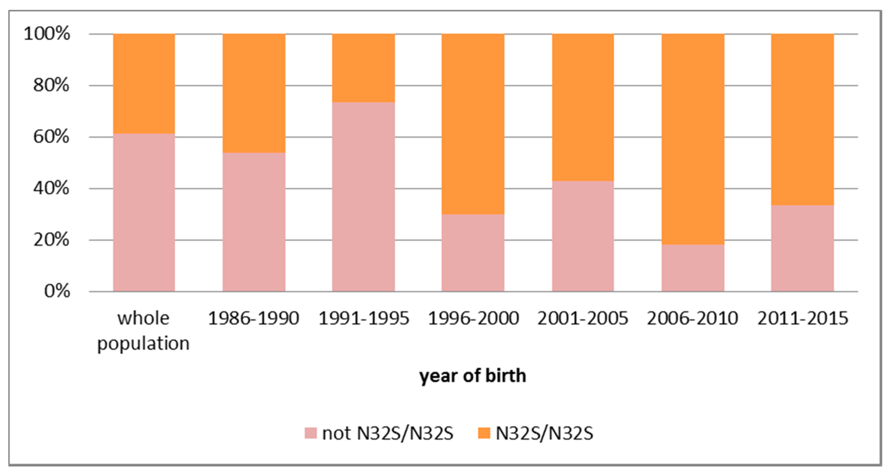
Children with somewhat higher residual OCTN2 carnitine transporter activity might escape diagnosis during neonatal screening. In the youngest age group (born 2011–2015), the apparent incidence of primary carnitine deficiency decreased to 1:512 births, but only 23% of newborns have been re-examined after two months of age and almost all identified infants had the most severe genotype (c.95A>G (p.N32S)/c.95A>G (p.N32S)) associated with lower residual carnitine transport activity (Figure 3). It is not known which level of residual OCTN2 carnitine transport activity is required to prevent severe complications such as cardiac arrhythmia and shortened life expectancy. From clinical experience, carnitine transport activities of 20% or less in fibroblasts are usually associated with very low carnitine levels [1]. For this reason, the current approach is to supplement all identified cases. Repeated attacks of hypoglycemia or sudden death from arrhythmia, even without cardiomyopathy, have been reported in patients discontinuing carnitine against medical advice [2]. For this reason, it seems prudent to continue this practice.
Reliable numbers for the true incidence of primary carnitine deficiency in the Faroe Islands will be available in some years, when hopefully all severe cases will have been identified and treated early, preventing premature deaths. This requires continued post-natal screening to identify those who have escaped diagnosis during neonatal screening and/or the addition of molecular analyses for the two mutations leading to the lowest residual activity of the OCTN2 transporter, c.95A>G (p.N32S), c.825-52G>A and the risk haplotype.

7. Summary

Primary carnitine deficiency has a very high prevalence in the Faroe Islands (1:300). This condition can be identified at birth by newborn screening, but some cases, especially the milder ones, might escape detection. In these cases, carnitine levels in a maternal carrier—even when further reduced by pregnancy—seem to be sufficient to provide enough carnitine to the affected neonate to prevent detection by early neonatal screening [39]. Regular treatment with oral carnitine, initiated early and continued for life, can result in low–normal levels of free and total carnitine levels in affected patients and prevent complications.

Acknowledgments

We would like to thank Lotte Risom and her colleagues at the Centre for Inherited Metabolic Disorders, Department of Clinical Genetics in Rigshospitalet University Hospital, Denmark for performing the genetic analyses.

Author Contributions

Ulrike Steuerwald and Nicola Longo collected the data and wrote the initial draft on the manuscript. David M. Hougaard and Nils Janzen were responsible for measuring carnitine levels. David M. Hougaard, Allan M. Lund, Jan Rasmussen and Nils Janzen participated in the review of newborn screening and clinical data on the patients and critically reviewed the manuscript.

Conflicts of Interest

Nicola Longo has received travel support for meeting attendance and honoraria from SigmaTau, Italy. The other authors declare that they do not have any conflicts of interest to be declared.

References

  1. Longo, N.; Frigeni, M.; Pasquali, M. Carnitine transport and fatty acid oxidation. Biochim. Biophys. Acta 2016, 1863, 2422–2435. [Google Scholar] [CrossRef] [PubMed]
  2. Longo, N.; Amat di San Filippo, C.; Pasquali, M. Disorders of carnitine transport and the carnitine cycle. Am. J. Med. Genet. C Semin. Med. Genet. 2006, 142C, 77–85. [Google Scholar] [CrossRef] [PubMed]
  3. El-Hattab, A.W.; Scaglia, F. Disorders of carnitine biosynthesis and transport. Mol. Genet. Metab. 2015, 116, 107–112. [Google Scholar] [CrossRef] [PubMed]
  4. El-Hattab, A.W.; Li, F.Y.; Shen, J.; Powell, B.R.; Bawle, E.V.; Adams, D.J.; Wahl, E.; Kobori, J.A.; Graham, B.; Scaglia, F.; et al. Maternal systemic primary carnitine deficiency uncovered by newborn screening: Clinical, biochemical, and molecular aspects. Genet. Med. 2010, 12, 19–24. [Google Scholar] [CrossRef] [PubMed]
  5. Lee, N.C.; Tang, N.L.; Chien, Y.H.; Chen, C.A.; Lin, S.J.; Chiu, P.C.; Huang, A.C.; Hwu, W.L. Diagnoses of newborns and mothers with carnitine uptake defects through newborn screening. Mol. Genet. Metab. 2010, 100, 46–50. [Google Scholar] [CrossRef] [PubMed]
  6. Schimmenti, L.A.; Crombez, E.A.; Schwahn, B.C.; Heese, B.A.; Wood, T.C.; Schroer, R.J.; Bentler, K.; Cederbaum, S.; Sarafoglou, K.; McCann, M.; et al. Expanded newborn screening identifies maternal primary carnitine deficiency. Mol. Genet. Metab. 2007, 90, 441–445. [Google Scholar] [CrossRef] [PubMed]
  7. De Biase, I.; Champaigne, N.L.; Schroer, R.; Pollard, L.M.; Longo, N.; Wood, T. Primary carnitine deficiency presents atypically with long qt syndrome: A case report. JIMD Rep. 2012, 2, 87–90. [Google Scholar] [PubMed]
  8. Schoderbeck, M.; Auer, B.; Legenstein, E.; Genger, H.; Sevelda, P.; Salzer, H.; Marz, R.; Lohninger, A. Pregnancy-related changes of carnitine and acylcarnitine concentrations of plasma and erythrocytes. J. Perinat. Med. 1995, 23, 477–485. [Google Scholar] [CrossRef] [PubMed]
  9. Roe, C.; Ding, J. Mitochondrial fatty acid oxidation disorders. In The Metabolic and Molecular Bases of Inherited Disease, 8th ed.; Scriver, C., Beaudet, A., Sly, W., Valle, D., Eds.; McGraw-Hill: New York, NY, USA, 2001; pp. 2297–2326. [Google Scholar]
  10. Stanley, C.A. Carnitine deficiency disorders in children. Ann. N. Y. Acad. Sci. 2004, 1033, 42–51. [Google Scholar] [CrossRef] [PubMed]
  11. Tamai, I.; Ohashi, R.; Nezu, J.; Yabuuchi, H.; Oku, A.; Shimane, M.; Sai, Y.; Tsuji, A. Molecular and functional identification of sodium ion-dependent, high affinity human carnitine transporter octn2. J. Biol. Chem. 1998, 273, 20378–20382. [Google Scholar] [CrossRef] [PubMed]
  12. Koizumi, A.; Nozaki, J.; Ohura, T.; Kayo, T.; Wada, Y.; Nezu, J.; Ohashi, R.; Tamai, I.; Shoji, Y.; Takada, G.; et al. Genetic epidemiology of the carnitine transporter octn2 gene in a japanese population and phenotypic characterization in japanese pedigrees with primary systemic carnitine deficiency. Hum. Mol. Genet. 1999, 8, 2247–2254. [Google Scholar] [CrossRef] [PubMed]
  13. Wilcken, B.; Wiley, V.; Sim, K.G.; Carpenter, K. Carnitine transporter defect diagnosed by newborn screening with electrospray tandem mass spectrometry. J. Pediatr. 2001, 138, 581–584. [Google Scholar] [CrossRef] [PubMed]
  14. Therrell, B.L., Jr.; Lloyd-Puryear, M.A.; Camp, K.M.; Mann, M.Y. Inborn errors of metabolism identified via newborn screening: Ten-year incidence data and costs of nutritional interventions for research agenda planning. Mol. Genet. Metab. 2014, 113, 14–26. [Google Scholar] [CrossRef] [PubMed]
  15. Rasmussen, J.; Kober, L.; Lund, A.M.; Nielsen, O.W. Primary carnitine deficiency in the faroe islands: Health and cardiac status in 76 adult patients diagnosed by screening. J. Inherit. Metab. Dis. 2014, 37, 223–230. [Google Scholar] [CrossRef] [PubMed]
  16. Wang, Y.; Taroni, F.; Garavaglia, B.; Longo, N. Functional analysis of mutations in the octn2 transporter causing primary carnitine deficiency: Lack of genotype-phenotype correlation. Hum. Mutat. 2000, 16, 401–407. [Google Scholar] [CrossRef]
  17. Wang, Y.; Ye, J.; Ganapathy, V.; Longo, N. Mutations in the organic cation/carnitine transporter octn2 in primary carnitine deficiency. Proc. Natl. Acad. Sci. USA 1999, 96, 2356–2360. [Google Scholar] [CrossRef] [PubMed]
  18. Rose, E.C.; di San Filippo, C.A.; Ndukwe Erlingsson, U.C.; Ardon, O.; Pasquali, M.; Longo, N. Genotype-phenotype correlation in primary carnitine deficiency. Hum. Mutat. 2012, 33, 118–123. [Google Scholar] [CrossRef] [PubMed]
  19. Wang, Y.; Korman, S.H.; Ye, J.; Gargus, J.J.; Gutman, A.; Taroni, F.; Garavaglia, B.; Longo, N. Phenotype and genotype variation in primary carnitine deficiency. Genet. Med. 2001, 3, 387–392. [Google Scholar] [CrossRef] [PubMed]
  20. Wang, Y.; Kelly, M.A.; Cowan, T.M.; Longo, N. A missense mutation in the OCTN2 gene associated with residual carnitine transport activity. Hum. Mutat. 2000, 15, 238–245. [Google Scholar] [CrossRef]
  21. Lamhonwah, A.M.; Olpin, S.E.; Pollitt, R.J.; Vianey-Saban, C.; Divry, P.; Guffon, N.; Besley, G.T.; Onizuka, R.; De Meirleir, L.J.; Cvitanovic-Sojat, L.; et al. Novel OCTN2 mutations: No genotype-phenotype correlations: Early carnitine therapy prevents cardiomyopathy. Am. J. Med. Genet. 2002, 111, 271–284. [Google Scholar] [CrossRef] [PubMed]
  22. De Sain-van der Velden, M.G.; Diekman, E.F.; Jans, J.J.; van der Ham, M.; Prinsen, B.H.; Visser, G.; Verhoeven-Duif, N.M. Differences between acylcarnitine profiles in plasma and bloodspots. Mol. Genet. Metab. 2013, 110, 116–121. [Google Scholar] [CrossRef] [PubMed]
  23. Scaglia, F.; Longo, N. Primary and secondary alterations of neonatal carnitine metabolism. Semin. Perinatol. 1999, 23, 152–161. [Google Scholar] [CrossRef]
  24. Vijay, S.; Patterson, A.; Olpin, S.; Henderson, M.J.; Clark, S.; Day, C.; Savill, G.; Walter, J.H. Carnitine transporter defect: Diagnosis in asymptomatic adult women following analysis of acylcarnitines in their newborn infants. J. Inherit. Metab. Dis. 2006, 29, 627–630. [Google Scholar] [CrossRef] [PubMed]
  25. Scaglia, F.; Wang, Y.; Singh, R.H.; Dembure, P.P.; Pasquali, M.; Fernhoff, P.M.; Longo, N. Defective urinary carnitine transport in heterozygotes for primary carnitine deficiency. Genet. Med. 1998, 1, 34–39. [Google Scholar] [CrossRef] [PubMed]
  26. Stanley, C.A. Carnitine disorders. Adv. Pediatr. 1995, 42, 209–242. [Google Scholar] [PubMed]
  27. Pochini, L.; Scalise, M.; Galluccio, M.; Indiveri, C. OCTN cation transporters in health and disease: Role as drug targets and assay development. J. Biomol. Screen. 2013, 18, 851–867. [Google Scholar] [CrossRef] [PubMed]
  28. Hirano, T.; Yasuda, S.; Osaka, Y.; Kobayashi, M.; Itagaki, S.; Iseki, K. Mechanism of the inhibitory effect of zwitterionic drugs (levofloxacin and grepafloxacin) on carnitine transporter (OCTN2) in caco-2 cells. Biochim. Biophys. Acta 2006, 1758, 1743–1750. [Google Scholar] [CrossRef] [PubMed]
  29. Hu, C.; Lancaster, C.S.; Zuo, Z.; Hu, S.; Chen, Z.; Rubnitz, J.E.; Baker, S.D.; Sparreboom, A. Inhibition of OCTN2-mediated transport of carnitine by etoposide. Mol. Cancer Ther. 2012, 11, 921–929. [Google Scholar] [CrossRef] [PubMed]
  30. Pochini, L.; Scalise, M.; Indiveri, C. Inactivation by omeprazole of the carnitine transporter (OCTN2) reconstituted in liposomes. Chem. Biol. Interact. 2009, 179, 394–401. [Google Scholar] [CrossRef] [PubMed]
  31. Rasmussen, J.; Nielsen, O.W.; Lund, A.M.; Kober, L.; Djurhuus, H. Primary carnitine deficiency and pivalic acid exposure causing encephalopathy and fatal cardiac events. J. Inherit. Metab. Dis. 2013, 36, 35–41. [Google Scholar] [CrossRef] [PubMed]
  32. Lund, A.M.; Hougaard, D.M.; Simonsen, H.; Andresen, B.S.; Christensen, M.; Duno, M.; Skogstrand, K.; Olsen, R.K.; Jensen, U.G.; Cohen, A.; et al. Biochemical screening of 504,049 newborns in denmark, the faroe islands and greenland--experience and development of a routine program for expanded newborn screening. Mol. Genet. Metab. 2012, 107, 281–293. [Google Scholar] [CrossRef] [PubMed]
  33. Lund, A.M.; Joensen, F.; Hougaard, D.M.; Jensen, L.K.; Christensen, E.; Christensen, M.; Norgaard-Petersen, B.; Schwartz, M.; Skovby, F. Carnitine transporter and holocarboxylase synthetase deficiencies in the faroe islands. J. Inherit. Metab. Dis. 2007, 30, 341–349. [Google Scholar] [CrossRef] [PubMed]
  34. Rasmussen, J.; Nielsen, O.W.; Janzen, N.; Duno, M.; Gislason, H.; Kober, L.; Steuerwald, U.; Lund, A.M. Carnitine levels in 26,462 individuals from the nationwide screening program for primary carnitine deficiency in the faroe islands. J. Inherit. Metab. Dis. 2014, 37, 215–222. [Google Scholar] [CrossRef] [PubMed]
  35. McGoey, R.R.; Marble, M. Positive newborn screen in a normal infant of a mother with asymptomatic very long-chain acyl-coa dehydrogenase deficiency. J. Pediatr. 2011, 158, 1031–1032. [Google Scholar] [CrossRef] [PubMed]
  36. Rasmussen, J.; Hougaard, D.M.; Sandhu, M.; Fjaellergaard, K.; Petersen, P.R.; Steuerwald, U.; Lund, A.M. Primary carnitine deficiency: Is fetal development affected and can newborn screening be improved? J. Inherit. Metab. Dis. 2017. [Google Scholar] [CrossRef]
  37. Rasmussen, J.; Lund, A.M.; Risom, L.; Wibrand, F.; Gislason, H.; Nielsen, O.W.; Kober, L.; Duno, M. Residual OCTN2 transporter activity, carnitine levels and symptoms correlate in patients with primary carnitine deficiency. Mol. Genet. Metab. Rep. 2014, 1, 241–248. [Google Scholar] [CrossRef] [PubMed]
  38. SLC22A5 gene. Available online: http://exac.broadinstitute.org/gene/ENSG00000197375 (accessed on 4 February 2017).
  39. De Boer, L.; Kluijtmans, L.A.; Morava, E. Primary carnitine (OCTN2) deficiency without neonatal carnitine deficiency. JIMD Rep. 2013, 10, 39–40. [Google Scholar] [PubMed]
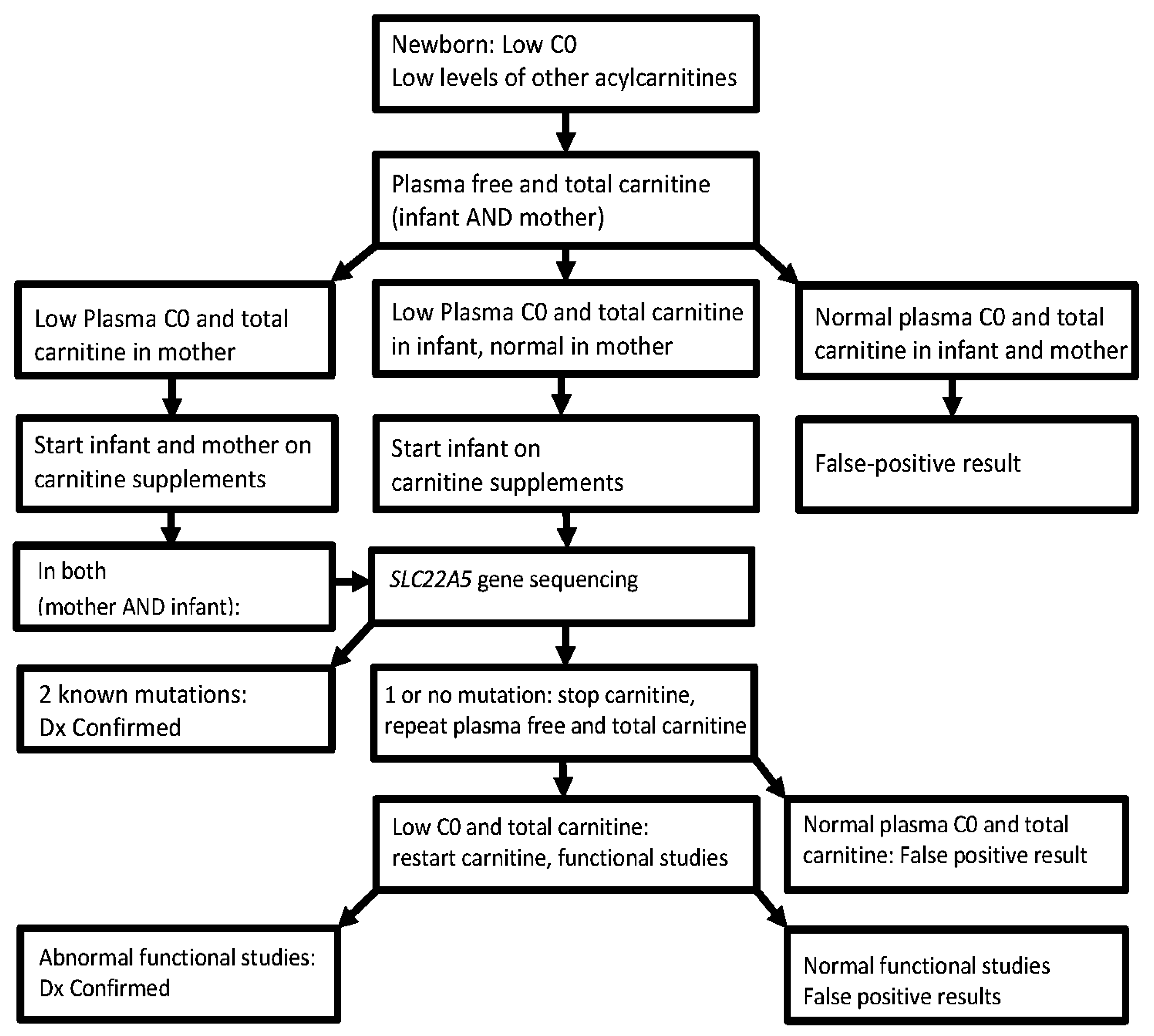
Figure 1. Diagnostic algorithm for the confirmation of a diagnosis of primary carnitine deficiency identified by low levels of free (C0) carnitine in the newborn screening blood spot.
Neonatalscreening 03 00001 g001
Figure 2. Subcellular distribution of normal and mutant OCTN2 carnitine transporters. The wild-type (A); and N32S-mutant (B) OCTN2 transporters were tagged with the green fluorescent protein and stably expressed in CHO cells. Cells were labeled in vivo with Bodipy-ceramide to visualize the Golgi. Phase contrast microscopy was used to define the cell borders and intracellular structures. Images were digitally overlaid to show the location of green transporters compared to the red Golgi and plasma membrane. Co-localization of membrane transporters with the Golgi is seen as a yellow signal. Note that both transporters localize normally to the plasma membrane. (C) Carnitine transport in CHO cells expressing normal and mutant carnitine transporters. CHO cells were transfected with normal and mutant OCTN2 cDNAs and selected for resistance to G418 (0.8 mg/mL). Carnitine (0.5 μM) transport was measured for 1 h in stably transfected cells and corrected for nonsaturable uptake (measured in the presence of 2 mM cold carnitine). Points are averages ± SE of three to five independent experiments (each in triplicate).
Neonatalscreening 03 00001 g002
Figure 3. Fraction of severe (c.95A>G (p.N32S)/c.95A>G (p.N32S)) versus other genotypes in the SLC22A5 gene encoding the OCTN2 carnitine transporter identified in different age groups.
Neonatalscreening 03 00001 g003
Table 1. Identification of primary carnitine deficiency by neonatal and by post-neonatal screening in the Faroe Islands by birth cohort.
Year of Birth1999–20002001–20052006–20102011–2015Overall
Cases detected by NBS [incidence of PCD]473 (+3) *4 (+2) *23
[1:330][1:496][1:543][1:512][1:483]
Additional cases (negative NBS) detected with population screening074 (+1) **012
Total incidence of PCD1:3301:2481:2961:5121:318
* Number in parenthesis indicates additional cases identified and treated at birth/prenatally by supplementing the mother with carnitine because of a positive family history; ** Number in parenthesis refers to a case in which the neonatal sample was not obtained.
Table 2. Primary carnitine deficiency in the Faroe Islands: prevalence found by population screening.
Year of Birth1901–20151986–19901991–19951996–20002001–20052006–20102011–2015
Individuals49,949432237483283347032603071
Screened at >2 months (%)33,33329542769280626932038713
66.7%68.3%73.9%85.5%77.6%62.5%23.2%
No. of PCD-cases16812151014116
Prevalence1:2971:3601:2501:3281:2481:2961:512
Table 3. Identification of primary carnitine deficiency by neonatal screening in the Faroe Islands (1999–2015).
GenotypeNumberPositive in Neonatal (Percent Identified)
c.95A>G, p.N32S/c.95A>G, p.N32S *20 **14 (70%)
c.95A>G, p.N32S/c.825-52G>A (splice)21 (50%)
c.95A>G, p.N32S/RH (risk haplotype)62 (33%)
Others21 (50%)
All cases30 *18 (60%)
* Five children with a positive family history were treated since birth and excluded from this table; ** Two cases had clearly normal carnitine levels in the neonatal sample. Three cases would have been detected applying the new, presently used algorithm. One child was not screened during the neonatal period.