Next Article in Journal
Acetaminophen, a Therapeutic or an Extremely Toxic Remedy—A Review
Previous Article in Journal
Modern Interpretation of Risk Factors in Breast Cancer of Women
 
 
Journal of Mind and Medical Sciences is published by MDPI from Volume 12 Issue 1 (2025). Previous articles were published by another publisher in Open Access under a CC-BY (or CC-BY-NC-ND) licence, and they are hosted by MDPI on mdpi.com as a courtesy and upon agreement with Valparaiso University (ValpoScholar).
Review

The Benign Mimickers of Carcinoma on Breast MRI

by
Irmak Durur-Subasi
1,2,*,
Adem Karaman
2,
Elif Demirci
3,
Sare Sipal
3 and
Mufide Nuran Akcay
4
1
Department of Radiology, International Faculty of Medicine, Istanbul Medipol University, Istanbul, Turkey
2
Department of Radiology, School of Medicine, Ataturk University, Erzurum, Turkey
3
Department of Pathology, School of Medicine, Ataturk University, Erzurum, Turkey
4
Department of General Surgery, School of Medicine, Ataturk University, Erzurum, Turkey
*
Author to whom correspondence should be addressed.
J. Mind Med. Sci. 2022, 9(1), 96-101; https://doi.org/10.22543/7674.91.P96101
Submission received: 8 December 2021 / Revised: 9 December 2021 / Accepted: 14 January 2022 / Published: 10 April 2022

Abstract

:
The similarity between benign and malignant pathologies on magnetic resonance imaging (MRI) and a wide-ranging variability of the lesions from benign proliferative changes to invasive breast carcinoma cause a lower and wide-ranging specificity of breast MRI relative to its surpass sensitivity. A wide range of tissue components such as the skin, the adipose tissue, vascular and neural tissues, connective tissues, glandular tissues, ducts, and muscle tissues are found here all together. This pictorial review was aimed at deliberating benign mimickers of breast carcinoma on MRI and trying to call attention to the overlapping and distinctive features.

Introduction

Magnetic resonance imaging (MRI) reaches a very high sensitivity in the detection of invasive breast cancer due to the typical appearance of malignant tumors, contrast kinetics and improved anatomic detail of the MRI with its high spatial resolution and the contrast-to-noise ratio. Contrary to the high sensitivity of MRI, the specificity is considered to be lower depending on several influences. One of them is the similarity of imaging findings between benign and malignant breast lesions. The wide variety of the lesions can contribute to this issue [1,2].
This pictorial review focuses on the nearness of benign breast pathologies to primary breast carcinoma in terms of MRI findings. Many of the lesions can be recognized only histologically.

Discussion

Adenomyoepithelioma

Adenomyoepithelioma arises from both myoepithelial and epithelial cells of the breast lobules and ducts. Most cases of the tumors are of benign origin. Benign adenomyoepitheliomas can display all suspicious features of breast carcinoma such as spiculated margins, a round shape, a fast enhancement, and a plateau kinetic pattern on the MRI [1] (Figure 1). The precise diagnosis can be made by biopsy only. The mitosis number by histopathology defines the exact malignant or benign characteristics of the lesion.

Amyloidosis

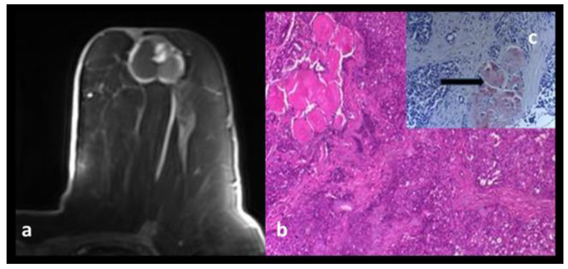
Amyloidosis of the breast is the accumulation of serum amyloid and it can be primary or secondary. Primary amyloidosis is idiopathic and the secondary one is triggered by a primary inflammatory pathology. The last one is the most seen form. Amyloidosis may be clinically a subtle or a stiff mass, along with extensive painfulness, and a peau d'orange look could also be seen. On the MRI, the tubular lesions, low T1, and high T2-short tau inversion recovery (STIR) signal, no enhancement, some weak marginal late enhancement can all be encountered [2]. The bizarre appearances can be appreciated as suspicious findings and the diagnosis is made by means of the histopathologic examination (Figure 2).

Angiolipoma

The breast angiolipoma is a rare lesion and contains mature adipose tissue and capillary vasculatures [3]. The peripheral enhancement and the rapid washout pattern due to peripherally located capillary vasculatures can cause suspicious appearances [3]. Lesions can be differentiated from breast carcinomas and angiosarcomas by means of histopathology only.

Apocrine Metaplasia

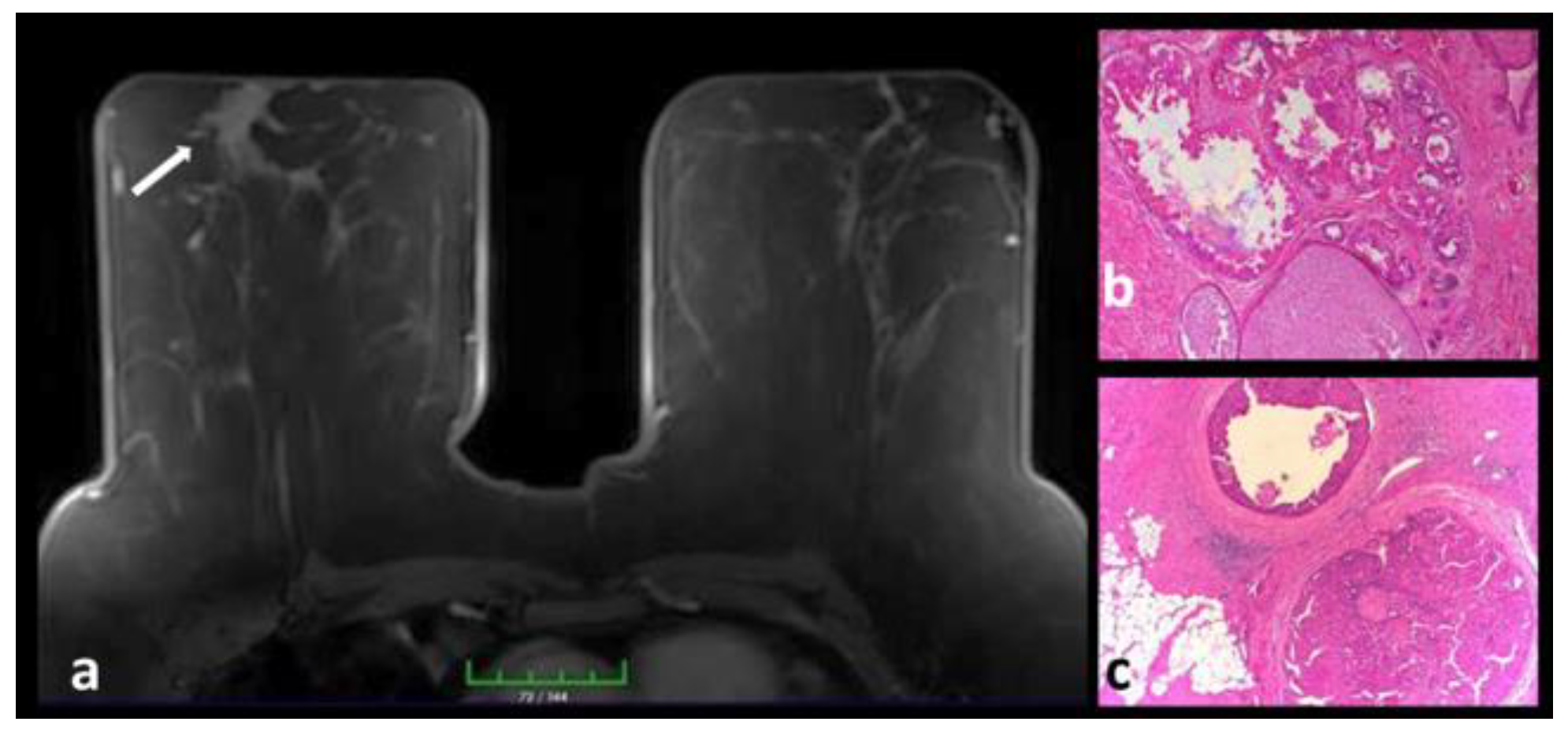
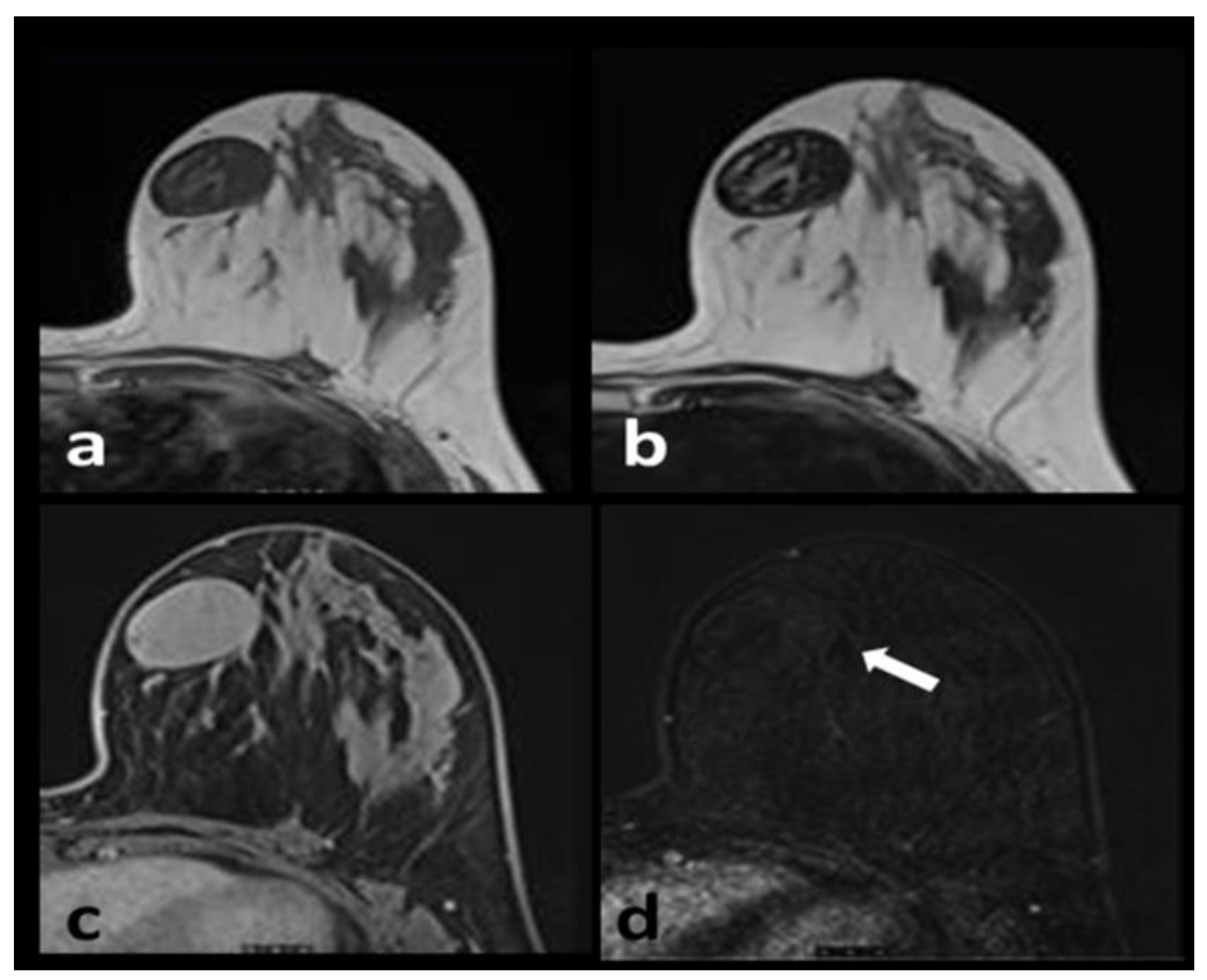
Apocrine metaplasia is composed of distended epithelial cells with abundant pink cytoplasm and round uniform nuclei with noticeable nucleoli. It can be realized in benign and malignant breast pathologies and it is a common histopathological finding in MRI-detected breast lesions [4]. Apocrine metaplasia improves the contrast enrichment, T2 brightness, and diffusion restriction [4] (Figure 3). Apocrine metaplasia frequently exists in the epithelial covering of cysts in gross cystic disease and it may be linked with painfulness and irregular nodularity that vary during the menstrual cycle.

Breast Abscesses

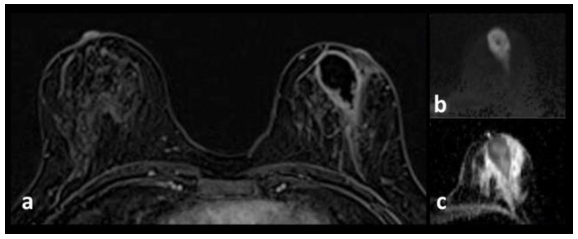
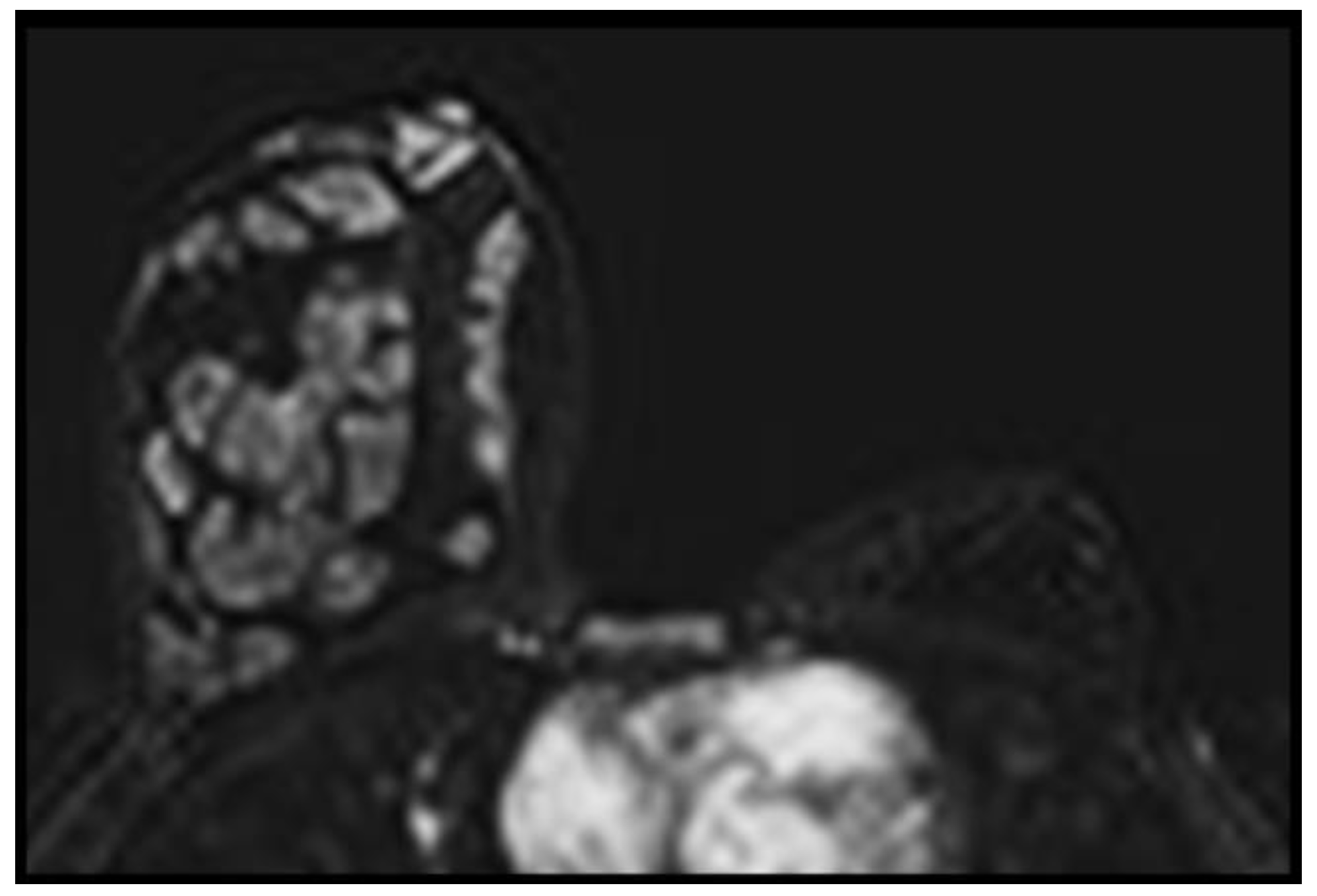
Breast abscesses are complications of the infectious or inflammatory processes. Its irregular morphology, peripheral and intense enhancement pattern, suspicious plateau or wash-out kinetics, diffusion characteristics may lead to misinterpretation. It is vital to evaluate MRIs from this perspective. Necrotic debris and viscosity within the abscess cavity, fibroblastic inflammatory proliferation and inflammatory cell migration within the abscess wall can show diffusion restriction [5] (Figure 4). The clinical scenario, the treatment response, and the histopathologic examination will help make the final diagnosis.

Diabetic Mastopathy

This tumor-like proliferation of fibrous tissue is an occasional condition and it mostly affects patients with Type I diabetes. On imaging studies, it may simulate carcinoma by parenchymal distortion and spiculated boundaries [6]. No enhancement, slight heterogeneous enhancement, patchy or diffuse homogeneous enhancement, a gradual kinetic pattern could also be encountered.

Fat Necrosis

Fat necrosis can mimic malignancy on MRI with irregular borders, parenchymal distortion, peripheral enhancement, and wash-out kinetic patterns. A relevant clinical history, the central fatty portion, the “black hole” finding can be considered an accurate diagnosis. However, typical features not always come across and biopsy may be necessary.

Fibroadenoma and Fibroadenoma-related Conditions

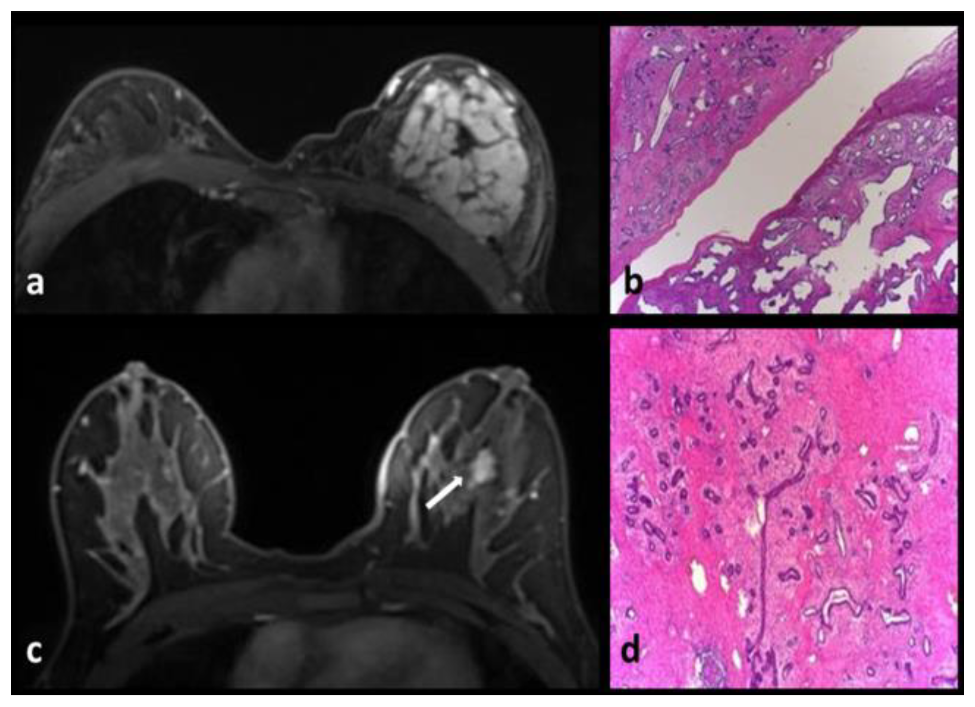
Fibroadenoma is a mostly detected benign lesion after fibrocystic changes. The myxoid fibroadenoma is generally a circumscribed, T2-hyperintense mass with un-enhancing septations (Figure 5). The enhancement pattern may be fast and robust and generally progressive [7]. The suspicious plateau or washout enhancement patterns can be faced as well. The centrifugal progression of enhancement from the center to the periphery may be misunderstood as an irregular mass in the early phases [7,8]. The degree of fibrosis in fibroadenomas can affect the amount of enhancement. Fibrosis can cause a weak or partial enhancement (Figure 6). This type of fibroadenomas may be more prominent on conventional sequences, can show bizarre appearances on contrast-enhanced images, and requires synchronized evaluation of both conventional sequences and contrast-enhanced images.
Fibroadenomas with infarction or mastopathy may cause suspicious imaging findings and may require biopsy (Figure 7).

Fibrocystic Changes

The fibrocystic breast has a wide-ranging spectrum of morphology and kinetics on the MRI, with a mass lesion or a non-mass like regional enhancing lesion or a focus. Regular, lobulated, or spiculated margins, suspicious kinetics with wash-out or plateau patterns, heterogeneous and homogeneous enhancement can all be seen and biopsy may be required [9].

Fibromatosis

Fibromatosis of the breast is a benign fibroblastic disease with no metastatic possibility, but a high propensity to relapse. The mass is isointense to muscle on T1-weighed images and heterogeneously hypo-iso-hyperintense on T2. The bright areas can be correlated with the prominent myxoid change, and the low-intensity area is correlated with condensed collagenous matter. The kinetic pattern has a generally benign progression [10,11]. The lesions are typically irregular.

Juvenile Papillomatosis

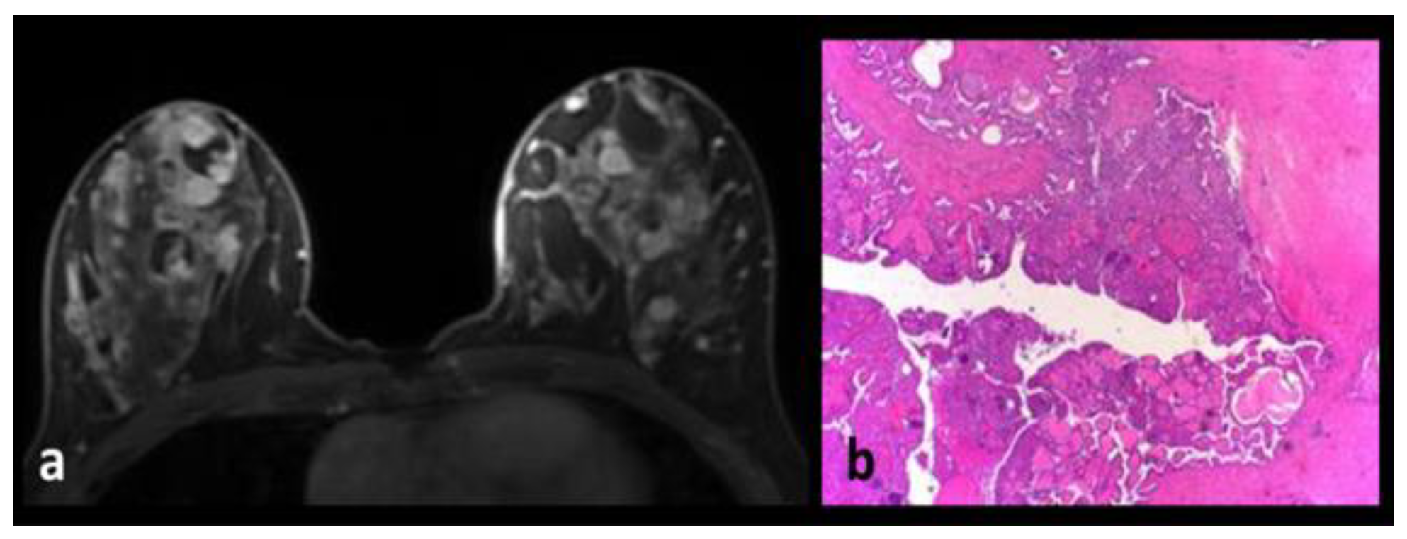
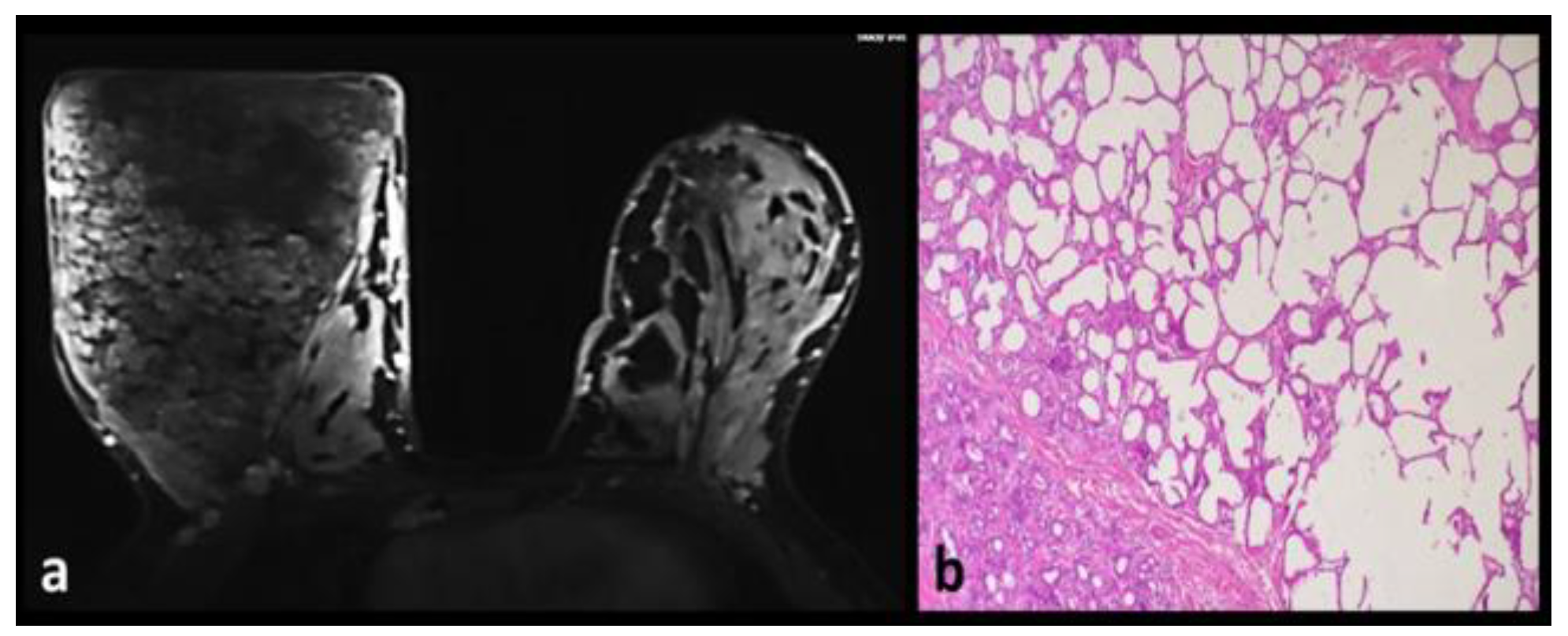
Juvenile papillomatosis (Swiss cheese disease) is an occasional, benign, and proliferative disease. It has been reported to be associated with carcinoma in up to 15% of the cases. The MRI findings of juvenile papillomatosis are multiple, bilateral, well-bordered complex cystic breast masses, cystic ductal dilatations with thick, nodular ductal walls, and clusters of solid lesions (Figure 8). The dynamic evaluation shows continuous and plateau patterns [12].

Idiopathic Granulomatous Mastitis

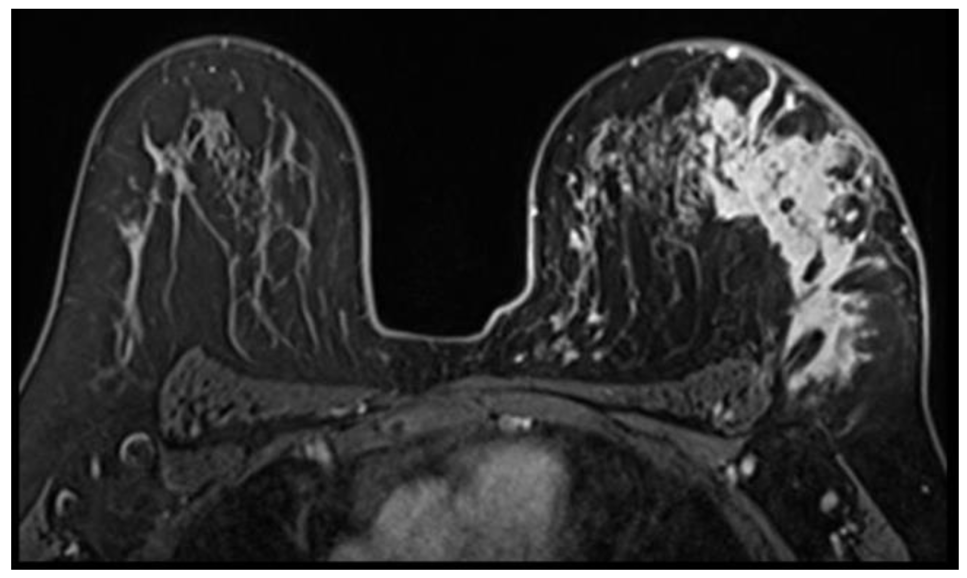
It can be seen as a mass lesion or a non-mass-like enhancement on MRI. Irregular and spiculated contours, homogenous-heterogeneous-rim enhancement can all be seen [13,14] (Figure 9). Lesions can display both non-suspicious and suspicious kinetic patterns. Diffusion restriction is seen.
The MRI will reveal skin edema, skin abscess, fistula tracts, parenchymal edema, necrosis-abscess, dilated-enhanced canals with intense contents, and lymphadenopathy. Advanced MRI findings have been described for idiopathic granulomatous mastitis. The T1 perfusions on the MRI findings of the disease are similar to those of malignant lesions [15].

Intraductal Papilloma-Intraductal Papillomatosis

Papillary breast lesions comprise a heterogeneous group and consists of a fibrovascular center, myoepithelium layer and outer cuboidal or columnar epithelium. Papillary lesions are classified as papillomas (solitary intraductal papilloma, multiple papillomas, papillomatosis, and juvenile papillomatosis), sclerosing papillary lesions, intraductal papillary carcinomas and invasive papillary carcinomas.
Solitary intraductal papillomas are usually located in the retroareolar region. On the MRI, an enhancing nodule, uniform enhancement, irregular morphology, washout, or plateau kinetics can be encountered. Intraductal papillomatosis or multiple intraductal papillomas arise from the terminal ductal lobular units and hence are generally peripherally located in the breast. They are usually associated with atypia, preinvasive, or invasive carcinoma. In addition to the suspicious findings of solitary papilloma, intraductal papillomatosis can show complex cystic morphology, non-mass like enhancement, and ductal wall enhancement patterns.

Intramammary Lymph Node

Intramammary lymph nodes generally have a wash-out kinetic pattern and diffusion restriction. Therefore, they must be differentiated from malignant nodules. Additionally, they are potential sites of locoregional spread for ipsilateral breast carcinoma [16].

Lactating Adenoma

Lactating adenomas are rare tumors and they usually exist as breast lumps in pregnancy or lactation. Morphologically, they are identical to fibroadenomas. However, they may contain milk-filled branching ducts. Histologically, cells with a vacuolated cytoplasm surrounding the secretory lobules of lactating adenoma or milk cause T1-hyperintensity. The lactating adenoma may look bright on T1, T2, and STIR. Secretory lobules show rigorous enhancement like lactational changes and may show a wash-out pattern, possibly due to augmented vascular properties [16] (Figure 10).

Phyllodes tumors

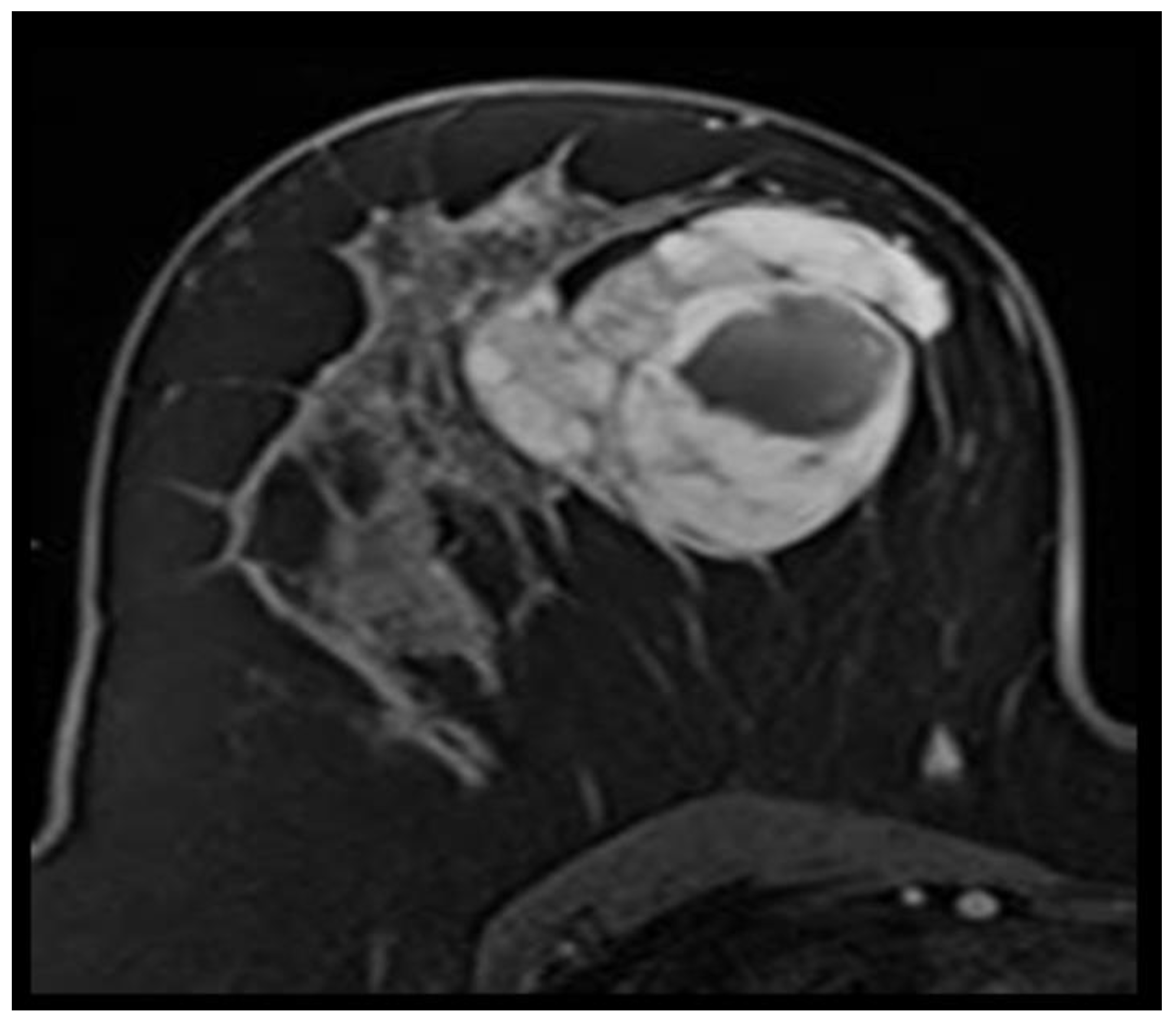
Generally, a Phyllodes tumor has analogous imaging characteristics to a fibroadenoma. A tumor’s size ≥3 cm, irregular configuration, micro-lobulated boundaries, intense enhancement, and necrosis on the MRI were suggestive of phyllodes tumors [17] (Figure 11). A histopathological analysis should be performed.

Pseudoangiomatous stromal hyperplasia

Pseudoangiomatous stromal hyperplasia (PASH) is a benign disease of the breast showing a complex web of slit-like spaces lined by slender spindle cells in stromal hyperplasia. A focal or segmental clumped enhancement, and irregular masses can be encountered [18].

Unilateral Lactation

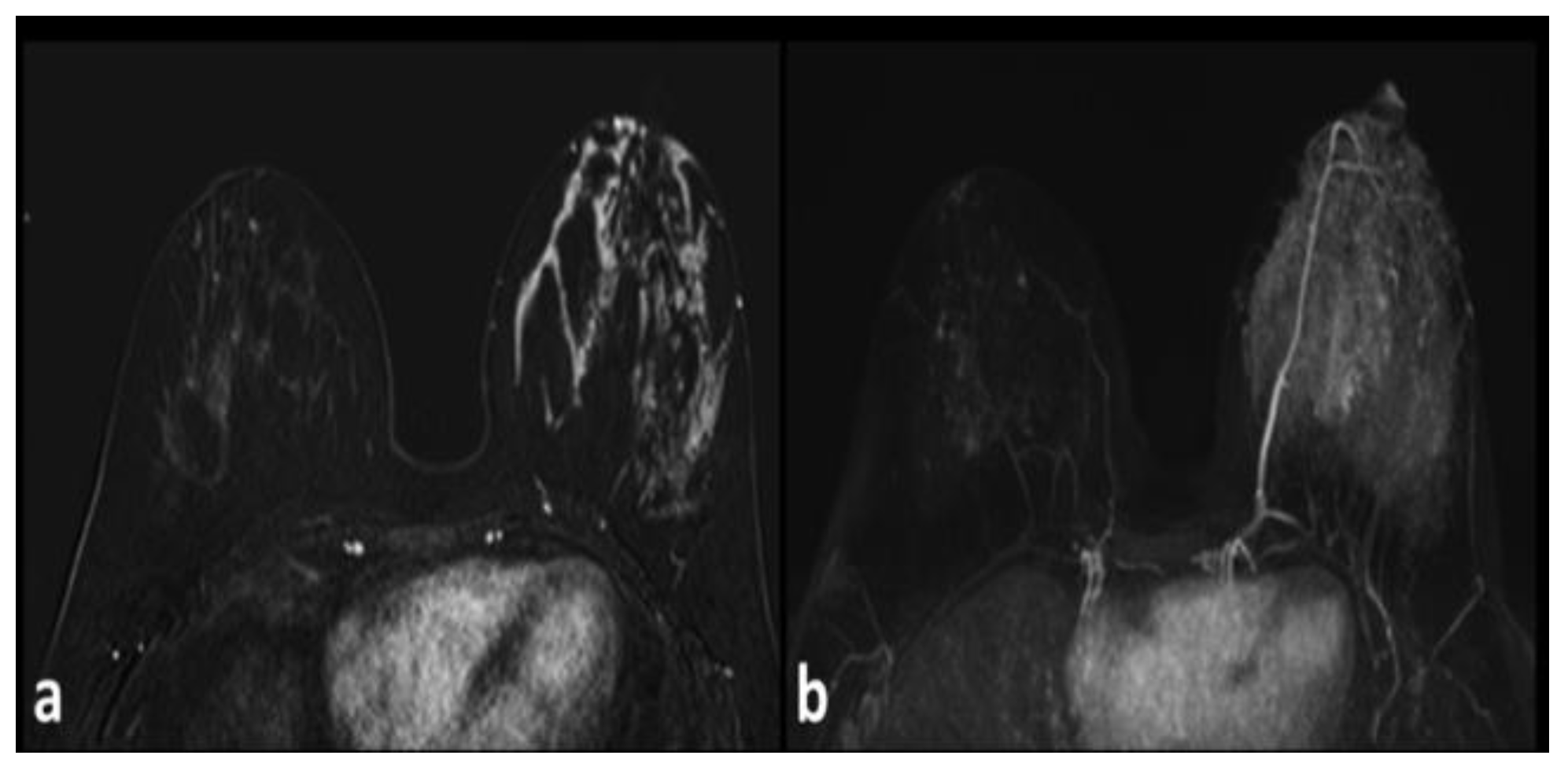
The lactational breast changes are the increase in glandular tissue size, diffuse T2 signal rise, fast early enhancement, an early plateau (Figure 12). MRI findings may be confused with infection, inflammation, or inflammatory carcinoma [19].

Sclerosing Adenosis

Sclerosing adenosis is a benign proliferative condition of the terminal duct lobular units characterized by an increased number of acini and their glands. A wash-out kinetic pattern has been reported [20].

Surgery and Radiotherapy

Post-radiation changes of edematous changes at the breast skin and parenchyma have been described. A postsurgical seroma also shrinks and architectural distortion and fibrosis remain. Stability in these findings usually occurs within 3 years after the treatment [21,22].

Conclusions

As a result, the breast can host many benign, borderline or malignant lesions. These lesions may mimic one another, perhaps due to their origin or their similar natures, or our imaging technology. Today, in this environment where an increase in the incidence of breast cancer is observed, it seems important to carefully evaluate the MRI, an advanced imaging that is able to refer patients to biopsy when necessary.

Conflict of interest disclosure

There are no known conflicts of interest in the publication of this article. The manuscript was read and approved by all authors.

Compliance with ethical standards

Any aspect of the work covered in this manuscript has been conducted with the ethical approval of all relevant bodies and that such approvals are acknowledged within the manuscript. Ethics approval and consent to participate: 03.07.2014-12.

Acknowledgments

The education of a physician continues throughout lifetime. I have to thank my professors, assistants, and patients who have trained me on this path. In these COVID-19 days, everything is for life, everything is for humanity. Dr. IDS

Author contribution statement

IDS, AK, ED, SS, MNA drafted and revised the manuscript, provided exemplar cases and prepared the figures, read and approved the final manuscript.

References

  1. Ruiz-Delgado, M.L.; López-Ruiz, J.A.; Eizaguirre, B.; Saiz, A.; Astigarraga, E.; Fernández-Temprano, Z. Benign adenomyoepithelioma of the breast: imaging findings mimicking malignancy and histopathological features. Acta Radiol. 2007, 48, 27–29. [Google Scholar] [CrossRef] [PubMed]
  2. O'Brien, J.; Aherne, S.; McCormack, O.; Jeffers, M.; McInerney, D. MRI features of bilateral amyloidosis of breast. Breast J. 2013, 19, 338–339. [Google Scholar] [CrossRef] [PubMed]
  3. Killian, J.K.; Merino, M.; Bondy, C.; Bakalov, V.; Chow, C. MRI of angiolipoma of the breast in Turner's syndrome. AJR Am J Roentgenol. 2004, 183, 1843–1844. [Google Scholar] [CrossRef]
  4. Gao, Y.; Dialani, V.; DeBenedectis, C.; Johnson, N.; Brachtel, E.; Slanetz, P. Apocrine Metaplasia Found at MR Biopsy: Is There Something to be Learned? Breast J. 2017, 23, 429–435. [Google Scholar] [CrossRef] [PubMed]
  5. Durur-Subasi, I.; Durur-Karakaya, A.; Karaman, A.; Seker, M.; Demirci, E.; Alper, F. Is the necrosis/wall ADC ratio useful for the differentiation of benign and malignant breast lesions? Br J Radiol. 2017, 90, 20160803. [Google Scholar] [CrossRef]
  6. Accurso, A.; Della Corte, G.A.; Rocco, N.; Varone, V.; Buonaiuto, R.; Compagna, R.; Tari, D.U.; Amato, B.; Riccardi, A. Unusual breast lesion mimicking cancer: diabetic mastopathy. Int J Surg. 2014, 12 (Suppl 1), S79–82. [Google Scholar] [CrossRef]
  7. Kuhl, C.K. Concepts for differential diagnosis in breast MR imaging. Magn Reson Imaging Clin N Am. 2006, 14, 305–328. [Google Scholar] [CrossRef]
  8. Lazar, A.L.; Vulturar, R.; Fodor, A.; et al. The molecular mechanisms linking metabolic syndrome to endometrial and breast cancers. J Mind Med Sci. 2021, 8, 167–178. [Google Scholar] [CrossRef]
  9. Chen, J.H.; Nalcioglu, O.; Su, M.Y. Fibrocystic change of the breast presenting as a focal lesion mimicking breast cancer in MR imaging. J Magn Reson Imaging. 2008, 28, 1499–1505. [Google Scholar] [CrossRef]
  10. Xu, Y.; Liu, P.; Lu, H.; Zhang, S.; Zhu, Y. Imaging manifestation of mammary fibromatosis. Breast J. 2013, 19, 673–675. [Google Scholar] [CrossRef]
  11. Yalagachin, G.; Lakshmikantha, N.; Mashal, S.B.; Yalagachin, G.; Lakshmikantha, N.; Mashal, S.B. Prevalence of nodular goiter in patients with breast diseases. J Clin Invest Surg. 2020, 5, 91–95. [Google Scholar] [CrossRef]
  12. Durur-Subasi, I.; Alper, F.; Akcay, M.N.; Demirci, E.; Gundogdu, C. Magnetic resonance imaging findings of breast juvenile papillomatosis. Jpn J Radiol. 2013, 31, 419–423. [Google Scholar] [CrossRef] [PubMed]
  13. Durur-Subasi, I. DW-MRI of the breast: a pictorial review. Insights Imaging. 2019, 10, 61. [Google Scholar] [CrossRef] [PubMed]
  14. van Oers, H.; Schlebusch, L. Indicators of psychological distress and body image disorders in female patients with breast cancer. J Mind Med Sci. 2020, 7, 179–187. [Google Scholar] [CrossRef]
  15. Ucar, E.A.; Durur-Subasi, I.; Yilmaz, K.B.; Arikok, A.T.; Hekimoglu, B. Quantitative perfusion parameters of benign inflammatory breast pathologies: A descriptive study. Clin Imaging. 2020, 68, 249–256. [Google Scholar] [CrossRef]
  16. Durur-Subasi, I.; Durur-Karakaya, A.; Alper, F.; Karaman, A.; Kılıc, R.M.; Sipal, S.; Demirci, E.; Akcay, M.N. Breast lesions with high signal intensity on T1-weighted MR images. Jpn J Radiol. 2013, 31, 653–661. [Google Scholar] [CrossRef]
  17. Duman, L.; Gezer, N.S.; Balcı, P.; Altay, C.; Başara, I.; Durak, M.G.; Sevinç, A.I. Differentiation between Phyllodes Tumors and Fibroadenomas Based on Mammographic Sonographic and MRI Features. Breast Care (Basel). 2016, 11, 123–127. [Google Scholar] [CrossRef]
  18. Jones, K.N.; Glazebrook, K.N.; Reynolds, C. Pseudoangiomatous stromal hyperplasia: imaging findings with pathologic and clinical correlation. AJR Am J Roentgenol. 2010, 195, 1036–1042. [Google Scholar] [CrossRef]
  19. Marinescu, S.A.; Bejinariu, C.G.; Şapte, E.; Marinaş, M.C.; Giuglea, C. Complications related to breast reconstruction after mastectomy using multiple surgical techniques - a national and international comparative analysis. Rom J Morphol Embryol. 2019, 60, 87–93. [Google Scholar]
  20. Motofei, I.G. Biology of cancer; from cellular and molecular mechanisms to developmental processes and adaptation. Semin Cancer Biol 2021, S1044-579X(21)00253-4. [Google Scholar] [CrossRef]
  21. Andrei, C.A.; Scăunașu, R.V.; Simionescu, A.A.; Burcoș, T.; Lupușoru, M.D.; Lica, G. The incidence of haemorrhagic and thrombo-embolic events after breast cancer surgery in patients treated with pharmacological thrombo-prophylaxis. J Clin Invest Surg. 2019, 4, 10–18. [Google Scholar] [CrossRef]
  22. Miricescu, D.; Diaconu, C.C.; Stefani, C.; Stanescu, A.M.A.; Totan, A.; Rusu, I.R.; et al. The Serine/Threonine Protein Kinase (Akt)/ Protein Kinase B (PkB) Signaling Pathway in Breast Cancer. J Mind Med Sci. 2020, 7, 34–39. [Google Scholar] [CrossRef]
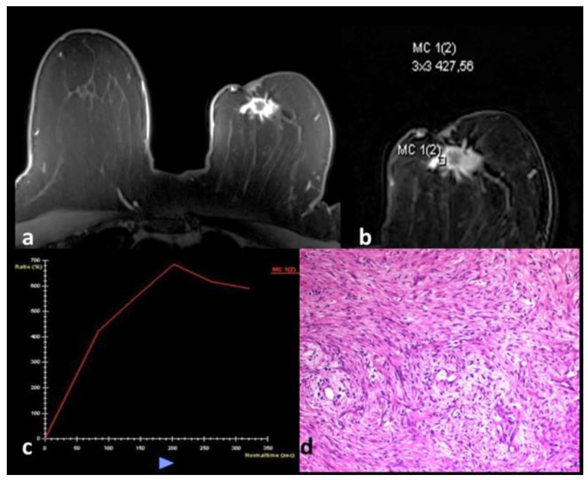
Figure 1. (a-d). A 55-year-old female patient with adenomyoepithelioma. A spiculated subareolar mass at the left breast (a). It causes nipple retraction and parenchymal distortion and contains central necrosis. The kinetic analysis shows the wash-out pattern (b, c). Although these findings are compatible with the BI-RADS category 5 lesion, the histopathological examination shows the benign spindle cell type adenomyoepithelioma (d).
Jmms 09 00008 g001
Figure 2. (a-c). A 48-year-old female patient with invasive papillary carcinoma. Post-contrast T1 weighed image shows a complex cystic mass at the subareolar region of the left breast (a). An enhancing nodule is seen at the anterolateral part of the tumors and the lateral component has an intense content. The wall of the tumor is also enhancing. Histopathology shows invasive papillary carcinoma and amyloid accumulation in the tumor (b). The arrow shows a Congo red positive area (A-HEX200 and B-Congo-RedX200) (c).
Jmms 09 00008 g002
Figure 3. (a-c). A 33-year-old female patient with apocrine metaplasia and atypical ductal hyperplasia. A palpable abnormality and no suspicious findings on the ultrasonography lead to an advanced evaluation by means of MRI. An irregular, minimally enhancing, superficially located, asymmetrical glandular intensity (arrow) at the outer part of the right breast has been detected (a). The histopathological analysis shows apocrine metaplasia and atypical ductal hyperplasia (b, c).
Jmms 09 00008 g003
Figure 4. (a-c). Breast abscess on the left side. Peripheral enhancement, irregular internal borders, central necrosis (a), and diffusion restriction are seen (b, c).
Jmms 09 00008 g004
Figure 5. (a-d). A 21-year-old woman with a giant fibroadenoma Post-contrast T1-weighed image shows a circumscribed, intensely enhancing mass with non-enhancing septations (a) The histopathologic examination reveals fibroadenoma having a benign phyllodes component (H-E x100) (b) A 28-year-old patient with a left central irregular mass (arrow) (c) and histopathologic analysis reveal benign fibroadenoma (H-E x100) (d).
Jmms 09 00008 g005
Figure 6. (a-d). A 43-year-old female patient with fibroadenoma. The mass is located at the inner part of the left breast, heterogeneously hypointense on both T1 (a) and T2 (b), which shows vague enhancement (c, d).
Jmms 09 00008 g006
Figure 7. An 18-year-old female patient with huge fibroadenoma. The mass fully occupies and enlarges the right breast. The normally appearing left breast is small in size.
Jmms 09 00008 g007
Figure 8. (a, b). A 26-year-old patient with juvenile papillomatosis. Complex cystic lesions and multiple nodules are seen (a). The histopathologic examination confirms the diagnosis (H-E x100) (b).
Jmms 09 00008 g008
Figure 9. A 38-year-old woman with idiopathic granulomatous mastitis. A swollen left breast. Regional intense enhancement is seen in the outer part. Subtle skin alterations are seen.
Jmms 09 00008 g009
Figure 10. (a, b). A 36-year-old with lactating adenoma. A huge mass enlarged during pregnancy. The mass fills and enlarges the right breast. After delivery, the surgical excision and the histopathology of the lesion are consistent with the diagnosis.
Jmms 09 00008 g010
Figure 11. A 45-year-old female patient with a phyllodes tumor. The huge mass shows macrolobulated contours and central necrosis.
Jmms 09 00008 g011
Figure 12. (a-b). Unilateral lactation causes the intense enhancement of the left breast on post-contrast T1 weighed image (a) and maximum intensity projection (b).
Jmms 09 00008 g012