Abstract
Epidermolysis bullosa (EB) is a rare, serious genetic disease, incurable through the current means. Apart from this initial definition, there was later some ease in the definition of the disease, including the manifestations of toxic epidermal necrolysis and Stevens Johnson syndrome in this entity. In medical practice, there are cases that do not overlap with the description in the literature, thus the treatment must be adapted and personalized to the particularities. We present the case of a female new-born, with "de novo" mutation for the early-onset antenatal epidermolysis and our personalized therapeutic management, based on collagen from bovine corneas by enzymatic process. The histological examination showed that the collagen membranes serve as a support for the epithelial cells that formed a surface monolayer after 48 hours. Therefore. this case report shows that collagen-based biomaterials could be used to accelerate the dermal-epidermal healing in various conditions of the child, such as Stevens Johnson syndrome, bullous epidermolysis and widespread burns.
Introduction
Epidermolysis bullosa is a rare condition, with genetic determinism, affecting less than 5 patients in 10,000 people in Europe [1]. There is a polymorphism of the clinical forms, the spectrum of manifestations varying from mild to serious forms and the basic pathophysiological mechanism consists in the exaggerated fragility of the intercellular connections with the appearance of cleavage planes that determine the occurrence of blisters on the skin and mucous membranes in response to heat, local pressure, and friction.
The term "epidermolysis bullosa" actually comprises a heterogeneous group of over 25-30 vesicular hereditary disorders that have been classified according to the level at which the cleavage occurs and the blisters appear, as follows [2]:
- The epidermolytic form (cleavage of keratinocytes with autosomal dominant, recessive or x-linked transmission);
- The dermolytic (or dystrophic) form cleavage in superficial dermal papillae and autosomal dominant or recessive transmission;
- The mixed form (Kindler syndrome).
Genetically, the mutations described are diverse:
- Autosomal dominant, recessive or x-linked intra- epithelial forms with loci in regions 8q24, 12q13, 17q12- q21, Xq 27-3qter;
- Junctional forms show autosomal recessive mutations in the region 1q31, 1q32, 1q25-q31, 10q24-3, 17q11-qter, 18q 11.2;
- The dermolytic forms are due to dominant or recessive mutations of the α1 polypeptide gene VII (COL7A) that determine collagen synthesis and are located in the region 3p 21.3. There are “de novo” mutations due to genes of unknown location.
According to the literature, the onset is immediately after birth, which can affect the skin of the whole body, as well as the mucous membranes, and trigger factors can be minor, such as: hits, scratches, even more energetic touches. The repercussions are reducible, joint damage occurs, periodontitis, edentation, nail damage, sclerosis, severe protein and mineral deficiency [3].
Case presentation
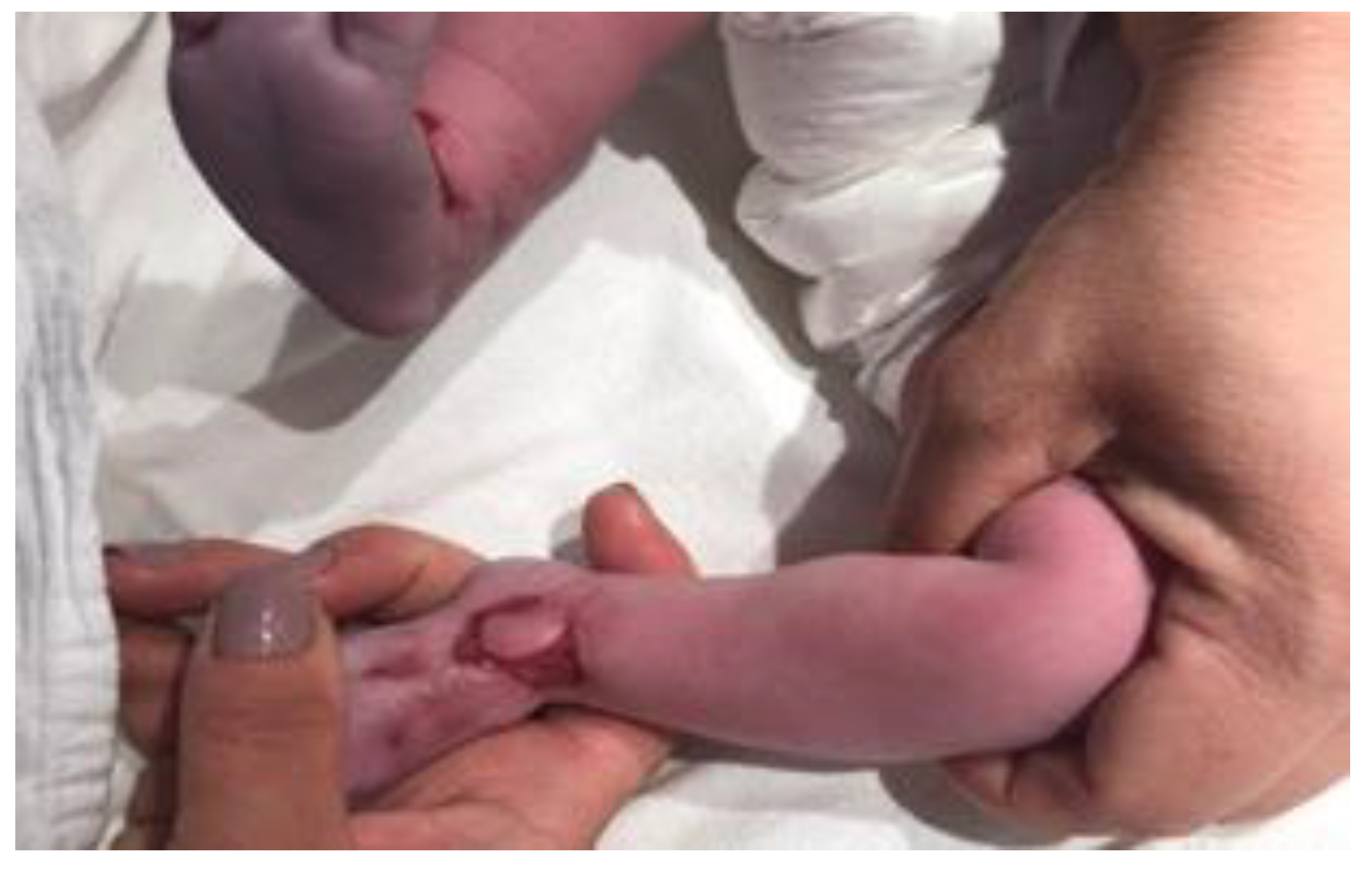
We present the case of a "de novo" mutation for the early-onset antenatal epidermolysis. The female child, from a carefully monitored clinical and ultrasound pregnancy, was born at the gestational age of 39 weeks. There was nothing suggestive in the family health history, both parents and the 2-year-old brother being absolutely healthy. As additional information, the feeding of the whole family, implicitly of the mother of the child during the antepartum period, is made with foods from strictly biological cultures, being very balanced in nutritional principles. The mother did not show any pathology during pregnancy. The only special mention was regarding the stress perceived by the mother during pregnancy, in part related to the COVID-19 pandemic. At the time around the birth, all the regulations regarding the prevention of intra- hospital dissemination of the Sars-Cov-2 infection were respected according to the protocols, including social distancing, facial mask wearing and frequent cleaning and disinfection of the surfaces [4,5,6]. The mother was tested negative for Sars-Cov-2 before admission. The postnatal adaptation was good, W = 3,200 g, balanced cardio- respiratory, spontaneous motions, intestinal tract present. In the first 12 hours postpartum, in the lumbar region, buttock, and at the level of the upper limbs, blistering lesions that break appeared, revealing large areas of dermis (Figure 1).

Figure 1.
The blistering lesions.
Due to the high septic risk, considering that in the first 24 hours about 50% of the area was affected, the decision to transfer the child to NICU for supervision and care was made. The hospitalization lasted for 40 days, during which time local antiseptic solutions were applied, along with hydration baths, emollients, local dressings, treatment for hydro electrolytic disorder and broad- spectrum antibiotics (meronem). During the hospitalization period, the blistering lesions occurred at the level of the entire skin, including at the level of the buccal mucosa, except for the hairy area of the head.
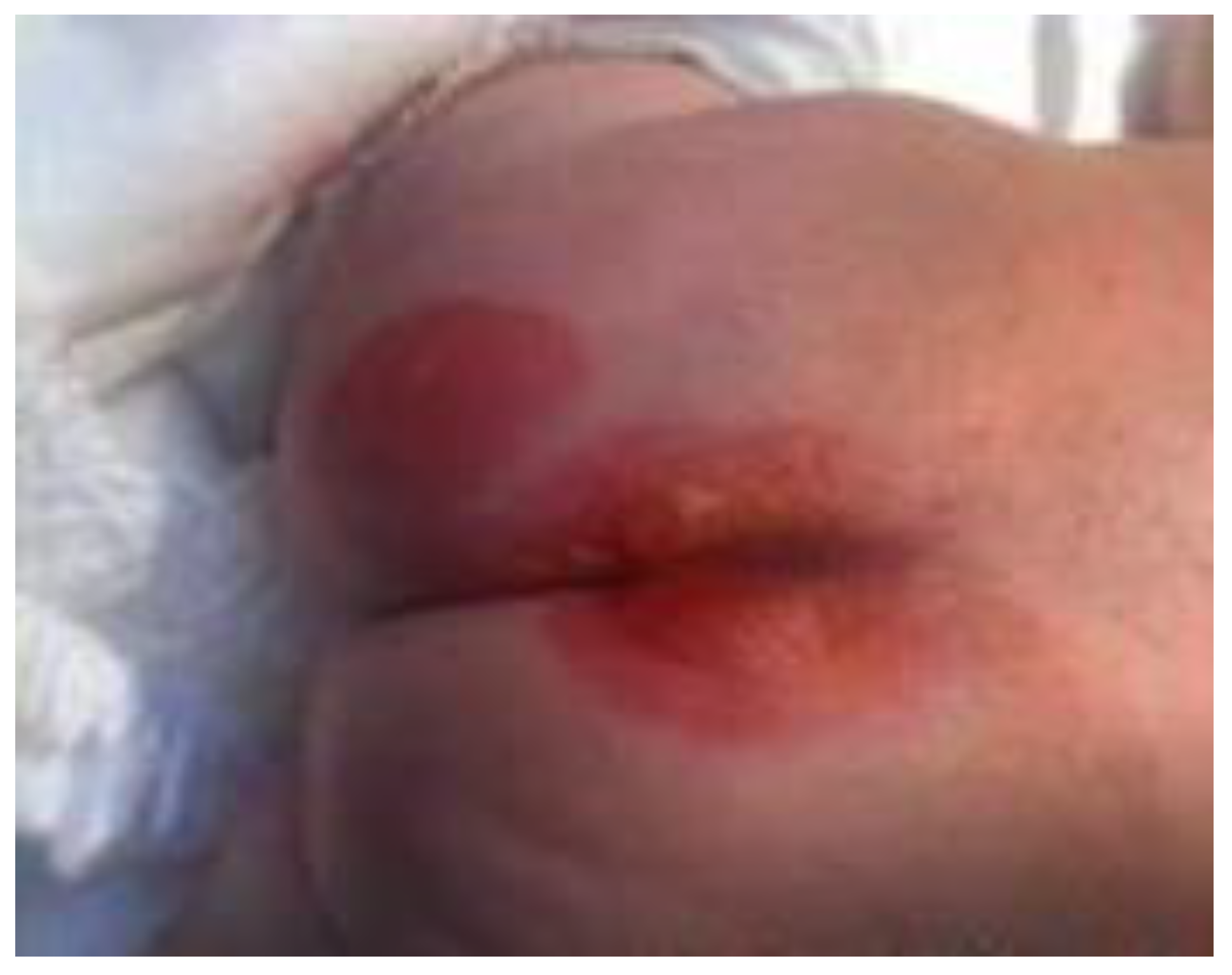
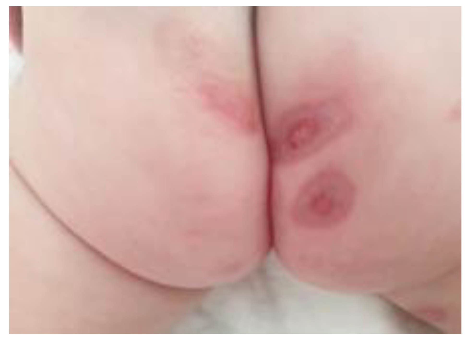
After discharge, at the age of 42 days, W=3,240g, the child receives home care recommendation for new and old lesions. During the 8-month period, new lesions are present, especially in the frictional areas. Two lesions resulted in keloid scars and there were many recurrences in the respective areas (region of the gluteal crease). At 9 months of age, the child has a severe degree of dystrophy (W = 5,700g, without being associated with psychomotor retardation) (Figure 2, Figure 3 and Figure 4).

Figure 2.
Early lesions in the gluteal region.
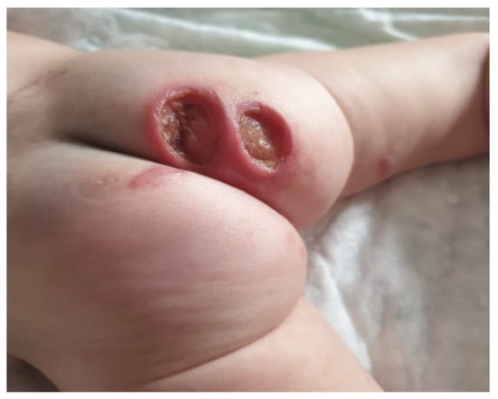
Figure 3.
Advanced lesions in the gluteal region.
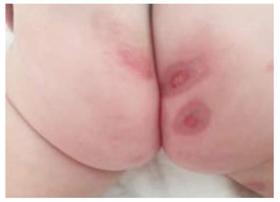
Figure 4.
Healing lesions.
Complementary examinations "targeted" on the condition (bullous epidermolysis) were performed. At the histological examination of the lesion, it was found that the intercellular connections in the normal integumentary architecture were altered by changes in collagen structures. Karyotyping of the child and parents (performed at a specialized institute in the UK) has not established genetic mutations, the diagnosis made by experts being "transient epidermolysis bullosa".
Blood tests revealed a significant degree of anemia (Hb: 9.7g/dl with serum iron ~ 18 μg%), hypoalbuminemia (2.9g/dl), hypocalcemia (7.9g/dl) and cultures from the superinfected lesions from the gluteal crease determined the presence of Pseudomonas aeruginosa.
A peculiarity of the case is that the child is still breastfed, has no respiratory or digestive intercourse and the complementary food has been extremely deficient without high animal protein (meat, egg) and the diet is deficient, of the lactovegetarian type.
The diagnosis of the child at 9 months is: epidermolysis bullosa, secondary pyodermal infection (pseudomonas), malnutrition II/III, iron deficiency, anemia, hypocalcemia. The follow-up and dispensation of the case is lasting for the chronic underlying disease (epidermolysis) and for the associated conditions (malnutrition, anemia). The hygiene rules are very strict, the disinfection and the continuous dressing of the injuries being mandatory. Nutrition recovery depends on the family's compliance, which unfortunately is supportive of restrictive diets.
The literature establishes that, depending on the severity of the disease, after the healing of the bullous lesions, keloid scars may occur, some of them mutilating, with limbs or segments in flexion, hair loss, nails, teeth, (temporary) impaired vision depending on the location of the typical lesions.
A unique feature of the case, in total discrepancy with the data in the literature, is the "failure to agree" to the early, antenatal onset of the severe postnatal evolution of the lesions with the lack of genetic modifications and to make the diagnosis of a "transient" form after karyotyping. This case highlights the importance of the signed informed consent, after presenting all possible incidents and complications, as well as the therapeutic alternatives, this being an important element in building a strong and confident patient-doctor relationship, increase adherence to treatment and prevent the possible medical errors [7,8].
Therapeutic options and decisions in our case
Type I collagen was prepared from bovine corneas by enzymatic processes, using pepsin (Merck) in acetic acid medium. For the solution obtained, the collagen content was determined by hydroxyproline (Hyp) dosage, according to the modified Woesneer method and the content in hexozoamines (by the Elson Morgan method).
GAG (KS-keratan sulfate mixture) and CS (chondroitin sulfate) were extracted from the same corneal tissue by papain enzymatic method and purified by ion exchange chromatography and ethanol precipitation. The composition of the GAG mixture was determined by measuring uronic acids (for CS) and neutral carbohydrates (for KS).
Membrane preparation: Type I collagen solution was diluted with 0.5M acetic acid to a final concentration of 0.4 % (g/g) and dialyzed relative to distilled water to its pH.
The GAG mixture, containing two parts KS and one CS was dissolved in distilled water at a concentration of 0.85 and added to the collagen solution, through continuous stirring.
Variants of COL-GAG (collagen-glycosaminoglycan) solutions in ratio 1: 1, 2: 1, 4: 1, 7: 7 and 10: 1 were prepared. The COL type I solution and COL-GAG composition were dried as a membrane by evaporation of the solvent at 35°C for 48 hours.
COL-GAG sponges were prepared identically to the membranes, with the exception that the dilution was made at a concentration of 0.8% (g/g). The drying of the COL- GAG composition was carried out by lyophilization, for 72 hours, obtaining elastic white foam foils.
The sponges based on fibrillary collagen and gelatin (COL F-Gel) were obtained according to the following procedure (Figure 5):
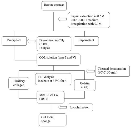
Figure 5.
Figure 5. The procedure used to obtain sponges based on fibrillar collagen and gelatin
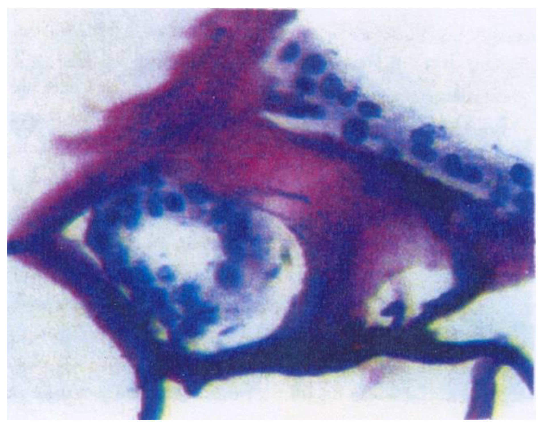
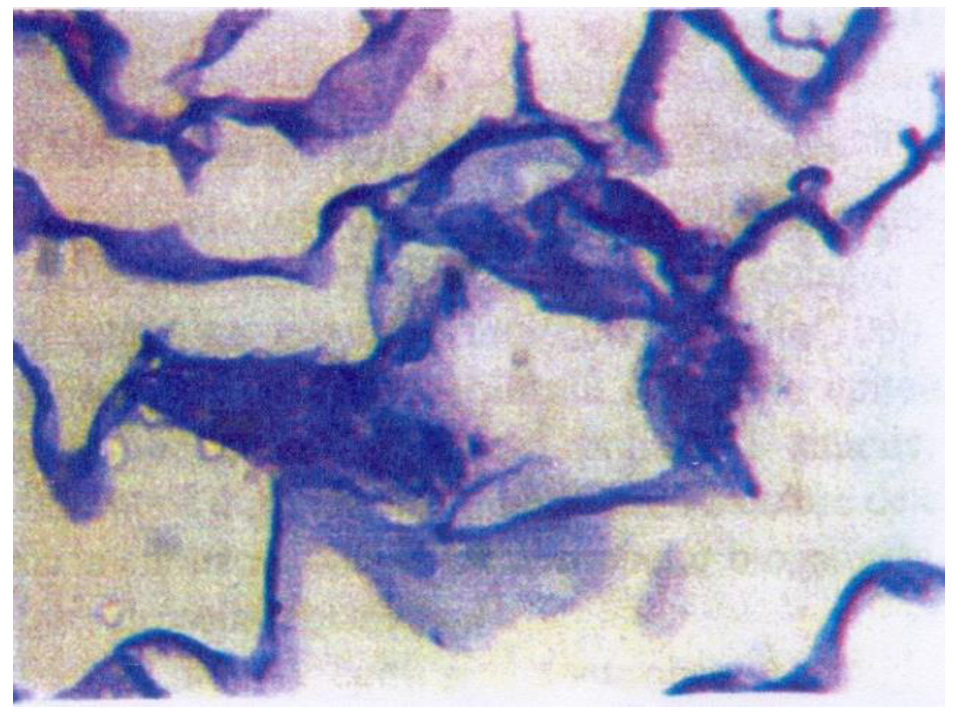
A stabilized line of calf kidney epithelial cells was introduced into Petri culture vessels in contact with collagen membranes and sponges at 37°C for 96 hours. The histological examination showed that the collagen membranes serve as a support for the epithelial cells that formed a surface monolayer after 48 hours after cultivation. At 96 hours of culture, the cells had a normal appearance (Figure 6 and Figure 7).
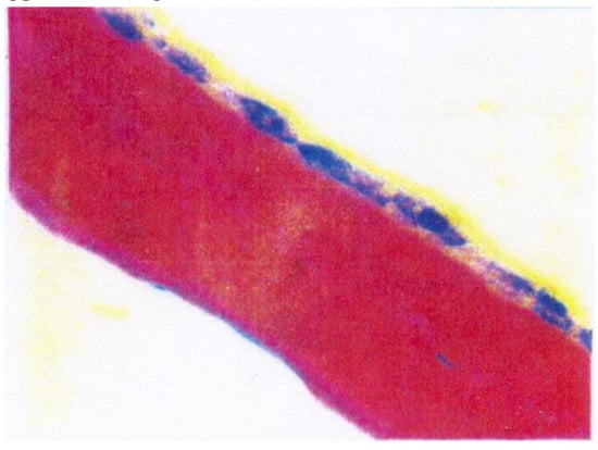
Figure 6.
Optical microscopy images of epithelial cells grown on collagen membranes, 40 hours.
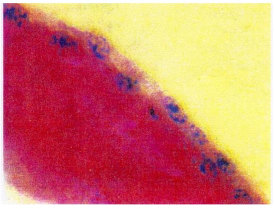
Figure 7.
Optical microscopy images of epithelial cells grown on collagen membranes, 96 hours.
Regarding the collagen sponges at 48 hours, cells were observed only on their surface, and at 96 hours, cells penetrated into the three-dimensional network structure and attached to the alveolar structure (much better in COL- GAG structures compared to COL) (Figure 8 and Figure 9).

Figure 8.
Epithelial cells grown on sponge by COL-GAG. After 96 hours, the cells migrated inside the sponge and attached to its alveolar structure

Figure 9.
Epithelial cells grown 96 hours on the COL F-Gel sponge.
Discussions
From the point of view of the clinical manifestations, certain types of dystrophic epidermolysis (EB) have been described, along with the most frequent type of multiple forms of epidermolysis [9].
- Hallopeau-Siemens autosomal recessive EB - the most severe form, with congenital blisters on a large percentage of the body surface and sometimes with portions absent from the integument (caused by traumas during birth). The blisters also affect the mucous membranes, subsequently causing integumentary scarring in the digestive tract (esophagus, buccal cavity) as well, thus leading to significant complications: post-keloid scar contraction, fusion of fingers, nail disappearance, eye inflammation, chronic malnutrition. On the long term, there is an increased risk of squamous, lethal carcinoma.
- Autosomal recessive non-Hallopeau-Siemens EB - the blisters are usually more limited on the hands and feet (elbows, knees), post-bulging scars occur, not very mutilating, as well as nail malformative changes.
- Autosomal dominant EB is the mildest form of EB, the blisters are usually confined to the hands, feet, knees elbows.
In less severe situations, abnormal nail growth is the only sign of the disease. In recent years, epigenetic causes have been most frequently discussed, as being incriminated in the action of the gene switch, which activates certain genes or suppresses the action of oth-ers, thus transforming them into dormant genes.
The generic known period of 1,000 days is defining in the development of a child. It practically starts at the time of conception and ends after 2 years of life. Excluding a multitude of triggering factors (diet, medicines, nuisances, etc.) special attention is paid to maternal stress during the antepartum period [10,11].
There has been a study for a period of 13 years (see book "1,000 days"), which shows an increase of cortisol (as, a stress hormone) during the antepartum period, during pregnancy, postpartum, associated with the increase of cortisol in the newborn and remote effects, including a change in neuroplasticity. Affected children showed an increase in the stress level and a decrease in adaptation to the beginning of the new school year or to public speaking [12,13].
In addition, stressors (e.g., noise, light) applied to batches of laboratory mice caused atopic dermatitis lesions, with changes in the rate of cell regeneration and in normal cellular architecture. Collagen, as a biomaterial component and support, has been recently used in medicine more and more often. Its properties have been exploited, such as the behavior of biological dressing and the non-stick and anti- adherence qualities [14,15,16,17].
In recent years, nutrition has been underlined as an important trigger of bullous skin diseases [18]. The wound management has been under debate in order to reach a consensus [19]. The physiological disappearance of the provisional matrix of a clot is as important as its formation. The inadequate removal of fibrin may interfere with the normal process of wound healing [20,21].
This study shows that biomaterials based on COL and GAG may be a possible future solution, at least in patients with extensive epithelial disorders (epidermolysis, Steven- Johnson syndrome, severe burns, corneal lesions and so on) [22,23].
Highlights
- ✓
- Epidermolysis bullosa is a rare condition with genetic determinism, with a significant polymorphism of the clinical forms.
- ✓
- The follow-up should carefully monitor the cutaneous mucosal relapses, the growth and development of the child and the possible sequelae.
- ✓
- Shortening the healing time with collagen membranes will reduce the risk of further septic complications, dehydration and hydro-electrolytic imbalance.
Conclusions
In conclusion, the presented case has the following features: early onset (antenatal), lack of possible triggering factors in the mother (except for stressful periods), lack of changes in the genetic karyotype, severity of the skin condition, association of deficient conditions (malnutrition, anemia, rickets).
Although diagnosed with the "mild" form from a genetic point of view, the evolution is autosomal recessive Hallopeau-Siemens type EB. The evolution will have to be carefully monitored in terms of cutaneous mucosal relapses, the growth and development of the child and of the possible sequelae.
Collagen-based biomaterials can be used to accelerate dermal-epidermal healing in various conditions of the child (Stevens Johnson syndrome, bullous epidermolysis, widespread burns). Shortening the healing time with collagen membranes will reduce the risk of further complications: septic - infections, hydro electrolytic - dehydration.
Conflicts of Interest disclosure
There are no known conflicts of interest in the publication of this article. The manuscript was read and approved by all authors.
Compliance with ethical standards
Any aspect of the work covered in this manuscript has been conducted with the ethical approval of all relevant bodies and that such approvals are acknowledged within the manuscript.
References
- Commission on the European Communities. Communication from the Commission to the European Parliament, the Council, the European Economic and Social Committee and the Committee of the Regions on Rare Diseases - Europe's challenges {SEC(2008)2713} {SEC(2008)2712}. 2008. Available online: https://op.europa.eu/en/publication-detail/-/publication/c8a042d8-ffb9-4b01-9c91- c1497a2b3fd7/language-en (accessed on 18 April 2021).
- Oliveira, Z.N.; Périgo, A.M.; Fukumori, L.M.; Aoki, V. Immunological mapping in hereditary epidermolysis bullosa. An Bras Dermatol. 2010, 85, 856–861. [Google Scholar] [CrossRef] [PubMed]
- Bello, Y.M.; Falabella, A.F.; Schachner, L.A. Management of epidermolysis bullosa in infants and children. Clin Dermatol. 2003, 21, 278–282. [Google Scholar] [CrossRef] [PubMed]
- Dascalu, A.M.; Tudosie, M.S.; Smarandache, G.C.; Serban, D. Impact of COVID-19 pandemic upon ophthalmological clinical practice. Rom J Leg Med. 2020, 28, 96–100. [Google Scholar] [CrossRef]
- Veeraraghavan, V.; Srinivasan, K. Work place impact on mental wellbeing of frontline doctors. J Mind Med Sci. 2020, 7, 188–192. [Google Scholar] [CrossRef]
- Popescu, B.; Doinița, O.I.; Bălălău, C.; Scăunașu, R.; Manole, F.; Domuța, M.; Oancea, A.L. Fibroscopic examination on ENT patients in COVID-19 era. J Clin Invest Surg. 2020, 5, 63–65. [Google Scholar] [CrossRef]
- Șerban, D.; Brănescu, C.M.; Smarandache, G.C.; Tudor, C.; Tănăsescu, C.; Tudosie, M.S.; Stana, D.; Costea, D.O.; Dascălu, A.M.; Spătaru, R.I. Safe surgery in day care centers: focus on preventing medical legal issues. Rom J Leg Med. 2021, 29, 60–64. [Google Scholar] [CrossRef]
- Serban, D.; Smarandache, A.M.; Cristian, D.; Tudor, C.; Duta, L.; Dascalu, A.M. Medical errors and patient safety culture - shifting the healthcare paradigm in Romanian hospitals. Rom J Leg Med. 2020, 28, 195–201. [Google Scholar] [CrossRef]
- Freeman, E.B.; Köglmeier, J.; Martinez, A.E.; Mellerio, J.E.; Haynes, L.; Sebire, N.J.; Lindley, K.J.; Shah, N. Gastrointestinal complications of epidermolysis bullosa in children. Br J Dermatol. 2008, 158, 1308–1314. [Google Scholar] [CrossRef]
- Gruskay, D.M. Nutritional management in the child with epidermolysis bullosa. Arch Dermatol. 1988, 124, 760–761. [Google Scholar]
- Bohiltea, R.; Turcan, N.; Cavinder, C.M.; Ducu, I.; Paunica, I.; Andronache, L.F.; Cirstoiu, M.M. Risk factors, predictive markers and prevention strategies for intrauterine fetal death. An integrative review. J Mind Med Sci. 2020, 7, 52–60. [Google Scholar] [CrossRef]
- Hubbard, L.; Haynes, L.; Sklar, M.; Martinez, A.E.; Mellerio, J.E. The challenges of meeting nutritional requirements in children and adults with epidermolysis bullosa: proceedings of a multidisciplinary team study day. Clin Exp Dermatol. 2011, 36, 579–583. [Google Scholar] [CrossRef] [PubMed]
- Birge, K. Nutrition management of patients with epidermolysis bullosa. J Am Diet Assoc. 1995, 95, 575–579. [Google Scholar] [CrossRef] [PubMed]
- Bobic, S.; Constantin, V.D.; Budu, V.A.; Socea, B.; Popescu, G.H.; Nica, E. Proactive therapeutical modulation of the postoperative intraperitoneal adhesions - the efficacy of the collagen-based biomaterials (simple and composite). Unified Journal of Medicine and Medical Sciences. 2016, 2, 1–9. [Google Scholar]
- Bobic, S.; Constantin, V.D.; Albu Kaya, M.; Marin, S; et al. Postoperative peritoneal adhesions prophylaxy using collagen-based biomaterials. ICAMS 2018 – 7th International Conference on Advanced Materials and Systems. 2018, 45–50. [Google Scholar] [CrossRef]
- Constantin, V.D.; Carâp, A.; Bobic, S.; Budu, V; et al. Tissue engineering-collagen sponge dressing for chronic wounds. ICAMS 2018 – 7th International Conference on Advanced Materials and Systems. 2018, 63–68. [Google Scholar] [CrossRef]
- Socea, B.; Carâp, A.; Bratu, O.G.; Diaconu, C.C.; Dimitriu, M.; Socea, L.I.; Bobic, S.; Constantin, V.D. The role of the composite and biologic meshes in the trocar site hernia repair following laparoscopic surgery. Materiale Plastice. 2018, 55, 146–148. [Google Scholar] [CrossRef]
- Fedeles, F.; Murphy, M.; Rothe, M.J.; Grant-Kels, J.M. Nutrition and bullous skin diseases. Clin Dermatol. 2010, 28, 627–643. [Google Scholar] [CrossRef]
- Pope, E.; Lara-Corrales, I.; Mellerio, J.; Martinez, A.; Schultz, G.; Burrell, R.; Goodman, L.; Coutts, P.; Wagner, J.; Allen, U.; Sibbald, G. A consensus approach to wound care in epidermolysis bullosa. J Am Acad Dermatol. 2012, 67, 904–917. [Google Scholar] [CrossRef]
- Fometescu, S.G.; Costache, M.; Coveney, A.; Oprescu, S.M.; Serban, D.; Savlovschi, C. Peritoneal fibrinolytic activity and adhesiogenesis. Chirurgia (Bucur). 2013, 108, 331–340. [Google Scholar]
- Shah, H.; Shah, R.; Sanghani, H.; Lakhani, N. Health related quality of life (HRQoL) and its associated surgical factors in diabetes foot ulcer patients. J Clin Invest Surg. 2020, 5, 83–90. [Google Scholar] [CrossRef]
- Kiritsi, D.; Nyström, A. Recent advances in understanding and managing epidermolysis bullosa. F1000Res. 2018, 7, F1000. [Google Scholar] [CrossRef] [PubMed]
- Kaur, J.; Budhwar, J.; Maheshwary, A.; Bhatti, K.S. Blistering in a newborn: a rare case report. Int J Res Dermatol. 2020, 6, 432. [Google Scholar] [CrossRef]