Next Article in Journal
A Rare Complication in a COVID-19 Positive Patient with Sigmoid Colon Cancer-Hemoperitoneum Due to Gallbladder Necrosis Following Micro-Thrombosis
Previous Article in Journal
Neuroendocrine Carcinoma of the Breast: A Case Report
 
 
Journal of Mind and Medical Sciences is published by MDPI from Volume 12 Issue 1 (2025). Previous articles were published by another publisher in Open Access under a CC-BY (or CC-BY-NC-ND) licence, and they are hosted by MDPI on mdpi.com as a courtesy and upon agreement with Valparaiso University (ValpoScholar).
Case Report

Periaortic Venous Necklace and Renal Right Double Arteries; Case Report

by
Petru Bordei
1,
Constantin Andrei Rusali
1,
Constantin Ionescu
1,
Dragos Serban
2,* and
Valeriu Ardeleanu
3,4,5,6
1
Department of Anatomy, Faculty of Medicine, Ovidius University from Constanta, Constanta, Romania
2
Department of General Surgery, Emergency University Hospital Bucharest, Carol Davila University of Medicine and Pharmacy, 050098 Bucharest, Romania
3
Doctoral School, Faculty of General Medicine, Ovidius University from Constanta, Constanta, Romania
4
Dunarea de Jos University from Galati, Galati, Romania
5
General Hospital CFR Galati, Galati, Romania
6
Arestetic Clinic, Galati, Romania
*
Author to whom correspondence should be addressed.
J. Mind Med. Sci. 2021, 8(2), 312-316; https://doi.org/10.22543/7674.82.P312316
Submission received: 19 April 2021 / Revised: 20 April 2021 / Accepted: 3 July 2021 / Published: 10 October 2021

Abstract

:
The case was found on an organic sample consisting of the two kidneys with the renal pedicles and the corresponding segments of the abdominal aorta and inferior vena cava. From the inferior face of the left renal vein, on the lower side of the aorta, a venous branch with an upward path of 8.02 mm was detached, passing on the anterior face of the aorta, passing before its right side, in order to end on the left side of the inferior vena cava, 13.9 mm above the end of the left renal vein in the inferior vena cava, this branch thus describing a periaortic ring (necklace), in which on the left side of the aorta the inferior adrenal vein ends. The periaortic ring (necklace) had a cross-sectional dimension of 3.2 mm and a vertical one of 1.7 cm. On the right side of the aorta, a 2.9 mm venous branch came out of the renal vein, ending on the left side of the inferior vena cava, 1.2 mm above the end of the left renal vein. At the level of the right kidney there were two renal arteries, superior and inferior. Between the two arteries there was an interval of 5.1 cm.

Introduction

The periaortic venous necklace is also referred to as the periaortic venous ring or renal collar, a name introduced by Huntington in 1920 [1]. The anterior modal venous branch receives the inferior adrenal vein, and the posterior venous branch, retroaortic, ends in the inferior vena cava more or less inferior to the anterior branch [2]. The supernumerary renal arteries represent the persistence of the embryonic vessels that appeared during the ascending migration of the kidney. They usually originate directly in the aorta and frequently go, either one or both, to the renal poles (polar arteries, upper or lower), sometimes penetrating the kidneys through the renal hilum, the renal arteries themselves [3]. The incidence of circumaortic renal vein 2 is reported to be ~ 10% (ranging from 2 to 17%) [2].
Regarding multiple renal arteries, the name given by the authors varies; we consider that the name would be correct according to their number: double, triple, quadruple or according to the vascularized territory (polar, upper or lower arteries). We do not agree with the name of accessory arteries, because they have a well-defined and sometimes quite extensive vascularization territory.

Case Presentation

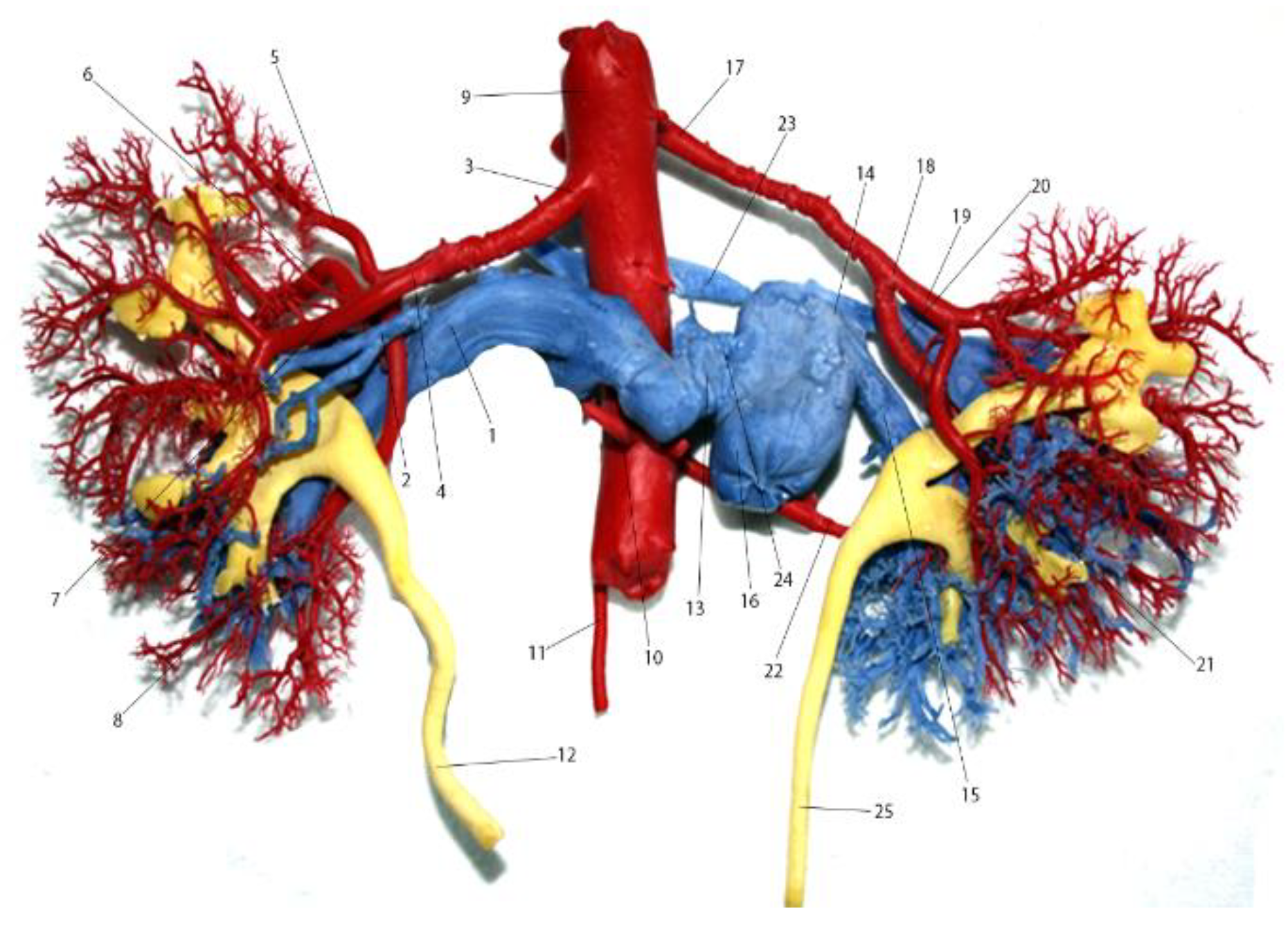
Our case was based on an organic sample consisting of the two kidneys collected in the autopsy room of the forensic laboratory, from a 72-year-old male patient, who died in a road accident. The kidneys were harvested together with the corresponding segments of the abdominal aorta and inferior vena cava, the intravascular injections being performed through the two vessels, the injection of the skin-caliceal system being made through the ureter. For the injection we used a German-made Technovit 7143, a self-curing resin based on methyl methacrylate, in powder and liquid form. For the preparation of the injectable substance, we used two parts of powder and one part of fluid, the powder being red for the arteries, blue for veins and yellow for the skin-caliceal system. After plastic injection and solidification, the resulting preparation was corroded with NaOH and photographed, anteriorly and posteriorly. The measurements were performed with the chisel, Figure 1 and Figure 2. The study was conducted within the anatomy department of the Faculty of Medicine in Constanta.
The left renal vein was present at the formation level with a diameter of 12.02 mm, forming juxtarenal from an anterior branch of 11.0 mm and a posterior branch of 4.0 mm. The anterior branch consisted of two branches, one superior which was 9.0 mm and the other one inferior 6.6 mm, which were united near the medial margin of the renal sinus, under the left renal artery. On the posterior face of the anterior venous branch, at a distance of 14.02 mm from its formation, the posterior branch ends, which forms postero-medially at 3 mm from the formation of the anterior venous branch, its formation being made of two venous branches, both 3 mm, upper and lower. At the end of the posterior branch in the anterior one, the left renal vein had a diameter of 12.02 mm. Upon formation, the left venous trunk has an ascending path, describing a wide circle arch with lower concavity.
On the left inferior lateral flank of the aorta, from the lower side of the left renal vein, a branch with an 8.02 mm ascending path was detached, passing on the anterior face of the aorta, passing before the right flank of it, to end on the left flank of the inferior vena cava, slightly posterior, 13.9 mm above the end of the left renal vein in the inferior vena cava, this branch thus describing a periaortic ring (necklace), in which on the left flank of the aorta the inferior adrenal vein ends. The periaortic ring (necklace) had a cross-sectional dimension of 3.2 mm and a vertical 1.7 cm. After issuing the branch that formed the periaortic ring, the left renal vein had a retroaortic pathway, describing a curve with the superior concavity on the posterior face of the aorta, the curve being superior and inferior between the origin of the aorta of two pairs of lumbar arteries. The upper branch of the curve ends on the right flank of the inferior vena cava, at this level the left renal vein having a diameter of 11 mm. At the union of the descending and ascending branches of the curve, a 2.9 mm venous branch came out of the renal vein, ending on the left flank of the inferior vena cava, 1.2 mm above the end of the left renal vein.
The left renal artery originates on the left postero- lateral flank of the aorta, 9.5 mm below the origin of the superior right renal artery, originally having a diameter of 7.0 mm, at the end the diameter being 6.9 mm. It ends extrarenally, approximately halfway between the left flank of the aorta and the medial margin of the left kidney in two branches: anteriorly 5.4 mm and posteriorly 5.02 mm. The anterior branch is terminated juxtarenally by four branches: upper polar 3.03 mm, upper mesorenal 6.01 mm, lower mesorenal 4 mm and lower polar 5.9 mm. The first three branches are preventive, the fourth branch being backwards. The posterior branch, rectilinear, was divided intrasinusally into a polarpostero-superior branch of 2.2 mm and a posterior mesorenal branch of 3 mm, for the posterior mesorinium.
In the right kidney, there were two renal arteries, upper and lower. The right upper renal artery originated in the right postero-lateral flank of the aorta, having a right inferior-lateral oblique pathway and was originally 5.9 mm in diameter. There was a distance of 5.1 cm between it and the artery of the lower right kidney. The artery had a retrograde groove, the periaortic ring being situated preaortically, at an equal distance between the two right renal arteries. The artery ends in an extrarenal bifurcation, closer to the kidneys, in the antero-superior 4.9 mm frame, for 2/3 antero-superior kidney and 4.03 mm posterior- inferior branch, for the posterior mesoriceum. The antero- superior branch divides into two 3.1 mm upper polar arteries, a superior mesorenal branch of 3.01 mm for the upper 1/3 of the kidney and a posterior mesorenal branch of 3.02 mm. The inferior renal artery, having a diameter of 4.0 mm, had a precavital and retroureteral route, being intended for the lower and lower thirds of the right kidney. The right renal vein was formed near the inferior vena cava, with a diameter of 10.02 mm, and at the end of the inferior vena cava, the diameter was 13.02 mm. It is formed by two anterior branches, 6.5 mm higher and 7.5 mm lower. The wreath superior venous was passing between the upper and lower terminal branches of the superior right renal artery.

Discussions

The incidence of the periaortic venous collar is rare, being variable, seen in 1.5% [4,5] and 16% of the cases [6]. It appears at the end of the eighth week of intrauterine life [6]. In his anatomy [1], the intersupracardinal anastomosis, together with intersubcardinal and sub-supracardinal anastomoses, forms a complete venous ring around the aorta, below the origin of the superior mesenteric artery, called the renal collar. The same aspect is also noted in human embryology [7]. The renal collar is formed from anastomoses between the supracardial (posterior) veins and the subcardinal (anterior) veins, with the posterior arm of the collar regressing during development [5]. On the right side, the anterior arm of the collar is embedded in the lateral wall of the renal segment of the inferior vena cava; and on the left side, the anterior arm of the collar forms the left renal vein. In this anomaly, the left renal vein surrounds the abdominal aorta, during the development of the inferior vena cava, anastomotic communications between the subcardinal and supracardinal veins forming a collar surrounding the aorta. Normally, the ventral portion of this periaortic collar persists as the left renal vein, and the dorsal portion regress. If the dorsal portion still persists, the periaortic collar results.
In our case, both right renal arteries were larger in diameter than the lower mesenteric artery (3.0 mm), but smaller than the corresponding left renal artery.
Double right renal arteries are less frequent than left ones, with variable differences from one author to another: 5.71% [8], 8.70% [9], 19.45% [10], 58.33% [11] or 62.5% [12]. There are authors who claim that the left double renal arteries are most commonly encountered with 2% [13], 7.55% or even 48% [14] percentages. The retroureteral route of the double renal arteries is also less commonly encountered than the preureteral route, in about a quarter of the cases [15] or even 1/3 of the cases [9], after [16] the preureteral route of the arteries being more frequently preureteral. The retrograde pathway of the double right renal arteries was encountered in 10% of the cases [16,17] or in 29.31% of the cases [10].
There are findings that the single artery on the opposite side has its origin of the aorta between the origins of the double renal arteries, frequently in the middle distance, and [10] also findings on the origin of the left single artery in 25.49% between the origins of the double renal arteries, in 60-78% of the cases above the upper right renal artery and in 13.73% of cases in the upper right renal artery [18]. It is worth mentioning the rarity of the cases of periaortic venous ring, as well as the fact that we did not find in the literature a case in which the periaortic venous ring is associated with the presence of two right renal arteries. The morphological characteristics of the vascular elements (arteries and veins) [19,20] were followed in detail from the point of view of the origin, the path and especially of the morphometry, the measurements being made up to tenths of a millimeter. Any further data on vascular malformations or abnormalities can be useful in the study of the processes of angiogenesis or development of some tumors, but also from the perspective of the treatment [21,22,23].

Highlights

The presence of the periaortic venous ring, depending on its size, can evolve asymptomatically if widened, being accidentally discovered at an angiographic examination or after death, at the necropsy examination.
When tightened, it can compress the aorta, which can cause adrenal aortic aneurysm, presenting the characteristic symptomatology.

Conclusions

The presence of the periaortic venous ring (necklace), depending on its size, can evolve asymptomatically when widened, as in the case we described, the abdominal aorta being 15.02 mm at this level, being accidentally discovered at an angiographic examination or after death, at the necropsy examination. When tightened, it can compress the aorta, which can lead to adrenal aortic aneurysm, presenting the characteristic symptomatology.
This vascular variant is potentially dangerous if it is not recognized before or during retroperitoneal surgery. During retroperitoneal surgery or nephrectomy, the surgeon may be falsely secured by the normal anterior renal vein, confidently grasping the aorta, and mobilizing the kidney. In this process, the retroaortic renal vein can be damaged, leading to fatal hemorrhage [2].

Conflicts of Interest disclosure

There are no known conflicts of interest in the publication of this article. The manuscript was read and approved by all authors.

Compliance with ethical standards

Any aspect of the work covered in this manuscript has been conducted with the ethical approval of all relevant bodies and that such approvals are acknowledged within the manuscript.

References

  1. Standring, S. Gray’s Anatomy. The Anatomical Basis of Clinical Practice. Ed. Elsevier, Churchill Livinngstone. Edinburgh. London. New York, 2015. ISBN 9780702068515.
  2. Ayaz, S.; Ayaz, Ü.Y. Detection of retroaortic left renal vein and circumaortic left renal vein by PET/CT images to avoid misdiagnosis and support possible surgical procedures. Hell J Nucl Med. 2016, 19, 135–9. [Google Scholar] [CrossRef] [PubMed]
  3. Karkos, C.D.; Bruce, I.A.; Thomson, G.J.; Lambert, M.E. Retroaortic left renal vein and its implications in abdominal aortic surgery. Ann Vasc Surg. 2001, 15, 703–8. [Google Scholar] [CrossRef] [PubMed]
  4. Ghandour, A.; Partovi, S.; Karuppasamy, K.; Rajiah, P. Congenital anomalies of the IVC-embryological perspective and clinical relevance. Cardiovasc Diagn Ther. 2016, 6, 482–492. [Google Scholar] [CrossRef] [PubMed]
  5. Trigaux, J.P.; Vandroogenbroek, S.; De Wispelaere, J.F.; Lacrosse, M.; Jamart, J. Congenital anomalies of the inferior vena cava and left renal vein: evaluation with spiral CT. J Vasc Interv Radiol. 1998, 9, 339–45. [Google Scholar] [CrossRef] [PubMed]
  6. Chircor, L.; Surdu, L. Morphogenesis of the inferior cav system. In: Human Embryology. Ed. Ex Ponto. Constanţa, 2014; 416–417ISBN 978-606-598-355-7. [Google Scholar]
  7. Chen, R.Y.; Chang, H. Renal dysplasia. Arch Pathol Lab Med. 2015, 139, 547–51. [Google Scholar] [CrossRef] [PubMed]
  8. Date, Y.; Takano, T.; Fujii, T.; Terasaki, T.; Sakaguchi, M. Double D Technique: An Innovative Modified Bifurcated Stent Graft Deployment Strategy for an Isolated Common Iliac Artery Aneurysm With a Challenging Renal Artery Anatomy. Vasc Endovascular Surg. 2019, 53, 613–616. [Google Scholar] [CrossRef] [PubMed]
  9. Matsumoto, K.; Miyajima, A.; Fukumoto, K.; Komatsuda, A.; Niwa, N.; Hattori, S.; Takeda, T.; Kikuchi, E.; Asanuma, H.; Oya, M. Factors influencing the operating time for single-port laparoscopic radical nephrectomy: focus on the anatomy and distribution of the renal artery and vein. Jpn J Clin Oncol. 2017, 47, 976–980. [Google Scholar] [CrossRef] [PubMed]
  10. Bordei, P.; Sapte, E.; Iliescu, D. Double renal arteries originating from the aorta. Surg Radiol Anat. 2004, 26, 474–9. [Google Scholar] [CrossRef] [PubMed]
  11. Mir, N.S.; Ul Hassan, A.; Rangrez, R.; Hamid, S.; Sabia, Tabish, S. A.; Iqbal, Suhaila, Masarat, Rasool, Z. Bilateral Duplication of Renal Vessels: Anatomical, Medical and Surgical perspective. Int J Health Sci (Qassim). 2008, 2, 179–85. [Google Scholar] [PubMed]
  12. Yokuş, A.; Toprak, N.; Gündüz, A.M.; Akdeniz, H.; Akdemir, Z.; Dündar, İ.; Arslan, H. Anterior Cerebral Artery and Anterior Communicating Artery Variations: Assessment with Magnetic Resonance Angiography. World Neurosurg. 2021, 155, e203–e209. [Google Scholar] [CrossRef] [PubMed]
  13. Regmi, P.R.; Amatya, I.; Kayastha, P.; Paudel, S.; Suwal, S.; Ghimire, R.K. Normal Anatomy and Variants of Renal Vasculature with Multidetector Computed Tomography in a Tertiary Care Hospital: A Descriptive Cross-sectional Study. JNMA J Nepal Med Assoc. 2020, 58, 911–914. [Google Scholar] [CrossRef] [PubMed]
  14. Resende, F.C.; Avelar, G.F. The sexual segment of the kidney of a tropical rattlesnake, Crotalus durissus (Reptilia, Squamata, Viperidae), and its relationship to seasonal testicular and androgen cycles. J Morphol. 2021, 282, 1402–1414. [Google Scholar] [CrossRef] [PubMed]
  15. Cordier, G.; Nguyen-Huu, B.M.H.S. Segmenttion Arterielle du rein [Arterial segmentation of the kidney]. Presse Med. 1964, 72, 2433–2438. [Google Scholar]
  16. Spinu Arsenie, D.; Bratu, O.G.; Marcu, D.R.; Stanciu, A.E.; Gherghiceanu, F.; Ionita-Radu, F.; Bungau, S.; Stanescu, A.M.A.; Mischianu, D. Underactive bladder - an underestimated entity. J Mind Med Sci. 2020, 7, 23–28. [Google Scholar] [CrossRef]
  17. Jun, K.W.; Cho, J.; Kim, M.H.; Hwang, J.K.; Park, S.C.; Moon, I.S.; Kim, J.I. Changes in hemostatic factors after kidney transplantation: A retrospective cohort study. Medicine (Baltimore). 2021, 100, e27179. [Google Scholar] [CrossRef] [PubMed]
  18. Singh, G.; Jolly, N. Peripheral Vascular Disease Involving Transplant Renal Artery. J Invasive Cardiol. 2021, 7, 257–260. [Google Scholar] [CrossRef]
  19. Onisâi, M.C.; Iordan, I.; Gaman, M.; Bumbea, H.; Vlădăreanu, A.M. Unexpected hope for a multiple myeloma patient. J Mind Med Sci. 2020, 7, 257–260. [Google Scholar] [CrossRef]
  20. Elian, V.; Cioca, G.; Pantea, A.S.; et al. Metabolic syndrome and chronic kidney disease: pathogenic, clinical, and therapeutic correlations. 1st International Conference on Interdisciplinary Management of Diabetes Mellitus and its Complications (INTERDIAB). Interdisciplinary approaches in diabetic chronic kidney disease. 2015, pp. 48–52.
  21. Ardeleanu, V.; Georgescu, C.; Frîncu, L.D.; Frâncu, L.L.; Vesa, D. Angiogenesis as Prospective Molecular Biology Technique for Cancer Study. Romanian Biotehnological Letters. 2014, 19, 9637–9648. [Google Scholar]
  22. Motofei, I.G.; Rowland, D.L.; Georgescu, S.R.; Tampa, M.; Paunica, S.; Constantin, V.D.; Balalau, C.; Manea, M.; Baleanu, B.C.; Sinescu, I. Post-Finasteride Adverse Effects in Male Androgenic Alopecia: A Case Report of Vitiligo. Skin Pharmacol Physiol. 2017, 30, 42–45. [Google Scholar] [CrossRef] [PubMed]
  23. Ardeleanu, V.; Francu, L.; Georgescu, C. Neoangiogenesis. Assessment in Esophageal Adenocarcinomas. Indian J Surg. 2015, 77 (Suppl 3), 971–976. [Google Scholar] [CrossRef] [PubMed]
Figure 1. Front view. 1. left renal vein at formation; 2. left renal vein at termination; 3. the periaortic venous ring; 4. left inferior adrenal vein; 5. venous branch from the left renal vein to the inferior vena cava; 6. inferior vena cava; 7. right renal vein at formation; 8. venous ramus superior to the right renal vein; 9. venous branch inferior to the right renal vein; 10. the right ureter; 11. upper right renal artery at origin; 12. upper right renal artery at termination; 13. lower right renal artery at origin; 14. left renal artery at origin; 15. left renal artery at termination, anterior branch; 16. left upper polar artery; 17. superior mesorenal artery; 18. inferior mesorenal artery; 19. left lower polar artery; 20. aorta; 21. inferior mesenteric artery; 22. the left ureter.
Jmms 08 00041 g001
Figure 2. Rear view. 1. left renal vein at formation; 2. ram veins posteriorly; 3. left renal artery at origin; 4. left renal artery at termination; 5. left upper polar artery; 6. Antero- superior mesorenal artery; 7. left posterior mesorenal artery; 8. left antero-inferior mesorenal artery; 9. aorta; 10. lumbar arteries; 11. inferior mesenteric artery; 12. the left ureter; 13. left renal vein at termination; 14. venous ramus superior to the right renal vein; 15. venous branch inferior to right renal vein; 16. inferior vena cava; 17. upper right renal artery at origin; 18. upper right renal artery at termination; 19. the anterior terminal branch of the superior right renal artery; 20. posterior terminal branch of the superior right renal artery; 21. Lower polar branch; 22. lower right renal artery; 23. the periaortic venous ring; 24. Venous branch from the left renal vein to the inferior vena cava; 25. the right ureter.
Jmms 08 00041 g002