Next Article in Journal
Modern Approaches for Antiandrogen-Resistant Prostate Cancer Therapy
Previous Article in Journal
The Analysis of Risk Factors Associated with Women’s Urinary Incontinence; Literature Review
 
 
Journal of Mind and Medical Sciences is published by MDPI from Volume 12 Issue 1 (2025). Previous articles were published by another publisher in Open Access under a CC-BY (or CC-BY-NC-ND) licence, and they are hosted by MDPI on mdpi.com as a courtesy and upon agreement with Valparaiso University (ValpoScholar).
Review

Non-Dental Oral Cavity Findings in Gastroesophageal Reflux Disease: A Systematic Review and Meta-Analysis

by
Madalina-Gabriela Indre
1,
Darius Sampelean
2,
Vlad Taru
2,*,
Angela Cozma
2,
Dorel Sampelean
2,
Mircea Vasile Milaciu
2 and
Olga Hilda Orasan
2
1
Prof. Dr. Octavian Fodor Regional Institute of Gastroenterology and Hepatology, Cluj-Napoca, Romania
2
4th Medical Department, Iuliu HațIeganu University of Medicine and Pharmacy, 18 Republicii Street, 400015 Cluj-Napoca, Romania
*
Author to whom correspondence should be addressed.
J. Mind Med. Sci. 2021, 8(1), 60-70; https://doi.org/10.22543/7674.81.P6070
Submission received: 2 October 2020 / Revised: 14 December 2020 / Accepted: 14 December 2020 / Published: 8 April 2021

Abstract

:
Gastroesophageal reflux disease (GERD) is known as the most prevalent gastrointestinal disorder in the United States, leading to substantial morbidity, although associated mortality is rare. Based on the appearance of esophageal mucosa on upper endoscopy, GERD is divided into erosive esophagitis (ERD) and nonerosive reflux disease (NERD). Heartburn and acid regurgitation are the typical symptoms of the disease, although some patients may present atypical manifestations such as epigastric pain, nausea, asthma, chronic cough, pharyngitis, laryngitis, sleep disturbances, otitis, and sinusitis. Other signs, such as oral mucosal lesions may result from GERD by direct acid or acidic vapor contact in the oral cavity. Oral manifestations such as tooth erosion, periodontitis, gingivitis, palatal erythema, ulceration, glossitis, oral acid burning sensation, halitosis, xerostomia have recently been reported in GERD patients. A considerable percentage of the patients are affected by oral manifestations before the onset of gastrointestinal symptoms, although in most cases the gastrointestinal signs and symptoms dominate the clinical picture. The injured oral mucosa negatively impacts the quality of life, especially functional limitation, physical inability and psychological disabilities, thus leading to social isolation. There is plenty of non-standardized information on the oral mucosal changes in GERD. In this context, we aimed at synthesizing and analyzing the current available evidence on non-dental oral cavity lesions and complaints that are present in patients diagnosed with GERD.

Introduction

Gastroesophageal reflux disease (GERD) is a condition that develops when the reflux of the stomach contents causes troublesome symptoms and/or complications [1]. GERD is known as the most prevalent gastrointestinal disorder in the United States, leading to substantial morbidity, although associated mortality is rare [2]. Based on the appearance of the esophageal mucosa on upper endoscopy, GERD is divided into erosive esophagitis (ERD) and nonerosive reflux disease (NERD) [3]. Heartburn and acid regurgitation are the typical symptoms of the disease, although some patients may present atypical manifestations such as epigastric pain, nausea, asthma, chronic cough, pharyngitis, laryngitis, sleep disturbances, otitis, sinusitis [4]. Other signs, such as oral mucosal lesions may result from GERD by direct acid or acidic vapor contact in the oral cavity. The oral manifestations, such as tooth erosion, periodontitis, gingivitis, palatal erythema, ulceration, glossitis, oral acid burning sensation, halitosis, xerostomia have recently been reported in GERD patients [5]. A considerable percentage of the patients are affected by oral manifestations before the onset of the gastrointestinal symptoms, although in most cases the gastrointestinal signs and symptoms dominate the clinical picture [6]. The injured oral mucosa negatively impacts the quality of life, especially functional limitation, physical inability and psychological disabilities, thus leading to social isolation [7]. There is plenty of non-standardized information on oral mucosal changes in GERD [8]. In this context, we aimed at synthesizing and analyzing the current available evidence on non-dental oral cavity lesions and complaints that are present in patients diagnosed with GERD.

Discussions

This review with meta-analysis is grounded in a structured protocol, developed using the Preferred Reporting Items for Systematic Review and Meta-Analysis Protocols (PRISMA) [9,10].
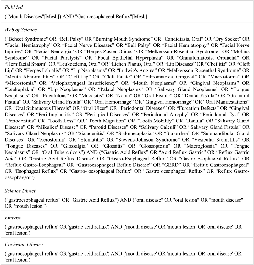
Research strategy. PubMed, Web of Science, Science Direct, Embase and Cochrane Library were searched by two independent researchers using a pre-piloted screening and selection tool in order to identify the eligible studies published between January 2000 and June 2020. The search was based upon Medical Subjective Heading (Mesh) terms of “Mouth Diseases” and “Gastroesophageal Reflux” (Table 1).
All articles relevant to the topic of this paper were retrieved and their bibliographies were hand searched for further references.
The screening and selection process consisted of two stages: (1) screening of title and abstracts and (2) screening and selection of full text papers (Figure 1).
Inclusion and exclusion criteria. The selected studies for the systematic review fulfilled the following criteria: (a) cross-sectional and case-control studies, (b) publications in English, (c) regardless of the publication status (published, in press, or in progress) (d) conducted on adults clinically diagnosed with GERD who underwent at least one additional evaluation for the confirmation of the diagnosis—upper gastrointestinal endoscopy, esophageal manometry or esophageal pH monitoring, (e) reporting results on the presence of non-dental oral cavity lesions or oral complaints. We excluded experimental studies, case presentations, case series, systematic reviews, articles published in other languages than English, or conducted among children populations.
The documents were handled using the Mendeley reference manager software. The selection process was conducted by two independent reviewers, while disagreements were settled by means of group discussions until a consensus was reached.
Data extraction. The data were extracted by two independent researchers using a pre-defined extraction form. Any disagreement between these two authors was resolved by means of group discussions. For each study, the following data were extracted: title, first author, year of publication, country, type of study, GERD diagnostic method, number of patients for the reported groups, gender, mean age, the use of PPI (proton pump inhibitor) therapy, reported exclusion criteria, data on the oral cavity assessment, type of lesion, type of complaint, odds ratio with confidence interval, mean plus standard deviation, p value.
Meta-analysis. For the studies reporting homogenous data, the relationship between the presence of GERD and a specific non-dental oral cavity lesion or a specific oral complaint was examined based on the odds ratio (OR) and its 95% confidence interval (CI). For studies measuring a specific score, the mean and standard deviation (SD) were used to calculate the standard mean difference (SMD) and the 95% CI. Effect sizes were interpreted using Cohen’s Standards for Interpreting Effect Sizes [11]. Assuming the differences in the methodology of the studies included in the meta-analysis, a random effect model was used. All calculations were conducted in the Comprehensive Meta- Analysis® software (v3).
The heterogeneity of the studies was analyzed using the I2 statistic, where a I2 ≥ 75% indicated important heterogeneity, with p < 0.05 defined statistical significance. Tau2 (τ2) was calculated using the restricted maximal likelihood method. Results showing important heterogeneity are reported in this article, but they cannot be used to generalize conclusions. The publication bias was analyzed using Egger’s regression, with p < 0.05 defining statistical significance and funnel plots for graphical description. In the event of publication bias, the Rosenthal method was used to calculate the fail-safe N statistic.
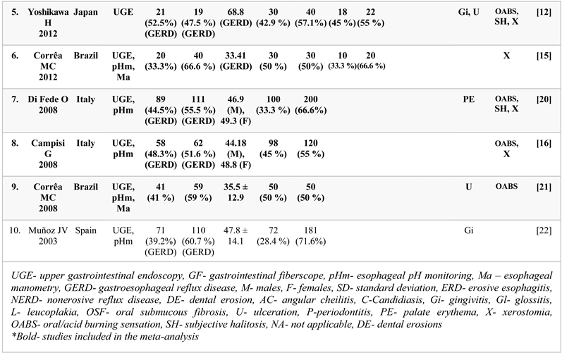
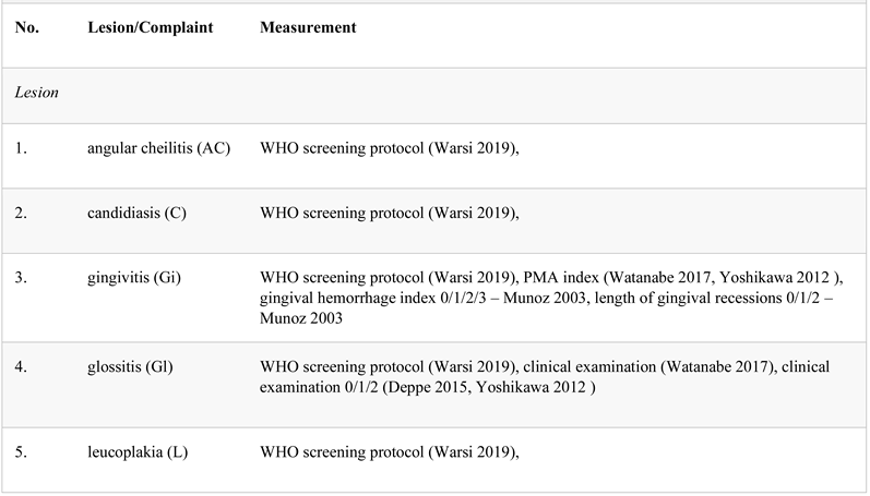
Some of the studies included in the meta-analysis compared GERD patients to different control subgroups, younger and older controls [12,13]. This problem was solved by combining the two control subgroups to form a single one, according to Cochrane Guidelines [14]. Some studies calculated scores to define oral lesions/symptoms. The PMA Index (papillary marginal attachment index: P was defined as “any degree of inflammation of the interdental papilla mesial to the tooth”; M was defined as “any degree of inflammation of the marginal gingiva on the facial aspect”; A was defined as “any disturbance of attachment as indicated by any degree of recession of the marginal gingiva from normal contour”) was used to define gingivitis in two studies [12,13] and Saxon test (simple, reproducible, and low-cost test for xerostomia, which involves chewing on a folded sterile sponge for 2 min; saliva production is quantitated by weighing the sponge before and after chewing and it was used to define xerostomia; normal control subjects produced greater than or equal to 2.75 gm of saliva in 2 min) in two studies [12,13]. Two of the studies [12,13] that quantify xerostomia measured the quantity of saliva in grams secreted in 2 min, while the other two in milliliters/minute [15,16]. The results were analyzed together, assuming that the difference in protocols were negligible.
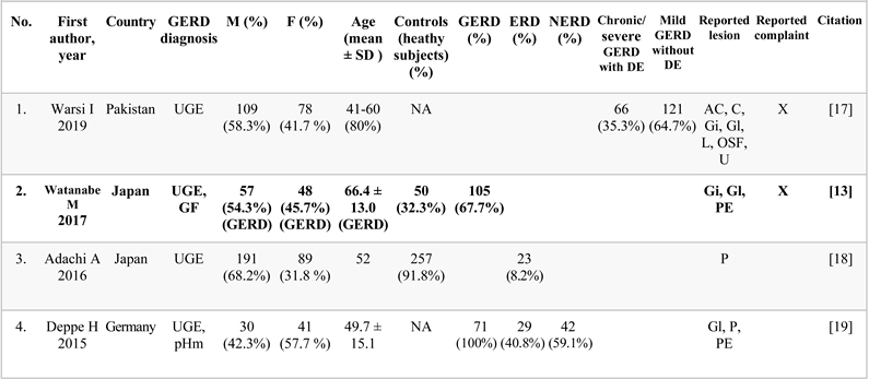
Study inclusion. The systematic search provided a total of 615 citations (Figure 1). Three additional citations were identified by means of manually searching the relevant references for the field published papers. After eliminating the duplicates, 599 studies remained. Out of these, 509 were discarded because they did not meet the inclusion criteria. The full text of the remaining 50 citations was examined in detail. Forty full-text published studies did not meet the inclusion criteria and were excluded from the analysis. The reasons for exclusion were: published in other languages than English (n = 8), no clinical examination of the oral cavity (n = 14), GERD diagnosis based only on clinical manifestations (n = 15), abstracts only (n = 2), children patients (n = 1). Experimental studies, case presentations, and case series were also excluded.
Ten studies were included for the narrative synthesis [12,13,15,16,17,18,19,20,21,22]. Two studies did not include a control group (healthy subjects) and were, therefore, excluded from the quantitative analysis [17,19]. Only one study reported periodontitis in both patients and healthy controls and was impossible to be analyzed quantitatively [18]. One study reported different measures of gingivitis and was consequently excluded from the meta-analysis [22]. Accordingly, 6 studies were deemed eligible for inclusion in the meta-analysis (Table 2).
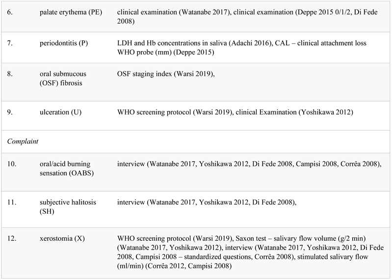
Narrative synthesis. The descriptive characteristics of the included studies are presented in Table 3. One study was case-control [20], and 9 were cross-sectional reports. The included studies were published between 2003 and 2019. All studies used UGE (upper gastrointestinal endoscopy) for the confirmation of GERD. Six (60%) studies also used pH monitoring [15,16,19,20,21,22], with further 2 (20%) including esophageal manometry [15,21] in the diagnosis. One study reported the use of a gastrointestinal fiberscope [13].

WHO- World Health Organization, PMA- papillary marginal attachment index, LDH- lactate dehydrogenase, Hb- hemoglobin, OSF- oral submucous fibrosis

Lesions. The most frequently reported oral lesions were gingivitis [12,13,17,22] and glossitis [12,13,17,19], followed by palatal erythema [13,19,20]. Two studies reported periodontitis [18,19] and another two oral ulcerations [12,17]. The reported prevalence of gingivitis in GERD patients ranged from 50.8 to 67.4% [21,22], while glossitis was reported in 5.6–7.6% of the cases [13,19]. One study reported a prevalence of 52.1% for periodontitis in GERD patients [19], while 14–21.5% of GERD patients presented palate erythema [19,20].
Complaints. The most reported prevalent complaint was xerostomia [12,13,15,16,17,19,21], followed by oral acid burning sensation [12,13,16,20,21] and subjective halitosis [12,13,20]. The reported prevalence of xerostomia in GERD patients ranged from 45.7 to 57.5% [12,13,16,20]. The oral acid burning sensation was present in 17.5–52% of GERD patients [12,13,16,20,21], while subjective halitosis was reported in 7.5–49.2% of the cases [12,13,20]. Quantitative synthesis. A total of 1694 subjects were included, with 687 in the control group and 1507 patients with GERD. A meta-analysis was performed separately for each type of lesion/complaint and each analysis included 2 to 5 articles, depending on the reporting data (Table 4).
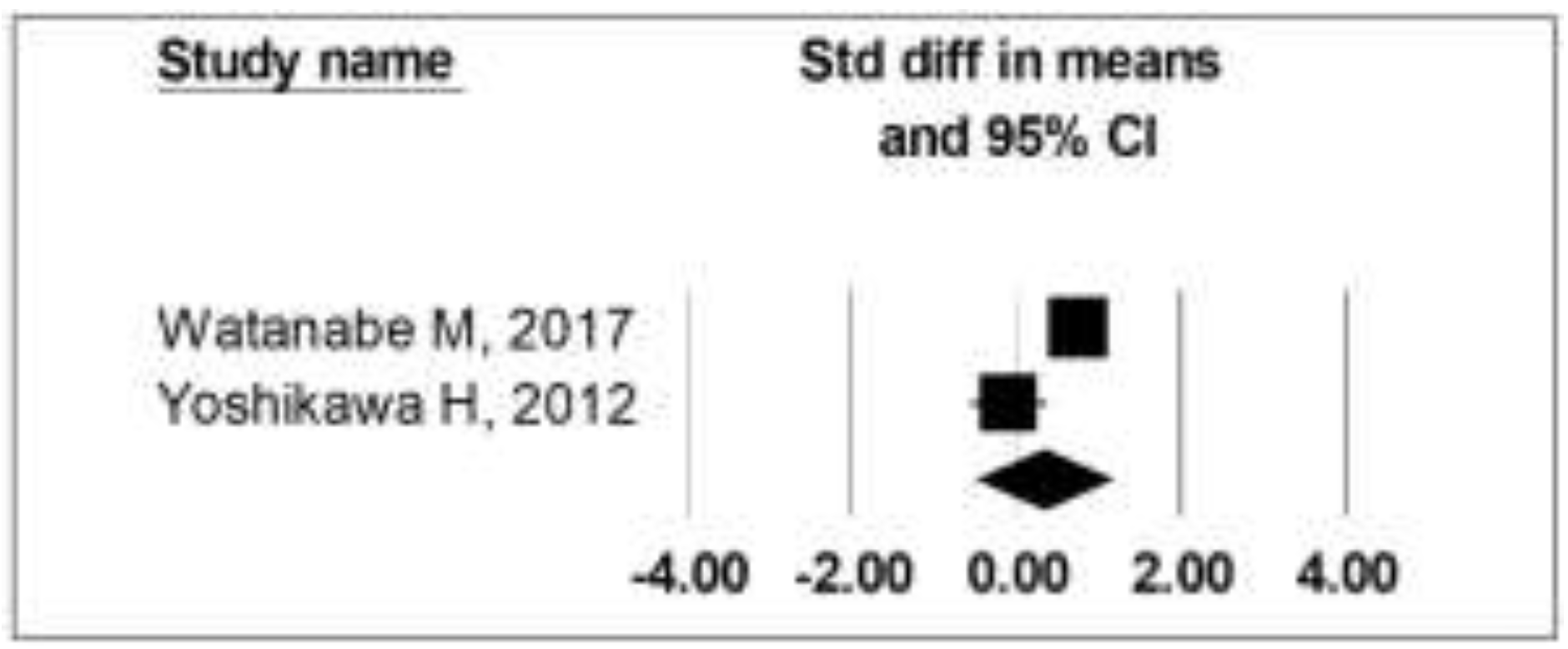
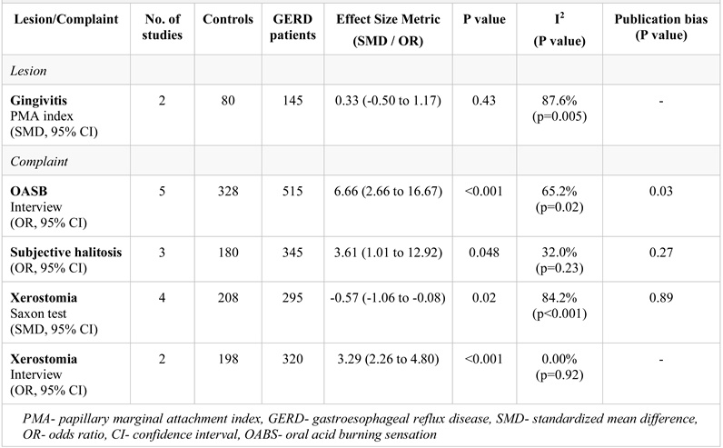
Gingivitis. Two studies, which reported gingivitis as PMA index, were eligible for the meta-analysis12,13. The cumulative analysis (SMD = 0.33, 95% CI: −0.50 to 1.17, p = 0.43) revealed a small, non-significant difference between patients with GERD and controls (Table 4, Figure 2).

PMA- papillary marginal attachment index, GERD- gastroesophageal reflux disease, CI- confidence interval

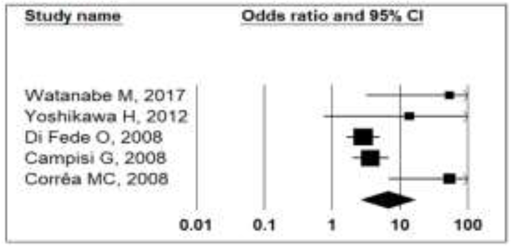
Oral acid burning sensation. Five studies, which reported oral acid burning sensation were analyzed quantitatively [12,13,16,20,21]. Patients with GERD had a high cumulative OR (6.66, 95% CI: 2.66 to 16.67, p < 0.001) of experiencing acid burning sensation compared to controls (Table 4, Figure 3).
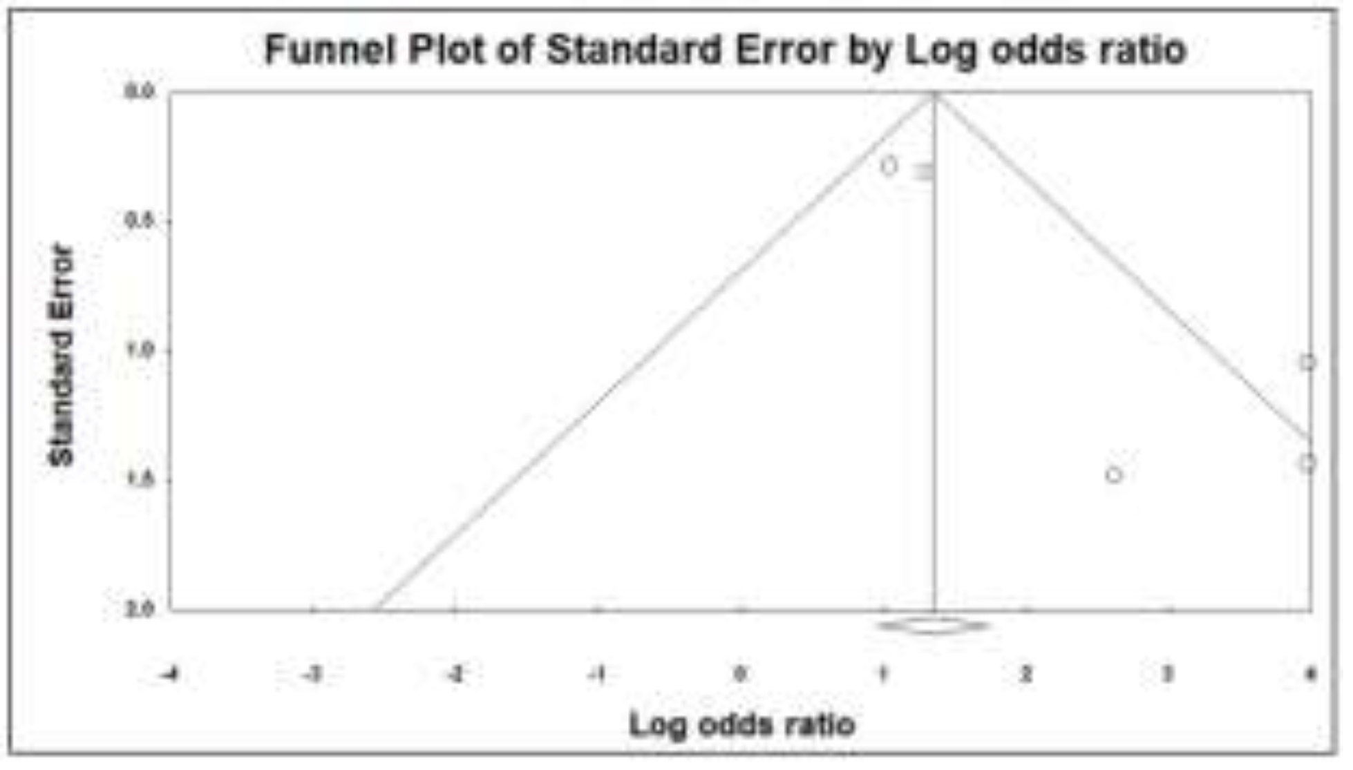
Despite the large effect size, a moderate heterogeneity (I2= 65.2%, p = 0.02) and a significant publication bias (p = 0.03) indicate a significant difference in the evidence (Figure 4).

OABS- oral acid burning sensation, GERD- gastroesophageal reflux disease

Subjective halitosis was reported and quantitatively analyzed in three studies [12,13,20]. GERD was associated with a moderate cumulative OR (3.61, 95% CI: 1.01 to 12.92, p = 0.048) of experiencing halitosis compared to controls (Table 4, Figure 5).

GERD- gastroesophageal reflux disease, CI- confidence interval

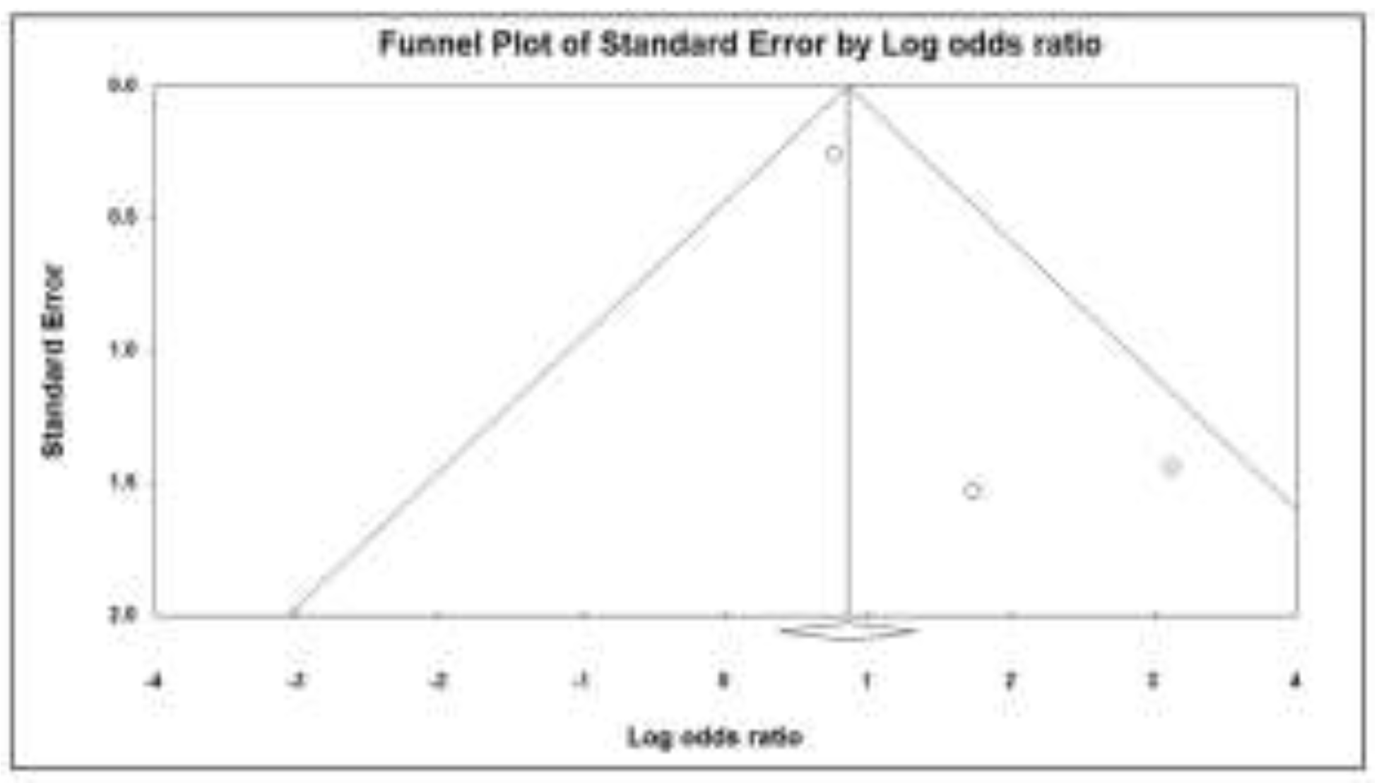
The results were robust with low heterogeneity (I2 = 32%, p = 0.23) and acceptable publication bias (p = 0.27) (Figure 6).

GERD- gastroesophageal reflux disease

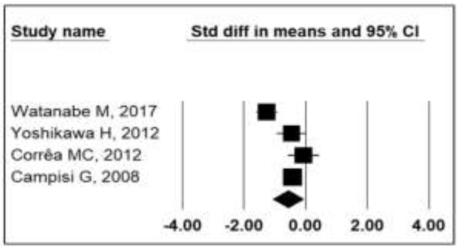
Xerostomia was reported by assessing the stimulated salivary function in four studies [12,13,15,16] and by interview, reporting the OR in two studies [16,20]. The SMD for the studies reporting the Saxon test score was moderately decreased in patients with GERD (SMD = −0.57, 95% CI: −1.06 to −0.08, p = 0.02) compared to controls (Table 4, Figure 7).

GERD- gastroesophageal reflux disease, CI- confidence interval

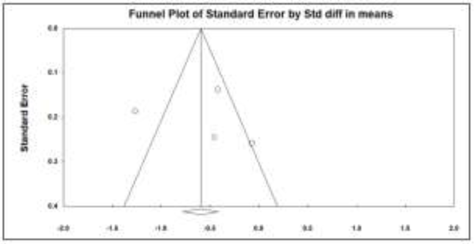
The studies showed significant heterogeneity (I2 = 84.2%, p < 0.001) and acceptable publication bias (p = 0.89) (Figure 8).

GERD- gastroesophageal reflux disease

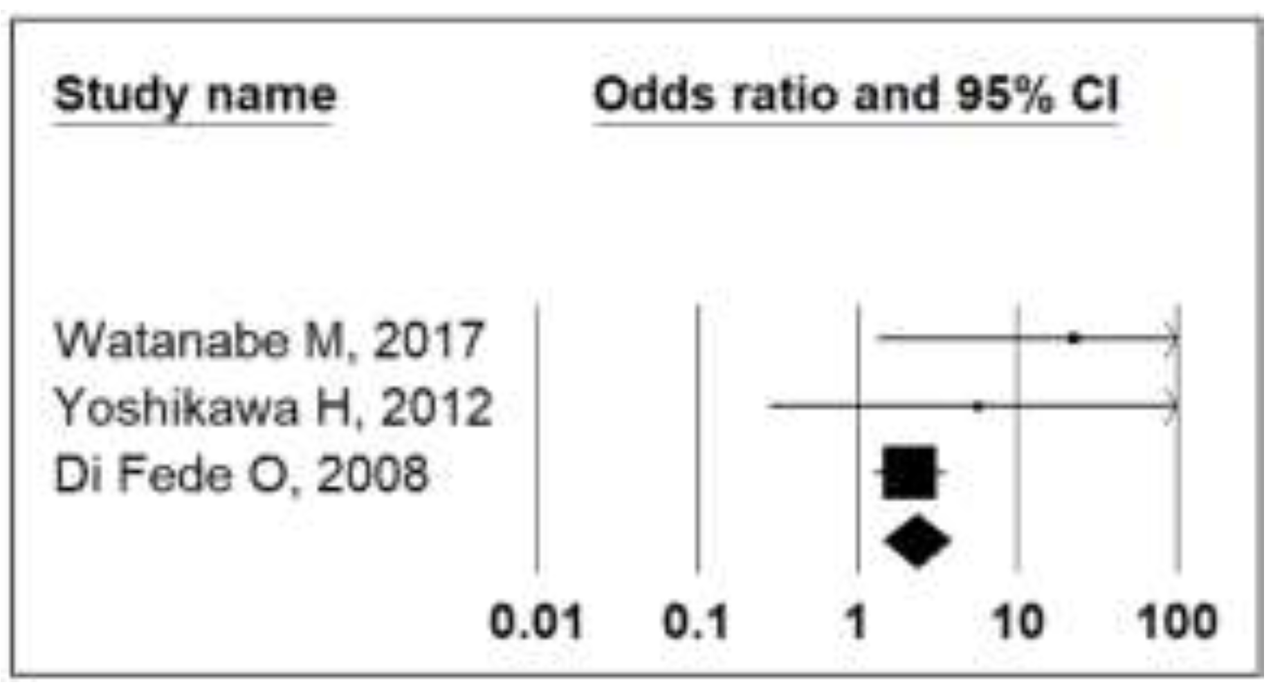
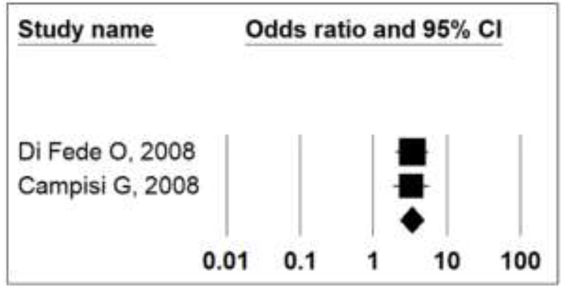
Meanwhile, the analysis of the two studies reporting OR showed a moderately increased risk (OR = 3.29, 95% CI: 2.26 to 4.80, p < 0.001) in patients with GERD versus controls, with low heterogeneity (p = 0.92) (Table 4, Figure 9).

OABS- oral acid burning sensation, GERD- gastroesophageal reflux disease, CI- confidence interval

This systematic review with meta-analysis indicates that individuals diagnosed with GERD are at an increased risk of presenting oral acid burning sensation, subjective halitosis or xerostomia compared to controls without GERD. Non-dental oral cavity lesions such as angular cheilitis, candidiasis, gingivitis, glossitis, leucoplakia, palate erythema, periodontitis, oral submucous fibrosis and ulceration may stand as extraesophageal manifestations of GERD. This review highlights the novel fact that non- dental oral cavity lesions are more frequent in patients diagnosed with GERD than in non-GERD controls. The risk of developing oral lesions such as dental erosions in these patients has been repeatedly investigated and numerous data supporting this association are available [23,24,25,26,27], but the association with non-dental oral cavity lesions/complaints is still insufficiently investigated and controversial.
The biologically possible explanation for the presence of non-dental oral cavity lesions in patients with GERD could stand in the repeated exposure to gastric acid or acidic vapors over a prolonged period of time [25]. Patients with GERD present a reduced tone of the lower esophageal sphincter, which potentates the backflow of the gastric content up to the mouth and airways [28]. Esophagitis stands as the most common complication of GERD, the condition being diagnosed and staged by means of upper digestive endoscopy [3]. Other complications are represented by Barret’s esophagus and adenocarcinoma [29]. Currently, an endoscopic examination is not routinely recommended for patients with oral lesions such as dental erosions due to its high cost and patient discomfort [30]. To our knowledge, a standardized oral examination has not yet been defined for GERD patients. Moreover, injured oral mucosa negatively impacts the quality of life, especially functional limitation, physical inability and psychological disabilities and could lead to social isolation [31].
The strengths of this systematic review are represented by the rigorous methodology that was applied for the study selection and data extraction following the PRISMA guidelines. A meta-analysis was conducted for the occurrence of gingivitis diagnosed by means of the PMA index, oral acid burning sensation, subjective halitosis and xerostomia, estimating the odds of developing these conditions in GERD patients. In all 10 included studies, the oral cavity assessment was performed by trained dentists and GERD was diagnosed by experienced gastroenterologists.
The limitations of the study include an important heterogeneity of methods for reporting non-dental oral cavity lesions and complaints, which prevented the quantitative analysis of some studies. Due to the small number of studies which met the inclusion criteria, those reporting ongoing IPP were not excluded. In some cases, subgroups were merged to form a single group to enable the comparative analysis. In some cases, the calculation of the publication bias was not possible. The majority of the studies included in the meta-analysis had high levels of heterogeneity, suggesting that the results should be interpreted with caution. Even though some studies reported data for NERD versus ERD patients, it could not be used to generate conclusive results. The studies included in the review had a cross-sectional or case report design which could not present the temporal association between GERD and non-dental oral cavity lesions. Finally, this review was limited to English publications, therefore it is possible to have missed relevant publications on the topic. This study highlights the need for the dental referral of patients diagnosed with GERD. We suggest that a standardized dental clinical examination should be included in the management of these patients. The results of this study indicate the importance of identifying non- dental oral cavity lesions and complaints in the diagnosis of GERD and possibly including a new subtype of GERD associated with oral non-dental manifestations in the Montreal consensus recommendations [32].

Highlights

Patients with GERD are at risk of developing oral acid burning sensation, subjective halitosis and xerostomia.
Non-dental oral cavity lesions such as angular cheilitis, candidiasis, gingivitis, glossitis, leucoplakia, palate erythema, periodontitis, oral submucous fibrosis and ulceration could represent extraesophageal manifestations of GERD.

Conclusions

The results of this systematic review with meta- analysis indicate that patients with GERD are at high risk of developing oral acid burning sensation, subjective halitosis and xerostomia compared to non-GERD controls. Non-dental oral cavity lesions such as angular cheilitis, candidiasis, gingivitis, glossitis, leucoplakia, palate erythema, periodontitis, oral submucous fibrosis and ulceration could represent extraesophageal manifestations of GERD.

Conflicts of Interest disclosure

There are no known conflicts of interest in the publication of this article. The manuscript was read and approved by all authors.

Compliance with ethical standards

Any aspect of the work covered in this manuscript has been conducted with the ethical approval of all relevant bodies and that such approvals are acknowledged within the manuscript.

References

  1. Vakil, N.; van Zanten, S.V.; Kahrilas, P.; Dent, J.; Jones, R.; Global Consensus Group. The Montreal definition and classification of gastroesophageal reflux disease: A global evidence-based consensus. Am J Gastroenterol. 2006, 101, 1900–1920. [Google Scholar] [CrossRef] [PubMed]
  2. Cohen, E.; Bolus, R.; Khanna, D.; Hays, R.D.; Chang, L.; Melmed, G.Y.; Khanna, P.; Spiegel, B. GERD symptoms in the general population: Prevalence and severity versus care-seeking patients. Dig Dis Sci. 2014, 59, 2488–2496. [Google Scholar] [CrossRef]
  3. Lundell, L.R.; Dent, J.; Bennett, J.R.; Blum, A.L.; Armstrong, D.; Galmiche, J.P.; Johnson, F.; Hongo, M.; Richter, J.E.; Spechler, S.J.; et al. Endoscopic assessment of oesophagitis: Clinical and functional correlates and further validation of the Los Angeles classification. Gut. 1999, 45, 172–180. [Google Scholar] [CrossRef] [PubMed]
  4. Giannini, E.G.; Zentilin, P.; Dulbecco, P.; Vigneri, S.; Scarlata, P.; Savarino, V. Management strategy for patients with gastroesophageal reflux disease: A comparison between empirical treatment with esomeprazole and endoscopy-oriented treatment. Am J Gastroenterol. 2008, 103, 267–275. [Google Scholar] [CrossRef]
  5. Vinesh, E.; Masthan, K.; Kumar, M.S.; Jeyapriya, S.M.; Babu, A.; Thinakaran, M. A Clinicopathologic Study of Oral Changes in Gastroesophageal Reflux Disease, Gastritis, and Ulcerative Colitis. J Contemp Dent Pract. 2016, 17, 943–947. [Google Scholar] [CrossRef]
  6. Jajam, M.; Bozzolo, P.; Niklander, S. Oral manifestations of gastrointestinal disorders. J Clin Exp Dent. 2017, 9, e1242–e1248. [Google Scholar] [CrossRef] [PubMed]
  7. Haag, D.G.; Peres, K.G.; Balasubramanian, M.; Brennan, D.S. Oral Conditions and Health-Related Quality of Life: A Systematic Review. J Dent Res. 2017, 96, 864–874. [Google Scholar] [CrossRef]
  8. Ranjitkar, S.; Smales, R.J.; Kaidonis, J.A. Oral manifestations of gastroesophageal reflux disease. J Gastroenterol Hepatol. 2012, 27, 21–27. [Google Scholar] [CrossRef]
  9. Liberati, A.; Altman, D.G.; Tetzlaff, J.; Mulrow, C.; Gøtzsche, P.C.; Ioannidis, J.P.; Clarke, M.; Devereaux, P.J.; Kleijnen, J.; Moher, D. The PRISMA statement for reporting systematic reviews and meta-analyses of studies that evaluate healthcare interventions: Explanation and elaboration. BMJ. 2009, 339, b2700. [Google Scholar] [CrossRef]
  10. Shamseer, L.; Moher, D.; Clarke, M.; Ghersi, D.; Liberati, A.; Petticrew, M.; Shekelle, P.; Stewart, L.A.; PRISMA-P Group. Preferred reporting items for systematic review and meta-analysis protocols (PRISMA-P) 2015: Elaboration and explanation. BMJ. 2015, 350, g7647. [Google Scholar] [CrossRef]
  11. Nakagawa, S.; Cuthill, I.C. Effect size, confidence interval and statistical significance: A practical guide for biologists. Biol Rev Camb Philos Soc. 2007, 82, 591–605. [Google Scholar] [CrossRef] [PubMed]
  12. Yoshikawa, H.; Furuta, K.; Ueno, M.; Egawa, M.; Yoshino, A.; Kondo, S.; Nariai, Y.; Ishibashi, H.; Kinoshita, Y.; Sekine, J. Oral symptoms including dental erosion in gastroesophageal reflux disease are associated with decreased salivary flow volume and swallowing function. J Gastroenterol. 2012, 47, 412–420. [Google Scholar] [CrossRef]
  13. Watanabe, M.; Nakatani, E.; Yoshikawa, H.; Kanno, T.; Nariai, Y.; Yoshino, A.; Vieth, M.; Kinoshita, Y.; Sekine, J. Oral soft tissue disorders are associated with gastroesophageal reflux disease: Retrospective study. BMC Gastroenterol. 2017, 17, 92. [Google Scholar] [CrossRef] [PubMed]
  14. Cumpston, M.; Li, T.; Page, M.J.; Chandler, J.; Welch, V.A.; Higgins, J.P.; Thomas, J. Updated guidance for trusted systematic reviews: A new edition of the Cochrane Handbook for Systematic Reviews of Interventions. Cochrane Database Syst Rev. 2019, 10, ED000142. [Google Scholar] [CrossRef] [PubMed]
  15. Corrêa, M.C.; Lerco, M.M.; Cunha Mde, L.; Henry, M.A. Salivary parameters and teeth erosions in patients with gastroesophageal reflux disease. Arq Gastroenterol. 2012, 49, 214–218. [Google Scholar] [CrossRef]
  16. Campisi, G.; Lo Russo, L.; Di Liberto, C.; Di Nicola, F.; Butera, D.; Vigneri, S.; Compilato, D.; Lo Muzio, L.; Di Fede, O. Saliva variations in gastro-oesophageal reflux disease. J Dent. 2008, 36, 268–271. [Google Scholar] [CrossRef]
  17. Warsi, I.; Ahmed, J.; Younus, A.; Rasheed, A.; Akhtar, T.S.; Ain, Q.U.; Khurshid, Z. Risk factors associated with oral manifestations and oral health impact of gastro- oesophageal reflux disease: A multicentre, cross- sectional study in Pakistan. BMJ Open. 2019, 9, e021458. [Google Scholar] [CrossRef]
  18. Adachi, K.; Mishiro, T.; Tanaka, S.; Yoshikawa, H.; Kinoshita, Y. A Study on the Relationship between Reflux Esophagitis and Periodontitis. Intern Med. 2016, 55, 2523–2528. [Google Scholar] [CrossRef]
  19. Deppe, H.; Mücke, T.; Wagenpfeil, S.; Kesting, M.; Rozej, A.; Bajbouj, M.; Sculean, A. Erosive esophageal reflux vs. non erosive esophageal reflux: Oral findings in 71 patients. BMC Oral Health. 2015, 15, 84. [Google Scholar] [CrossRef]
  20. Orhan, U.; Aziz Serkan, S.; Selçuk, G.; Sinan, Ö.; Cem Batuhan, O.; Ayhan, Ö.; Erdal, P.; Mustafa, D. Evaluating the effect of tumor size on survival and its prognostic significance among gastric cancer patients. J Clin Invest Surg. 2020, 5, 76–82. [Google Scholar] [CrossRef]
  21. Corrêa, M.C.; Lerco, M.M.; Henry, M.A. Study in oral cavity alterations in patients with gastroesophageal reflux disease. Arq Gastroenterol. 2008, 45, 132–136. [Google Scholar] [CrossRef] [PubMed]
  22. Muñoz, J.V.; Herreros, B.; Sanchiz, V.; Amoros, C.; Hernandez, V.; Pascual, I.; Mora, F.; Minguez, M.; Bagan, J.V.; Benages, A. Dental and periodontal lesions in patients with gastro-oesophageal reflux disease. Dig Liver Dis. 2003, 35, 461–467. [Google Scholar] [CrossRef] [PubMed]
  23. Li, W.; Liu, J.; Chen, S.; Wang, Y.; Zhang, Z. Prevalence of dental erosion among people with gastroesophageal reflux disease in China. J Prosthet Dent. 2017, 117, 48–54. [Google Scholar] [CrossRef]
  24. Pace, F.; Pallotta, S.; Tonini, M.; Vakil, N.; Bianchi Porro, G. Systematic review: Gastro-oesophageal reflux disease and dental lesions. Aliment Pharmacol Ther. 2008, 27, 1179–1186. [Google Scholar] [CrossRef] [PubMed]
  25. Jordão, H.W.T.; Coleman, H.G.; Kunzmann, A.T.; McKenna, G. The association between erosive toothwear and gastro-oesophageal reflux-related symptoms and disease: A systematic review and meta-analysis. J Dent. 2020, 95, 103284. [Google Scholar] [CrossRef]
  26. Picos, A.; Lasserre, J.F.; Chisnoiu, A.M.; Berar, A.M.; d’Incau, E.; Picos, A.M.; Chira, A.; des Varannes, S.B.; Dumitrascu, D.L. Factors associated with dental erosions in gastroesophageal reflux disease: A cross-sectional study in patients with heartburn. Med Pharm Rep. 2020, 93, 23–29. [Google Scholar] [CrossRef]
  27. Ganesh, M.; Hertzberg, A.; Nurko, S.; Needleman, H.; Rosen, R. Acid Rather Than Nonacid Reflux Burden Is a Predictor of Tooth Erosion. J Pediatr Gastroenterol Nutr. 2016, 62, 309–313. [Google Scholar] [CrossRef]
  28. Moazzez, R.; Anggiansah, A.; Bartlett, D.W. The association of acidic reflux above the upper oesophageal sphincter with palatal tooth wear. Caries Res. 2005, 39, 475–478. [Google Scholar] [CrossRef]
  29. Malfertheiner, P.; Hallerbäck, B. Clinical manifestations and complications of gastroesophageal reflux disease (GERD). Int J Clin Pract. 2005, 59, 346–355. [Google Scholar] [CrossRef]
  30. Smyth, E.C.; Lagergren, J.; Fitzgerald, R.C.; Lordick, F.; Shah, M.A.; Lagergren, P.; Cunningham, D. Oesophageal cancer. Nat Rev Dis Primers. 2017, 3, 17048. [Google Scholar] [CrossRef]
  31. Villanueva-Vilchis, M.D.; López-Ríos, P.; García, I.M.; Gaitán-Cepeda, L.A. Impact of oral mucosa lesions on the quality of life related to oral health. An etiopathogenic study. Med Oral Patol Oral Cir Bucal. 2016, 21, e178–e184. [Google Scholar] [CrossRef] [PubMed]
  32. DeVault, K.R.; Castell, D.O.; American College of Gastroenterology. Updated guidelines for the diagnosis and treatment of gastroesophageal reflux disease. Am J Gastroenterol. 2005, 100, 190–200. [Google Scholar] [CrossRef] [PubMed]
Figure 1. PRISMA Flow Diagram.
Jmms 08 00008 g001
Figure 2. Forest plot summarizing PMA index in GERD patients versus controls.
Jmms 08 00008 g002
Figure 3. Forest plot summarizing OABS in GERD patients versus controls. OABS- oral acid burning sensation, GERD- gastroesophageal reflux disease, CI- confidence interval.
Jmms 08 00008 g003
Figure 4. Funnel plot summarizing publication bias of studies which reported OABS in GERD patients versus controls.
Jmms 08 00008 g004
Figure 5. Forest plot summarizing reported subjective halitosis in GERD patients versus controls.
Jmms 08 00008 g005
Figure 6. Funnel plot summarizing publication bias of studies which reported subjective halitosis in GERD patients versus controls.
Jmms 08 00008 g006
Figure 7. Forest plot summarizing the Saxon test in GERD patients versus controls.
Jmms 08 00008 g007
Figure 8. Funnel plot summarizing publication bias of studies which reported the Saxon test in GERD patients versus controls.
Jmms 08 00008 g008
Figure 9. Forest plot summarizing reported xerostomia in GERD patients versus controls.
Jmms 08 00008 g009
Table 1. Search strategy.
Jmms 08 00008 i001
Table 2. Studies included for the narrative synthesis.
Jmms 08 00008 i002aJmms 08 00008 i002b
Table 3. Measurement characteristics.
Jmms 08 00008 i003aJmms 08 00008 i003b
Table 4. Quantitative analysis comparing non-dental oral cavity lesions in GERD patients versus healthy controls.
Jmms 08 00008 i004