Abstract
Calcium and magnesium are minerals with important functions throughout the body. The deficiency has been associated with an increased risk of cardiovascular disease, diabetes, and prostate cancer, and affects the skin and teeth. Some studies have associated it with osteoarthritis. Knee osteoarthritis is a degenerative pathology with a high prevalence that affects the knee joint, and prevention is necessary in the context of the lack of understanding of pathophysiology. The role of serum calcium and magnesium levels was considered in this regard. The study included a group of 371 hospitalized patients for unilateral or bilateral knee pain, of whom 326 patients had knee osteoarthritis and were the subject of the research. The risk factors such as age, gender, body mass index, weight status, and certain anatomical changes were analyzed, including the varus and valgus alignment. The results show the inverse relationship of Ca values with the radiological classification of knee osteoarthritis and the importance of risk factors such as age, gender, and obesity for the onset and progression of the pathology. Serum Mg values were not statistically significant in this study group.
Introduction
Calcium (Ca) and magnesium (Mg) are minerals with important functions throughout the body [1], especially in the skeletal and muscular system where, along with other components, they ensure the biological, metabolic and mechanical functions [2]. They also maintain healthy skin and teeth and its deficiency causes dehydration of the skin and yellowing of tooth enamel [3].
Calcium has implications in cell divisions, protein secretion, glycogen metabolism, and muscle contractions [2], including heart muscle. Calcium has a role in digestion and regulating blood pressure [3] and makes an important contribution to maintaining bone homeostasis, as well as in bone remodeling. The bone consists of an organic component containing mainly type I collagen with a role in flexibility and an inorganic component with a role in ensuring resistance to compressive forces, consisting mainly of Ca and other minerals [4]. Calcium homeostasis in the body is dependent on parathyroid hormone, vitamin D [5,6,7], calcitonin [3], and kidney function. Calcium and Mg absorption is achieved in the intestine [8], and is stimulated by the active form of vitamin D with the recommendation of a daily intake of 400 IU in adults. Magnesium helps to convert vitamin D into its active form so that they are combined in the treatment of various resistant forms of rickets [9,10,11].
Magnesium has a role in energy metabolism [12], protein synthesis, nucleic acids, and maintaining the electrical potential of tissues [13]. It is involved in the differentiation and proliferation of chondrocytes as well as reducing serum levels of inflammatory cells such as interleukin-1 or tumor necrosis factor- α and free radicals [14]. By preventing Ca and phosphorus precipitation, Mg with an inhibitor of interleukin-6 has a role in reducing the formation of large hydroxyapatite crystals [15].
Ca and Mg deficiency has been associated with an increased risk of cardiovascular diseases, diabetes [16], prostate cancer, and even osteoarthritis [17]. Some of the drugs widely used to treat the patients’ comorbidities may be involved in Ca and Mg metabolism [18,19,20]. For example, certain calcium channel blockers have a role in reducing the process of cartilage destruction by inhibiting genes involved in maintaining inflammation and stimulating the production of proteoglycans [21]. Drugs such as diuretics can increase renal excretion of Ca and Mg. This can generate disturbances in serum levels, with implications in bone homeostasis [22].
Knee osteoarthritis (KOA) is a degenerative pathology that progressively affects the anatomical structures of the knee joint and represents one of the main causes of functional impairment worldwide. Risk factors such as age, weight, gender, systemic inflammatory mediators, cellular and biochemical processes that may influence bone homeostasis or anatomical changes and local trauma are usually associated [23]. Thus, the onset of the disease and its slow progression make it possible to have radiological changes even at the first consultation [24].
The prevalence of KOA is alarmingly increasing and it is estimated that in 2032, compared to 2012, the consultations may increase by 15.7% [25], the costs of multiple hospitalizations is high, and improvement in quality of life is unfortunately not significant [26]. The pathophysiology of KOA has not been fully understood and in this context, it is essential to detect and reduce potentially modifiable risk factors, especially with individualized prevention strategies and the detection of factors that may have a protective role [27]. Early administration of mineral supplements should be considered, which may slow the progression of the disease and improve the symptoms [28]. The recommended daily intake is for Mg 4–6 mg/kg/day [29] and for Ca the ideal recommended dose is 1000 mg/day, but in European countries the daily mean is 687–1171 mg/day for men and 508/1047 mg/day for women [30]. Calcium and Mg overdose may cause gastrointestinal or cardiovascular symptoms including arrhythmias [31] or myocardial infarction [32].
Summarizing these data, the researchers argue that serum Ca and Mg levels could influence the imaging changes in ostheoarthritis [28].
The aim of the study is to evaluate the influence of serum Ca and Mg values in the radiological evolution of KOA, in relation to specific risk factors.
Materials and Methods
A retrospective study included 371 patients admitted to the Military Emergency Clinical Hospital “Dr. Iacob Czihac” Iasi between July 2017–July 2018. Criteria for inclusion were: uni- or bilateral knee pain, knee radiography performed for diagnostic purpose, and Ca and Mg serum levels present in medical records. Criteria for exclusion included previous knee surgery, or renal, intestinal, or parathyroid diseases.
The collected blood samples were stored at room temperature and processed within 2–3 hours of collection. Values were obtained using two automated biochemistry analyzers with spectrophotometric and turbidimetric reading methods (BA 400 and ILAB 650). Laboratory reference levels were: total serum Ca—8.5–10 mg/dl and Mg—1.6–2.4 mg/ dl.
Radiographs were taken with the Philips Optimies Telediagnostic conventional radiology device. The measurements of the narrowing of the tibio-femoral joint space were performed on radiographs, digitized in the FCR Prima Console Viewer program. We performed the measurements using as a reference the middle portion of the lateral and medial joint spaces of each knee and we determined the maximum height of the radiotransparent area between the edges of the tibio-femoral articular surfaces. Radiographs that showed a joint space of less than 5 mm were graded according to the Kellgren-Lawrence (KL) classification:
- Grade 0: Absence of radiological changes;
- Grade 1: possible narrowing of the joint space with a tendency for osteophyte formation;
- Grade 2: detecting osteophytes and possible narrowing of the joint space;
- Grade 3: definite narrowing of the joint space, significant osteophytosis and possible bone deformities;
- Grade 4: marked narrowing of the joint space accompanied by deformations, bone sclerosis and major osteophytes [33].
According to the KL classification, two groups were established. The control group comprised 45 patients without KOA (grade 0) and the cases group 326 patients with KOA in grades I–IV.
Body mass index (BMI) was calculated using the formula: BMI = weight (kg) / height (m) 2 and we classified patients in the following degrees of obesity: underweight—BMI <18.5; normal weight - BMI between 18.51–24.99; overweight - BMI between 25.00–29.99; obesity degree I—BMI between 30.00–34.99; grade II obesity - BMI between 35.00–39.99; morbid obesity— BMI of 40.00 or more.
We correlated the serum values of Ca and Mg with the radiological classification KL in association with specific risk factors that have significant influence for KOA severity.
Statistical analyses
Statistical analysis was performed in SPSS 24.0. Continuous data were characterized by mean values and standard deviations, and categorical data were expressed as percentages. We performed univariate data analysis using the Chi-square, t-Student, Mann-Whitney U and Kruskal Wallis tests. Next, we used a multinomial logistic regression model of Forward Entry type that included the variables that showed statistically significant differences in the univariate analysis; the dependent variable in the model was knee osteoarthritis, characterized by the 4 degrees analyzed compared to grade 0, used as a reference category and the characterization of the predictors was performed by calculating odds ratios (ORs) with the 95% confidence interval. All tests were 2-tailed; a p value ≤ 0.05 was considered statistically significant. Ethical clearance for the study was obtained from the institutional ethical committee.
Results
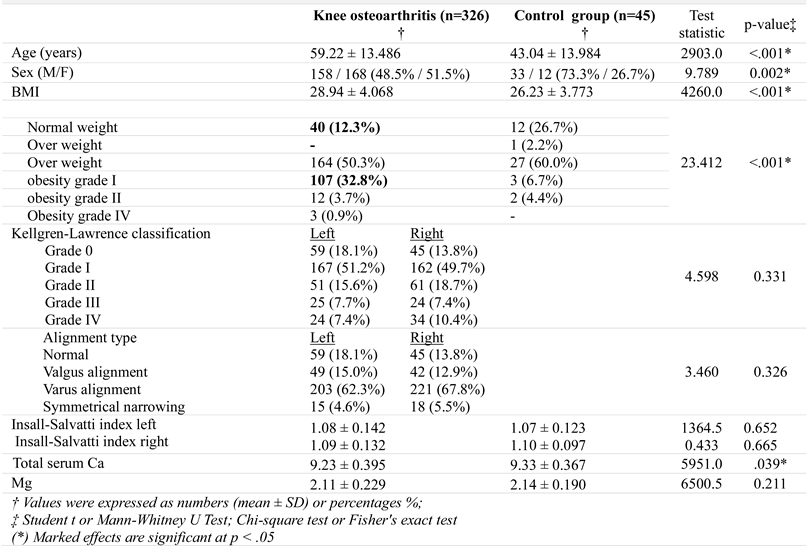
In the study group there were no statistically significant differences between the KOA diagnosis at the left and the right knee, 51.2% of cases having grade I KOA on the left, while 49.7% having grade I KOA on the right. The KOA in grade II was found in a proportion of 15.6% on the left and 18.7% on the right; grade III at 7.7% on the left and 7.4% on the right, and grade IV 7.4% on the left and 10.4% on the right (Table 1). Statistically significant differences were not found between types of knee alignment, valgus alignment being more common in the left knee (15.0%) than the right knee (12.9%) and varus alignment being more common in the right knee (67.8%) than the left (62.3%), and the symmetrical narrowing were (5.5% compared to 4.6). The distribution by gender was approximately balanced (48.5% men and 51.5% women). The mean age of patients was significantly higher (p < 0.001) for patients with KOA (59.22 ± 13,486) than patients in the control group (43.04 ± 13.984). The BMI was significantly higher (p < 0.001) in patients with KOA (28.94 ± 4.068) compared to those in the control group (26.23 ± 3.773).

Table 1.
Characteristics of patients in the study group and the control group.
Obesity show statistically significant differences between the two groups: grade I obesity was more frequently found in KOA patient group (32.8% compared to 6.7% in the control group) and normal weight was less found in KOA patients group (12.3% compared to 26.7% in the control group).
The Insall-Salvatti index could influence normal biomechanics, but it was not significantly different between patients with KOA and those in the control group in either knee (1.08 ± 0.142 compared to 1.07 ± 0.123 in the control group) for the left knee and (1.09 ± 0.132 compared to 1.10 ± 0.097 in the control group) for the right. However, it was noted that total serum Ca levels were significant lower (p = 0.039 *) in patients with KOA (9.23 ± 0.395) compared to the control group (9.33 ± 0.367) and Mg levels were lower in patients with KOA (2.11 ± 0.229) compared to those in the control group (2.14 ± 0.190), though the difference was not statistically significant.
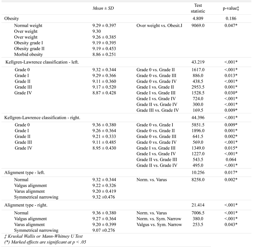
We performed univariate analysis of serum and Mg values compared on samples of interest, defined by the degree of obesity of patients, the degree of KOA, and the type of varus and valgus alignment for both knees (Table 2 and Table 3). Table 2 shows serum Ca levels in the following: a significant decrease in patients with grade I obesity compared to overweight patients (which, however, is not reflected in other obesity groups, so it may not have clinical relevance).

Table 2.
Comparative study of serum Ca values in groups (only the statistically significant comparisons between samples are enlisted).

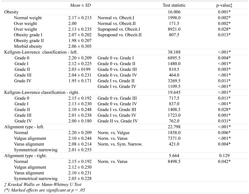
Table 3.
Comparative study of Mg values in groups (only the statistically significant comparisons between samples are enlisted).
Calcium levels decrease significantly as the degree of KOA worsens, which is also correlated with the varus and valgus alignment bilateral. In the case of left knee alignment, the serum Ca level is significantly lower only in patients with varus alignment, compared to normal knees—the other discrepancies, although present, have no statistical significance.
The comparative study of Mg levels is detailed in Table 3. There is a significant decrease in Mg levels as the degree of obesity worsens, respectively the degrees of KOA and the type of knees alignment. Regarding knee alignment, there is a tendency for lowered serum Mg levels, but with a statistically significant difference only for the right knee with varus alignment, compared to the control group.
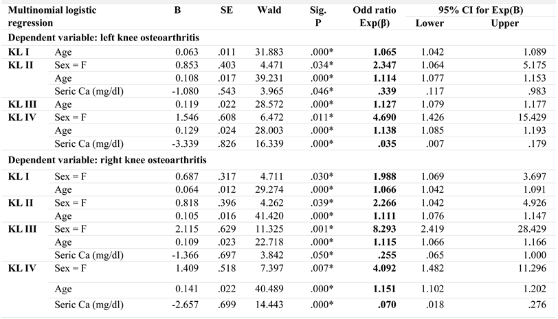
We used a multinomial logistic regression model, forward entry, to identify the predictive power of several independent variables on KL classification; the independent variables tested were those for which we identified statistically significant differences between the control group and the group of patients with KOA in the univariate analysis (Table 1): gender, age, obesity, and serum Ca levels. The type of knee alignment would also have been interesting to test, but the univariate analysis did not show a significant association between its presence and KL classification. The analysis was performed separately for KL classification for each knee and the reference category used for comparisons was KL 0, respectively healthy persons. The results are presented in Table 4.

Table 4.
Model coefficients and Wald test in multimodal logistic regression on predictive factors of osteoarthritis.
The regression model constructed for the left knee KOA assessment is valid, and is characterized by a correct case classification percentage of 51.2%, superior to the null model (without any predictor), in which the same percentage is 45.0%. The model identifies 3 significant predictors out of the 4 tested, namely the age, gender, and serum Ca values of the patients. The results in Table 4 show that the age of patients has an important role in KOA prediction, in all its degrees; female sex is an important predictor for KOA grade II and IV KL, with associated highest OR values. The serum Ca values are also significantly involved only in the case of grade II or IV KOA, having a protective role: as the level of serum Ca increases, the risk of KOA progression decreases significantly—this phenomenon is observed especially in grade IV KOA KL, where a one-unit increase in serum Ca results in a 0.035-fold decrease in OR risk.
The regression model constructed for the KOA assessment at the right knee is also valid, and is characterized by a percentage of correct case classification of 51.5%, superior to the null model (without any predictor), in which the same percentage is 43.7%. The model identifies the same 3 significant predictors among the 4 tested, namely age, gender, and serum Ca values of the patients. The results in Table 4 show that the female patients and age play an important role in the prediction of KOA, in all its degrees; being female is in fact the most important predictor, having associated the highest OR values. Serum Ca values are significantly involved only in the case of KOA grade III or IV KL, having a protective role: as the level of serum Ca increases, the risk of KOA progression decreases significantly - this phenomenon is observed especially in KOA grade IV KL, where a one-unit increase in serum Ca results in a 0.070-fold decrease in OR risk.
Discussions
Knee osteoarthritis is a degenerative pathology that affects the knee joint, for which medical services are increasingly required [34]. Significant structural and functional changes occur in cartilage that has a limited regenerative capacity [35] and because the pathophysiology of KOA is not completely understood, the importance of detecting the modifiable risk factors and those with a protective role for the joint has been emphasized [27].
The changes in KOA are closely related to a proinflammatory status [36,37] so it is important to evaluate the protective role that Ca could have for cartilage, as it is used in various forms in the treatment of periapical dental inflammation, acute edema or urticaria.
The researchers have demonstrated by experimental administration of calcium gluconate supplements its role in preventing the reduction of cartilage thickness by inhibiting chondrocytes and prostaglandin apoptosis [35]. Salem et al. have shown an inverse association between serum Ca values and prostate cancer risk [38] and other authors associate elevated serum Mg levels with oral cancer [39], providing new research opportunities.
The importance of calcium for the skin is also emphasized in the context of psoriasis, when lower levels of serum Ca were detected compared to the control group [40]). It is interesting to study the influence of Ca and Mg serum levels in radiologic changes in psoriatic arthritis.
Consequently, the hypothesis was also taken into account for KOA. Yazmalar et al. did not obtain statistically significant differences between serum Ca levels in KOA patients compared to the control group [41], the same conclusions being reported in a study that looked at the role of serum Ca levels in hand osteoarthritis [42].
Our results contradict these studies, the total serum Ca levels for patients with KOA (9.23 ± 0.395) was significantly lower (p = 0.039 *) compared to the control group (9.33 ± 0.367) and thus underline the conclusions of Hui Li et al., which support an inverse association between serum Ca values and radiological progression of KOA [17].
The role of serum Ca levels in the evolution of KOA has been demonstrated by studies that have highlighted the effects of Ca on chondrocytes in terms of matrix protein synthesis, cytoskeletal remodeling and apoptosis and regulation of proteoglycans involved in static cartilage compression [43].
Laboratory mice testing show that Mg deficiency generated a decrease in size and number of chondrocytes and proteoglycans that led to a reduction in size of articular cartilage [44]. Although some studies on human subjects have indicated an important role of Mg intake in preventing the onset and progression of KOA [45], they have been contradicted by studies that did not obtain a lower risk of KOA in increased Mg intake [28], or that noticed only modest inverse associations between them [46]. On the other hand, Wang and colleagues claim a lower risk of KOA in association with higher serum Mg values, but more evaluations are needed to confirm this hypothesis [47].
Consistently, the results developed from our research group show slightly lower Mg values in patients with KOA (2.11 ± 0.229) than in the control group (2.14 ± 0.190), but without a statistically significant difference. The protective role of Mg against the installation or progression of KOA could be explained by its participation in the preservation of muscular mass and strength that ensures a normal biomechanics [48].
Biomechanics can be altered especially by local risk factors such as anatomical changes in position involving the patella or abnormalities in the femoral or tibial extremities [49,50].
A number of risk factors associated with the onset or progression of KOA are age, sex, body mass index, or anatomical changes (Table 1), as evidenced by the literature [51]. Changes in chondrocytes and articular cartilage are exacerbated by aging and unfortunately support local inflammation [52], so that most studies associate an increased frequency of osteoarthritis in older people [24,53]. Our results support these conclusions, patients with KOA have a mean age of (59.22 ± 13,486) compared to the control group (43.04 ± 13,984).
Although it has reported that females have an increased frequency [54], we found a relatively balanced frequency though with a slight predominance of women (48.5% men and 51.5% women). In the research group, BMI was significantly higher (p <.001) in patients with KOA (28.94 ± 4.068) compared to the control group (26.23 ± 3.773). The degrees of obesity was statistically significant for grade I obesity in the case of patients with KOA (32.8% compared to 6.7% in the control group). Most researchers agree that obesity is the main risk factor for KOA [51,55], with potential for change through lifestyle and diet modification [56,57].
Another potential risk factor, though not statistically significant, was the Insall-Salvatti index, which did not show very different values between patients with KOA and the control group. The most common type of varus and valgus alignment in patients with KOA was varus alignment (62.3% left and 67.8% right). Valgus alignment was found in 15.0% in the left knee and 12.9% in the right knee and symmetrical narrowing in 5.5% left and 4.6% right. These differences are caused by the individualized biomechanics and load distribution.
We obtained an inverse association of the serum Ca levels with the varus alignment on the left side, and of Mg levels with the varus alignment on the right side. The Mg level, although not significant, showed an inverse relationship between KOA degrees and obesity. Factors with that significantly predicted bilateral KOA grades were gender (with the highest OR values for females), age, obesity, and serum Ca level of the patients [58,59,60]. Especially for patients with KL grade IV in both knees, we found that an increase of one unit of serum Ca level leads to a decrease in the risk of KOA (OR = 0.035) for the left knee and (OR = 0.07) for the right knee. Analysis of serum calcium and magnesium levels could be correlated with other pathologies in the future [61,62,63]. Also, it would be interesting to correlate the pathologies with other biomarkers that may have important predictive or diagnostic roles [64,65].
Conclusions
In conclusion, the serum Ca levels have an inverse relationship with the severity of KOA and could have a protective role. As the Mg serum levels did not present statistically significant differences between the two groups, and there are no definite conclusions regarding its protective role for KOA in the specialized literature, new research opportunities are open.
Conflict of interest disclosure
There are no known conflicts of interest in the publication of this article. The manuscript was read and approved by all authors.
Compliance with ethical standards
Any aspect of the work covered in this manuscript has been conducted with the ethical approval of all relevant bodies and that such approvals are acknowledged within the manuscript.
Acknowledgments
All authors have had equal contributions, participation and equal rights to this article.
References
- Checheriță, L.E.; Trandafir, V.; Stamatin, O.; Cărăuşu, E.M. Study of Biochemical Levels of Magnesium in Serum and Saliva in Patients with Stomatognathic System Dysfunctional Syndrome Determined by Compromised Bone Integrity and Prosthetic Treatment. Rev Chim 2016, 67, 1415–1420. [Google Scholar]
- Li, K.; Wang, X.; Li, D.; et al. The good, the bad, and the ugly of calcium supplementation: A review of calcium intake on human health. Clin Interv Aging. 2018, 13, 2443–2452. [Google Scholar] [CrossRef] [PubMed]
- Pravina, P.; Sayaji, D.; Avinash, M. Calcium and its Role in Human Body. Int. J. Res. Pharm. Biomed. Sci. 2013, 4, 659–668. [Google Scholar]
- Fan, P.; Chen, N.; Xue, S. Calcium intake, calcium homeostasis and health. Food Sci. Hum. Wellness 2016, 5, 8–16. [Google Scholar] [CrossRef]
- Pantea-Stoian, A.; Mitrofan, G.; Colceag, F.; Suceveanu, A.I.; Hainarosie, R.; Pituru, S.; Diaconu, C.C.; Timofte, D.; Nitipir, C.; Poiana, C.; et al. Oxidative Stress in Diabetes A model of complex thinking applied in medicine. Rev. Chim. 2018, 69, 2515–2519. [Google Scholar] [CrossRef]
- Stoian-Pantea, A.; Bala, C.; Rusu, A.; et al. Gender Differences in the Association of Ferritin and 25-hydroxyvitamin D. Rev Chim 2018, 69, 864–869. [Google Scholar] [CrossRef]
- Stănescu, A.M.A.; Grăjdeanu, I.V.; Iancu, M.A.; et al. Correlation of Oral Vitamin D Administration with the Severity of Psoriasis and the Presence of Metabolic Syndrome. Rev Chim 2018, 69, 1668–1672. [Google Scholar] [CrossRef]
- Vaishali, V.; Ran, W.; Leyla, O.; Puneet, D.; Yong, J.H.; Sylvia, C. Vitamin D, calcium homeostasis and aging. Bone Res. 2016, 4, 16041. [Google Scholar] [CrossRef]
- Christakos, S.; Dhawan, P.; Porta, A.; Mady, J.L.; Seth, T. Vitamin D and Intestinal Calcium Absorption. Mol Cell Endocrinol. 2011, 347, 25–9. [Google Scholar] [CrossRef]
- Tebben, J.P.; Singh, J.R.; Kumar, R. Vitamin D-Mediated Hypercalcemia: Mechanisms, Diagnosis, and Treatment. Endocr Rev. 2016, 37, 521–547. [Google Scholar] [CrossRef]
- Gröber, U.; Schmidt, J.; Kisters, K. Magnesium in Prevention and Therapy. Nutrients 2015, 7, 8199–8226. [Google Scholar] [CrossRef]
- Fontes-Pereira, A.; Rosa, P.; Thiago, B.; et al. Monitoring bone changes due to calcium, magnesium, and phosphorus loss in rat femurs using Quantitative Ultrasound. Sci Rep. 2018, 8, 11963. [Google Scholar] [CrossRef]
- de Baaij, H.F.J.; Hoenderop, G.J.J.; Bindels, J.M.R. Regulation of magnesium balance: Lessons learned from human genetic disease. Clin Kidney J. 2012, 5, i15–i24. [Google Scholar] [CrossRef]
- Forrest, H.N. Magnesium, inflammation and obesity in chronic disease. Nutr Rev. 2010, 68, 333–340. [Google Scholar] [CrossRef]
- Nasi, S.; So, A.; Combes, C.; Daudon, M.; Busso, N. Interleukin-6 and chondrocyte mineralisation act in tandem to promote experimental osteoarthritis. Ann Rheum Dis. 2016, 75, 1372–1379. [Google Scholar] [CrossRef] [PubMed]
- Zeng, C.; Wei, J.; Li, H.; et al. Relationship between Serum Magnesium Concentration and Radiographic Knee Osteoarthritis. J Rheumatol. 2015, 42, 1231–6. [Google Scholar] [CrossRef]
- Hui, L.; Chao, Z.; Jie, W.; et al. Serum Calcium Concentration Is Inversely Associated With Radiographic Knee Osteoarthritis. Medicine 2016, 95, e2838. [Google Scholar] [CrossRef]
- Tatu, A.L.; Ciobotaru, O.R.; Miulescu, M.; Buzia, O.D.; Elisei, A.M.; Mardare, N.; Diaconu, C.; Robu, S.; Nwabudike, L.C. Hydrochlorothiazide: Chemical Structure, Therapeutic, Phototoxic and Carcinogenetic Effects in Dermatology. Rev Chim 2018, 69, 2110–2114. [Google Scholar] [CrossRef]
- Takamatsu, A.; Ohkawara, B.; Ito, M. Verapamil Protects against Cartilage Degradation in Osteoarthritis by Inhibiting Wnt/β-Catenin Signaling. PLoS ONE. 2014, 9, e92699. [Google Scholar] [CrossRef]
- Tatu, A.L.; Ionescu, M.A. Multiple autoimmune syndrome type III- thyroiditis, vitiligo and alopecia areata. Acta Endo 2017, 13, 124–125. [Google Scholar] [CrossRef]
- Tatu, A.L.; Elisei, A.M.; Chioncel, V.; Miulescu, M.; Nwabudike, L.C. Immunologic adverse reactions of β-blockers and the skin (Review). Exp Ther Med. 2019, 18, 955–959. [Google Scholar] [CrossRef] [PubMed]
- Alexander, R.T.; Dimke, H. Effect of diuretics on renal tubular transport of calcium and magnesium. Am J Physiol Renal Physiol. 2017, 312, F998–F1015. [Google Scholar] [CrossRef] [PubMed]
- Lespasio, J.M.; Piuzzi, S.N.; Husni, M.E.; Muschler, F.G.; Guarino, A.J.; Mont, A.M. Knee Osteoarthritis: A Primer. Perm J. 2017, 21, 16–183. [Google Scholar] [CrossRef] [PubMed]
- Johnson, V.L.; Hunter, D.J. The epidemiology of osteoarthritis. Best Pract Res Clin Rheumatol. 2014, 28, 5–15. [Google Scholar] [CrossRef]
- Turkiewicz, A.; Petersson, I.F.; Bjork, J.; et al. Current and future impact of osteoarthritis on healthcare: A population-based study with projections to year 2032. Osteoarthr. Cartil. 2014, 22, 1826–1832. [Google Scholar] [CrossRef]
- Xie, F.; Kovic, B.; Jin, X.; He, X.; Wang, M.; Silvestre, C. Economic and Humanistic Burden of Osteoarthritis: A Systematic Review of Large Sample Studies. Pharmacoeconomics 2016, 34, 1087–1100. [Google Scholar] [CrossRef]
- Roos, E.M.; Arden, N.K. Strategies for the prevention of knee osteoarthritis. Nat Rev Rheumatol. 2016, 12, 92–101. [Google Scholar] [CrossRef]
- Wu, Z.; Yang, J.; Liu, J.; Lian, K. The relationship between magnesium and osteoarthritis of knee: A MOOSE guided systematic review and meta-analysis. Medicine 2019, 98, e17774. [Google Scholar] [CrossRef]
- Kisters, K. What is the correct magnesium supplement? Magnes Res. 2013, 26, 41–42. [Google Scholar] [CrossRef]
- Rozenberg, S.; Body, J.J.; Bruyère, O.; et al. Effects of Dairy Products Consumption on Health: Benefits and Beliefs--A Commentary from the Belgian Bone Club and the European Society for Clinical and Economic Aspects of Osteoporosis, Osteoarthritis and Musculoskeletal Diseases. Calcif Tissue Int. 2016, 98, 1–17. [Google Scholar] [CrossRef]
- Morisaki, H.; Yamamoto, S.; Morita, Y.; et al. Hypermagnesaemia-induced cardiopulmonary arrest before induction of anaesthesia for emergency caesarean section. J Clin Anaesth. 2000, 12, 224–226. [Google Scholar] [CrossRef] [PubMed]
- Harvey, C.N.; Biver, E.; Kaufman, J.M.; et al. The role of calcium supplementation in healthy musculoskeletal ageing: An Experts consensus meeting of the European Society for Clinical and Economic Aspects of Osteoporosis, Osteoarthritis and Musculoskeletal Diseases (ESCEO) and the International Foundation for Osteoporosis (IOF). Osteoporos Int. 2017, 28, 447–462. [Google Scholar] [CrossRef]
- Kohn, M.D.; Sassoon, A.A.; Fernando, N.D. Classifications in Brief: Kellgren-Lawrence Classification of Osteoarthritis. Clin Orthop Relat Res. 2016, 474, 1886–1893. [Google Scholar] [CrossRef]
- Hensor, E.M.; Dube, B.; Kingsbury, S.R.; Tennant, A.; Conaghan, P.G. Toward a clinical definition of early osteoarthritis: Onset of patient-reported knee pain begins on stairs. Data from the osteoarthritis initiative. Arthritis Care Res 2015, 67, 40–47. [Google Scholar] [CrossRef]
- Kang, S.J.; Kim, J.W.; Kim, K.Y.; Ku, S.K.; Lee, Y.J. Protective effects of calcium gluconate on osteoarthritis induced by anterior cruciate ligament transection and partial medial meniscectomy in Sprague-Dawley rats. J Orthop Surg Res. 2014, 9, 14. [Google Scholar] [CrossRef] [PubMed]
- Scanzello, R.C. Chemokines and Inflammation in Osteoarthritis: Insights from Patients and Animal Models. J Orthop Res. 2017, 35, 735–739. [Google Scholar] [CrossRef]
- Monibi, F.; Roller, B.L.; Stoker, A.; et al. Identification of synovial fluid biomarkers for knee osteoarthritis and correlation with radiographic assessment. J Knee Surg. 2016, 29, 242–247. [Google Scholar] [CrossRef]
- Salem, S.; Hosseini, M.; Allameh, F.; Babakoohi, S.; Mehrsai, A.; Pourmand, G. Serum calcium concentration and prostate cancer risk: A multicenter study. Nutr Cancer. 2013, 65, 961–968. [Google Scholar] [CrossRef] [PubMed]
- Cărăuşu, E.M.; Checheriță, L.E.; Stamatin, O.; Manuc, D. Study of Biochemical Level for Mg and Ca-Mg Imbalance in Patients with Oral Cancer and Potentially Malignant Disorder and their Prostetical and DSSS Treatment. Rev Chim 2016, 67, 2087–2090. [Google Scholar]
- Chaudhari, S.; Rathi, S. Correlation of serum calcium levels with severity of psoriasis. IJORD 2018, 4, 591–594. [Google Scholar] [CrossRef]
- Yazmalar, L.; Ediz, L.; Alpayci, M.; Hiz, O.; Toprak, M.; Tekeoglu, I. Seasonal disease activity and serum vitamin D levels in rheumatoid arthritis, ankylosing spondylitis and osteoarthritis. Afr Health Sci. 2013, 13, 47–5. [Google Scholar] [CrossRef] [PubMed][Green Version]
- Zoli, A.; Lizzio, M.M.; Capuano, A.; Massafra, U.; Barini, A.; Ferraccioli, G. Osteoporosis and bone metabolism in postmenopausal women with osteoarthritis of the hand. Menopause 2006, 13, 462–466. [Google Scholar] [CrossRef] [PubMed]
- Valhmu, W.B.; Raia, F.J. Myo-Inositol 1,4,5-trisphosphate and Ca(2+)/calmodulin-dependent factors mediate transduction of compression-induced signals in bovine articular chondrocytes. Biochem J. 2002, 361, 689–696. [Google Scholar] [CrossRef]
- Georgescu, M.; Tăpăloagă, P.R.; Tăpăloagă, D.; Furnaris, F.; Ginghină, O.; Negrei, C.; Giuglea, C.; Bălălău, C.; Ștefănescu, E.; Popescu, I.A.; et al. Evaluation of antimicrobial potential of nigella sativa oil in a model food matrix. Farmacia. 2018, 66, 1028–1036. [Google Scholar] [CrossRef]
- Zeng, C.; Li, H.; Wei, J.; Yang, T.; Deng, Z.H.; Yang, Y.; Zhang, Y.; Yang, T.B.; Lei, G.H. Association between Dietary Magnesium Intake and Radiographic Knee Osteoarthritis. PLoS ONE 2015, 26, 0127666. [Google Scholar] [CrossRef]
- Qin, B.; Shi, X.; Samai, P.S.; Renner, J.B.; Jordan, J.M.; He, K. Association of dietary magnesium intake with radiographic knee osteoarthritis: Results from a population-based study. Arthritis Care Res 2012, 64, 1306–1311. [Google Scholar] [CrossRef]
- Wang, Y.; Wei, J.; Zeng, C.; et al. Association between serum magnesium concentration and metabolic syndrome, diabetes, hypertension and hyperuricaemia in knee osteoarthritis: A cross-sectional study in Hunan Province, China. BMJ Open. 2018, 8, 019159. [Google Scholar] [CrossRef]
- Veronese, N.; Berton, L.; Carraro, S.; et al. Effect of oral magnesium supplementation on physical performance in healthy elderly women involved in a weekly exercise program: A randomized controlled trial. Am J Clin Nutr. 2014, 100, 974–981. [Google Scholar] [CrossRef]
- Motofei, I.G. A bihormonal model of normal sexual stimulation; the etiology of premature ejaculation. Med Hypotheses. 2001, 57, 93–95. [Google Scholar] [CrossRef]
- Duncan, S.T.; Noehren, B.S.; Lattermann, C. The role of trochleoplasty in patellofemoral instability. Sports Med Arthrosc Rev. 2012, 20, 171–180. [Google Scholar] [CrossRef]
- Silverwood, V.; Blagojevic-Bucknall, M.; Jinks, C.; Jordan, J.L.; Protheroe, J.; Jordan, K.P. Current Evidence on Risk Factors for Knee Osteoarthritis in Older Adults: A Systematic Review and Meta-Analysis. Osteoarthr. Cartil. 2015, 23, 507–515. [Google Scholar] [CrossRef]
- Musumeci, G.; Szychlinska, M.A.; Mobasheri, A. Age-related degeneration of articular cartilage in the pathogenesis of osteoarthritis: Molecular markers of senescent chondrocytes. Histol Histopathol. 2015, 30, 1–12. [Google Scholar] [CrossRef] [PubMed]
- Leyland, K.M.; Hart, D.J.; Javaid, M.K.; et al. The natural history of radiographic knee osteoarthritis: A fourteen- year population-based cohort study. Arthritis Rheum. 2012, 64, 2243–51. [Google Scholar] [CrossRef] [PubMed]
- Plotnikoff, R.; Karunamuni, N.; Lytvyak, E.; et al. Osteoarthritis prevalence and modifiable factors: A population study. BMC Public Health. 2015, 15, 1195. [Google Scholar] [CrossRef]
- Fowler-Brown, A.; Kim, D.H.; Shi, L.; et al. The mediating effect of leptin on the relationship between body weight and knee osteoarthritis in older adults. Arthritis Rheumatol. 2015, 67, 169–175. [Google Scholar] [CrossRef] [PubMed]
- Berbece, S.; Iliescu, D.; Ardeleanu, V.; Nicolau, A.; Jecan, R.C. Use of Phosphatidylcholine in the Treatment of Localized Fat Deposits- Results and expectations. Rev Chim 2017, 68, 1438–1441. [Google Scholar] [CrossRef]
- Brosseau, L.; Taki, J.; Desjardins, B.; et al. The Ottawa panel clinical practice guidelines for the management of knee osteoarthritis. Part II: Strengthening exercise programs. Clin Rehabil. 2017, 31, 596–11. [Google Scholar] [CrossRef]
- Gaman, M.A.; Dobrica, E.C.; Pascu, E.G.; et al. Cardio-metabolic risk factors for atrial fibrillation in type 2 diabetes mellitus: Focus on hypertension, metabolic syndrome and obesity. J Mind Med Sci. 2019, 6, 157–161. [Google Scholar] [CrossRef]
- Stoian Pantea, A.; Mitrofan, G.; Colceag, F.; Serafinceanu, C.; Totu, E.E.; Mocanu, V.; Mănuc, D.; Cărăuşu, E.M. Oxidative stress applied in Diabetes Mellitus-A new paradigm. MDPI Proc. 2019, 11, 7. [Google Scholar] [CrossRef]
- Ardeleanu, V.; Francu, L.L.; Georgescu, C. Neoangiogenesis- Assessment in esophageal adenocarcinomas. Indian J Surg. 2015, 77, 971–976. [Google Scholar] [CrossRef][Green Version]
- Gradisteanu, G.P.; Stoica, R.A.; Petcu, L.; et al. Microbiota signatures in type-2 diabetic patients with chronic kidney disease- A Pilot Study. J Mind Med Sci. 2019, 6, 130–136. [Google Scholar] [CrossRef]
- Ardeleanu, V.; Chebac, G.R.; Georgescu, C.; Vesa, D.; Frîncu, L.; Frâncu, L.D.; Păduraru, D. The modifications suffered by the peri-esophageal anatomical structures in the hiatal hernia disease: A qualitative and quantitative micro-anatomic study. RJME. 2010, 51, 765–770. [Google Scholar]
- Ardeleanu, V.; Georgescu, C.; Frîncu, L.D.; Frâncu, L.L.; Vesa, D. Angiogenesis as Prospective Molecular Biology Technique for Cancer Study. Romanian Biotehnological Letters. 2014, 19, 9637–9648. [Google Scholar]
- Cărăuşu, E.M.; Checheriță, L.E.; Stamatin, O.; Albu, A. Study of Serum and Saliva Biochemical Levels for Copper, Zinc and Copper-Zinc Imbalance in Patients with Oral Cancer and Oral Potentially Malignant Disorders and their Prostetical and DSSS (Disfunctional Syndrome of Stomatognathic System) Treatment. Rev Chim (Bucharest). 2016, 67, 1832–1836. [Google Scholar]
- Checheriță, L.E.; Trandafir, D.; Stamatin, O.; Cărăuşu, E.M. Study of Biochemical Levels in Serum and Saliva of Zinc and Copper in Patients with Stomatognathic System Dysfunctional Syndrome Following Bone Injury and Prosthetical Treatment. Rev Chim 2016, 67, 1628–1632. [Google Scholar]
© 2020 by the author. 2020 Nicoleta-Bianca Tudorachi, Iuliana Eva, Cristina Gena Dascalu, Rami AL-Hiary, Bogdan Barbieru, Mihai Paunica, Catalina Motofei, Aurelian-Corneliu Moraru


