Next Article in Journal
Xanthogranulomatous Pyelonephritis: Presentation and Management
Previous Article in Journal
Cardio Metabolic Risk Factors for Atrial Fibrillation in Type 2 Diabetes Mellitus: Focus on Hypertension, Metabolic Syndrome and Obesity
 
 
Journal of Mind and Medical Sciences is published by MDPI from Volume 12 Issue 1 (2025). Previous articles were published by another publisher in Open Access under a CC-BY (or CC-BY-NC-ND) licence, and they are hosted by MDPI on mdpi.com as a courtesy and upon agreement with Valparaiso University (ValpoScholar).
Article

The Effect of Glucosamine, Chondroitin and Harpagophytum Procumbens on Femoral Hyaline Cartilage Thickness in Patients with Knee Osteoarthritis—An MRI Versus Ultrasonography Study

by
Florentin A. Vreju
1,
Paulina L. Ciurea
1,
Anca Rosu
2,
Beatrice A. Chisalau
2,
Cristina D. Parvanescu
2,
Sineta C. Firulescu
2,
Adina Turcu-Stiolica
3,
Andreea L. Barbulescu
4,
Stefan C. Dinescu
1,
Cristiana I. Dumitrescu
4,*,
Roxana Mihaela Dumitrascu
2,
Criveanu Cristina
1,
Lucretiu Radu
5,
Mihai Tusaliu
6 and
Daniela Dumitrescu
7
1
Department of Rheumatology, Craiova University of Medicine and Pharmacy, Craiova, Romania
2
Craiova University of Medicine and Pharmacy, Craiova, Romania
3
Department of Pharmaco-Economics, University of Medicine and Pharmacy, Craiova, Romania
4
Department of Pharmacology, University of Medicine and Pharmacy, 2 Petru Rareş Street, Craiova, Romania
5
Department of Nursing & Medical Care, Craiova University of Medicine and Pharmacy, Craiova, Romania
6
Carol Davila University of Medicine and Pharmacy, Bucharest, Romania
7
Department of Radiology, Craiova University of Medicine and Pharmacy, Craiova, Romania
*
Author to whom correspondence should be addressed.
J. Mind Med. Sci. 2019, 6(1), 162-168; https://doi.org/10.22543/7674.61.P162168
Submission received: 15 October 2018 / Revised: 20 December 2018 / Accepted: 17 January 2019 / Published: 27 April 2019

Highlights

  • The combination with HPc could be able to delay progression of the knee osteoarthritis.
  • US and MRI represent important techniques with comparable results on patients with osteoarthritis, but with the remark that US is a much cheaper and more accessible tool.

Abstract

:

Highlights

  • The combination with HPc could be able to delay progression of the knee osteoarthritis.
  • US and MRI represent important techniques with comparable results on patients with osteoarthritis, but with the remark that US is a much cheaper and more accessible tool.

Abstract

Background: the evaluation of cartilage thickness has become possible with new techniques such as musculoskeletal ultrasonography (US) and magnetic resonance imagining (MRI), making the evaluation of the treatment response and the progression of the disease more accurate. Objective: to evaluate the efficacy of a Symptomatic Slow Acting Drug for Osteoarthritis using both US and MRI for measuring cartilage thickness at baseline and after 1 year. Methods: The study included the clinical evaluation of 20 patients at baseline, at 6 and 12 months as well as imaging exams (US and MRI) at baseline and after 1 year. Measurements were performed in both knees, in lateral and medial condyles, and in the intercondylar area. After the baseline visit, patients underwent a SYSADOA treatment which included Harpagophytum procumbens (HPc) administered on a daily basis, in a specific regimen. Results and discussions: The US examination permitted the detailed evaluation of the femoral hyaline cartilage thickness, with statistically significant differences before and after treatment at the level of the medial compartment, both in the dominant (1.59 ± 0.49 vs. 1.68 ± 0.49, p = 0.0013) and non-dominant knee (1.73 ± 0.53 vs. 1.79 ± 0.52, p = 0.0106). The US and the MRI correlated well (r = 0.63) and showed no radiographic progression in knee osteoarthritis after one year of treatment with specific SYSADOA. Moreover, the US showed improvement in the cartilage thickness of the medial compartment. Conclusions: The combination with HPc could increase the delay in the radiographic progression of the knee osteoarthritis, with improvement of femoral hyaline cartilage thickness in the medial and lateral compartment. The US might be an important tool in OA evaluation and monitoring.

Introduction

Osteoarthritis represents the most frequent type of articular involvement and is the result of altered metabolic processes at the level of the joint cartilage, leading to a higher rate of destruction relative to synthesis. The result is the thinning of the protective cartilage along with joint space narrowing, with consecutive pain and functional impairment in the early stages of the disease, as well as joint deformities and even ankyloses in later stages.
The treatment for osteoarthritis includes both fast acting symptomatic drugs (analgesics, nonsteroidal anti-inflammatory drugs) and slow acting drugs (SYSADOA – Symptomatic Slow Acting Drug for Osteoarthritis) – with a chondrotrophic and chondroprotective roles, such as glucosamine or chondroitin, involved in the balance between the synthesis and degradation of cartilage. The outcome is less pain and improved mobility and functioning at the joint; however, the effects tend to appear only after 6 months of treatment [1].
EULAR 2003 guidelines position glucosamine and chondroitin at the maximum level of evidence for pharmacological actions in case of knee osteoarthritis, being classified as 1A for clinical studies and at level A of recommendation [2,3]. More recently, the 2018 EULAR update for the management of hand OA includes chondroitin and chondroitin compounds among the best nutraceuticals which may be used for the improvement of articular functioning [4]. Most studies (over 300) evaluated products with a glucosamine/chondroitin rate of 500/400mg, most frequent dosages involving 1500 mg of glucosamine and 1200 mg of chondroitin, but for a short period of time, such as 3 to 6 months [5,6,7].
The American College of Rheumatology (ACR) conditionally (depending on the site of involvement) recommended avoiding glucosamine and chondroitin in cases of osteoarthritis [8], based mostly on the GAIT study that showed no difference in the joint space irrespective of the combination administered - glucosamine, chondroitin, celecoxib, the combination of glucosamine and chondroitin, or placebo [9].
Thus, the evaluation of cartilage thickness has become a requirement in the evaluation of treatment response and disease progression with the new techniques such as musculoskeletal ultrasonography (US) or magnetic resonance imagining (MRI). Currently, although consensus is lacking, most studies recommend MRI as the gold standard imaging method in rheumatology, since it can visualize all articular and peri-articular structures in great detail [10,11]. US might identify and evaluate cartilage thickness, as a hypoechoic structure, superficial to cortical bone [12,13,14,15].

Materials and Methods

Objective

The aim of our study was to evaluate the efficacy of a SYSADOA product that consists of a combination of 500g glucosamine sulfate, 400mg chondroitin sulfate, 10mg collagen type II and 40mg Harpagophytum procumbens per day (ed.), using both US and MRI to measure articular thickness. As a secondary objective, we proposed to confirm the level of agreement between US and MRI in cartilage thickness measurements.

Patients and Methods

This longitudinal prospective open study included 20 patients, aged 40-75 years, diagnosed with knee osteoarthritis according to clinical and imaging criteria [16]. Informed consent had been signed and medical history records were reviewed for previous joint disease or comorbidities. Patients underwent clinical evaluation, saving anamnestic and clinical data and establishing the stage of the disease using Kellgren and Lawrence criteria [17].
Excluded patients were those with severe knee osteoarthritis (Kellgren & Lawrence stage 4), aged over 75yr, with local trauma or in need for knee surgery, with inflammatory joint diseases or organ failure, with a change in the NSAID treatment in the past week, or with history of recent use (less than 2 months) of SYSADOA drugs. Pain was quantified on the VAS scale and functional impairment was assessed using the WOMAC osteoarthritis index and HAQ 20 item questionnaires [18].
The study design included clinical evaluation at baseline, at 6 and 12 months, and imaging exams (US and MRI) at baseline and after 1 year. After the baseline visit, which included both clinical and imaging evaluations, the patients received the SYSADOA treatment which consisted of a combination of 500g glucosamine sulfate, 400mg chondroitin sulfate, 10mg collagen type II, and 40mg Harpagophytum procumbens administered daily after lunch, for 2 months, alternated with 2 weeks of pause. In case of pain, patients were allowed to use escape medication (500 mg to 3000 mg paracetamol). Patient adherence was evaluated by counting capsules returned at each visit.

The ultrasound (US) evaluation

The ultrasound (US) evaluation was performed by the same examiner, an expert in musculoskeletal ultrasonography, using an ESAOTE Biomedica MyLab25 machine, equipped with a 10-18 MHz broadband, linear array transducer. Each patient was evaluated in grayscale (GS), being positioned in a supine position with the knees fully flexed in order to examine the femoral hyaline cartilage, in both the short and long axis of the femur. The US was performed according to EULAR guidelines [19]. The hyaline cartilage was identified as a hypoechoic line of varying thickness, which was lying on top of the hyperechoic femoral cortical bone, covered by a thin hyperechoic line, defined as cartilage interface. The measurements were performed in both knees with the transducer in transverse scan, in both lateral and medial condyles, and in the intercondylar area, assuring a sharp horizontal superficial hyperechoic demarcation of the cartilage (Figure 1).
Cartilage thickness was recorded and measured in all three areas. Moreover, any irregularities in the cartilage and subchondral bone, and any change in the hypoechoic aspect of the cartilage or in the cartilage interface that might have been indicative of crystal arthropathies, were noted. The interpretation of all findings was based on OMERACT definitions for US pathology [20].

The MRI examination

The scans were performed by an experienced radiology technician and were analyzed by an expert radiologist, with over 10 years of experience in rheumatologic MRI scans, which were both blinded to US and clinical data. The study was performed on a 1.5 T MR system (Siemens Magnetom Symphony, 1.5 T) equipped with CP Flex large coil.
The positioning of the patient in the magnet was supine with feet forward; the image protocol consisted of: COR_T2_TIRM, SAG_SE_T1, SAG_PD_TSE_FS and AX_T2_ME2D. The sequence parameters were: on COR_T2_TIRM: TR/TE: 7160 / 77; SAG_SE_T1: TR/TE: 526/12; SAG_PD_TSE_FS: TR/TE: 3960/14; AX_T2_ME2D: TR/TE: 758/22. The vision field was 170 mm and the slice thickness ranged between 0.5-1.5mm.
The measurement of cartilage thickness was performed first, on the COR_T2_TIRM sequence at three sites: in the middle of the medial femoral condyle, of the lateral femoral condyle, and the intercondylar area. The same medial and lateral femoral condyles were measured on SAG_SE_T to confirm the results.

Statistical analysis

Statistical analysis was performed using Graph Pad Prism 5.00 and data were expressed as the mean ± standard deviation (SD) unless specified otherwise. The values of cartilage thickness in both US and MRI evaluations were analyzed using paired t-test to verify differences between dominant and non-dominant sides and non-paired t-test to compare different imaging methods. Pearson’s correlation coefficient and linear regression models assessed the possible correlation between US findings and variables such as age, HAQ, or WOMAC. The significance level was set at p ≤ 0.05.

Results

The baseline evaluation included 40 knees in 20 patients (11 females and 9 males), with a mean age of 59.3±9.12yo and mean disease duration at the time of inclusion of 7.35±3.45 years, ranging from to 2 to 15 years. The mean VAS at baseline was 76±9.90 mm.
The US evaluation at baseline revealed that cartilage loss was more significant in the medial compartment, especially when compared to the intercondylar area. Thus, cartilage thickness was significantly higher in the central, intercondylar area (1.944±0.68 mm) when compared to the medial compartment (1.50±0.46, p=0.0037). However, we did not find any statistically significant differences between the intercondylar area and the lateral compartment, but a tendency for higher values in cartilage thickness in the central area (1.944±0.68 vs. 1.708±0.53, p=0.127). We also did not find any differences between the dominant versus non-dominant knee.
The MRI measurements correlated well with the values of cartilage thickness obtained by means of US, especially for the medial compartment (r=0.63), suggesting that ultrasonography could be an important evaluation method for the femoral hyaline cartilage. The minimal cartilage thickness value was 0.9 mm through MRI and corresponded to the minimal thickness of 0.83 mm measured through US, in the same patient.
From the point of view of articular functionality, it seems that there is a strong correlation with cartilage loss in general and not with specific medial compartment cartilage thinning (r=0.98). On the other hand, disability index correlates better with cartilage thickness in the dominant knee (r=0.82).

Imaging evaluation of treatment response after 1 year

The US and MRI evaluation 1 year after treatment initiation with combined therapy included 17 patients, since 3 patients were lost at follow-up: one patient underwent knee surgery secondary to meniscus and anterior cruciate ligament lesions, and 2 patients decided to leave the study for subjective reasons, not related to medication.
The MRI results showed a difference in the femoral hyaline cartilage thickness, both in medial and lateral compartments, with an improvement at this level after the treatment. Thus, in the medial compartment of the dominant knee, there was a higher difference between baseline and 1-year later visit, though still not statistically significance (p=0.09). Femoral cartilage thickness seems to improve after SYSADOA treatment, especially in the non-dominant knee (non-dominant, p=0.05 vs. dominant, p=0.08).
There was no difference in cartilage thickness before and after treatment, in the intercondylar area, both for the dominant (1.316±0.289 vs. 1.30±0.288mm, p=0.824) and non-dominant knee (1.232±0.264 vs. 1.276±0.258mm, p=0.500).
The US examination permitted detailed evaluation of the femoral hyaline cartilage thickness, with a statistically significant difference before and after treatment at the level of the medial compartment, both in the dominant (1.59±0.49 vs. 1.68±0.49, p=0.0013) and non-dominant knee (1.73±0.53 vs. 1.79±0.52, p=0.0106). The improvement was even more visible in the lateral compartment, without depending on the dominant/non-dominant joint (Table 1).

Discussions

This study highlighted two important facts: one concerning novel possibilities for the treatment of knee osteoarthritis and the other related to the role of US in the evaluation of the femoral hyaline cartilage, compared to the gold standard represented by MRI.
Multiple long-term studies have shown that glucosamine administration might delay the progression of knee osteoarthritis [21] and may even determine modifying changes in the course of the disease (reversion). The study published in 2002 by Pavelka et al., which included 202 patients with knee osteoarthritis, showed that after the daily administration for 3 years of 1500 mg of glucosamine or placebo, the progression of joint space narrowing was different (+0.04 vs. -0.19) [21]. Regarding the clinical evaluation, the placebo group showed no significant difference, in comparison with the active treatment group [13]. In 2003, Bruyere et al. published a study on 212 patients with knee osteoarthritis, with similar results, with the remark that patients with mild to moderate disease benefited more after glucosamine treatment [23].
Most of those studies tested only trophic compounds (glucosamine, chondroitin), in monotherapy or combination, without analgesic or anti-inflammatory drug association. Actually, it was considered that one of the benefits of SYSADOA treatment was the NSAID and analgesic drug sparing, in order to limit their cardiovascular, renal, or gastrointestinal adverse effects. However, the fact that NSAIDs and even glucocorticoids, in some specific doses, might influence joint cartilage metabolism was not evaluated. NSAIDs, especially, seem to inhibit the proteoglycan synthesis by chondrocytes [24].
In the last years, there has been increasing evidence of the favorable effects of Harpagophytum procumbens on pain and inflammation as an alternative to NSAIDs, with an improved safety profile [25]. The standardized daily extract of 60mg Harpagophytum procumbens seems to have a similar effect as selective COX-2 inhibitors, but with fewer adverse reactions. It also seems to have a chondroprotective role by inhibiting NO, TNF-α, interleukin 1-β, leukotriene formation, and matrix metalloproteinase (MMP), which are responsible for cartilage destruction. Nevertheless, the anti-inflammatory and chondroprotective actions could be explained by inhibition of lipid peroxidase [26].
Although, the association of those drugs was known, it was based only on the individual therapeutic properties of each of these compounds, as there is only one study in the literature showing that the combination of glucosamine-chondroitin-Harpagophytum procumbens could lead to the inhibition of MMP metabolism [27,28].
Our study shows an increased delay in the progression of osteoarthritis, by adding Harpagophytum procumbens to the known chondroprotective combination of glucosamine-chondroitin, leading to benefits even before the 3-year period of treatment.
The other result of the study showed a high level of agreement between US and MRI measurements of cartilage thickness, both on condyles and intercondylar area, in accordance with previous studies [29]. Similar to the study of Pradsgaard et al., we have found constantly higher values of cartilage thickness measured by MRI, compared to the US-measured ones, a fact that could be explained by the higher sound speed inside the cartilage, in comparison with other tissues (roughly 1696m/s vs 1540m/s) [29]. Pradsgaard et al. suggested multiplying the results of the US measurement by 1.10 in order to obtain an accurate cartilage thickness, a finding confirmed by our study. Thus, our study also revealed no differences in the speed of sound within cartilage when comparing the US results to the MRI results. Therefore, once again, US might prove an important and accessible tool in the diagnosis and evaluation of osteoarthritis, besides other inflammatory and non-inflammatory joint diseases [30,31].
We are aware of the limitations of this work. Because participants were drawn from a single center, the study risks selection bias. Moreover, our study is not placebo-controlled and is limited to the small number of patients evaluated by a single US examiner, but it can be considered a pilot for further extension research, with intra- and inter-reliability studies.

Conclusions

The combination of 500g glucosamine sulfate, 400mg chondroitin sulfate, 10mg collagen type II and 40mg Harpagophytum procumbens determined a delay in the radiographic progression of knee osteoarthritis, in all compartments, with improvement of the femoral hyaline cartilage thickness in the medial and lateral compartment, visible both through US and MRI imaging techniques.
Furthermore, US and MRI represent important techniques, with comparable results in the diagnosis, evaluation, treatment, and monitoring of patients with osteoarthritis, but with the remark that US is a much less costly and more accessible tool which offers the possibility of multiple joint evaluation.

Acknowledgement

This work was supported by a grant 966/2013 supported by Fiterman Pharma SRL.

Conflict of interest disclosure

There are no known conflicts of interest in the publication of this article. The manuscript was read and approved by all authors.

Compliance with ethical standards

Any aspect of the work covered in this manuscript has been conducted with the ethical approval of all relevant bodies and that such approvals are acknowledged within the manuscript.

References

  1. Biro, A.; Georgescu, L.; Nedelcut, C.; Marinescu, C.; Bolosiu, H. Effect of the combination glucosamine hydrochloride and chondroitin sulphate on knee osteoarthritis symptoms: a randomized, double blind, placebo-controlled study. Ro J Rheumatol. 2009, 18, 105–113. [Google Scholar]
  2. Jordan, K.; Arden, N.; Doherty, M.; et al. EULAR Recommendations 2003: an evidence based approach to the management of knee osteoarthritis: report of a task force of the standing committee for international clinical studies including therapeutic trials (ESCISIT). Ann Rheum Dis. 2003, 62, 1145–1155. [Google Scholar] [CrossRef] [PubMed]
  3. Zhang, W.; Moskowitz, R.W.; Nuki, G.; et al. OARSI recommendations for the management of hip and knee osteoarthritis, Part II: OARSI evidence-based, expert consensus guidelines. Osteoarthritis Cartilage. 2008, 16, 137–162. [Google Scholar] [CrossRef] [PubMed]
  4. Kloppenburg, M.; Kroon, F.P.; Blanco, F.J.; et al. 2018 update of the EULAR recommendations for the management of hand osteoarthritis. Ann Rheum Dis. 2019, 78, 16–24. [Google Scholar] [CrossRef]
  5. Henrotin, Y.; Marty, M.; Mobasheri, A. What is the current status of chondroitin sulfate and glucosamine for the treatment of knee osteoarthritis? Maturitas. 2014, 78, 184–187. [Google Scholar] [CrossRef]
  6. Roman-Blas, J.A.; Mediero, A.; Tardío, L.; et al. The combined therapy with chondroitin sulfate plus glucosamine sulfate or chondroitin sulfate plus glucosamine hydrochloride does not improve joint damage in an experimental model of knee osteoarthritis in rabbits. Eur J Pharmacol. 2017, 794, 8–14. [Google Scholar] [CrossRef]
  7. Silva, F.S., Jr.; Yoshinari, N.H.; Castro, R.R.; et al. Combined glucosamine and chondroitin sulfate provides functional and structural benefit in the anterior cruciate ligament transection model. Clin Rheumatol. 2009, 28, 109–117. [Google Scholar] [CrossRef]
  8. Hochberg, M.C.; Altman, R.D.; April, K.T.; et al. American College of Rheumatology 2012 recommendations for the use of nonpharmacologic and pharmacologic therapies in osteoarthritis of the hand, hip, and knee. Arthritis Care Res. 2012, 64, 465–474. [Google Scholar] [CrossRef]
  9. Sawitzke, A.D.; Shi, H.; Finco, M.F.; et al. The effect of glucosamine and/or chondroitin sulfate on the progression of knee osteoarthritis: a report from the glucosamine/chondroitin arthritis intervention trial. Arthritis Rheum. 2008, 58, 3183–3191. [Google Scholar] [CrossRef]
  10. Malattia, C.; Damasio, M.B.; Magnaguagno, F.; Pistorio, A.; Valle, M.; Martinoli, C.; et al. Magnetic resonance imaging, ultrasonography and conventional radiography in the assessment of bone erosions in juvenile idiopathic arthritis. Arthritis Rheum. 2008, 59, 1764–1772. [Google Scholar] [CrossRef]
  11. Zhang, W.; Doherty, M.; Peat, G.; et al. EULAR evidence-based recommendations for the diagnosis of knee osteoarthritis. Ann Rheum Dis. 2010, 69, 483–489. [Google Scholar] [CrossRef] [PubMed]
  12. Torp-Pedersen, S.; Bartels, E.M.; Wilhjelm, J.; Bliddal, H. Articular cartilage thickness measured with US is not as easy as it appears: a systematic review of measurement techniques and image interpretation. Ultraschall Med. 2011, 32, 54–61. [Google Scholar] [CrossRef] [PubMed]
  13. Patil, S.G.; Zheng, Y.P.; Wu, J.Y.; Shi, J. Measurement of depth-dependence and anisotropy of ultrasound speed of bovine articular cartilage in vitro. Ultrasound Med Biol. 2004, 30, 953–963. [Google Scholar] [CrossRef] [PubMed]
  14. Yao, J.Q.; Seedhom, B.B. Ultrasonic measurement of the thickness of human articular cartilage in situ. Rheumatology 1999, 38, 1269–1271. [Google Scholar] [CrossRef]
  15. Colebatch, A.N.; Edwards, C.J.; Ostergaard, M.; et al. EULAR recommendations for the use of imaging of the joints in the clinical management of rheumatoid arthritis. Ann Rheum Dis. 2013, 72, 804–814. [Google Scholar] [CrossRef]
  16. Altman, R.; Asch, E.; Bloch, D.; et al. Development of criteria for the classification and reporting of osteoarthritis. Classification of osteoarthritis of the knee. Diagnostic and Therapeutic Criteria Committee of the American Rheumatism Association. Arthritis Rheum. 1986, 29, 1039–1049. [Google Scholar] [CrossRef]
  17. Kellgren, J.H.; Lawrence, J.S. Radiological assessment of osteoarthrosis. Ann Rheum Dis. 1957, 16, 494–502. [Google Scholar] [CrossRef]
  18. Bellamy, N.; Buchanan, W.W.; Goldsmith, C.H.; Campbell, J.; Stitt, L.W. Validation study of WOMAC: a health status instrument for measuring clinically important patient relevant outcomes to antirheumatic drug therapy in patients with osteoarthritis of the hip or knee. J Rheumatol. 1988, 15, 1833–1840. [Google Scholar]
  19. Brown, A.K.; O'Connor, P.J.; Roberts, T.E.; Wakefield, R.J.; Karim, Z.; Emery, P. Recommendations for musculoskeletal ultrasonography by rheumatologists: setting global standards for best practice by expert consensus. Arthritis Rheum. 2005, 53, 83–92. [Google Scholar] [CrossRef]
  20. Wakefield, R.J.; Balint, P.V.; Szkudlarek, M.; et al. Musculoskeletal ultrasound including definitions for u ltrasonographic pathology. J Rheumatol 2005, 32, 2485–2487. [Google Scholar]
  21. Gavrilă, M.T.; Ștefan, C. Arthroscopic treatment for elbow intraarticular loose bodies. J Clin Invest Surg. 2018, 3, 100–104. [Google Scholar] [CrossRef]
  22. Reginster, J.Y.; Deroisy, R.; Rovati, L.C.; et al. Long-term effects of glucosamine sulphate on osteoarthritis progression: a randomised, placebo-controlled clinical trial. Lancet 2001, 357, 251–6. [Google Scholar] [CrossRef] [PubMed]
  23. Pavelká, K.; Gatterová, J.; Olejarová, M.; Machacek, S.; Giacovelli, G.; Rovati, L.C. Glucosamine sulfate use and delay of progression of knee osteoarthritis: a 3-year, randomized, placebo-controlled, double-blind study. Arch Intern Med. 2002, 162, 2113–2123. [Google Scholar] [CrossRef] [PubMed]
  24. Bruyere, O.; Honore, A.; Ethgen, O.; et al. Correlation between radiographic severity of knee osteoarthritis and future disease progression. Results from a 3-year prospective, placebo-controlled study evaluating the effect of glucosamine sulfate. Osteoarthritis Cartilage 2003, 11, 1–5. [Google Scholar] [CrossRef]
  25. Brandt, K.D. Effects of nonsteroidal anti-inflammatory drugs on chondrocyte metabolism in vitro and in vivo. Am J Med. 1987, 83, 29–34. [Google Scholar] [CrossRef]
  26. Chrubasik, S.; Model, A.; Black, A.; Pollak, S. A randomised double-blind pilot study comparing Doloteffin and Vioxx in the treatment of low back pain. Rheumatology 2003, 42, 141–148. [Google Scholar] [CrossRef]
  27. Grant, L.; McBean, D.; Fyfe, L.; Warnock, M. ffects of Harpagophytum procumbens (Devils claw) on the cyclooxygenase and lipoxygense pathways of the arachidonic acid cascade. In: Govil JN, Singh VK, Bhardwaj R (eds). Recent progress in medicinal plants volume 24, Studium Press, 2009: 203-19.
  28. Schulsse, S.; Wiggers, L.; Daix, M.; Kirschvink, N. Effects of an oral supplementation with glucosamine and chondroitin sulphate on MMP-2 activity in supernatants of IL-1 stimulated-equine chondrocytes. Acta physiologica. 2008, 194, P–15. [Google Scholar]
  29. Pradsgaard, D.Ø.; Fiirgaard, B.; Spannow, A.H.; Heuck, C.; Herlin, T. Cartilage thickness of the knee joint in juvenile idiopathic arthritis: comparative assessment by ultrasonography and magnetic resonance imaging. J Rheumatol. 2015, 42, 534–540. [Google Scholar] [CrossRef]
  30. Filippou, G.; Scirè, C.A.; Adinolfi, A.; et al. Identification of calcium pyrophosphate deposition disease (CPPD) by ultrasound: reliability of the OMERACT definitions in an extended set of joints-an international multiobserver study by the OMERACT Calcium Pyrophosphate Deposition Disease Ultrasound Subtask Force. Ann Rheum Dis. 2018, 77, 1194–1199. [Google Scholar] [CrossRef]
  31. Vreju, A.F.; Ciurea, M.E.; Popa, D.; et al. Ultrasonography in diagnosis and management of non-inflammatory conditions of the hand and wrist. Med Ultrason. 2016, 18, 90–95. [Google Scholar] [CrossRef]
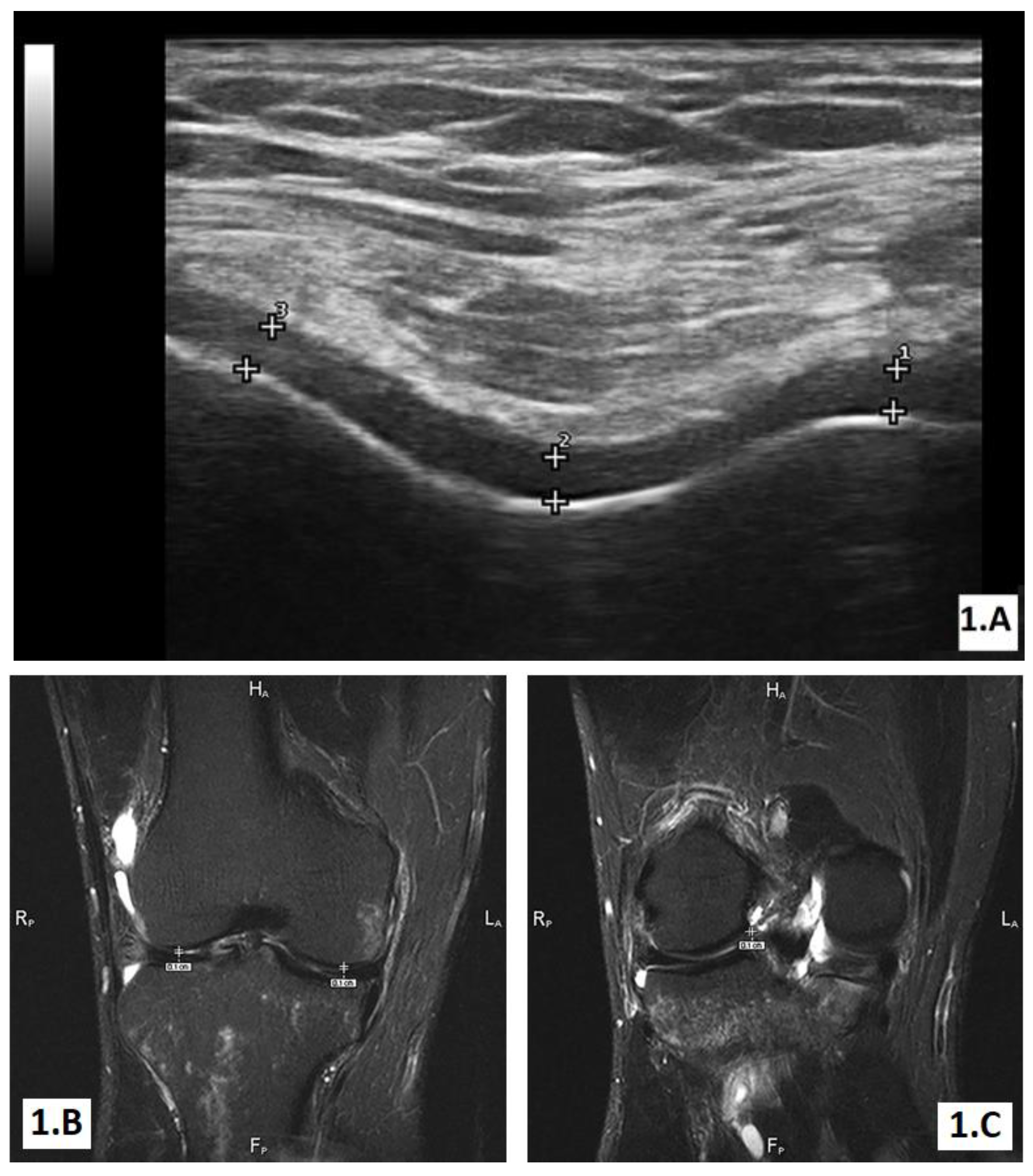
Figure 1. A. Ultrasound GS short axis image of the femoral hyaline cartilage, with the knee fully flexed. B, C. T2-weighed MRI images of the knee, with visible femoral cartilage and measurements of condyles (B) and intercondylar area (C).
Jmms 06 00024 g001
Table 1. US cartilage thickness evolution from baseline to one year
Jmms 06 00024 i001