Next Article in Journal
Vulnerability and Physical Well-Being of Caregivers: What Relationship?
Previous Article in Journal
Serum Markers of Bone Fragility in Type-2 Diabetes Mellitus
 
 
Journal of Mind and Medical Sciences is published by MDPI from Volume 12 Issue 1 (2025). Previous articles were published by another publisher in Open Access under a CC-BY (or CC-BY-NC-ND) licence, and they are hosted by MDPI on mdpi.com as a courtesy and upon agreement with Valparaiso University (ValpoScholar).
Review

Microleakage—The Main Culprit in Bracket Bond Failure?

by
Ioana Roxana Bordea
,
Adina Sîrbu
,
Ondine Lucaciu
,
Aranka Ilea
,
Radu Septimiu Câmpian
,
Doina Adina Todea
,
Teodora Gabriela Alexescu
*,
Maria Aluaș
,
Corina Budin
and
Andreea Simona Pop
The University of Medicine and Pharmacy, Cluj Napoca, Romania
*
Author to whom correspondence should be addressed.
J. Mind Med. Sci. 2019, 6(1), 86-94; https://doi.org/10.22543/7674.61.P8694
Submission received: 5 May 2018 / Revised: 20 July 2018 / Accepted: 18 August 2018 / Published: 27 April 2019

Highlights

  • In vitro microleakage value was higher in the gingival margin at the enamel-adhesive interfaces and in the occlusal margin at the adhesive-metal bracket interfaces.
  • Bracket debonding remains the main concern during the orthodontic treatment, despite the new techniques that may improve the conventional orthodontic treatment.

Abstract

:
Microleakage is the most common cause of bracket debonding. Moreover, different thermal expansion coefficients between the enamel, the adhesive, and the bracket bases will cause repeated expansion and contraction, adding more stress to the bonding strength. Debonding represents the failure of the adhesion between the brackets and the tooth enamel. The debonding of brackets from the enamel surface is the result of several factors, such as acid-etching and drying, adhesive application, and the time and type of photo activation. The under polymerization process of composite photo activation may lead to early bracket debonding. Objective. The aim of this research is to review the available studies assessing bracket debonding due to microleakage. Material and Methods. An electronic search in Pub Med database and Web of Science was conducted between September-October 2018. The inclusion criteria were articles written in English, full-text articles, studies published in the last 5 years, studies in vivo, ex vivo, and in vitro. The outcome measures in this research were the conditions that determine orthodontic bracket debonding due to microleakage. Results. The MEDLINE search resulted in 510 titles and abstracts that were relevant to the present topic; after selecting the articles published in the last five years, 74 were available for further selection. After the exclusion of all the studies irrelevant for the aim of the paper, 13 articles were finally included in this research. In vitro studies showed that microleakage score was higher in the gingival margin at the enamel-adhesive interfaces and in the occlusal margin at the adhesive-metal bracket interfaces. Conclusion. Bracket debonding remains the main concern during the orthodontic treatment, despite the new techniques.

Highlights

  • In vitro microleakage value was higher in the gingival margin at the enamel-adhesive interfaces and in the occlusal margin at the adhesive-metal bracket interfaces.
  • Bracket debonding remains the main concern during the orthodontic treatment, despite the new techniques that may improve the conventional orthodontic treatment.

Introduction

The first studies published on the bonding techniques used for bonding brackets to the enamel surface were conducted during the 1960s and those techniques have constantly improved ever since [1,2].
Different materials have been used in order to produce esthetic and non-esthetic brackets, such as stainless steel, ceramics, titanium, and polymers. In order to select the most suitable bracket adhesive combinations, in vitro studies are performed to evaluate the orthodontic bonding strength.
The laboratory tests evaluating the shear and tensile bond strength are the most used tests in the detection of the fulfillment of the orthodontic bonding system [3–5].
Debonding represents the failure of adhesion between the brackets and the tooth enamel. The debonding of brackets from the enamel surface is the result of several factors, such as acid-etching and drying, adhesive application, and the time and type of photo activation. The under polymerization process of composite photo activation may lead to early bracket debonding [6–8].
The bond strength between the bracket base and the enamel surface is essential in orthodontics. Microleakage is the most common cause of bracket debonding, representing the reduction in the marginal integrity, thus permitting the diffusion of bacteria, oral fluids, ions, and different types of molecules between the marginal gaps. Different thermal expansion coefficients between the enamel (α = 12 ppm/˚C), the adhesive (α= 20–55 ppm/˚C) and the bracket base (α = 16 ppm/˚C) will cause repeated expansion and contraction, adding additional stress to the bonding strength [9–13].
The minimum acceptable shear bond strength values of orthodontic appliances range between 5.8 MPa and 7.8 MPa; however, when the bond strength exceeds 10 MPa, the enamel is damaged [14].
Nowadays, direct and indirect bonding methods are used in orthodontics, both having advantages and disadvantages, and that correlate with bracket detachment [15]. The systems that can be used in orthodontics for the shear bond strength are acid primer, light-curing glass ionomer, light-cured and self-cured composite adhesive systems [16,17].

Objective

The aim of the current research is to systematically review available studies assessing bracket detachment due to microleakage.

Materials and Methods

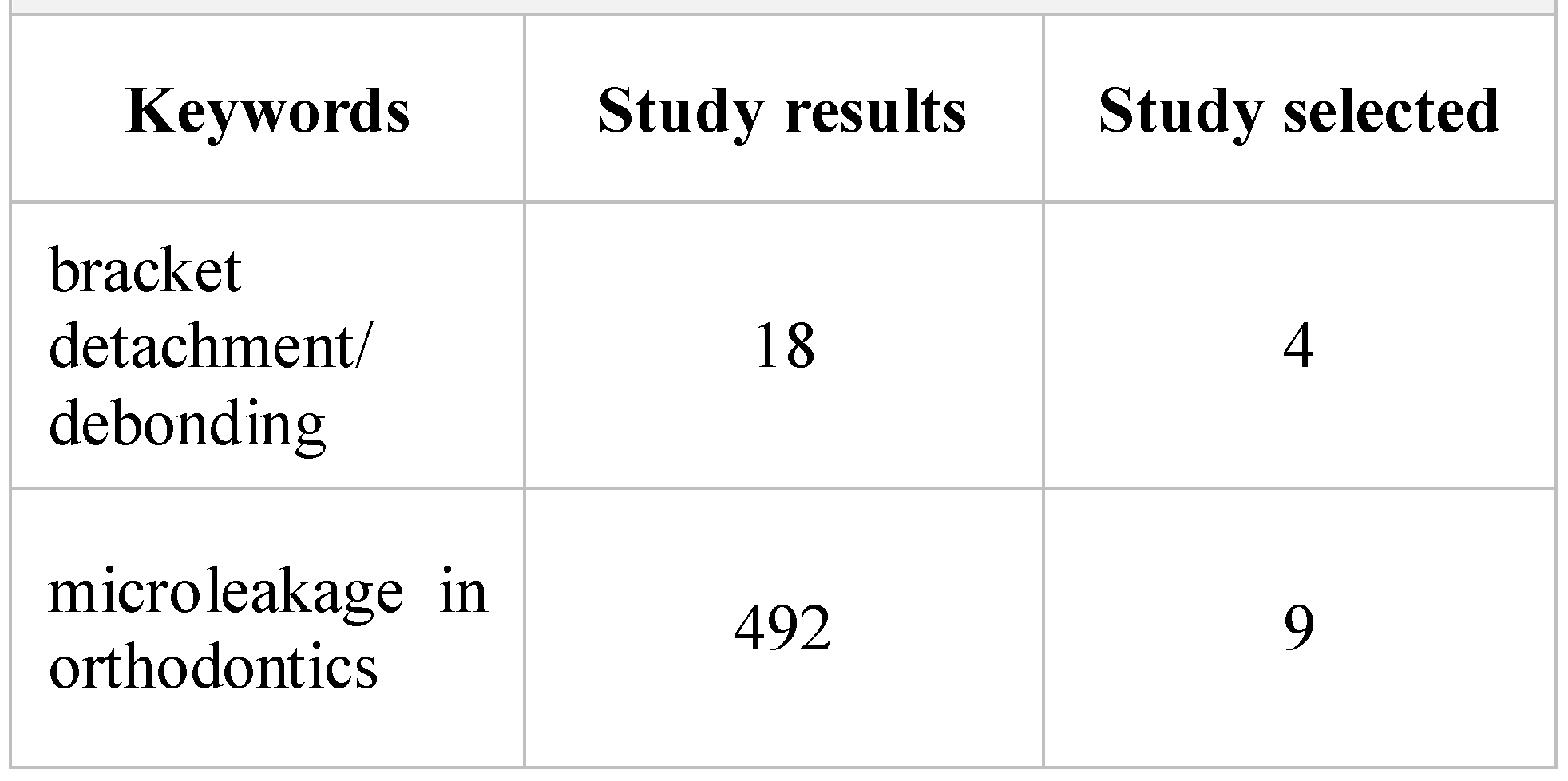
The purpose of this research is to summarize the current literature regarding bracket detachment due to microleakage. An electronic search in Pub Med database and Web of Science was conducted through September 2018. Only studies published in English were included in this research. The search in the databases used the following keywords: “bracket detachment/ debonding” OR “microleakage in orthodontics”. The studies from the reference list of the selected ones were then searched manually in the databases.
The inclusion criteria were: articles written in English, full-text articles, studies published in the last 5 years, and all the studies performed in vitro, ex vivo and in vivo. The exclusion criteria were: reviews of literature and studies about bonding that were correlated with other dental specialties.
The full-text articles remaining after the application of the inclusion and exclusion criteria were then evaluated in order to identify the eligible ones. From the studies included, we extracted the following data: the author(s), the study design, the total number of teeth used, the bonding technique used, the cause of bracket failure (detachment/ debonding and microleakage), results, and conclusions.
The outcome measure in this research was the incidence of Orthodontic Bracket Detachment due to microleakage.

Results and Discussions

MEDLINE search resulted in 510 titles and abstracts that were relevant for the present topic; after selecting the articles published in the last five years, 74 articles remained—Table 1. After the exclusion of all the studies irrelevant for the current aim, 13 articles were finally included in this analysis—Figure 1.
Sha et al. conducted a study on 30 extracted human maxillary premolars, using CAD/CAM techniques and customized bracket systems. They formed 5 groups of six teeth each in order to measure the debonding force (DF; N) and shear bond strength (SBS; N). The control group, Group 1, underwent direct bonding with a pre-adjusted bracket (Clippy M, Tomy); Group 2 underwent indirect bonding with Harmony bracket (American Orthodontic, Sheboygan); Group 3 underwent Incognito bracket (3 m Unitek); Group 4 underwent indirect bonding with Insignia bracket (Ormco) and Group 5 underwent indirect bonding with Orapix bracket (Orapix). Transbond XT and dual-curing self-adhesive resin cements (RelyX, ESPE) were used. Adhesive remnants were then analyzed with SEM. The results revealed that Group 2 (lingual self- ligating methods) had significantly higher DF than group 1 (pre-adjustable self-ligating labial metal bracket). Also, customized brackets exhibited larger deviations in DF and SBS. All customized bracket systems exhibited DF that was equivalent or superior to pre-adjustable brackets, even when placed by indirect bonding [18].
In the study conducted by Piccoli L et al., 60 dental elements were studied, both maxillary and mandibular, previously extracted for orthodontic or periodontal reasons. They used two different methods of orthodontic debonding: cutters for orthodontics and pliers for debonding. Three different materials for the adhesions of brackets were studied: light-curing adhesive system (Transbond XT primer, Transbond XT Adhesive Paste), self-curing adhesive system (Ortho-one No Mix Primer and paste) and glass ionomer cement (Fuji Ortho liquid and paste). Metallic self-ligating brackets were used in all 6 groups. There was a significant correlation between the debonding techniques, the materials for membership, and the ARI index. In the first survey among the elements in which a glass ionomer cement was used, 61% of the sample presented value 0 in the ARI index, compared to 8% of the items for which a light-curing composite was used and 31% among the elements for which a self-curing composite was used. The second survey investigation showed no significant values (p value >α). The results showed that adhesive bond failure site during debonding varies according to the material used for bonding. The highest values of the ARI index were recorded with the use of a light-cured composite; the same behavior was observed for the self-curing composites [19].
Arash V et al. conducted a study on 120 extracted human maxillary premolar teeth, which were randomly divided into 4 groups: HM group (metallic bracket/conventional bonding agents), SM group (metallic bracket (Standard-022, Dentaurum)/ Transbond self-etching primer), HC group (ceramic bracket/ conventional bonding agent Transbond XT), SC group (ceramic bracket/ Transbond self-etching primer). The ARI index was determined under stereomicroscope and the enamel detachment index was evaluated with SEM. The mean shear bond strength values were (MPa+/- SD): HM group = 12.59, SM group = 11.15, HC group = 7.7, SC group = 7.41. The conclusion was that the bond strength showed significant differences between groups: HM and HC, SM, and SC (p < 0.05) [20].
Kaneshima et al. used 60 human molars. Orthodontic tubes (3 M) were bound on teeth using the following adhesive systems: O-Opaque (Enlight, Ormco), LF-low fluorescence (Transbond Color Change, 3 M), HF-high fluorescence (Orthocem UV Trac, FGM). After debonding, the groups were subdivided according to the AR removal method: with/ without UV light. They used direct visual analysis, SEM, and time quantification for AR removal. AR removal with light was significantly faster compared to without UV light (p < 0.0001). The use of UV light may aid orthodontists in removing AR more thoroughly and in a shorter period of time [21].
In the study of Hedayati et al., 40 human premolars were used and divided into 4 equal samples: Group I: Acid etch plus Transbond XT primer and Transbond XT adhesive, Group II: Acid etch plus Transbond XT primer and nanocomposite (Filtek Z350), Group III: Scotchbond™ Universal primer plus Transbond XT, and Group IV: Scotchbond™ and nanocomposite. The sections were prepared in order to compare the microleakage values in the groups at occlusal and gingival margins under the stainless steel brackets. Statistical analysis was done using the ANOVA test. The results showed that the gingival side had a statistically higher value of micro- leakage. The nanocomposite Filtek Z350 presented higher values of microleakage in the occlusal and gingival side of the brackets related to Transbond XT.
The brackets that were bound using acid etch showed higher values in comparison with the group in which Scotchbond was used. In the groups that were bound with nanocomposites, the values of microleakage were higher [22].
Öztürk et al. conducted a study on 30 human maxillary premolars that were divided into five groups and ceramic brackets were bound. One group of teeth had the bonding performed with Transbond XT and the other groups were bound through an indirect technique with Custom I.Q. (Reliance Orthodontic Products), Sondhi Rapid-Set (3 M Unitek), RMbond (RMO), and Transbond IDB (3 M Unitek). In order to evaluate microleakage, the Skyscan Micro Ct system model 1172 was used. The Kruskal- Wallis test and Wilcoxon signed rank test were used for the statistical analysis. As for the values of microleakage, there was no significantly statistical difference between the studied groups according to the Kruskal-Wallis test, but the Wilcoxon signed rank test indicated different values regarding the coronal microleakage volume and the percentage in the groups with RM bond and Transbond IDB [23].
Pakshir et al. used 120 bovine deciduous lower incisors that were divided in four groups and bound with metallic brackets: Group I: Acid etching + Transbond XT primer + direct illumination, group II: acid etching + Transbond XT primer + transillumination, group III: Transbond XT self-etching primer + direct illumination and Group IV: Transbond XT self-etching primer + transillumination. In order to assess the values of microleakage, dye penetration was used and sections at the enamel-adhesive and adhesive-bracket interfaces were made and then observed under the stereomicroscope. Statistical evaluation was performed using the Kruskal- Wallis and Mann-Whitney U tests. All the compared groups presented higher values at the gingival margin compared to the incisal one, with statistically significant differences in groups where the transillumination was performed [24].
Kim et al. conducted a study on 40 human maxillary premolars, comparing the microleakage values under 3 M Unitek APC Flash-Free Adhesive Coated System bracket and the APC PLUS Adhesive Coated System bracket after thermal cycling. Afterwards, the samples were preserved in a water bath for 24 h and thermocycled for 5000 cycles and immersed in 2% methylene blue solution. The teeth were then put in acrylic and sectioned. The Mann- Whitney U test was applied. The values of microleakage were observed at the enamel-adhesive interface from both sides (occlusal and gingival) and microleakage was higher in the Flash-Free group [25].
In 2015, Alkis et al. studied 144 human maxillary premolar teeth with metallic bracket bonding, that were divided into four groups and further on subdivided into three sub-groups. Group 1- Transbond XT, GreenGloo and Kurasper F, Group 2- Transbond Plus SEP, Bond Force and Clearfil S3 with Transbond XT composite resin, Group 3- three two-step self-etching bonding systems (Clearfil SE Bond, Clearfil Protectbond and Clearfil Liner Bond with Transbond XT composite resin) and Group 4- three self-adhesive resin cements (Maxcem Elite, Relyx U 100 and Clearfil SA Cement). The teeth were then sealed with nail varnish, stained with 0.5% basic fuchsine for 24 h and then evaluated at the adhesive- enamel, adhesive-bracket interfaces from the occlusal and gingival margins. The statistical analyses were done using Kruskal–Wallis and Wilcoxon signed-rank tests. The results showed no statistically significant differences regarding microleakage, with higher values at the enamel- adhesive interface. The authors concluded that microleakage was not influenced by the type of adhesive used [26].
In the study performed by Tudehzaeim, 60 human premolar teeth were analyzed and divided into three groups. The first group was the control group. Metal brackets were bound and, after that, debound in groups 2 and 3. The adhesive was removed at the base of the bound brackets by sandblasting and Er-YAG laser. The brackets were than rebound and the teeth were stained with 2% methylene blue for 24 h, sectioned and examined under a stereomicroscope. The values of microleakage were evaluated. The Kruskal-Wallis test was used for the statistical analysis. The microleakage values showed no statistically significant difference (p > 0.05). As for the microleakage at the enamel-adhesive interface, the gingival margins exhibited higher micro- leakage values and, in the adhesive bracket interface, the occlusal margin showed higher micro leakage values. Er-YAG laser irradiation and sandblasting for the removal of the adhesive from brackets exhibited acceptable microleakage values [27].
Toodehzaeim et al. conducted a study on 90 human premolars that were divided into six groups bound with metallic brackets. G1 (control): After acid etching, assure primer and assure adhesive were applied on non- contaminated enamel surfaces. G2 (contaminated after etching): The etched enamel surface was exposed to saliva and then assure primer and assure adhesive were applied. G3 (contaminated after priming): Saliva contamination was done after the use of assure primer. The teeth were stained with 2% methylene blue for 24 h, sectioned and examined under a stereomicroscope at ×16 magnification. The statistical analysis was performed using the Fisher’s exact test. In dry conditions, Assure and TMIP revealed insignificant differences regarding microleakage values. The contaminated groups showed higher values of microleakage at the enamel/adhesive interface (p< 0.01). In wet conditions, assure groups revealed higher values of microleakage at the enamel-adhesive interface (p < 0.05). The micro- leakage values at the enamel-adhesive interface were higher compared to the adhesive-bracket interface because of saliva contamination [28].
In 2014, Toodehzaeim et al. conducted a study on 33 human premolar teeth that were divided into three groups bound with stainless steel brackets, acid etching group (group 1), laser etching with Er: YAG at 100 mJ and 15 Hz for 15 s (group 2), and laser etching with Er: YAG at 140 mJ and 15 Hz for 15 s (group 3). Significant differences were not detected between the three groups. The teeth were sealed with nail varnish, stained with 2% methylene blue for 24 h, sectioned and examined under a stereomicroscope. The statistical analysis was performed using the Kruskal-Wallis test. The microleakage values at the bracket-adhesive interface showed no significant difference in saliva contaminated groups. No significant differences were observed for the adhesive-enamel and bracket-adhesive surfaces either. The conclusion from this research was that the Er: YAG laser with 1.5 and 2.1 watt may be used as an adjunctive in order to perfect the surface for orthodontic bracket bonding [29].
In 2014, Shahabi et al. studied 100 human premolar teeth, divided into 5 groups and bound with stainless steel brackets. The teeth were kept in a cariogenic solution for 12 weeks. The teeth for groups 1 and 2 underwent acid etching for 30 and 120 s, while the group 3 underwent laser and acid etching. In groups 4 and 5, a self-etch primer (SEP) was used and the specimens were put in acidulated phosphate fluoride (APF) for 4 min before the etching process. The brackets were bound on the enamel surface, and then the specimens were put in methylene blue for 12 h and placed in acrylic resin. The teeth SBS was determined with an Instron Universal Testing Machine and the value of microleakage was determined under a stereomicroscope. The highest values were observed in the specimens prepared by APF + acid etching. A significant difference in SBS (p = 0.009) was observed. A high frequency of bond failure in the enamel- adhesive interface was observed in the SEP group. The conclusion of this study was that the enamel preparation with SEP displayed the lowest SBS of all the groups that were studied. The correlation between SBS and microleakage was not significant even though all the groups presented some amount of microleakage [30].
The incidence of bracket detachment/ debonding is increased during orthodontic treatment due to several factors, although progress in this field has been significant in the last years. On this basis, we conducted the present study which has focused only on the latest publications from the past five years.
Orthodontic treatment requires the use of various removable and fixed appliances to correct different malocclusions of the teeth, also improving the oral and general health of the treated patients.
The main components of the fixed treatment are ceramic or metal brackets that are attached to teeth with different types of adhesives. Wires and springs attached to these brackets determine the movement of the teeth, therefore it is essential for the brackets to remain attached to the enamel surface during the entire course of treatment. However, bracket debonding still remains the main concern in case the movement takes place.
At present, new techniques based on three- dimensional scanning, computer-aided design, computer- aided manufacturing (CAD-CAM) techniques, customized bracket systems and lasers have come to improve conventional orthodontic treatment. However, literature data remain limited regarding these recent techniques. The customized types of brackets have shown larger deviations in the debonding force and shear bond strength that is equal or superior to pre-adjustable brackets placed by indirect techniques [18].
In a recent study, Piccoli et al. showed that the use of orthodontic cutters or debonding pliers does not affect the adhesive bond failure site and both techniques leave an important quantity of adhesive on the enamel’s surface. Also, in resin reinforced glass ionomer cements, the pattern of the debonding presents a higher risk of enamel damage. When photopolymerizing or self-curing composite resins are used, the values of the ARI Index are higher, so the remaining adhesive needs to be removed by other methods, thus increasing the risk of iatrogenic injuries [19].
Some of the studies attempted to investigate whether adhesive bond varied in relation to the material used in bonding and debonding methods. Most of these studies have shown that the metallic brackets presented a higher bond strength compared to ceramic brackets, also the self-etching primer used determined fewer bonds in comparison with conventional techniques [20].
In 2017, Kaneshima et al. demonstrated that AR removal with UV light was significantly faster in comparison with the no UV light method (p < 0.0001), removing AR more efficiently and in less time [21].
The studies included in this research regarding microleakage showed that, when comparing the occlusal and the gingival sides of brackets, the gingival side displayed statistically higher microleakage values than the occlusal side.
In 2018, Hedayati et al. reported the superiority and efficiency of Transbond XT when combined with Scotchbond primer adhesive over Filtek Z350 regarding the limitation of the microleakage under bound stainless steel brackets [22].
The study performed by Öztürk et al. showed no significant difference between the type of bonding techniques and the adhesive material used for the microleakage between the enamel-composite-bracket complexes examined under ceramic brackets. Microleakage occurred more in the coronal region in RM bond and Transbond IBD in indirect bonding groups [23]. A study by Pakshir et al. on the effect of enamel preparation and light curing methods on microleakage found that microleakage is minimized if all the margins of the stainless steel brackets are cured directly [24].
In 2016, Kim et al. concluded that there is no significant difference regarding the microleakege values on APC Flash-Free and APC Plus adhesive coated systems [25].
The in vitro study performed by Alkis et al. showed a higher microleakage value in the adhesive-enamel interface that in the adhesive-bracket interface [26].
Toodehzaeim et al. found that the microleakage value was higher in bracket-adhesive interfaces in all groups except for the sandblast group. The microleakage VALUE was higher in the gingival margin at the enamel-adhesive interfaces and in the occlusal margin at the adhesive- metal bracket interfaces [27].
Toodehzaeim et al. found no significant difference between Assure and TMIP. Regarding the enamel- adhesive interface, a higher microleakage VALUE following saliva contamination was evidenced compared to bracket-adhesive interface. In the groups contaminated with saliva, a lower microleakage score was observed at the enamel-adhesive interface of Transbond Plus/TIMP compared to Assure. Another study in which laser was used for etching showed that Er Yag laser may be used as an adjunctive technique in order to prepare the surface for orthodontic stainless steel bracket bonding [28,29].
There was no correlation between shear bond strength and microleakage as showed in the study conducted by Shahabi et al. [30].
The existing close bi-directional relationship between oral, the general health, and its impact on the health and quality of the individual’s life supports a strong conceptual basis for integration between oral healthcare and general healthcare perspectives. The oral health status of a population is of great importance and it can be associated with chronic diseases or common risk factors such as hypertension, diabetes and obesity (31–37).
Patients that undergo orthodontic treatments may be healthy patients or may be suffering from different pathologies of the cardiovascular system, the respiratory system (one of the most common would be sleep apnea), and the digestive system. These types of pathologies may or may not interfere with the orthodontic treatment (38- 45).
The reasons that determine the choice of patients to experience orthodontic treatments is the desire for straight, aligned, and whiter teeth, thus focusing on the esthetic choice of modern society. The color of the teeth and their position are very important aspects and, because of that, patients try to reach lighter shades (46–48).
A study on a target group of 1517 children showed a prevalence of 51% dento-maxillary anomalies. In addition to the prevalence of dento-maxillary anomalies, this study has also assessed the need for orthodontic treatment: 22%—high orthodontic treatment, 28%—mean orthodontic treatment, 49%—no orthodontic treatment [49].

Limitations

The limitations of this research are that only 13 articles could be analyzed and the meta- analysis could not be realized because of the lack of homogenous studies.

Conclusions and future directions

In vitro studies have shown that the microleakage value was higher in the gingival margin at the enamel- adhesive interfaces and in the occlusal margin at the adhesive-metal bracket interfaces.
Bracket debonding remains the main concern during the orthodontic treatment, despite new techniques based on three-dimensional scanning, computer aided design, computer aided manufacturing (CAD-CAM techniques), customized bracket systems and lasers, which may improve the conventional orthodontic treatment. Literature data remain limited regarding these ultimate techniques and this is why it is imperatively necessary to conduct further studies on this subject.

Compliance with Ethical Standards

Any aspect of the work covered in this manuscript has been conducted with the ethical approval of all relevant bodies and that such approvals are acknowledged within the manuscript.

Acknowledgments

All authors have equal rights as first author on this paper.

Conflicts of Interest

There are no known conflicts of interest in the publication of this article. The manuscript was read and approved by all authors.

References

  1. Isber, H.; Ambrosio, A.R.; Carvalho, P.E.; Valle-Corotti, K.M.; Siqueira, D.F. Comparative in vitro study of the shear bond strength of brackets bonded with restorative and orthodontic resins. Braz Oral Res. 2011, 25, 49–55. [Google Scholar] [CrossRef] [PubMed]
  2. Elsaka, S.E.; Hammad, S.M.; Ibrahim, N.F. Evaluation of stresses developed in different bracket-cement-enamel systems using finite element analysis with in vitro bond strength tests. Prog Orthod. 2014, 15, 33. [Google Scholar] [CrossRef]
  3. Gioka, C.; Bourauel, C.; Zinelis, S.; Eliades, T.; Silikas, N.; Eliades, G. Titanium orthodontic brackets: structure, composition, hardness and ionic release. Dentl Mater. 2004, 20, 693–700. [Google Scholar] [CrossRef]
  4. Kusy, R.P.; O’grady, P.W. Evaluation of titanium brackets for orthodontic treatment: part II—the active configuration. Am J Orthod Dentofacial Orthop. 2000, 118, 675–684. [Google Scholar] [CrossRef] [PubMed]
  5. Klocke, A.; Kahl-Nieke, B. Influence of force location in orthodontic shear bond strength testing. Dent Mater. 2005, 21, 391–396. [Google Scholar] [CrossRef]
  6. Gama, A.C.; Moraes, A.G.; Yamasaki, L.C.; Loguercio, A.D.; Carvalho, C.N.; Bauer, J. Properties of composite materials used for bracket bonding. Braz Dent, J. 2013, 24, 279–283. [Google Scholar] [CrossRef] [PubMed]
  7. Parrish, B.C.; Katona, T.R.; Isikbay, S.C.; Stewart, K.T.; Kula, K.S. The effects of application time of a self-etching primer and debonding methods on bracket bond strength. Angle Orthod. 2012, 82, 131–136. [Google Scholar] [CrossRef]
  8. Dall’Igna, C.M.; Marchioro, E.M.; Spohr, A.M.; Mota, E.G. Effect of curing time on the bond strength of a bracket-bonding system cured with a light-emitting diode or plasma arc light. Eur J Orthod. 2011, 33, 55–59. [Google Scholar] [CrossRef]
  9. Karandish, M. Relevance of Micro-leakage to Orthodontic Bonding- a Review. J Dent. Biomater. 2016, 3, 254–260. [Google Scholar]
  10. Youssef, M.N.; Youssef, F.A.; Souza-Zaroni, W.C.; Turbino, M.L.; Vieira, M.M. Effect of enamel preparation method on in vitro marginal microleakage of a flowable composite used as pit and fissure sealant. Int J Paediatr Dent. 2006, 16, 342–347. [Google Scholar] [CrossRef]
  11. Mahal, R.-D.S. A Standardized Approach to Determine the Effect of Thermocycling and Long Term Storage on the Shear Bond Strength of Orthodontic Brackets Cemented to bovine Enamel; National Library of Canada=Bibliothèque Nationale du: Ottawa, ON, Canada, 2000. [Google Scholar]
  12. Canbek, K.; Karbach, M.; Gottschalk, F.; Erbe, C.; Wehrbein, H. Evaluation of bovine and human teeth exposed to thermocycling for microleakage under bonded metal brackets. J Orofac Orthop. 2013, 74, 102–112. [Google Scholar] [CrossRef]
  13. Bishara, S.E.; Ostby, A.W.; Laffoon, J.F.; Warren, J. Shear bond strength comparison of two adhesive systems following thermocycling: a new self-etch primer and a resin-modified glass ionomer. Angle Orthod. 2007, 77, 337–341. [Google Scholar] [CrossRef] [PubMed]
  14. Lamper, T.; Ilie, N.; Huth, K.C.; Rudzki, I.; Wichelhaus, A.; Paschos, E. Self-etch adhesives for the bonding of orthodontic brackets: faster, stronger, safer? Clin Oral Investig. 2014, 18, 313–319. [Google Scholar] [CrossRef]
  15. Almosa, N.; Zafar, H. Incidence of orthodontic brackets detachment during orthodontic treatment: A systematic review. Pak J Med Sci. 2018, 34, 744–750. [Google Scholar] [CrossRef] [PubMed]
  16. Reddy, K.; Kishore MS, V.; Safeena, S. Shear bond strength of acidic primer, light-cure glass ionomer, light-cure and self-cure composite adhesive systems-an in vitro study. J Int Oral Health. 2013, 5, 73–78. [Google Scholar]
  17. Ahmed, T.; Rahman, N.A.; Alam, M.K. Assessment of in vivo bond strength studies of the orthodontic bracket- adhesive system: A systematic review. Eur J Dent. 2018, 12, 602–609. [Google Scholar] [CrossRef]
  18. Sha, H.N.; Choi, S.H.; Yu, H.S.; Hwang, C.J.; Cha, J.Y.; Kim, K.M. Debonding force and shear bond strength of an array of CAD/CAM-based customized orthodontic brackets, placed by indirect bonding—An in Vitro study. PLoS One. 2018, 13, e0202952. [Google Scholar] [CrossRef]
  19. Piccoli, L.; Migliau, G.; Besharat, L.K.; Di Carlo, S.; Pompa, G.; Di Giorgio, R. Comparison of two different debonding techniques in orthodontic treatment. Ann Stomatol (Roma). 2017, 8, 71–78. [Google Scholar] [CrossRef]
  20. Arash, V.; Naghipour, F.; Ravadgar, M.; Karkhah, A.; Barati, M.S. Shear bond strength of ceramic and metallic orthodontic brackets bonded with self-etching primer and conventional bonding adhesives. Electron Physician. 2017, 9, 3584–3591. [Google Scholar] [CrossRef]
  21. Kaneshima, E.N.; Berger, S.B.; Fernandes, T.M.F.; Navarro, M.F.L.; Oltramari, P.V.P. Using UV light for adhesive remnant removal after debonding of orthodontic accessories. Braz Oral Res. 2018, 32, e47. [Google Scholar] [CrossRef]
  22. Hedayati, Z.; Farjood, A. Evaluation of Microleakage under Orthodontic Brackets Bonded with Nanocomposites. Contemp Clin Dent. 2018, 9, 361–366. [Google Scholar] [CrossRef] [PubMed]
  23. Öztürk, F.; Ersöz, M.; Öztürk, S.A.; Hatunoğlu, E.; Malkoç Micro, C.T. Evaluation of microleakage under orthodontic ceramic brackets bonded with different bonding techniques and adhesives. Eur J Orthod. 2016, 38, 163–169. [Google Scholar] [CrossRef]
  24. Pakshir, H.; Ajami, S. Effect of Enamel Preparation and Light Curing Methods on Microleakage under Orthodontic Brackets. J Dent (Tehran). 2015, 12, 436–446. [Google Scholar]
  25. Kim, J.; Kanavakis, G.; Finkelman, M.D.; Lee, M. Microleakage under ceramic flash-free orthodontic brackets after thermal cycling. Angle Orthod. 2016, 86, 905–908. [Google Scholar] [CrossRef] [PubMed]
  26. Alkis, H.; Turkkahraman, H.; Adanir, N. Microleakage under orthodontic brackets bonded with different adhesive systems. Eur J Dent. 2015, 9, 117–121. [Google Scholar] [CrossRef]
  27. Tudehzaeim, M.H.; Yassaei, S.; Taherimoghadam, S. Comparison of Microleakage under Rebonded Stainless Steel Orthodontic Brackets Using Two Methods of Adhesive Removal: Sandblast and Laser. J Dent (Tehran). 2015, 12, 118–124. [Google Scholar] [PubMed]
  28. Toodehzaeim, M.H.; Rezaie, N. Effect of Saliva Contamination on Microleakage Beneath Bonded Brackets: A Comparison Between Two Moisture- Tolerant Bonding Systems. J Dent (Tehran). 2015, 12, 747–755. [Google Scholar]
  29. Toodehzaeim, M.H.; Yassaei, S.; Karandish, M.; Farzaneh, S. In vitro evaluation of microleakage around orthodontic brackets using laser etching and Acid etching methods. J Dent (Tehran). 2014, 11, 263–269. [Google Scholar]
  30. Shahabi, M.; Ahrari, F.; Mohamadipour, H.; Moosavi, H. Microleakage and shear bond strength of orthodontc brackets bonded to hypomineralized enamel following different surface preparations. J Clin Exp Dent. 2014, 6, e110–e115. [Google Scholar] [CrossRef]
  31. Dong, Q.; Chen, C.; Shan, L. A study on the relationship between the orthopedics of mandibular retrognathia and hypertension. Journal of Modern Stomatology. 2004, 1, 036. [Google Scholar]
  32. Rusu, A.; Todea, D.; Rosca, L.; Nita, C.; Bala, C. The development of a sleep apnea screening program in Romanian type 2 diabetic patients: a pilot study. Acta Diabetol. 2012, 49, 105–109. [Google Scholar] [CrossRef] [PubMed]
  33. Bensch, L.; Braem, M.; Van Acker, K.; Willems, G. Orthodontic treatment considerations in patients with diabetes mellitus. Am J Orthod Dentofacial Orthop. 2003, 123, 74–78. [Google Scholar]
  34. Nita, R.A.; Todea, D.; Rosca, L.; Bala, C.; Hancu, N. Correlation of the daytime sleepiness with respiratory sleep parameters in patients with sleep apnea and type 2 diabetes. Acta Endo (Buc). 2011, 7, 163–171. [Google Scholar]
  35. Neeley WW 2nd Gonzales, D.A. Obesity in adolescence: implications in orthodontic treatment. Am J Orthod Dentofacial Orthop. 2007, 131, 581–588. [Google Scholar] [CrossRef]
  36. Alexescu, T.; Motocu, M.; Negrean, V.; Tarmure, S.; Lencu, M. Obezitatea si Sindromul metabolic. Epidemiologie si Etiopatogenie. Clujul Medical. 2009, 82, 353–359. [Google Scholar]
  37. Vremaroiu-Coman, A.; Alexescu, T.G.; Negrean, V.; Milaciu, M.V.; Buzoianu, A.D.; Ciumarnean, L.; Todea, A.D. Ethical aspects of smoking cessation among the population from Transylvania. Balneo Research Journal. 2018, 9, 254–259. [Google Scholar]
  38. Burden, D.; Mullally, B.; Sandler, J. Orthodontic treatment of patients with medical disorders. Eur J Orthod. 2001, 23, 363–372. [Google Scholar] [PubMed]
  39. Ionescu, R.; Bertesteanu, S.V.; Popescu, R.C.; Balalau, C.; Scaunasu, R.V.; Popescu, B. Standard or individualized quality of life for larynx cancer patients? J Clin Invest Surg. 2018, 3, 62–65. [Google Scholar] [CrossRef]
  40. Lucas, V.S.; Omar, J.; Vieira, A.; Roberts, G.J. The relationship between odontogenic bacteraemia and orthodontic treatment procedures. Eur J Orthod. 2002, 24, 293–301. [Google Scholar]
  41. Radescu, D.O.; et al. Results in the Treatment with Twin Block Polymeric Appliance of the Retrognathic Mandible in Sleep Apnea Patients. Materiale Plastice 2017, 54, 473–476. [Google Scholar]
  42. Banabilh, S.M. Orthodontic view in the diagnoses of obstructive sleep apnea. J Orthod Sci. 2017, 6, 81–85. [Google Scholar] [CrossRef]
  43. Coman, A.C.; et al. Multilateral characterization of masks and tubes surfaces in contact with respiratory system through ventilation. Journal of Optoelectronics and Advanced Materials 2015, 17, 1563–1571. [Google Scholar]
  44. Daley, T.D.; Armstrong, J.E. Oral manifestations of gastrointestinal diseases. Can J Gastroenterol. 2007, 21, 241–244. [Google Scholar] [PubMed]
  45. Todea, D.; Cadar, O.; Simedru, D.; Roman, C.; Tanaselia, C.; Suatean, I.; Naghiu, A. Determination of Major-to- Trace Minerals and Polyphenols in Different Apple Cultivars. Not Bot Horti Agrobo. 2014, 42, 523–529. [Google Scholar]
  46. Gomes, M.N.; Dutra, H.; Morais, A.; Sgura, R.; Devito-Moraes, A.G. In-Office Bleaching During Orthodontic Treatment. J Esthet Restor Dent. 2017, 29, 83–92. [Google Scholar] [CrossRef] [PubMed]
  47. Bordea, I.R.; et al. The influence of chromophore presence in an experimental bleaching gel on laser assisted tooth whitening efficiency. Studia Universitatis Babes-Bolyai. Chemia 2016, 61, 215–224. [Google Scholar]
  48. Yadav, D.; et al. Effect of tooth bleaching on orthodontic stainless steel bracket bond strength. J Orthod Sci. 2015, 4, 72–76. [Google Scholar] [CrossRef]
  49. Andreea, S.; et al. Correlations Between pH Values of Oral Fluid and Dental Caries Epidemiologic Indicators in Children Aged within 6-12 Years. Revista de Chimie 2018, 69, 484–487. [Google Scholar]
Figure 1. Types of Articles.
Jmms 06 00014 g001
Table 1. Articles in Medline database.
Jmms 06 00014 i001