Review

Deep Caries Lesions Revisited: A Narrative Review

by
Irina Maria Gheorghiu
1,
Sergiu Ciobanu
2,
Ion Roman
2,
Stana Păunică
3,*,
Anca Silvia Dumitriu
3,* and
Alexandru Andrei Iliescu
4
1
Department of Restorative Odontotherapy, Faculty of Stomatology, Carol Davila University of Medicine and Pharmacy, 17-23 Calea Plevnei Street, 010232 Bucharest, Romania
2
Department of Odontology, Periodontology and Oral Pathology, Nicolae Testemiţanu State University of Medicine and Pharmacy, 165 Ștefan cel Mare și Sfânt Boulevard, MD-2004 Chișinău, Moldova
3
Department of Periodontology, Faculty of Stomatology, Carol Davila University of Medicine and Pharmacy, 17-23 Calea Plevnei Street, 010232 Bucharest, Romania
4
Department of Oral Rehabilitation, Faculty of Dental Medicine, University of Medicine and Pharmacy of Craiova, 68, 1 Mai 66 Boulevard, 200638 Craiova, Romania
*
Authors to whom correspondence should be addressed.
J. Mind Med. Sci. 2025, 12(1), 37; https://doi.org/10.3390/jmms12010037
Submission received: 20 February 2025 / Revised: 15 April 2025 / Accepted: 14 May 2025 / Published: 23 May 2025

Abstract

:
Background/Objectives: Deep caries lesions represent an actual concern in preserving tooth vitality and preventing irreversible pulpitis. As presently the non-selective approach is considered an overtreatment, the concept of selective caries removal is highly recommended. The goal of this narrative review is to focus on current trends in carious dentine excavation and adjunctive therapies. Methods: A keyword-based selection of scientific publications issued in the last six years, i.e., 2019–2024, was conducted with the search engine of PubMed (MEDLINE) and ScienceDirect databases, using the following keywords: deep carious lesion; caries removal; indirect pulp capping; adjunctive antimicrobial therapy; adjunctive anti-enzymatic therapy; biomimetic restorative dentistry. Discussions: In deep caries management, the current trends of carious dentine excavation recommend preferentially partial caries removal technique as less risky to pulp exposure and more conservative compared to the stepwise technique (SW). Presently, advanced additional procedures such as antimicrobial photodynamic therapy and an anti-enzymatic approach are also considered for caries arrest. Conclusions: Selective caries removal and adjunctive photodynamic antimicrobial therapy are procedures of choice in preserving pulp vitality. Anti-enzymatic therapies impede decoupling with time of adhesive restorations from the smear layer. Biomimetic restorative dentistry and smart materials introduce the principles of artificial intelligence in the therapeutic approach of deep caries.

1. Introduction

Dental caries is a multifactorial disease widespread throughout the world that involves all categories of teeth and ages and presently is estimated the main burden of oral health [1,2,3,4]. According to the Organization for Caries Research (ORCA) terminology, this dynamic pathologic condition, though both biofilm-mediated and -driven, is a non-communicable one eventually resulting in the demineralization of dental hard tissues [5,6].
Dental caries is a diet-modulated disease caused by the ecological imbalance of dental biofilm milieu as a result of frequent and lasting sugar exposure of tooth hard tissues that support acidogenic activity [7]. Streptococcus mutans and Lactobacilli spp. are the most known bacterial inhabitants of deeper caries biofilms that locally produce acids from sugar fermentation [3,8,9].
The following demineralization of enamel, dentin, and cementum eventually lead to cavitated loss of tooth structure [3]. Caries’ advancement degrades the tooth hard tissues in a conglomerate of soft organic material, dead microbiota, and their secondary turnover compounds, commonly termed in practice as soft dentine [4].
Depending on the various stages of tooth decay progress illustrated by net mineral loss, the caries lesions clinically can be detected as non-cavitated, micro-cavitated, and cavitated [5]. Furthermore, in deep carious lesions, clinically, it is essential to assess the extension to the pulp chamber, as the preservation of pulp vitality is pivotal for any conservative treatment aiming to achieve long-term tooth survival [4,10,11,12].
According to the European Society of Endodontology (ESE) position statement, clinically, cavitated tooth decay occurs as deep caries or extremely deep caries. In deep caries, the destructive lesion is located in the inner quarter of dentine whereas in extremely deep caries, the whole dentine thickness is completely penetrated [13].
For practical diagnosis and treatment decisions, radiographic imaging is crucial. When located occlusal or proximal, in deep caries, a distinct hard radiopaque barrier between cavitated lesion and pulp chamber contour can be observed while in extremely deep caries, the entire thickness of dentine shows various degrees of radiolucency [13].
In operative dentistry, it is paramount to decide the amount of carious biomass that should be removed, avoiding pulp exposure, and equally the amount of dentine that needs to be preserved beneath the prepared cavity for maintaining vital uninflamed dental pulp [2,14].
The aim of this narrative review was to focus on the management of deep caries lesions regarding carious dentine excavation and adjunctive therapies.
Moreover, we intended to trigger the attention of health professionals on caries–host plausible relationships, as the improper treatment of deep caries over time gradually results in chronic apical periodontitis, which nowadays is confirmed to interfere with oral and general health.
The objective of this review article was to investigate current trends in carious dentine excavation according to position statements in deep caries management to avoid pulp exposure and to present advanced aspects related to the following issues such as the adjunctive antimicrobial and anti-enzymatic therapies as well as the biomimetic concept in deep caries.

2. Materials and Methods

A systematic search was conducted to identify the appropriate publications in the field of deep caries lesions from the PubMed (MEDLINE) and ScienceDirect databases.
We performed a search for specific keywords for deep caries lesions management as follows: “deep carious lesion”; “caries removal”; “indirect pulp capping”; “adjunctive antimicrobial therapy”; adjunctive anti-enzymatic therapy”; “biomimetic restorative dentistry”. For the keywords “adjunctive antimicrobial therapy” and “adjunctive anti-enzymatic therapy”, we added “in carious lesions” to obtain results only for the operative dentistry field, as adjunctive therapies are applied in all medical fields.
The following inclusion and exclusion criteria were applied in our study:
  • Inclusion Criteria
Publication date of investigated articles: within the last six years (2019–2024). We completed the search in December 2024.
We included articles that provided full-text access in order to obtain a complete and comprehensive understanding of the discussed topic.
We mainly included publications in English, and used Google Translate for articles in other languages.
  • Exclusion criteria
We excluded the articles available only as abstracts (only providing a general idea of the content of the article) as well as correspondence and editorials due to a possible lack of objectivity.

3. Results

The total number of full text results identified was 471.
After the initial search, we removed the duplicates and selected the obtained results to be directly related to our topic and to ensure that they met the eligibility criteria we used. Subsequently, we analyzed and systematized them into the main directions related to the modern management of deep carious lesions: current trends in carious dentine excavation, position statements in deep caries management related to endpoint of carious dentine excavation, modern approach of indirect pulp capping, adjunctive antimicrobial, and anti-enzymatic therapies and biomimetic concepts in deep caries.
The review was conducted using a standardized tool for a review methodological study (Preferred Reporting Items for Systematic Reviews and Meta-Analyses PRISMA) (Figure 1).

4. Discussion

4.1. Current Trends in Carious Dentine Excavation

Commonly in dental practice, the excavation of carious dentine in deep caries may be complete, partial, or in two steps (stepwise) [7,8,9]. However, over time, the philosophy for removing the carious dentine shifted from extensive, complete, instrumental excavation until sound hard dentine was reached on the pulpal wall of the cavity toward a more conservative approach, limited to taking away only the outer layer of infected soft carious dentine and preserving the inner zone of affected dentine, which surrounds the pulp chamber [7,14].
The ICCC (International Caries Consensus Collaboration) and World Dental Federation (FDI) strongly support a minimally invasive approach in deep carious lesions, recommending a preferential selective removal of carious tissue to soft dentine by preserving more carious dentine next to the pulp chamber [15].
Regardless of the depth of the carious process, the treatment of cavitated lesions is oriented to be minimally invasive. The conservative excavation techniques are similar, being dedicated to removing only a part of carious dentine [7,8]. Except the non-selective caries removal, in practice currently, two approaches are operating for excavating the carious dentine: selective caries removal to both soft and firm dentine and stepwise caries removal [4,7].
The atraumatic restorative treatment (ART), as a full operative method for restoring the decayed teeth under specific dental settings, also relies on selective caries removal. By using hand instruments and final coronal restoration with high viscosity glass ionomer, the clinical long-lasting outcome of ART in permanent teeth proved to be comparable to former non-selective caries removal followed by silver amalgam or composite resin restorations. When placed on one coronal surface, the recorded survival rates of restorations were 86% at 3 years and 72% at 6 years [8,16].
Non-selective or complete caries removal corresponds to traditional one appointment whole carious lesion excavation of both soft and firm pigmented dentine by using hand instruments until on the whole depth of the cavity reaches a sound border of hard dentine. Clinically, the status of hard dentine is recognized as resistant to scratching by hand instruments or to probe penetration. The final restoration is immediately placed [4,13,15].
Presently, this managing approach of deep cavitated caries is essentially considered an overtreatment and based on its drawbacks such as the risk of pulp exposure, unnecessary mechanical weakening of the tooth and transit facilitation of noxious medication or dental material compounds through the remaining dentine wall to the dental pulp, it is no longer recommended by the European Society of Endodontology (ESE) [13].
The attitude in dental caries treatment is a noteworthy challenge worldwide. Though in PBRN (National Dental Practice-Based Research Network) studies, an increased interest of practitioners for selective caries removal strategies was reported, the American Dental Association (ADA) is still working to finalize the clinical guidelines dedicated to selective caries removal for deciding the endpoint of excavation, soft or firm dentine. According to ADA the concept of selective caries removal should be encouraged in everyday practice by continuing education and further studies [15].
Histologically, the soft dentine is a necrotic and contaminated tissue. However, even its total removal does not leave the underneath healthy dentine free from bacteria. Since clinically it is hard to appreciate the limit between soft and leathery dentine, deep caries management is rather an over-excavation [17].
Conventionally this treatment approach, also termed indirect pulp capping (IPC), relies on maintaining a thin layer of dentine protected by a line of calcium hydroxide or adhesive resin for avoiding pulp exposure. Final tooth restoration follows in the same appointment [8,18]. The main disadvantage of IPC is the deep maneuver of soft dentine excavation that jeopardizes pulp vitality. That happens mainly when the aimed conservative approach fails due to undetected pulp exposure left untreated before providing the permanent coronal filling material [4,8].
The concept of selective caries removal is defined as targeted and non-invasive excavation of carious dentine in deep cavities and presumes the initial complete removal of soft dentine at the cavity border until hard, healthy dentine is confirmed at probing. Concerning the pulpal wall of the cavity, regardless of occlusal or axial location, this approach preserves in the proximity of the pulp chamber either the still soft dentine or the pigmented firm dentine, avoiding pulp tissue exposure [4,13,15].
As a selective approach for caries removal, the most popular is the stepwise technique (SW), a procedure intending the selective excavation of whole carious tissues that runs progressively in two steps. The first operative one consists of the removal of the greater part of carious tissue from the cavitated lesion, leaving soft carious dentine merely on the pulpal floor. In the same appointment, the cavity is temporarily restored for approximately 12 months, allowing the inactivation of carious lesion intentionally left behind that becomes in between both solid and desiccated. By preserving pulp vitality, this first operative intervention of SW aims to support the biological defense mechanisms of the pulp–dentine complex in order to stimulate the secretion of tertiary reparatory dentine [4,8].
The second step of SW includes the re-entry, the final removal of remaining soft dentine to firm dentine and the placement of final coronal restoration [4,8]. The favorable outcomes of SW are illustrated by the enhanced defensive reaction of the pulp–dentine complex expressed in dentinal tubules sclerosis and tertiary dentine and first of all by operative prevention of pulp exposure. However, high caution at re-entry is recommended even in the second step as the opening risk of the pulp chamber is still present due to unavoidable anatomic proximity [4,8,19].
More recently, along with common stepwise excavation of carious dentine in two stages, another selective caries removal concept is running, namely the partial caries removal technique. This hopeful safe treatment procedure indicated in cavitated deep caries, which is not based on selective caries removal to hard dentine but only to soft or to a firm one, allowing the final restoration in the same patient visit [7].
As a minimally invasive approach, it seems that this novel technique of selective removal to soft dentine is less risky to pulp exposure and simultaneously more conservative than conventional IPC and is currently the most adopted stepwise technique [8]. Moreover, it may be promoted at both levels, educational in dental schools and therapeutically in the dental health care system. Unreserved patients should also be properly informed [20,21].
Additional advantages are the lower cost treatment by avoiding multiple appointments, minimal discomfort to the patient, the radiographic confirmed tertiary dentine deposition, similar reduced number of residual microorganisms after cavity filling as in complete removal of soft dentine, and the superior survival outcome compared to the stepwise technique [8].
Dissimilar to the classical rules of G.V. Black, the revolutionary concept of minimally invasive techniques is based on the immediate sealing of the cavity in one visit by preserving infected dentine, either permanently in the case of ART or temporarily in case of SW [4,8].
Unfortunately, clinical and radiological survey is not relevant in the assessment of biological responses of the dental pulp subsequent to selective caries removal. Though both reasonable and enthusiastic, presently, at a molecular level, the techniques pointed toward the selective removal of soft dentine in deep carious lesions did not yet succeed to elucidate the complex interplay between microbial causal biofilms and the immune response of pulp tissue involved in suppressing the inflammation and inducing the reactionary dentine [4,22]. Specifically, patients’ specific requirements and their time are important and depend on their willingness to accept the dentist’s treatment decision [15].

4.2. Position Statements in Deep Caries Management

Clinical studies highlighted that less invasive carious tissue removal techniques are effective in reducing the risk of iatrogenic pulp exposure [8]. The practical field of operative dentistry badly needs a consensus regarding the routine contemporary treatment of deep caries [9,13,23,24,25,26].
The last position statements of the European Society of Endodontology (ESE) and American Association of Endodontics (AAE) are pivotal in dental practice and education as they guide a more conservative biological approach in the proper management of deep carious lesions [27,28,29].
However, in some currently disputed directions such as the diagnosis of pulpal diseases, vital pulp treatments, adequate pulp capping materials, and restoration, a beneficial consensus between ESE and AAE was established, concerning the issue of non-selective caries removal in cavitated lesions, which unfortunately raised strong disagreement [27,28].
Advising for caries excavation close to the pulp chamber the use of magnification and caries detectors or laser fluorescence, according to AAE, is more fundamental to entirely removing the carious dentine than to avoid pulp exposure. This allegation relies on the hindering effect of residual infected dentine regarding the detection of real condition in recently damaged dental pulp, inflamed or necrotic that would result in an unpredictable outcome of vital pulp management [28].
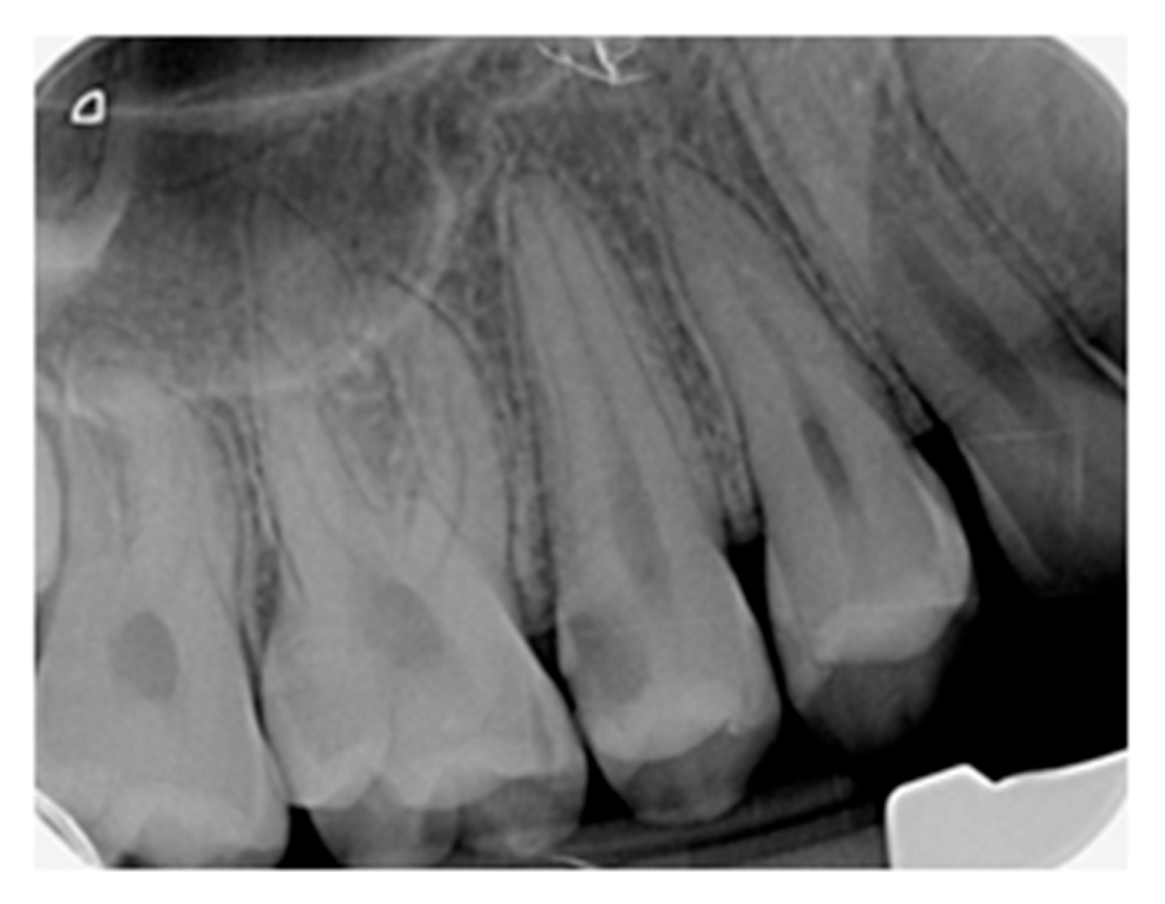
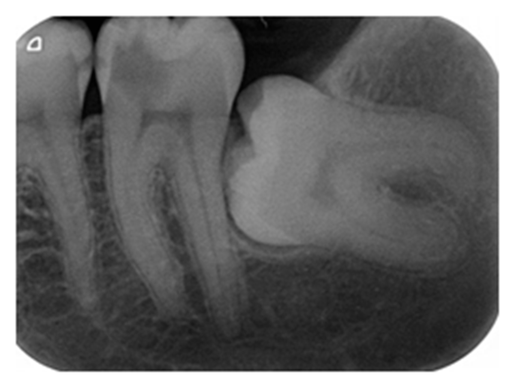
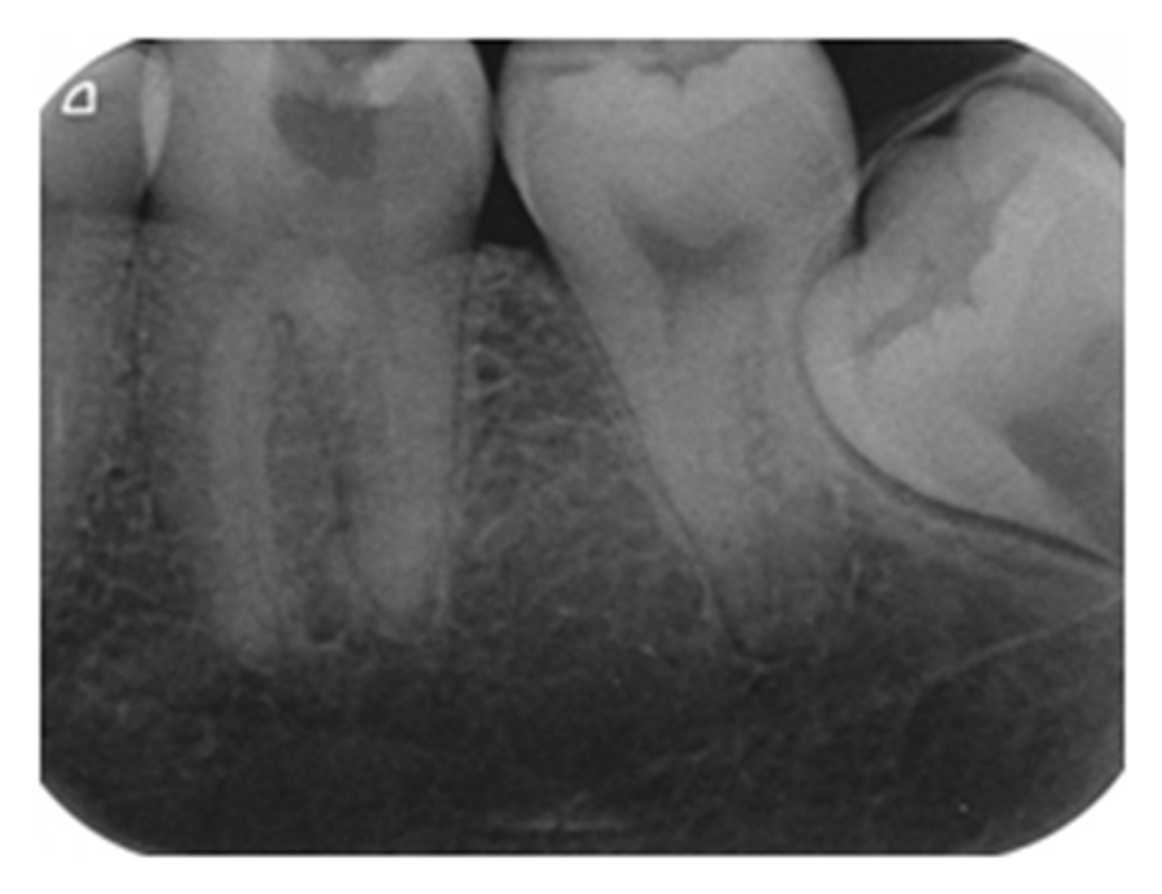
When the penetration depth of a carious lesion cannot be assessed clinically, in contrast, the radiographs illustrate two types of images, deep lesions and extremely deep lesions. A distinctive tait for deep lesions (Figure 2), which show a dentine demineralization extended pulp-ward round 3/4 of dentine thickness, is the presence of a higher radiodensity rim between the caries and pulp chamber compared to extremely deep lesions (Figure 3 and Figure 4), which are characterized by complete homogenous penetration without any tendency of increasing peripheral density [30].
Since persistent inflammation occurs in extremely deep caries, either symptomless or symptomatic, the radiodensity rim plays a role in orientating the marker of both, bacterial pulp-ward dissemination and histological pulp reaction [30,31,32,33,34,35].
The rationale of conservative treatment in deep caries is looking for the best comprehension of pulp pathophysiological status as well as of carious activity. Accordingly, such an imagistic orientating marker might be the starting point in the decision on the deep caries excavation technique [27,30].
Rejecting non-selective caries removal in deep caries of both vital asymptomatic teeth and those presenting signs and symptoms of reversible pulpitis, ESE recommends either one-stage selective caries removal or the stepwise technique. However, it has to be stressed that in reversible pulpitis with the goal of stable pulp recovery, selective caries removal is strictly limited to those patients where the radiographs confirmed caries that did not progress to the pulp chamber by over a quarter of the dentine thickness [13].
In cavitated lesions, the stepwise technique allows the conversion of the initial cariogenic environment into an inactive one, as, following the first step of this approach, the carious process is stopped. The reduced bacterial load found at the second step of excavation substantiates with time the biologic efficacy of stepwise caries removal [30,36].
However, despite the current biological approach in deep caries management, some inquiries are still carrying on, as, in a vital tooth, no information can be obtained about the initial status of pulp inflammation, before leaving behind the carious dentine in the first stage of the stepwise excavation procedure [30,36].
An encouraging clinical follow-up based on the preserved pulp vitality does not allow a simultaneous insight into the histological process of pulp repair. The pulp–dentine complex orchestrates multiple pathways of healing due to its extremely intricate molecular relationships [13,30,33,34,35,36,37,38,39].
A methodological item remains still unsolved for oral health providers, meaning the abilities they have for performing proper treatment in deep carious lesions are limited. In particular, whether the general practitioners are sufficiently qualified as endodontists to perform accurate treatment in deep carious lesions affects the treatment decision [27].
Related to the ESE position statement (2019), it is essential to know if an expertise-less general practitioner, working without magnification, is allowed to non-selectively remove the carious dentine in deep cavitated lesions [13,27]. Furthermore, in the case of choosing selective caries removal, it is essential to know if it is valid for a general practitioner to obtain alleged agreement for applying class I procedure (indirect pulp capping or direct pulp capping in iatrogenic exposure) and unsuitable to class II procedure (infective carious pulp exposure) [13,27,40,41,42].
It seems recommendable for the time being that this dispute may be optimally solved by a general practitioner making an immediate decision of referral to an endodontist before starting the operative procedure [27,41,42].

4.3. Endpoint of Carious Dentine Excavation

According to traditional techniques of complete soft dentine, the endpoint of excavation should be the sound dentine surface. Conversely, the minimally invasive strategy relies on two operative alternatives, either stepwise carious dentine removal or selective, partial removal [43].
As some reports mention, a higher successful outcome to preserving pulp vitality in the stepwise method (60.2%) vs. the non-selective removal of carious dentine (40.3%) relies on appropriate case selection, critical when aiming to avoid risky pulp exposure, which would compromise pulp vitality and subsequently, over time, tooth survival as well [43].
Nevertheless, continued controversies concerning the definition of caries endpoint between the ESE and AAE position statements drive more complex strategies in the management of deep caries [2,8,13,43].
Moreover, the operative steps of bonding techniques in creating the hybrid layer non-affecting the pulp status, the adequate pulp protection in various and hard to assess patho-physiological conditions, the new bioactive dental materials as well the concept of biomimetic restorative dentistry, are open windows to finding biologically improved clinical decisions [43,44].
Presently, the high success rate of MTA (mineral trioxide aggregate) and hydraulic calcium silicates substituted calcium hydroxide in pulp protection is being investigated [41]. However, for optimal clinical outcomes, hydraulic cements are demanding both the moment of final tooth restoration, required by prolonged material setting time, and the etching procedure that requires previous cement maturation [43].
Even though in deep caries, the selective removal of soft dentine is prioritized, the final goal of extending tooth longevity cannot be achieved if the choused dental materials for restoration do not long-term, effectively seal the residual carious dentine intentionally left behind to preclude the pulp exposure [44].
The removal of carious dentine may be appreciated subjectively when hand excavators are used or objectively in the case of self-limiting polymer-based (PEEK) burs such as Polybur P1 (Komet, Lemgo, Germany), avoiding unnecessary hard dentine loss [45]. Obviously, in the selective removal of carious dentine, it is crucial to establish the right moment of arrival at the endpoint of the excavation maneuver, namely at soft or leathery dentine. The aimed outcome is more easily objectified by using self-limiting burs whose hardness is between that of hard and soft dentine [46].
At least in primary molars a rather similar high tooth survival rate was observed in subjective selective carious dentine removal by hand excavators and objective excavation using self-limiting polymer-based burs [46].
Another self-limiting procedure is chemomechanical deep caries management under magnification, based on the chemical action of Carisolv gel associated with mechanical hand excavation aimed to preserve the affected dentine, eliminating solely the infected dentine [47]. However, it should be stressed that the use of caries-detecting dye is required to avoid excessive removal either of the affected caries or sound dentin [48].
A more biological approach of self-limiting procedures in deep carious lesions is crucial when the normal status of pulp tissue shifts to inflammation and signs and symptoms of reversible pulpitis are perceived. It should be stressed that in reversible pulpitis, the conventional without magnification non-selective technique of soft dentine removal to leathery dentine by rotary burs recorded a statistically significant lower rate of pulp survival compared to self-limiting Carisolv-based procedure [47].
Although direct pulp capping is considered the first option preserving tooth vitality in the case of unintentional pulp exposure during caries excavation, this outcome is appropriately achieved only if the practitioner has the suitable professional expertise in endodontics, including the diagnosis of authentic pulp tissue condition, adequate instruments, and pulp capping materials [28,29,49].

4.4. Indirect Pulp Capping

In current dental practice, once finalized, the carious dentine excavation of the tooth pulp is farther subjected to various harmful circumstances such as etching procedures, setting temperature of dental materials, toxicity of leaching components from restorations, polymerization shrinkage resulting in microleakage, as well as nanoleakage due to incomplete bonding penetration into previously demineralized dentine by etching [50].
Even though some decades ago, IPC was considered only a physical barrier against cariogenic bacteria and noxious agents, including those emerged from restorative dental materials, steadily, IPC has become crucial as a therapeutic approach in stimulating the protective tertiary dentine, stopping residual caries, and particularly providing long-lasting favorable context for pulp stem cell survival [50,51,52,53].
As clinically it is not easy to determine the dentine thickness, it is beneficial to protect the pulp with a biocompatible dental material [13]. Even though no preference is expressed, presently in IPC, calcium silicate cements, including dicalcium and tricalcium silicates, hydraulic calcium silicate cements, and bioceramics, are selective materials [13,28,49]. Moreover, as a critical step of the finalized vital treatment, IPC requires in the same visit, the immediate placement of permanent restoration [28].
Assessing the biocompatible materials, according to a 4-year follow-up study of IPC, it was emphasized that after non-selective complete caries debridement in deep cavitated lesions, MTA (mineral trioxide aggregate) proved to be clinically superior to calcium hydroxide [15]. Over time, simultaneously with composite resins restorations, adhesive systems were included among IPC materials. Used without or with cavity liners, adhesive systems also shield pulp vitality, attempting to impede the bacterial microleakage [18,50].
Considering the adhesive systems, one of the key decisional issues is linked to the quality of dentine subjected to upcoming bonding since numerous studies claim higher adhesion on healthy dentine compared to caries-affected dentine [28]. A degree of uncertainty is also raised when caries-affected dentine is preserved or a small amount of residual carious dentine is left over as the pulpal aspect of the prepared cavity [27,54].
After infected dentine excavation, compared to normal hard dentine, the freshly exposed surface of caries-affected dentine is softer and rather rough but is equally bacteria-free. Even though it exhibits a higher porosity of intertubular dentine due to partial demineralization, the caries-affected dentine still has numerous hydroxyapatite crystals and its collagen fibrils preserve, unmodified, their periodic banding [54].
Therefore, not surprisingly, the hybrid layer, generated by micromechanical locking and chemical bonding, after adhesive application on caries-affected dentine is homogenous and similar to that formed on hard, sound dentine [54]. Moreover, though normal dentine and caries-affected are obviously different in their physical and chemical properties, regarding the outcome of bonding procedures, their biomechanical behavior proved to be rather similar, with both of them resulting in cohesive failure [54,55].
Advances in adhesion research highlighted that the bonding strength of dentine depends on both the morphological and biochemical parameters of dentine and bonding systems [54,55]. In 60% of cases using etch-and-rinse systems, no bonding strength difference was found between sound and caries-affected dentine. Conversely, in 85% of studies applying self-etch adhesives on caries-affected dentine, the bonding strength was lower [54].
The status of bonding strength exhibited in caries-affected dentine can be explained by different mechanisms. Subsequent to initial bacterial demineralization, chemical metamorphosis from genuine hydroxyapatite to calcium phosphate, which is more etching resistant, occurs [54].
Therefore, the use of etch-and-rinse systems results in acid higher penetration in predominantly hydroxyapatite-based layers of the deepest dentine unlike the resin monomer that cannot pursue an equivalent trail. Coupled with higher porosity of intertubular dentine, in deep layers of etched dentine, obviously, the bonding strength will be minimal [54].
Conversely, in self-etching systems relying on mild acid resin monomers, the demanded bonding is impeded by occluded dentinal tubules with etching-resistant whitlockite crystals precipitated during repeated alternation of de- and re-mineralization, common processes that occur in deep caries progress [54].
Another frequent challenging operative attitude in clinical settings is to decide on dry-bonding or wet-bonding protocol, as the majority perception shows a higher bond strength following dry-bonding [56,57].
According to notorious rules of operative dentistry, before bonding, the etch-and-rinse systems remove the smear layer and enhance the dentine permeability, unlike the self-etch systems, which preserve and incorporate the smear layer into the novel hybrid layer. It seems that for maintaining pulp vitality, the etch-and-rinse adhesive systems clinically prevail in sealing ability over self-etch ones (100% vs. 85.5%) [50].
Reiterating the accurate management of etch-and-rinse systems, after removal of the smear layer, it is paramount to keep the exposed dentin collagen fibrils moist to avoid their collapse. This way, the subsequent adhesive infiltration results in a well-developed hybrid layer [56,57].
Though the wet-bonding protocol is thought to be superior, its clinical approach is quite challenging as it depends on various not easy to be appraised factors. Starting with the demanding assessment of adequate moisture, further crucial issues include the remoteness of air blast and its stroke duration, as well as the solvent chemical composition and drying time [56,57].
Wet-bonding is technically sensitive and especially requires good clinical expertise of the practitioner in assessing the real degree of moisture [56]. However, it seems that randomized clinical trials exploring the failure over time of this bonding technique regarding loss of retention, postoperative sensitivity, marginal adaptation, and discoloration as well as recurrent caries do not support the recommendation of the strict maintenance of dentine moisture [56].
Noteworthy, dry-bonding should not be confused with dentine dehydration. The basic principle is to eliminate the water layer covering the dentine surface without the evaporation of intratubular dentinal fluid [56]. Occasionally, under applicator pressure, dentinal fluid may emerge. However, combined with universal adhesives usually containing 10% to 20% water, the emerging dentinal fluid facilitates sufficient monomer penetration among the collagen fibrils of demineralized dentine to generate the hybrid layer [56].
Moreover, the outcomes of in vitro studies highlighting the superiority of wet-bonding over dry-bonding should not be considered critical since, in clinical settings, compared to moisture itself, other various known and unknown issues might be more significant [56].

4.5. Adjunctive Antimicrobial and Anti-Enzymatic Therapies

A mounting trend in recent years in deep caries lesions occurs for antimicrobial photodynamic therapy (aPDT) as an adjunctive disinfection strategy of soft dentine left behind in selective caries removal [58,59,60]. aPDT is a non-invasive therapeutic technique, simple and inexpensive, without side effects but promoting tissue repair of the dentine–pulp complex, owing to its anti-inflammatory outcomes [58].
Antibacterial photodynamic therapy rarely causes bacterial resistance as its bactericidal effect is based on reactive oxygen species (ROS) [43]. Even though, on the whole, aPDT is a three-component interactive technique (photosensitizer, visible light of resonant wavelength, and oxygen), the decontamination relies on singlet oxygen and free radicals termed reactive oxygen species (ROS). When released in a carious lesion microenvironment, ROS are targeting and killing the causal cariogenic bacteria aiming to facilitate the affected dentine re-mineralization [58].
In vitro studies of adhesive restorations evaluating the combined use of aPDT in selective caries excavation mentioned the highest occurrence of bond failures predominantly when it was used for infected dentine [20,61]. In turn, in vivo studies assessing the longevity of adhesive restorations, due to the high diversity of laser or LED light (light-emitting diode) and photosensitizer protocols, demonstrated the necessity of elaborating proper operative guidelines as well as prolonged clinical follow-up duration [20].
Nevertheless, the hitherto published studies should be cautiously analyzed as the delivered information about long-lasting clinical outcomes in deep caries management involving the combined operative procedure of selective caries removal with dentine aPTD is still limited. Moreover, based on the judged risk of bias, inconsistency, and imprecision, statistically, these studies show a moderate certainty of evidence [20].
Insufficiently studied issues of adjunctive therapies related to the restoration’s longevity are also those concerning the putative involvement of encrypted dentine matrix metalloproteinases (MMPs) on dentine bonding mechanisms [17,62]. To the already established effect of modeling and degradation of extracellular matrix components, recently, the inquiring implication of MMPs is linked to the enzymatic degradation of the hybrid layer [62].
In situ zymography studies demonstrated the presence of active dentine MMPs within the resin-bonded hybrid layers in areas where the collagen matrix was partially or at all infiltrated by bonding resin monomer [56]. Diagnostic markers of active dentinal caries that might compromise the integrity of the smear layer are telopeptides ICTP (C terminal telopeptide) and CTX (C terminal crosslinking telopeptide) of type I collagen [62].
Presently, the examination of the gene expression pattern of tissue inhibitors of metalloproteinases (TIMPs) resulted in no significant differences between sound and carious dentine, concluding that the regulation of the response of TIMPs might not be a real defense mechanism to caries progress. Nonetheless, the ongoing research on tissue inhibitors of metalloproteinases aims to arrive at effective techniques for protecting the hybrid layer adhesive interface against the harmful MMP-mediated proteolysis [62].

4.6. Biomimetic Concept in Deep Caries

The foremost endeavor of biomimetic restorative dentistry (BRD) is to upgrade caries management by less dental hard tissue excavation and the prolonged survival of restorations. The quadruple decision-making intention is focused on the preservation of pulp vitality, the maximal bond strength of restorative materials to the tooth hard tissues, long-lasting marginal seal, and minimal polymerization stress [46,63].
Until more rigorous research is performed, allegedly, biomimetic restorative dentistry (BRD) outclasses traditional adhesive techniques due to various advancements, such as the excessive removal of carious dentine following dyes warning, overtreatment caused by aluminum oxide air abrasion, no better restoration longevity due to deactivation of dentine matrix metalloproteinases, or using selected gold standard adhesive systems [7,63,64].
Though BRD is closely aligned to principles of minimal invasive dentistry, unlike the attitude regarding the carious dentine removal in deep cavitated lesions of former mentioned technique pointing toward the visual–tactile approach, the diagnosis and excavation of carious dentine BRD is oriented toward caries-detecting dyes [45,63].
Regrettably, caries-detecting dye recommendation fails in selectively staining exclusively denatured collagen and irreversible demineralized dentine, inadvertently guiding to less biological, more extensive dentine excavation, including sound circumpulpal dentine and potentially re-mineralizable dentine after cavity sealing. Similar drawbacks are encountered in aluminum oxide air abrasion, whereas the pulp exposure is definitely higher [63].
Noteworthy, a mounting current trend is to substitute the conventional terms of caries-infected and caries-affected dentine with clinically illustrating features which depict the carious dentine as soft, leathery, firm, or hard [7,63,65].
Though the attractive hypothesis of improving bond strength by the deactivation of dentine matrix metalloproteinases using in vitro inhibitors such as chlorhexidine, doxycycline, or EDTA (ethylenediaminetetraacetic acid) recorded some positive results, the clinically relevant outcomes failed.
Regarding the gold standard adhesive systems, though OptiBond FL (Kerr, CA 92821, USA) and Clearfil SE Bond (Kurashiki, Okayama 710-0801, Japan) proved to be exceedingly appreciated in dental practice, they should not be alleged as gold standards [63,66,67,68,69,70,71].
Still inquiring and debatable is the so-called concept of decouple with time based on a presumed hierarchy in running the bonding process during the restoration material setting. Starting from the false conviction that different bonding strengths depend on various topographical hardnesses of dentine, maximal setting shrinkage toward the mineralized and dry walls of restoration cavity occurs, whereas gap creation occurs against the soft weekly mineralized and wet areas [63,72,73].
Though the supporters of the decouple with time concept claim an 80–90% increase in bonding strength if a minimum of 5 min of polymerizing break is assigned to allow the maturation of hybrid layer, no clinical evidence proves that this technique would be superior to commonly used protocols [63].
The current trend toward smart materials in deep caries management aims to find responsive materials for etiological factors that eventually allow a targeted therapeutic approach [74,75,76,77,78,79]. However, the indiscriminate use of such materials like ion-releasing restorative ones under the axiomatic umbrella of bioactive might be faulty until their biological distribution and affinity to specific tissue accumulation is controlled by long-term clinical surveillance [43,80,81].
From the perspective of artificial intelligence, presently, smart materials are at most considered weakly intelligent as presently they may successfully manage either only one or a limited amount of tasks [43,82]. Contemporary smart dental materials are still not able to obtain a comprehensive intelligence that would allow for orchestrating complex and numerous clinical approaches [43,83].

4.7. Limitations of the Study

As the present study is a narrative review, it is subject to limitation and our results should be interpreted with caution. Though our aim was to globally discuss the current status in the field of deep caries lesions for the future research of related literature, a systematic review should be the method of choice.

5. Conclusions

Radiographic imaging is crucial for differentiating deep caries from extremely deep caries. The selective removal of carious tissue may be partial or stepwise. Both general practitioners and specialists in endodontics may manage soft dentine removal in class I procedure but the class II procedure should be exclusively performed by a specialist in endodontics as inadvertent pulp exposure occurs in infective carious dentine. The wet-bonding protocol is technically sensitive and requires good clinical expertise in assessing the real degree of moisture but dry-bonding should not be confused with dentine dehydration. An ongoing evaluation is focused on combining selective caries removal with antimicrobial adjunctive photodynamic therapy of soft dentine as well as on the involvement of dentine matrix metalloproteinases in the enzymatic degradation of the hybrid layer. Decoupling with time of restoration is on a hierarchy-based basis. Biomimetic restorative dentistry upgrades caries management by less dental hard tissue excavation and the prolonged survival of restorations. Smart materials in deep caries management aim to provide a targeted therapeutic approach by using responsive substances for etiological factors. Presently, smart materials may be considered at most weakly intelligent since they cannot orchestrate complex and numerous clinical approaches in deep caries management.

Author Contributions

Conceptualization, I.M.G. and S.P.; methodology, S.P.; software, I.R. and A.A.I.; validation, S.P. and I.R.; formal analysis, S.C., I.R. and A.S.D.; resources, I.R. and A.S.D.; writing—original draft preparation, I.M.G. and S.C.; writing—review and editing, I.M.G. and A.A.I.; supervision, A.A.I. All authors have read and agreed to the published version of the manuscript.

Funding

This study received no external funding.

Conflicts of Interest

The authors declare no conflicts of interest.

Abbreviations

The following abbreviations are used in this manuscript:
ORCAOrganization for Caries Research
ESEEuropean Society of Endodontology
ICCCInternational Caries Consensus Collaboration
FDIWorld Dental Federation
ARTatraumatic restorative treatment
PBRN National Dental Practice-Based Research Network
ADAAmerican Dental Association
IPCindirect pulp capping
SWstepwise technique
AAEAmerican Association of Endodontics
MTAmineral trioxide aggregate
Apdtantimicrobial photodynamic therapy
ROSreactive oxygen species
LEDlight emitting diode
MMPsmatrix metalloproteinases
ICTPcross-linked telopeptide of type I collagen
CTXC terminal crosslinking telopeptide
TIMPstissue inhibitors of metalloproteinases
BRDbiomimetic restorative dentistry
EDTAethylenediaminetetraacetic acid

References

  1. Pitts, N.B.; Twetman, S.; Fisher, J.; Marsh, P.D. Understanding dental caries as a non-communicable disease. Br. Dent. J. 2021, 231, 749–753. [Google Scholar] [CrossRef] [PubMed]
  2. Gill, K.; Stevenson, R.G. Navigating through caries excavation and pulp capping techniques in permanent teeth. Dent. Rev. 2024, 4, 100078. [Google Scholar] [CrossRef]
  3. Veneri, F.; Vinceti, S.R.; Filippini, T. Fluoride and caries prevention: A scoping review of public health policies. Ann. Ig. 2024, 36, 270–280. [Google Scholar] [CrossRef]
  4. Widbiller, M.; Weiler, R.; Knüttel, H.; Galler, K.M.; Buchalla, W.; Scholz, K.J. Biology of selective caries removal: A systematic scoping review protocol. BMJ Open 2022, 12, e061119. [Google Scholar] [CrossRef]
  5. Machiulskiene, V.; Campus, G.; Carvalho, J.C.; Dige, I.; Ekstrand, K.R.; Jablonski-Momeni, A.; Maltz, M.; Manton, D.J.; Martignon, S.; Martinez-Mier, E.A.; et al. Terminology of dental caries and dental caries management: Consensus report of a workshop organized by ORCA and cariology research group of IADR. Caries Res. 2020, 54, 7–14. [Google Scholar] [CrossRef]
  6. Giacaman, R.A.; Fernandez, C.E.; Munoz-Sandoval, C.; Leon, S.; Garcia-Manriquez, N.; Echeverria, C.; Valdes, S.; Castro, R.J.; Gambetta-Tessini, K. Understanding dental caries as a non-communicable and behavioral disease: Management implications. Front. Oral Health 2022, 3, 764479. [Google Scholar] [CrossRef]
  7. Spatafora, G.; Li, Y.; He, X.; Cowan, A.; Tanner, A.C.R. The Evolving Microbiome of Dental Caries. Microorganisms 2024, 7, 121. [Google Scholar] [CrossRef]
  8. Warreth, A. Dental Caries and Its Management. Int. J. Dent. 2023, 2023, 9365845. [Google Scholar] [CrossRef]
  9. Bjørndal, L.; Simon, S.; Tomson, P.L.; Duncan, H.F. Management of deep caries and the exposed pulp. Int. Endod. J. 2019, 52, 949–973. [Google Scholar] [CrossRef]
  10. Maldupa, I.; Al-Yaseen, W.; Giese, J.; Elagami, R.A.; Raggio, D.P. Recommended procedures for managing carious lesions in primary teeth with pulp involvement—Scoping review. BDJ Open 2024, 10, 74. [Google Scholar] [CrossRef]
  11. Taha, N.A.; Ali, M.M.; Abidin, I.Z.; Khader, Y.S. Pulp survival and postoperative treatment needs following selective vs. total caries removal in mature permanent teeth with reversible pulpitis: A randomized clinical trial. J. Dent. 2024, 151, 105408. [Google Scholar] [CrossRef] [PubMed]
  12. Zeng, Q.; Chen, M.; Zheng, S.; Wei, X.; Liu, H. Efficacy of vital pulp therapy for carious pulp injury in permanent teeth: A study protocol for an open-label randomized controlled noninferiority trial. Trials 2024, 25, 700. [Google Scholar] [CrossRef]
  13. Duncan, H.F.; Galler, K.M.; Tomson, P.L.; Simon, S.; El-Karim, I.; Kundzina, R.; Krastl, G.; Dammaschke, T.; Fransson, H.; Markvart, M.; et al. European Society of Endodontology position statement: Management of deep caries and the exposed pulp. Int. Endod. J. 2019, 52, 923–934. [Google Scholar] [CrossRef]
  14. Gonzalez-Gil, D.; Flores-Fraile, J.; Vera-Rodriguez, V.; Martin-Vacas, A.; Lopez-Marcos, J. Comparative meta-analysis of minimally invasive and conventional approaches for caries removal in permanent dentition. Medicina 2024, 60, 402. [Google Scholar] [CrossRef]
  15. Jurasic, M.M.; Gillespie, S.; Sorbara, P.; Clarkson, J.; Ramsay, C.; Nyongesa, D.; McEdward, D. Deep caries removal strategies: Findings from the national dental PBRN. J. Am. Dent. Assoc. 2022, 153, 1078–1088. [Google Scholar] [CrossRef]
  16. Barros, M.M.A.F.; Rodrigues, M.I.Q.; Muniz, F.W.M.C.; Rodrigues, L.K.A. Selective, stepwise, or nonselective removal of carious tissue: Which technique offers lower risk for the treatment of dental caries in permanent teeth? A systematic review and meta-analysis. Clin. Oral. Investig. 2020, 24, 521–532. [Google Scholar] [CrossRef]
  17. Ferreira, L.A.Q.; Anestino, T.A.; Branco, N.T.T.; Diniz, L.A.; Diniz, M.G.; de Magalhães, C.S.; Peixoto, R.T.R.D.C.; Moreira, A.N.; Dias, D.R.; Madeira, M.F.M.; et al. Adjunctive therapies for in vitro carious lesions: Antimicrobial activity, activation of dentin metalloproteinases and effects on dental pulp cells. Photodiagnosis. Photodyn. Ther. 2022, 40, 103168. [Google Scholar] [CrossRef]
  18. Edwards, D.; Stone, S.; Bailey, O.; Tomson, P. Preserving pulp vitality: Part one—Strategies for managing deep caries in permanent teeth. Br. Dent. J. 2021, 230, 77–82. [Google Scholar] [CrossRef]
  19. Iliescu, A.-A.; Gheorghiu, I.M.; Tănase, M.; Iliescu, A.; Mitran, L.; Mitran, M.; Perlea, P. Reactionary versus reparative dentine in deep caries. ARS Medica Tomitana 2019, 25, 15–21. [Google Scholar] [CrossRef]
  20. Veiga Faria, L.; de Oliveira Fernandes, T.; Silva Guimaraes, L.; Rodrigues Cajazeira, M.R.; Santos Antunes, L.; Azeredo Alves Antunes, L. Does selective caries removal in combination with antimicrobial photodynamic therapy affect the clinical performance of adhesive restorations of primary or permanent teeth? A systematic review with meta-analysis. J. Clin. Pediatr. Dent. 2022, 46, 1–14. [Google Scholar] [CrossRef]
  21. Jardim, J.J.; Mestrinho, H.D.; Koppe, B.; de Paula, L.M.; Alves, L.S.; Yamaguti, P.M.; Almeida, J.C.F.; Maltz, M. Restorations after selective caries removal: 5-year randomized trial. J. Dent. 2020, 99, 103416. [Google Scholar] [CrossRef] [PubMed]
  22. Iliescu, A.-A.; Perlea, P.; Gheorghiu, I.M.; Mitran, M.; Mitran, L.; Moraru, I.; Scarlatescu, S.; Erdoğan, S.; Iliescu, A. Molecular mechanisms of dentine-pulp complex response induced by microbiome of deep caries. ARS Medica Tomitana 2019, 25, 53–60. [Google Scholar] [CrossRef]
  23. Schwendicke, F.; Walsh, T.; Lamont, T.; Al-yaseen, W.; Bjørndal, L.; Clarkson, J.E.; Fontana, M.; Gomez Rossi, J.; Göstemeyer, G.; Levey, C.; et al. Interventions for treating cavitated or dentine carious lesions. Cochrane Database Syst. Rev. 2021, 7, CD013039. [Google Scholar] [CrossRef]
  24. Careddu, R.; Plotino, G.; Cotti, E.; Duncan, H.F. The management of deep carious lesions and the exposed pulp amongst members of two European endodontic societies: A questionnaire-based study. Int. Endod. J. 2021, 54, 366–376. [Google Scholar] [CrossRef]
  25. Chevalier, V.; Le Fur, B.A.; Duncan, H.F. Frightened of the pulp? A qualitative analysis of undergraduate student confidence and stress during the management of deep caries and exposed pulp. Int. Endod. J. 2021, 54, 130–146. [Google Scholar] [CrossRef]
  26. Edwards, D.; Bailey, O.; Stone, S.J.; Duncan, H. How is carious pulp exposure and symptomatic irreversible pulpitis managed in UK primary dental care? Int. Endod. J. 2021, 54, 2256–2275. [Google Scholar] [CrossRef]
  27. Duncan, H.F.; Tomson, P.L.; Simon, S.; Bjørndal, L. Endodontic position statements in deep caries management highlight need for clarification and consensus for patient benefit. Int. Endod. J. 2021, 52, 2145–2149. [Google Scholar] [CrossRef]
  28. Hirschberg, C.S.; Bogen, G.; Galicia, J.C.; Lemon, R.R.; Peters, O.A.; Ruparel, N.B.; Tay, F.R.; Whiterspoon, D.E. AAE position statement on vital pulp therapy. J. Endod. 2021, 47, 1340–1344. [Google Scholar] [CrossRef]
  29. Arroyo-Bote, S.; Ribas-Perez, D.; Bennasar Verges, C.; Rodriguez Menacho, D.; Villalva Hernandez-Franch, P.; Barbero Navarro, I.; Castano Seiquer, A. Influence of academic training and professional experience on the management of deep caries lesions. Healthcare 2024, 12, 1907. [Google Scholar] [CrossRef]
  30. Demant, S.; Dabelsteen, S.; Bjørndal, L. A macroscopic and histological analysis of radiographically well-defined deep and extremely deep carious lesions: Carious lesion characteristics as indicators of the level of bacterial penetration and pulp response. Int. Endod. J. 2021, 54, 319–330. [Google Scholar] [CrossRef]
  31. Liu, Y.; Wang, J.; Dong, B.; Zhai, Y.; Zhou, L.; Sun, S.; Li, X.; Wu, L. Prediction and validation of microbial community function from normal pulp to pulpitis caused by deep dentinal caries. Int. Endod. J. 2023, 56, 608–621. [Google Scholar] [CrossRef] [PubMed]
  32. Selvakumar, D.R.; Krishnamoorthy, S.; Venkatesan, K.; Ramanathan, A.; Abbott, P.V.; Rajasekaran, P.K.A. Active bacteria in carious dentin of mandibular molars with different pulp conditions: An in vivo study. J. Endod. 2021, 47, 1883–1889. [Google Scholar] [CrossRef] [PubMed]
  33. Li, J.; Wang, S.; Dong, Y. Regeneration of pulp-dentine complex-like tissue in a rat experimental model under an inflammatory microenvironment using high phosphorus-containing bioactive glasses. Int. Endod. J. 2021, 54, 1129–1141. [Google Scholar] [CrossRef]
  34. Shayegan, A.; Zucchi, A.; De Swert, K.; Balau, B.; Truyens, C.; Nicaise, C. Lipoteichoic acid stimulates the proliferation, migration and cytokine production of adult dental pulp stem cells without affecting osteogenic differentiation. Int. Endod. J. 2021, 54, 585–600. [Google Scholar] [CrossRef]
  35. Wen, B.; Huang, Y.; Qiu, T.; Huo, F.; Xie, L.; Liao, L.; Tian, W.; Guo, W. Reparative dentin formation by dentin matrix proteins and small extracellular vesicles. J. Endod. 2021, 47, 253–262. [Google Scholar] [CrossRef]
  36. Ricucci, D.; Siqueira, J.F.; Rộças, I.N.; Lipski, M.; Shiban, A.; Tay, F.R. Pulp and dentine responses to selective caries excavation: A histological and histobacteriological human study. J. Dent. 2020, 100, 103430. [Google Scholar] [CrossRef]
  37. de Farias, J.O.; da Costa Sousa, M.G.; Martins, D.C.M.; de Oliveira, M.A.; Takahashi, I.; de Sousa, L.B.; da Silva, I.G.M.; Correa, J.R.; Carvalho, A.E.S.; Saldanha-Araujo, F.; et al. Senescence on dental pulp cells: Effects on morphology, migration, proliferation, and immune response. J. Endod. 2024, 50, 362–369. [Google Scholar] [CrossRef]
  38. Yang, C.; Gao, Q.; Yang, K.; Bian, Z. Human dental pulp stem cells are subjected to metabolic reprogramming and repressed proliferation and migration by the sympathetic nervous system via α1B-adrenergic receptor. J. Endod. 2023, 49, 1641–1651. [Google Scholar] [CrossRef]
  39. Galler, K.M.; Weber, M.; Korkmaz, Y.; Widbiller, M.; Feuerer, M. Inflammatory response mechanisms of the dentine-pulp complex and the periapical tissues. Int. J. Mol. Sci. 2021, 22, 1480. [Google Scholar] [CrossRef]
  40. Hanna, S.N.; Perez Alfayate, R.; Prichard, J. Vital pulp therapy an insight over the available literature and future expectations. Eur. Endod. J. 2020, 5, 46–53. [Google Scholar] [CrossRef]
  41. Cushley, S.; Duncan, H.F.; Lundy, F.T.; Nagendrababu, V.; Clarke, M.; El Karim, I. Outcomes reporting in systematic reviews on vital pulp treatment: A scoping review for the development of a core outcome set. Int. Endod. J. 2022, 55, 891–909. [Google Scholar] [CrossRef] [PubMed]
  42. Pandya, J.K.; Wheatley, J.; Bailey, O.; Taylor, G.; Geddis-Regan, A.; Edwards, D. Exploring deep caries management and barriers to the use of vital pulp treatments by primary care dental practitioners. Int. Endod. J. 2024, 57, 1453–1464. [Google Scholar] [CrossRef]
  43. Par, M.; Cheng, L.; Camilleri, J.; Lingström, P. Applications of smart materials in minimally invasive dentistry—Some research and clinical perspectives. Dent. Mater. 2024, 40, 2008–2016. [Google Scholar] [CrossRef]
  44. Chou, Y.F.; Pires, P.M.; Alambiaga-Caravaca, A.M.; Spagnuolo, G.; Hibbits, A.; Sauro, S. Remineralisation of mineral-deficient dentine induced by experimental ion-releasing materials in combination with a biomimetic dual-analogue primer. J. Dent. 2025, 152, 105468. [Google Scholar] [CrossRef]
  45. Schwendicke, F.; Badakksh, P.; Gomes Marques, M.; Medeiros Demarchi, K.; Ramos Rezende Brant, A.; Moreira, C.L.; Dias Ribeiro, A.P.; Coelho Leal, S.; Hilgert, L.A. Subjective versus objective, polymer bur-based selective carious tissue removal: 2-year randomized clinical trial. J. Dent. 2023, 138, 104728. [Google Scholar] [CrossRef]
  46. Ali, A.H.; Thani, F.B.; Foschi, F.; Banerjee, A.; Mannocci, F. Self-limiting versus rotary subjective carious tissue removal: A randomized controlled clinical trial—2-year results. J. Clin. Med. 2020, 9, 2738. [Google Scholar] [CrossRef]
  47. Choudhary, K.; Gouraha, A.; Sharma, M.; Sharma, P.; Tiwari, M.; Chouksey, A. Clinical and Microbiological Evaluation of the Chemomechanical Caries Removal Agents in Primary Molars. Cureus. 2022, 12, e31422. [Google Scholar] [CrossRef]
  48. Saber, A.M.; El-Housseiny, A.A.; Alamoudi, N.M. Atraumatic restorative treatment and interim therapeutic restoration: A review of the literature. Dent. J. 2019, 7, 28. [Google Scholar] [CrossRef]
  49. Kunert, M.; Lukomska-Szymanska, M. Bio-inductive materials in direct and indirect pulp capping—A review article. Materials 2020, 13, 1204. [Google Scholar] [CrossRef]
  50. Alovisi, M.; Baldi, A.; Comba, A.; Gamerro, R.; Paolone, G.; Mandurino, M.; Dioguardi, M.; Roggia, A.; Scotti, N. Long-term evaluation of pulp vitality preservation in direct and indirect pulp capping: A retrospective clinical study. J. Clin. Med. 2024, 13, 3962. [Google Scholar] [CrossRef]
  51. Chansaenroj, A.; Kornsuthisopon, C.; Suwittayarak, R.; Rochanavibhata, S.; Loi, L.K.; Lin, Y.C.; Osathanon, T. IWP-2 modulates the immunomodulatory properties of human dental pulp stem cells in vitro. Int. Endod. J. 2024, 57, 219–236. [Google Scholar] [CrossRef] [PubMed]
  52. Harrell, C.R.; Djonov, V.; Volarevic, V. The cross-talk between-mesenchymal stem cells and immune cells in tissue repair and regeneration. Int. J. Mol. Sci. 2021, 22, 2472. [Google Scholar] [CrossRef] [PubMed]
  53. Bucchi, C.; Bucchi, A.; Martinez-Rodriguez, P. Biological properties of dental pulp stem cells isolated from inflamed and healthy pulp and cultured in an inflammatory microenvironment. J. Endod. 2023, 49, 395–401. [Google Scholar] [CrossRef]
  54. Sun, Q.; Shu, C.; Wang, C.; Shi, M.; Zu, J.; Luo, Q.; Wu, M.; Chen, Y.; Li, X. Bonding effectiveness of the universal adhesive on caries-affected dentin of deciduous teeth. Materialia 2020, 14, 100885. [Google Scholar] [CrossRef]
  55. Steiner, R.; Edelhoff, D.; Stawarczyk, B.; Dumfahrt, H.; Lente, I. Effect of Dentin Bonding Agents, Various Resin Composites and Curing Modes on Bond Strength to Human Dentin. Materials 2019, 17, 3395. [Google Scholar] [CrossRef]
  56. Forville, H.; Wendlinger, M.; Cochinski, G.D.; Favoreto, M.W.; Wambier, L.M.; Chibinski, A.C.; Loguercio, A.D.; Reis, A. Re-evaluating the role of moist dentin of adhesive systems used in the etch-and-rinse bonding strategy. A systematic review and meta-analysis. J. Dent. 2025, 152, 105495. [Google Scholar] [CrossRef]
  57. Naupari-Villasante, R.; Falconi-Paez, C.; Castro, A.S.; Gutierez, M.F.; Mendez-Bauer, M.L.; Aliaga, P.; Davilla-Sanchez, A.; Arrais, C.; Reis, A.; Loguercio, M.F. Clinical performance of posterior restorations using a universal adhesive over moist and dry dentin: A 36-month double-blind split-mouth randomized clinical trial. J. Dent. 2024, 147, 105080. [Google Scholar] [CrossRef]
  58. Bahrami, R.; Pourhajibagher, M.; Nikparto, N.; Bahador, A. The impact of antimicrobial photodynamic therapy on pain and oral health-related quality of life: A literature review. J. Dent. Sci. 2024, 19, 1924–1933. [Google Scholar] [CrossRef]
  59. Coelho, A.; Amaro, I.; Rascao, B.; Marcelino, I.; Paula, A.; Saraiva, J.; Spagnuolo, G.; Ferreira, M.M.; Marto, C.M.; Carilho, E. Effect of cavity disinfectants on dentin bond strength and clinical success of composite restorations—A systematic review of in vitro, in situ and clinical studies. Int. J. Mol. Sci. 2021, 22, 353. [Google Scholar] [CrossRef]
  60. Santos, G.M.; Pacheco, R.L.; Bussadori, S.K.; Santos, E.M.; Riera, R.; de Oliveira Cruz Latorraca, C.; Mota, P.; Benavent Caldas Belloto, E.F.; Martimbianco, A.L.C. Effectiveness and safety of ozone therapy in dental caries treatment: Systematic review and meta-analysis. J. Evid. Based Dent. Pract. 2020, 20, 101472. [Google Scholar] [CrossRef]
  61. Alrahlah, A.; Niaz, M.O.; Abrar, E.; Vohra, F.; Rashid, H. Treatment of caries affected dentin with different photosensitizers and its adhesive bond integrity to resin composite. Photodiagnosis Photodyn. Ther. 2020, 31, 101865. [Google Scholar] [CrossRef] [PubMed]
  62. Anumula, L.; Ramesh, S.; Kolaparthi, V.S.K. Matrix metalloproteinases in dentin: Assessing their presence, activity and inhibitors—A review of current trends. Dent. Mater. 2024, 40, 2051–2073. [Google Scholar] [CrossRef] [PubMed]
  63. Reis, A.; Feitosa, V.P.; Chibinski, A.C.; Favoreto, M.W.; Gutierez, M.F.; Loguercio, A.D. Biomimetic restorative dentistry: An evidence-based discussion of common myths. J. Appl. Oral Sci. 2024, 32, e20240271. [Google Scholar] [CrossRef]
  64. Peumans, M.; Politano, G.; Van Meerbeek, B. Effective protocol for daily high-quality direct posterior composite restorations. Cavity preparation and design. J. Adhes. Dent. 2020, 22, 581–596. [Google Scholar] [CrossRef]
  65. Elhennawy, K.; Finke, C.; Paris, S.; Reda, S.; Jost-Brinkmann, P.G.; Schwendicke, F. Selective vs stepwise removal of deep carious lesions in primary molars: 24 months follow-up from a randomized controlled trial. Clin. Oral Investig. 2021, 25, 645–652. [Google Scholar] [CrossRef]
  66. Dreweck, F.; Burey, A.; Dreweck, M.O.; Fernandez, E.; Loguercio, A.D.; Reis, A. Challenging the concept that OptiBond FL and Clearfil SE Bond in NCCLs are gold standard adhesives: A systematic review and meta-analysis. Oper. Dent. 2021, 46, E276–E295. [Google Scholar] [CrossRef]
  67. de Albuquerque, E.G.; Warol, F.; Calazans, F.S.; Poubel, L.A.; Marins, S.S.; Matos, T.; de Souza, J.J.; Reis, A.; Barceleiro, M.O.; Loguercio, A.D. A new dual-cure universal simplified adhesive: 19-month randomized multicenter clinical trial. Oper. Dent. 2020, 45, e255–e270. [Google Scholar] [CrossRef]
  68. de Albuquerque, E.G.; Warol, F.; Tardem, C.; Calazans, F.S.; Poubel, L.A.; Matos, T.P.; Souza, J.J.; Reis, A.; Barceleiro, M.O. Universal simplified adhesive applied under different bonding techniques; 36-month randomized multicenter clinical trial. J. Dent. 2022, 122, 104120. [Google Scholar] [CrossRef]
  69. Barceleiro, M.O.; Lopes, L.S.; Tardem, C.; Calazans, F.S.; Matos, T.; Reis, A.; Calixto, L.; Loguercio, A.D. Thirty-six-month follow-up of cervical composite restorations placed with MDP-free universal adhesive system using different adhesive protocols: A randomized clinical trial. Clin. Oral Investig. 2022, 26, 4337–4350. [Google Scholar] [CrossRef]
  70. Naupari-Villasante, R.; Matos, T.P.; de Albuquerque, E.G.; Warol, F.; Tardem, C.; Calazans, F.S.; Poubel, L.A.; Reis, A.; Barceleiro, M.O.; Loguercio, A.D. Five-year clinical evaluation of universal adhesive applied following different bonding techniques: A randomized multicenter clinical trial. Dent. Mater. 2023, 39, 586–594. [Google Scholar] [CrossRef]
  71. Cascales, A.F.; Moscardo, A.P.; Toledano, M.; Banerjee, A.; Sauro, S. An in-vitro investigation of the bond-strength of experimental ion-releasing dental adhesive to caries-affected dentine after 1 year of water storage. J. Dent. 2022, 119, 104075. [Google Scholar] [CrossRef]
  72. Adams, K.R.; Savett, D.A.; Lien, W.; Raimondi, C.; Vandewalle, K.S. Evaluation of a novel “Quad” wavelength light curing unit. J. Clin. Exp. Dent. 2022, 14, e815–e821. [Google Scholar] [CrossRef]
  73. Montoya, C.; Babariya, M.; Ogwo, C.; Querido, W.; Patel, J.S.; Melo, M.A.; Orrego, S. Synergistic effects of bacteria, enzymes, and cyclic mechanical stresses on the bond strength of composite restorations. Biomater. Adv. 2025, 166, 214049. [Google Scholar] [CrossRef]
  74. Yang, Q.; Zheng, W.; Zhao, Y.; Shi, Y.; Wang, Y.; Sun, H.; Xu, X. Advancing dentin remineralisation: Exploring amorphous calcium phosphate and its stabilizers in biomimetic approaches. Dent. Mater. 2024, 40, 1282–1295. [Google Scholar] [CrossRef]
  75. Maciel Pires, P.; Ionescu, A.C.; Perez-Garcia, M.T.; Vezzoli, E.; Soares, I.P.M.; Brambilla, E.; de Almeida Neves, A.; Sauro, S. Assessment of the remineralisation induced by contemporary ion-releasing materials in mineral-depleted dentine. Clin. Oral Investig. 2022, 26, 6195–6207. [Google Scholar] [CrossRef]
  76. Alambiaga-Caravaca, A.M.; Chou, Y.F.; Moreno, D.; Aparicio, A.; Lopez-Castellano, A.; Feitosa, V.P.; Tezvergil-Mutluay, A.; Sauro, S. Characterization of experimental flowable composite containing fluoride-doped calcium phosphates as promising remineralising materials. J. Dent. 2024, 143, 104906. [Google Scholar] [CrossRef]
  77. Sauro, S.; Spagnuolo, G.; Del Giudice, C.; Neto, D.M.A.; Fechine, P.B.A.; Chen, X.; Rengo, S.; Chen, X.; Feitosa, V.P. Chemical, structural and cytotoxicity characterization of experimental fluoride-doped calcium phosphates as promising remineralising materials for dental applications. Dent. Mater. 2023, 39, 391–401. [Google Scholar] [CrossRef]
  78. Pires, P.M.; Nunes Monteiro, A.S.; de Accioly Costa, P.H.; Silva, A.S.; Lopes, R.T.; Yoshihara, K.; Sauro, S.; de Almeida Neves, A. Dentine mineral changes induced by polyalkenoate cements after different selective caries removal techniques: An in vitro study. Caries Res. 2023, 57, 21–31. [Google Scholar] [CrossRef]
  79. Valizadeh, S.; Kamangar, S.H.; Nekoofar, M.H.; Behroozibakhsh, M.; Shahidi, Z. Comparison of dentin caries remineralization with four bioactive cements. Eur. J. Prosthodont. Restor. Dent. 2022, 30, 223–229. [Google Scholar] [CrossRef]
  80. Giannini, M.; Sauro, S. Bioactivity in restorative dentistry: Standing for the use of innovative materials to improve the longevity of restorations in routine dental practice. J. Adhes. Dent. 2021, 23, 176–178. [Google Scholar] [CrossRef]
  81. Braga, R.R.; Fronza, B.M. The use of bioactive particle and biomimetic analogues for increasing the longevity of resin dentin interface. A literature review. Dent. Mater. J. 2020, 39, 62–68. [Google Scholar] [CrossRef] [PubMed]
  82. Sauro, S. Dental materials—Future active resins? Br. Dent. J. 2024, 236, 460–461. [Google Scholar] [CrossRef] [PubMed]
  83. Nguyen, P.Q.; Courchesne, N.D.; Duraj-Thatte, A.; Praveschotinunt, P.; Joshi, N.S. Engineered living materials: Prospects and challenges for using biological systems to direct the assembly of smart materials. Adv. Mater. 2018, 30, e1704847. [Google Scholar] [CrossRef] [PubMed]
Figure 1. PRISMA flow diagram.
Jmms 12 00037 g001
Figure 2. Distal deep caries in tooth 15—area of unchanged radiodensity of dentine separating carious lesion from pulp.
Jmms 12 00037 g002
Figure 3. Extremely deep caries (tooth 37)—lesion completely penetrates dentine in area of mesial pulp horn.
Jmms 12 00037 g003
Figure 4. Extremely deep occlusal caries (tooth 36)—carious lesion penetrates entire thickness of dentine without opening pulp chamber. Tooth is still vital and asymptomatic.
Jmms 12 00037 g004
Disclaimer/Publisher’s Note: The statements, opinions and data contained in all publications are solely those of the individual author(s) and contributor(s) and not of MDPI and/or the editor(s). MDPI and/or the editor(s) disclaim responsibility for any injury to people or property resulting from any ideas, methods, instructions or products referred to in the content.