Review

Current Insight into the Dynamics of Secondary Endodontic Infections

by
Alexandru Andrei Iliescu
1,
Irina Maria Gheorghiu
2,*,
Sergiu Ciobanu
3,
Ion Roman
3,
Anca Silvia Dumitriu
4,† and
Stana Păunică
4,†
1
Department of Oral Rehabilitation, Faculty of Dental Medicine, University of Medicine and Pharmacy of Craiova, 200638 Craiova, Romania
2
Department of Restorative Odontotherapy, Faculty of Stomatology, Carol Davila University of Medicine and Pharmacy, 010232 Bucharest, Romania
3
Department of Odontology, Periodontology and Oral Pathology, Nicolae Testemiţanu State University of Medicine and Pharmacy, MD-2004 Chișinău, Moldova
4
Department of Periodontology, Faculty of Stomatology, Carol Davila University of Medicine and Pharmacy, 010232 Bucharest, Romania
*
Author to whom correspondence should be addressed.
These authors contributed equally to this work.
J. Mind Med. Sci. 2025, 12(1), 28; https://doi.org/10.3390/jmms12010028
Submission received: 5 March 2025 / Revised: 28 April 2025 / Accepted: 30 April 2025 / Published: 4 May 2025

Abstract

:
Background/Objectives: The aim of this narrative review is to perform an updated literature review of the root canal microbiome in secondary endodontic infections and the bacterial dynamics that govern the processes leading to the development of these persistent endodontic infections and periapical lesions. Methods: A literature search of scientific publications issued in the last 8 years, i.e., 2017–2024, was conducted in PubMed (MEDLINE) and ScienceDirect databases, using the following keywords: endodontic microbiome; endodontic pathogens; periapical lesion; primary endodontic infection; secondary/persistent endodontic infection; functional redundancy. Discussions: Secondary endodontic infections (SEIs) are a highly prevalent pathological condition affecting a minimum of one tooth in more than half of adults worldwide. The transition from primary endodontic infection (PEI) to secondary endodontic infection (SEI) is mainly governed by Enterococus faecalis (EF) that invades and dominates the previous endodontic biofilm initiated by Fusobacterium nucleatum (FN). Conclusions: The findings from different studies indicate that secondary endodontic infections are polymicrobial. In SEIs, the microbial species interactions are crucial in influencing the ecology of infected root canals. The issue of the keynote pathogen is still under debate. Both EF and FN pathogens cooperate with neighboring residents. Functional redundancy of the endodontic microbiome explains how the ecological diversity modulates its pathogenicity.

1. Introduction

Primary endodontic infections (PEIs) are the final outcome of microbial colonization in root canals of pulpless teeth, regardless of the cause of losing their vitality. Secondary endodontic infections (SEIs) are also established in non-vital teeth. However, unlike the primary ones, they are installed following to a more or less incorrectly managed previous endodontic treatment, this explains why these secondary endodontic infections are also named persistent root canal infections [1].
In dental practice, secondary root canal infections are commonly diagnosed and managed as chronic apical periodontitis. Since this chronic inflammatory process usually evolves clinically rather than asymptomatically, as a rule, it is an imagistic-based diagnosis following mostly from the periapical radiographic examination [2].
As a rule, in daily endodontic practice, a tooth with chronic apical inflammatory involvement is hard to escape in routine clinical screenings, as any experienced endodontist is aware that focus should be either on already root-filled and restored teeth or on teeth with progressing deep carious lesions [3].
Epidemiological studies conclude that chronic apical periodontitis is a highly prevalent pathological condition worldwide, with more than half of adults having at least one tooth affected by this disease. Moreover, chronic apical periodontitis occurs more frequently in endodontically treated teeth than in non-treated ones [3,4].
Over the past two decades, it has been agreed that the best healing outcome might be achieved when endodontic treatment relies on an adequate understanding of its etiology and pathogenesis and eventually results in effective elimination of causative microorganisms harbored in infected root canals [5,6,7].
The various types of microbiomes in infected apical root canals mirror a high individual variability expressed by the prominent diversity of bacterial genera and species. Furthermore, significant differences occur between microorganisms residing in the coronal and apical thirds of root canals [8].
The reduced oxygen tension and nutrients provided by periapical inflammatory exudates are among the most favorable, encouraging factors that decide the establishment of obligate anaerobes in the apical third of infected root canals. Therefore, the rigorous endodontic treatment of the apical root canal third, corresponding to more or less 5 mm of the tooth root length, is critical for removing the infective microbiome, since it is the main culprit of persistent chronic apical periodontitis [8,9,10].
The prognosis of healing in chronic apical periodontitis is highly influenced by its multifaceted factors guiding the pathologic progress, both systemic, such as immune status, and co-morbidities or local ones, depending on the diversity of the root canal microbiome and the persistent endodontic infection or the size of the periapical bone lesion and tooth type—mono- or multi-rooted [11,12].
Various issues should be discussed. First, the apical lesion might be illustrated on radiograph as a stage of healing in progress, which is the best treatment follow-up. Conversely, some unwanted outcomes should also be highlighted, such as the failure of primary root canal treatment or of conservative retreatment.
An imagistic new emerging pathology may mask the proper healing of the initial lesion or even the progression over time of a primary chronic apical pathology despite the well-managed endodontic treatment of previously infected root canals [2,4].
The aim of this narrative review is to perform an updated literature review of the root canal microbiome in secondary endodontic infections and the bacterial dynamics that govern the processes leading to the development of these persistent endodontic infections and periapical lesions.

2. Materials and Methods

The objective of this paper was to investigate and analyze the role of bacteria in secondary/persistent endodontic infections, reviewing recent trends in the detection of endodontic microorganisms, persistent taxa and the complex interactions, both synergistic and antagonistic, of endodontic biofilm components.

2.1. Research Question

This review addressed the following research question:
What are the main characteristics regarding composition and bacterial dynamics of the endodontic microbiome in secondary/persistent endodontic infections?

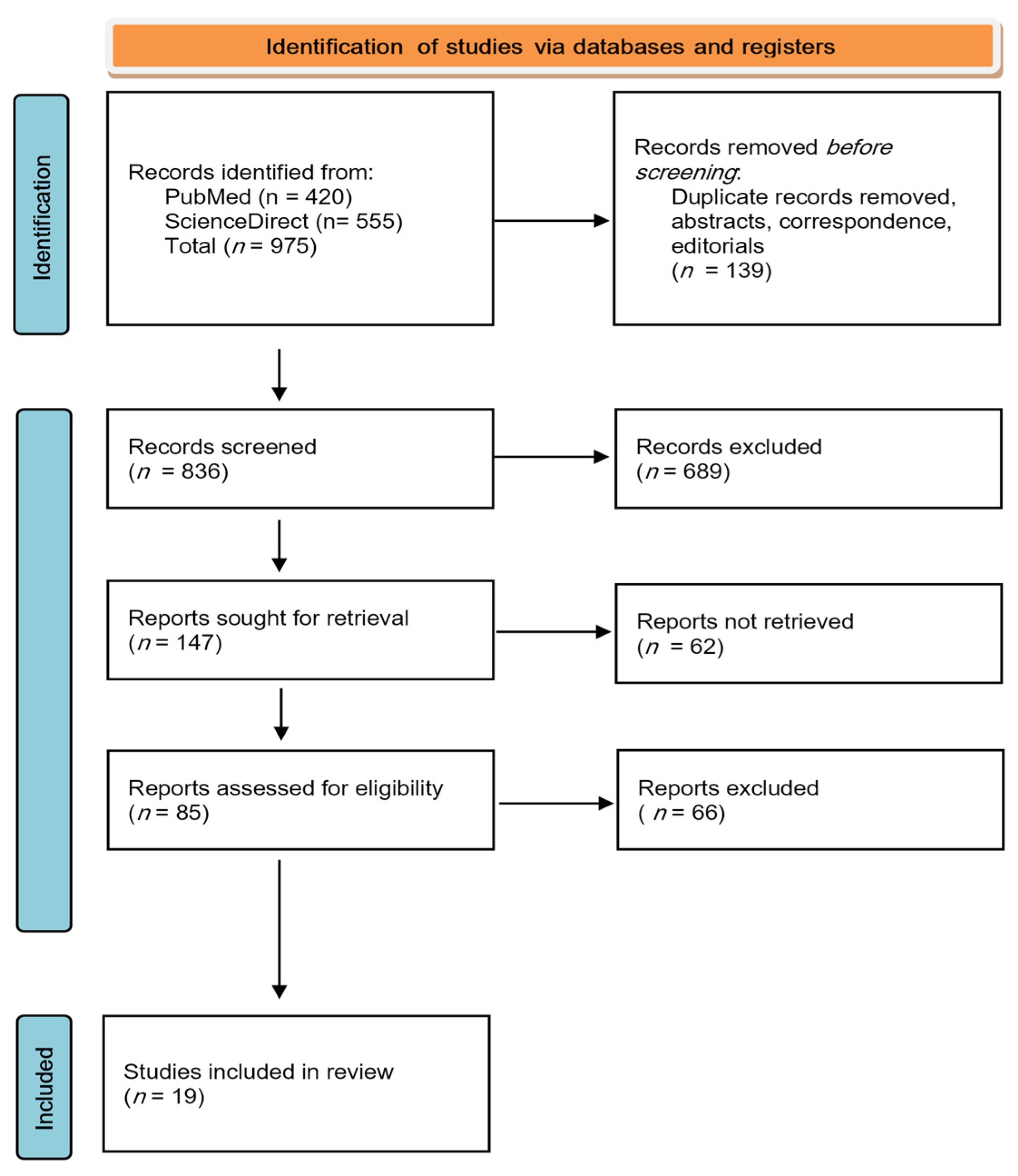
2.2. Search Strategy

The present study was conducted in accordance with PRISMA (Preferred Reporting Items for Systematic Reviews and Meta-Analyses) criteria.
In order to identify the relevant publications, we accordingly performed a search in the biomedical databases PubMed (MEDLINE) and ScienceDirect, using the following keywords: “endodontic microbiome”, “endodontic pathogens”, “periapical lesion”, “primary endodontic infection”, “secondary/persistent endodontic infection”, and “functional redundancy”. For an appropriate search for the “secondary/persistent endodontic infection”, we completed separate searches for “secondary endodontic infection” and “persistent endodontic infection”. Also, when using the keywords “functional redundancy,” we added “of endodontic microbiome” to obtain results related to the root canal microbiome, knowing that this term is also specific to other systems of the human body.
The following inclusion and exclusion criteria were applied in our study:

2.2.1. Inclusion Criteria

Publication date of investigated articles: within the last 8 years (2017–2024). We completed the search in November 2024.
Full-text availability: We included articles that provided full-text access to the research question and study topic.
Language: We included those publications in English to avoid any translation issues.

2.2.2. Exclusion Criteria

We excluded studies not relevant to the topic.
We excluded non-English publications.
We excluded articles available only as abstracts (only providing a general idea of the article’s topic and not enough details).
We also excluded duplicate articles, as well as letters/correspondence, editorials/interviews and opinion pieces, as they contain information that has already been covered, have little significance or could present a possible lack of objectivity.

3. Results

3.1. Quality Assessment

Our search led to a total of 975 records identified from the investigated databases (PubMed n = 420, ScienceDirect n = 555).
The selection process was performed by the authors as follows: formal analysis, S.C., I.R., and A.S.D., and validation, S.P. and I.R. They independently screened the research papers to assess their trustworthiness, relevance, and high quality, and compared their results.
After removing duplicates, abstracts, correspondence, and editorials, 836 studies remained for the screening stage. This stage initially included a review of the title and the abstract so that only publications directly related to endodontic bacteria and periapical lesions were selected.
In the second screening stage, we performed a comprehensive full-text review of the previously selected articles. We analyzed and systematized them in the main directions related to our research question regarding endodontic flora and endodontic microbiome dynamics in secondary/persistent endodontic infections.

3.2. Summary of Findings

The results of the included studies are presented in Table 1.
During the entire process, we followed the PRISMA guidelines in order to ensure the quality assessment of the studies to be included in our review (Figure 1).
The quality of the studies was assessed by the authors in terms of precision, directness, limitations, and consistency of the results, and only studies that met these criteria were included in this paper. We have summarized the main findings of this paper in the tables presented below.

3.3. Interpreting the Findings

The previously systematized findings of this study were then interpreted, according to the specific theme, following the complex interconnections between them. The results are presented in the Section 4.

4. Discussion

4.1. Current Insight into Microbial Colonization of Necrotic Root Canals

Root canal infections are polymicrobial and opportunistic [1,13], as we presented in Table 2. The current concept of the endodontic microbiome relies on an organized structural complex of multispecies microbial communities. Accordingly, the oral microflora is considered a mixture of mini microbiomes located in various anatomical sites rather than a core homogenous association of microorganisms. Particularly, the sheltered environment of infected root canals confers optimal developmental facilities for a unique oral microbiome that develops an intricate pathogenic interplay with the endodontic system and tissues surrounding the tooth root. Modern machine-learning models disclosed both the pathogenic signature of infected root canal microflora and its specific metabolic pathways, resulting in improved knowledge of the chronic apical periodontitis mechanism [17].
The surviving modality of multispecies microbial communities in the endodontic system relies on their morphologically and functionally organized architecture in the infected root canal milieu. This optimal structure, bacterial biofilm, is attached mostly to root canal walls but also to its wide assortment of morphologies, such as lateral canals, isthmuses, apical delta, and dentinal tubules [18,30]. In the apical area of root canals, the microbial biofilms range from 74% in endodontically treated teeth to 80% in untreated ones [31].
Nevertheless, the infection progress impedes the microbiome profile from arriving at an established status since, in order to better adapt to the versatile metabolic milieu of root canals, the microbial communities might alter their abundance, consistency, and diversity within the microbiome architecture itself. Not surprisingly, the unwanted stress of environmental changes in the endodontic system, above all the imbalances between some microbial taxa, might induce a dormant state for certain microbial populations [30].
Over time, it was unveiled that this ecological shift relies on metabolic by-products involvement, namely the sulfur and aromatic radicals released from amino acids enzymatic degradation or carbon dioxide, hydrogen, and butyrate as final compounds of anaerobic fermentation [14].
The outcome is the establishment of the unique ecological niche of the infected endodontic system, with particular nutritional demands that select the compliant microbial communities [32]. Furthermore, the site-specific kind of colonization may also explain the various differences in the dominant phyla and genera [33,34].
Unfortunately, both the upregulation and downregulation of metabolic microbial by-products released in infected root canals cannot be assessed by modern NGS (next-generation sequencing) techniques. In order to determine the complex mechanisms triggered by the endodontic microbiome in chronic apical periodontitis, additional metagenomic techniques to fulfill these objectives are required [35].
Up to 94% of the human oral microbiome is characterized by the presence of six major phyla: Firmicutes (36.7%), Bacteroidetes (17.1%), Protobacteria (17.1%), Actinobacteria (16.6%), Spirochetes (7.9%), and Fusobacteria (5.2%) [15].
However, the endodontic system of necrotic teeth harbors particular microorganisms, mainly Clostridiales and Arachnia, which are hardly encountered in other ecological niches of the oral cavity, such as plaque, saliva and periodontal pockets, due to putative different nutritional demands [36].
Endodontic infection is the outcome of usually 10–30 microbial species accessing and colonizing untreated root canals in necrotic teeth [15]. In the early stages of root canal infections, a mixed microbial community, including commensals and potential pathogens, results in a range of microbial profiles in individual clinical cases [14].
In dental practice focusing on the infected root canal microbiome, the clinical diagnosis considers two pathologies, primary apical periodontitis and secondary/persistent apical periodontitis, as the presence of endodontic pathogens correlates with a radiographic image of chronic apical lesions.
Although both primary (PEIs) and secondary root canal infections (SEIs) are basically polymicrobial, expressing some predominant genera, they still demonstrate great individual variations [35]. The microbiomes of infected root canals are usually similar, except for the microbial diversity, which is higher in primary infections [10,16].
Nevertheless, a genus abundance imbalance was recorded between both infections. In the PEI, Parvimonas was in the upper position, while in the SEI, Fusobacterium was in the upper position.
The PEI/SEI dichotomy in microbial proportion may emerge from an imbalance inside the root canal communities that prompts the infection due to the domination of some members.
Moreover, it should be underscored that, depending on various inclusion/exclusion criteria, sampling methodologies, and root canal treatments, the reported presence of certain taxa, the difference in diversity and proportion between PEIs and SEIs is oscillating [1,17]. For instance, given the evolution in technologies for bacterial detection, it was disclosed that Cutibacterium acnes and Delftia acidovorans are increased in abundance exclusively in SEIs, though they have not been identified by 16S rRNA investigations, highlighting the requirement of unbiased meta-analyses to abolish investigational errors [15].

4.2. Decipher the Endodontic Microbiome

Microbiology studies of infected root canals shifted over the last two decades from initially culture-based laboratory techniques to molecular approaches and some of the most relevant findings are presented in Table 3. Among them, next-generation sequencing (NGS) is currently considered a more culture-independent strategy than a microbiome-based strategy [13,16,30,37,38,39].
Microbiological methods are commonly divided into open-ended and closed-ended, chronologically covering roughly five generations [12]. The first generation of microbiological methods, namely the culture studies, recognized the main cultivable bacterial species that dominated the infected root canals and their susceptibility to customary antimicrobial and antibiotics used in current endodontic treatment [18,31].
The rigorous anaerobic methodology allowed the identification of strict (Prevotella sp., Porphyromonas sp., Fusobacterium nucleatum, Parvimonas micra) or facultative anaerobes (Enterococcus faecalis) (EN) that are in charge of coming out of chronic apical periodontitis either as primary or secondary clinical expression [30,36].
Additionally, it was suggested that, occasionally, communities of Gram-negative anaerobic bacteria could set off acute apical periodontitis, whereas the Gram-positive bacteria are highly abundant in secondary root canal infections. However, the bacterial culture method failed to evaluate the multispecies components in biofilms and to unveil the non-cultivable bacteria [16,18].
The following four generations of bacterial laboratory study comprise the newcomers relying on molecular principles of identification [37]. Commonly, molecular techniques are more sensitive and specific in detecting particular taxa than previous ones, revealing difficult-to-grow or uncultivable microbiota [8].
Polymerase chain reaction (PCR), either species or group specific, and conventional checkerboard hybridization belonging to the closed-ended molecular methods, in their position of second-generation studies, opened the era of molecular studies in contemporary endodontic microbiology [17].
Using whole genomic probes, these methods identified in root canal infections difficult-to-grow bacteria, such as Treponema denticola and Tannerella forsythia, or fastidious ones, namely Filifactor alocis and Dialister sp. [16]. Although mainly proficient in disclosing the complex and abundant microbial communities of the endodontic microbiome, PCR methods confront some limits as they require specific primers to increase the demand for specific microorganisms [13].
Excluding closed-ended methods aimed at recognizing targeted bacterial species, the third generation of microbiological methods, consisting of open-ended DNA-based (deoxyribonucleic acid-based) assays, enabled distinguishing at least the most dominant taxa, if not all of them, including both the cultivable and as-yet-uncultivable/difficult-to-culture microorganisms [8,10]. However, they recognized merely the dominant bacterial species succeeded in resolutely contributing to outlining the profile of endodontic microbiome, underscoring the concept of bacterial community as an endopathogen [37].
The fourth generation is characterized by using PCR, reverse-capture checkerboard hybridization and microarrays as closed-ended molecular methods. Owing to these laboratory approaches, culture-independent molecular methods were identified in infected root canals as candidate pathogens, some as-yet-uncultivable/difficult-to-culture bacteria belonging to the phyla Synergistetes and Bacteroidetes [20,40]. Unfortunately, these PCR methods can be influenced by various technical aspects (differences in DNA extraction methods, preferential DNA amplification), which can lead to distorted results.
The last generation, namely NGS (next-generation sequencing), uses a deep-coverage open-ended analysis of microbiota involved in both primary and secondary/persistent root canal infections. Despite the exhaustive unraveling of the endodontic microbiome, it was also crucial that it underscored its high diversity and identified many scarce, unexpected microbial species [19].
Currently, the gene sequence provided by targeted next-generation sequencing is more frequently analyzed using operational taxonomic units (OTUs), which signifies a cluster of closely related individuals usually having similar 16S rRNA genes [41].
Considering the present status of both primary and secondary root canal infections, DNA sequencing affords the exploration of microbiome genetic potential and its taxonomic identification [36,41,42].
However, NGS should be considered just a screening tool, since the final goal of deciphering endodontic symptoms expressing different dynamic stages of pulp degradation depends not only on the microbiome functions and its individual variations related to bacterial load and virulence factors but also on host reactivity and genetic predisposition [37,43].
For a deeper understanding of the complex relationship between the microbiome of infected root canals and clinical signs and symptoms of chronic apical periodontitis, more comprehensive methods, such as whole genomic sequencing or meta-transcriptomics, should be approached [36].
Except for the valuable data concerning the identification of taxa constituting the endodontic microbial community, it should be highlighted that transcriptomics also enhance knowledge about microbiota interactions and pathogenic consequences [20,24].
Currently, based on cross-sectional datasets, deep learning methods have proven efficient in obtaining appropriate information about the microbiome of infected root canals and derived functional pathways that support initiation and progression in primary apical periodontitis (PAP) and/or symptomatic apical periodontitis (SAP).
Moreover, by using meta-analysis and machine learning of 16S rRNA sequencing that avoids sample contamination, it is also possible both to manage and to systematically quantify large-scale microbiome data [17].
These methods can reveal the essential taxa of infected root canals as well as the whole microbiome functional pathways involved in the generation and progression of chronic apical periodontitis [24,44].
The generations of microbiological methods for identifying the endodontic bacteria and their main characteristics are presented in Table 4 and Table 5.

4.3. Primary Versus Secondary Endodontic Infections

Many studies have been carried out on microbiome characteristics in primary versus secondary infections and the main results are presented in Table 6.

4.3.1. Microbial Communities in Primary Endodontic Infections

In primary root canal infections, the initial values of the bacterial load in the apical canal range from 105 to 106 [6,43]. Subsequent to chemomechanical treatment, both the microbiota abundance and the number of taxa descend, arriving at a bacterial load of merely 103 to 104 [6].
Classic bacteriological culture-based studies highlighted that of the 200 to 300 inhabitants of the oral cavity, only 5–12 genera were uncovered in infected root canals as strictly anaerobes. Later, the broad-range culture and 16S rRNA sequencing studies identified additional species, such as Peptostreptoccus, Prevotella, Porphyromonas, Propionibacterium, Parvimonas, Fusobacterium, Eubacterium, Pseudoramibacter, Olsenella, Lactobacillus, and Actynomices, along with facultative anaerobic streptococci. The predominant microbial genera comprise Parvimonas, Peptostreptoccus, Campylobacter, Prevotella, Fusobacterium, Pseudoramibacterium, Eubacterium, Arachnia, Fretibacterium [14,17].
Differences were also noticed at the phylum level between PEIs and SEIs. Three out of 16 phyla, namely Bacteroidetes, Fusobacteria, and Spirochaetes, had higher values in PEIs than SEIs. Looking for operational taxonomic units (OTUs), in PEIs, an abundance of OTUs belonging to obligate anaerobes was observed: Fusobacterium nucleatum (OTU18), Parvimonas micra (OTU10), Porphyromonas endodontalis (OTU24), Prevotella oris (OTU96), Slackia exigua (OTU47), Dialister pneumosintes (OTU57), and Schwartzia_AF287991 (OTU101) [45].
Genus Dialister is also frequently involved in PEIs via Dialister pneumosintes (OTU57) and Dialister invisus (OTU15) by facilitating multiple microbial interactions [25].

4.3.2. Microbial Communities in Secondary Endodontic Infections

Culture-based studies usually isolate in treated roots with SEIs no more than 1–3 genera of Gram-positive anaerobes, such as Streptococcus, Lactobacillus, and Enterococcus [17].
The microbiota found in secondary root canal infections are mainly fastidious anaerobic bacteria belonging to the Clostridiales (Parvimonas, Peptostreptoccus, Pseudoramibacterium, Eubacterium) [41]. Actinobacteria are also increased in SEIs.
Although Fusobacterium is recognized as the dominant genus, it should be remembered that Prevotella and Gram-positive anaerobic cocci, such as Parvimonas, are also included among the most abundant taxa [21].
Significant members of the Fusobacteria phylum, namely Fusobacterium nucleatum (FN) and Leptotrichia buccalis, are pivotal in the genesis of endodontic biofilms since they occupy the position of initial microflora, supporting the dynamic relationship between early and late microbial colonizers (Bacteroidetes, Spirochaetes, Synergistetes). FN, Parvimonas micra and Prevotella intermedia are opportunistic pathogens with high proteolytic potential. Although taxonomically distinct due to co-aggregation mechanism, both FN and Parvimonas micra cooperate synergistically in endodontic biofilms, including their common pathogenic objectives [20].
As expected, in contrast to obligate anaerobes, the facultative anaerobe (EF)(OTU6) was significantly higher in SEIs compared to PEIs, mostly in previously treated root canals. The survival in a fastidious environment relies on the propensity to form endodontic biofilms and their high resistance to antimicrobial medication, including calcium hydroxide, which allows them to become viable-but-non-cultivable microorganisms [1,17].
In SEIs, EF (OTU6) was negatively correlated with FN (OTU18). The only organism that positively correlated in both PEIs and SEIs was Pyramidobacter piscolens (OTU7). However, its bacterial associations differed between PEIs and SEIs [37].
Less investigated until now, more attention should be paid to bacteria associated with the dysbiotic oral microbiome, such as Fretibacterium fastidiosum (OTU17), isolated in both PEIs and SEIs, which is resistant to conventional root canal management. Although the bacterial composition in both PEIs and SEIs was fairly similar to 16S rRNA sequencing, the proportion of taxa between PEIs and SEIs oscillated depending on methodologies [16].

4.3.3. Persisting Taxa in Post-Instrumentation Secondary Infections

Over time, the clinical failures in root canal treatments are usually associated with SEIs. Regarding the bacterial load, for comparison, the total bacterial load per infected root canal is assessed to reach 103 to 108 in primary endodontic infections and 103 to 107 in post-treatment infections [31]. However, it has to be emphasized that weather at the phylum level does not show any significant inconsistency; conversely, obvious differences emerge both at the species and genus levels [8].
Despite the complexity of polymicrobial endodontic infections, which highlights some predominant genera, as individual manifestations, they are essentially variable [33,46]. Fretibacterium Fastidiosum and Pyramidobacter piscolens, major members of phylum Sinergistetes, proved to be vulnerable to root canal treatment, suggesting that the bacterial resistance depends at least partially on bacterial interchanging capability within the endodontic biofilm [20].
Finally, the endodontic treatment does not eliminate the initial bacterial taxa, though it usually reduces their number, except for the enrichment in Proteobacteria [33]. Therefore, the molecular-based diagnosis of post-instrumentation conditions identified the most abundant phylum as Firmicutes [33,44,47,48]. This was followed by these seven phyla, in descending order of prevalence: Bacteroidetes, Actinobacteria, Proteobacteria, Fusobacteria, Spirochaete, Synergistetes, and Saccharibacteria [6,14].
The high prevalence of Firmicutes after root canal treatment should be highlighted, as it is presumed that some of its members were underestimated in the culture-based era, such as Dialister sp., Pseudoramibacter alactolyticus, Filifactor alocis, and Solobacterium moorei, which might be putative endodontic pathogens. These were followed by these seven phyla, in descending order of prevalence: Bacteroidetes, Actinobacteria, Proteobacteria, Fusobacteria, Spirochaete, Synergistetes, and Saccharibacteria [8,44].
The second most prevalent phylum, Bacteriodetes, orchestrates the activity of the microbial community in biofilms, modulating the pathogenicity of other species. Regarding Actinobacteria as the third most prevalent post-instrumentation phylum, it is noteworthy to mention the presence of Cutibacterium acnes, which actively participates in persistent secondary endodontic infections [20,33].
Furthermore, some potential resistant species, such as EF, Streptococci, Selenomonas noxia, Solobacterium moori, Capnocytophaga gingivalis, Arachnia propionica, Prevotella sp., Parvimonas micra and Peptostreptococcaceae, increased in SEIs [22].
However, fungi detected in PEIs, such as Candida and Mallasezia, were not found after chemomechanical treatment [8,38,49].
SEIs are characterized by a mixed flora that is usually organized under various microbial combinations. Typically, a difference occurs between teeth with radiologically adequate and inadequate root fillings, illustrating in failed endodontic treatment a bacterial abundance similar to root canal infections in PEIs [34,50].
The microbial population in infected root canals of SAP is distinct compared to untreated infected root canals (PEIs), as conventional endodontic antimicrobials may not be effective in the endodontic milieu of root-filled teeth.
According to the taxonomic classification at the OTU level reported in a recent paper, the most dominant OTUs found in descending order in post-treatment chronic apical periodontitis were Fusobacterium nucleatum, Prevotella loescheii, Streptococcus intermedius, Porphyromonas gingivalis, Parvimonas micra, Synergistetes bacterium, Tannerella forsythia, Peptostreptococcus stomatis, Pseudomonas gessardii, and Pseudoramibacter alactolyticus [36,48,51,52,53,54].
Modern investigation techniques identified some additional bacteria (Cutibacterium acnes, Enterococcus faecalis, Delftia acidovorans) that are definitely implied in post-treatment SEIs, since they proved to have antibiotic resistance genes and a high potential to generate complex biofilms [17].
Obviously, the center of attention is EF, frequently found in root-filled teeth and seldom in untreated, already infected root canals. EF proved to be a notable survivor of the harsh environment typical for the endodontic system of root-filled teeth [33,55]. Furthermore, sometimes EF is not detected or its presence is scarce due to particularities associated with patient pathology, site-specific sampling, and detection methods.
The last four decades of molecular diagnostics in infectious diseases have greatly improved our knowledge about the microflora of infected pulpless teeth. Presently, it is recognized that more than 55% of taxa in posttreatment SEIs comprise as-yet-uncultivable/difficult-to-culture and uncharacterized phylotypes. Moreover, some of them may become dominant taxa [22,46,49].
With the exception of the dominant genus Streptococcus, some other genera, such as Bacillus and Marinlactibacillus, were identified both before and after chemomechanical treatment, suggesting that they are partners in the residual microbiome. Marinlactibacillus is a novel Gram-positive facultative anaerobic genus and one of its species, Marinpsychrotolerans, was found associated with infected root canals. On the other hand, the increased post-instrumentation level of Streptococci belonging to the Streptococcus anginosus group (S. anginosus, S. constellatus, S. intermedius), which are well-known invaders of dentinal tubules, is the outcome of both the expanded surface of exposed infected tubules and the resistance ability to chemical disinfection [20].

4.3.4. Putative Correlation Between Root Canal Microbiome and Clinical Symptoms

Currently, this issue is still under debate, since rigorous studies on correlation between polymicrobial complex of infected root canals and clinical signs and symptoms elicited in apical pathologies regrettably failed [13,54].
Clinically, SEIs may persist asymptomatically and emerge as an acute apical abscess or recur after the root canal treatment. The key sign of an SEI is the occurrence of periapical radiolucency certifying the chronic inflammatory involvement of periradicular bone [38]. Numerous studies emphasized the radiographic evidence of SEIs in 30–65% of root-filled teeth [1].
According to Ørstavik, when the periapical index (PAI) is ≥3 in infected root canals, facultative anaerobe Gram-positive microorganisms dominate, namely Lactobacillus, Cutibacterium, Pseudomonas, and Streptococcus [13].
Additional signs or symptoms of persistent endodontic infection that prompted an SEI usually may be tenderness or pain to percussion and chewing, sinus tracts and even swelling. However, no correlation was found between the overt clinical symptoms and the components of involved microbial communities [33].
Some surveys support a significantly higher abundance of bacterial genera, such as Campylobacter, Porphyromonas, Fusobacterium, Olsenella, Tannerella, and Fretibacterium, in symptomatic patients, whereas in asymptomatic cases, Sphingomonas would be the most representative [13].
A particular mention is made of Cutibacterium, which seems to be an “imagistic marker” in chronic apical periodontitis, since its prevalence proved to be directly related with the augmentation of apical radiolucency (PAI index) [13].
Nevertheless, even in proper endodontic treatments, some secondary infections, such as those with Proteobacteria, mostly Pseudomonas genus, cannot be radiologically identified as the image of the periapex is unmodified despite the existence of microorganisms in the endodontic system [36].
Another rather abundant Proteobacteria in SEIs is Campylobacter, a typical genus characterizing aggressive periodontal disease [36,40]. The risky potential involvement of the Streptococcus anginosus group in acute apical abscesses also has to be underscored [20].
EF may also be found in root-filled teeth without apical radiolucency and, to an insignificant extent, in still untreated primary infections [31]. Despite the severe nutritional milieu of filled root canals, over time, this dormant residual bacterium restarts to flourish when both coronal and apical seals become permeable, allowing the percolation of nutritive glycoproteins from saliva or tissue fluids, including apical inflammatory exudates [8,39].
Once the endodontic infection is reactivated, either the former yet unhealed chronic apical periodontitis resumes its evolution, or a new apical lesion could be generated.
The detection of saccharolytic bacteria Prevotella loescheii and Streptococcus intermedius might decipher the presence of a serum-degrading bacterial consortium due to the local availability of glycoproteins and proteins, which are recognized nutrients emerging from inflammatory periapical serum.
Hence, in acute apical abscesses, the virulence of FN and Parvimonas Micra is expressed via methionine and cysteine metabolism, which enable tripeptides’ enzymatic degradation of amino acids, finally arriving at a tissue-toxic metabolite, hydrogen sulfide [20,40].
On the same nutrient basis, this time provided by initial serum proteins and by resulting proteins from the enzymatic degradation of glycoprotein, the high abundance of FN, Porphyromonas gingivalis, Peptostreptococcus stomatis, Parvimonas micra and Tannerella forsythia as typical proteolytic bacteria may be explained [23,56].
Notably, particularly in endodontic-periodontal lesions characterized by severe alveolar resorption, the prevalent species, such as Prevotella intermedia, Parvimonas micra, Eikenella nodatum and Fusobacterium nodatum, attained a relative abundance of over 50% [33]. An attractive hypothesis for explaining the intricate relationships between clinical signs and symptoms and the complex composition of the endodontic microbiome might rely on its architecture, illustrated by an abundant core constant microbial population seeded with scattered mini-isles of microbial sub-populations [13].

4.4. Dynamic Transition from Primary to Secondary Endodontic Infection

Primary endodontic infections (PEIs) result from the oral bacteria’s invasion and colonization of necrotic pulp. In contrast, secondary endodontic infections (SEIs) result from the persistence of initial root canal infection after endodontic treatment due to insufficient chemomechanical treatment, defective root canal filling, or later access of microorganisms via marginal micro-infiltration [26]. The main studies findings about dynamic transition from primary to secondary endodontic infection are presented in Table 7.
Co-occurrence network analysis of Fusobacterium nucleatum and Enterococcus faecalis depends on existing pathology. In PAP, the highest proportion belongs to Fusobacterium nucleatum, which is the most prevalent and abundant anaerobic bacterium compared to SAP, where Enterococcus faecalis prevails. FN significantly contributes to the development of polymicrobial communities harbored in infected root canals [57]. Although chiefly present in PEIs it was reported that FN may be also closely related to SEIs due its detection rate of 32.5% [1].
In PEIs, the most abundant and prevalent microorganism is the Gram-negative strict anaerobe Fusobacterium nucleatum (FN), mainly in symptomatic cases [1]. The central position of FN is also endorsed at the transcriptomic level by its appreciable number of homologous antibiotic resistance genes [48].
FN releases various virulence factors, such as outer membrane proteins (FomA, RadD, Fap2, Aid1, CmpA), lipopolysaccharides, butyric acid, hydrogen sulfide, endotoxins, and serine proteases. Moreover, its outer membrane proteins facilitate the co-aggregation of FN with biofilm colonizers, which is a pivotal mechanism for endodontic biofilm debut and its later maturation [20].
Although FN is basically mutualistic with other microbiota members, it may shift from neutral to pathogenic features, and it is pivotal to understand how it interacts within the microbial community [45].
While a PEI moves gradually and asymptomatically to an SEI, the proportion between FN and EF (Enterococcus faecalis) that time rarely identified in infected but untreated root canals, is inverted and now EF prevails. However, though FN still displays in SEIs a relatively high prevalence, in contrast, its abundance drops noticeably [1,26].
Whether EF is seldom detected in PEIs, during SEIs, its prevalence increases up to 20–77%, and could reach 90%. Moreover, EF is among the highest detected bacteria in endodontic failures, reaching 28–89.6% [24,43].
It should be remembered that in the clinical setting, a special form of SEI is called refractory apical periodontitis (RAP), as after repeated therapeutic approaches to the root canals, it remains symptomatic and shows no radiographic evidence of periapical bone healing. RAP is triggered mostly by microbes infecting necrotic root canals, as well as bacterial toxins and metabolic byproducts.
Unless the infected root canals in RAP are untreated, the endodontic milieu is changed by repeated chemomechanical treatments. Accordingly, EF could be instead called a primary pathogen in RAP, since it not only survives under the co-aggregation state but also grows in a harsh root canal milieu despite the environmental stress, characterized by starvation, alkalinity, hyperosmosis, low oxygen pressure, and endodontic antimicrobial challenges [25].
Moreover, SEIs should not be mandatory in the outcome of EF pathogenicity but rather for persistent post-treatment biofilms. In addition, the samples from those infected root canals did not prove the FN presence, and some other microbial species living in endodontic biofilms have been incriminated. Thus, there were formerly suggested underestimated species, Cutibacterium acnes and Delftia acidovorans. Current advances in molecular microbiology allow, if attesting the enrichment of phosphotransferase system and peptidoglycan biosynthesis pathway within biofilm, a proper evaluation of PEIs, suggesting a sufficient microbial potential for survival. Clinically, this means microbial recrudescence and translation from PEI to SEI [18].
Presently, omics data underscore that information about the microbial world of infected root canals is far from being achieved due to the range of unexamined microorganisms and their still incompletely known role in human pathology [45].
Relationships between FN and EF are complex. EF and FN colonize both the oral cavity and infected root canals. Moreover, it is pivotal to notice that FN binds physically to EF either in planktonic state or in endodontic biofilms, mediated by the FN protein Fap2 of the outer membrane [25,26,45].
The close physical contact of coaggregated EF and FN compels them to change the pathogenicity by activating specific signal transduction cascades and modulating transcriptome changes. Given the strong support for coaggregation-based FN in EF survival, currently, in SEI, there has emerged the necessity of changing the conventional mono-aim therapeutic strategy targeted to EF with a dual-aim strategy, including both EF and its partner FN [25].
Commonly, the microbial species interactions are crucial for influencing the ecology of infected root canals and managing both apical lesion unveiling and progression. Similar features are displayed in soft but dynamic translation from PEI to SEI. Although initially, FN was the dominant pathogen in PEIs and crucial for facilitating EF colonization, over time, antagonistic relationships developed. However, later in SEI, an increased prevalence of EF is observed based on its capability to produce hydrogen peroxide and acidic milieu, resulting in the killing of FN and installing its final domination [26].
In the dynamic transition from PEI to SEI, the pivotal issue of bacterial relationships within the microbiome is the tendency of EF to invade and dominate previous biofilms initiated by FN. Clinically, from a rarely detected bacterium in PEIs, as a result of the microbiome transition, EF becomes one of the most abundant in SEIs [45].
The definite microbial shift from PEI to SEI relies on ever-changing events in the infected root canal milieu, which compels pathogens EF and FN to interact constantly with neighboring residents [44].
For surviving in a multispecies microbial community of infected root canals, EF tries to use in SEIs its advantages in the antagonistic relation with other pathogens, such as the former partner in PEIs, which was FN. Nevertheless, caution should be addressed to this main pathogen shift from PEI to SEI, since the majority of data are provided by in vitro studies, and these desperately need in vivo data to be certified [26].
A better understanding of EF and PN interactions within microbial communities at the molecular and genetic levels, both in PEIs and SEIs, and, above all, their role in apical lesion progression, definitely can improve diagnosis and therapeutic approach of root canal infections [8].
It is also highly recommended that clinical samples be examined using additional DNA extraction and deep sequencing. Animal model studies of infected root canals, such as Forsyth’s mouse model, would also be appreciated in evaluating the complex and dynamic process of transition from PEI to SEI. Moreover, there is a special interest in elucidating the real potential of EF to invade and dominate the pioneer biofilm generated by FN [1,24].

4.5. Putative Endodontic Pathogens vs. Functional Redundancy

Post-treatment SEI proved to be a polymicrobial persistent root canal infection that shows ample inter-individual variability of diverse constituent taxa. Because each individual microorganism is not evenly involved in the functional activity of the endodontic ecosystem, the role of microbial interactions turns out to be pivotal [36,48,51,56,58,59].
Table 8 summarizes some results of studies regarding putative endodontic pathogens versus functional redundancy.
Sequencing studies suggested that the presence of residual bacteria is explained by the complexity of their intimate interactions within the biofilm resulting in an increased resistance to the new challenges encountered after chemomechanical treatment and root canal filling [20].
Based on quorum sensing and cross-feeding, these diversified and complex correlations also result in synergistic or antagonistic effects of endodontic microbiota [60,61,62,63]. The nutrients accessibility in concert with virulence driving factors performing proteolytic activity, cell invasion, or activation of proinflammatory cytokines are in charge of the selection of the main abundant bacteria found in SEIs [51,52,53,54]. However, the correlation with clinical signs and symptoms induced by residual bacteria actually should be investigated in longitudinal clinical studies [20,64].
The rather frequent typical proteolytic bacteria, such as Fusobacterium nucleatum, Porphyromonas gingivalis, Peptostreptococcus stomatis, Parvimonas micra and Tannerella forsythia, residing in infected root canals, in conjunction with actively involved peptidases and increased metabolic pathways of proline and arginine, could suggest a putative role as endodontic pathogens [23].
Root canal infections are definitely of polymicrobial origin. However, regardless of the prevalence, until now, no species with central positions in the interaction network within endodontic microbiomes may be considered as possible analogs of keystone pathogens. Although these organisms are not considered “classical pathogens” that provide clear-cut mono-infections, they are putative targets for endodontic management [1,64].
Currently, there is growing evidence that prolonged root canal infections may initiate systemic damage or exacerbate already existing pathologies, including cardiovascular disease, diabetes mellitus, chronic liver disease, and blood disorders [65,66,67]. A better knowledge of the microbiota involved in either PEIs or SEIs would help to more precisely define endodontic conditions with a predisposition to systemic diseases. A contrary correlation proved that systemic perturbations could also generate and support the progression of endodontic infections, including, above all, chronic apical lesions [68].
In this position of the most prevalent and proportionally abundant bacteria in PEIs, FN was also found to prevail in colorectal cancer [1]. On the other hand, commonly found EF in many, but not all, SEIs is also one of the frequent causes of nosocomial infections, causing difficulties in proper management given its increased resistance to antibiotics [69].
The mouth is the gateway to the gastrointestinal tract. It is interesting to note that EF’s natural habitat is the gut rather than the oral cavity, yet it manages to colonize inside the tooth root canal and become one of the most dominant endodontic pathogens in secondary infections through the interaction with FN [18,26].
This shows the deep connection between the oral and gut microbiomes, especially under disease conditions. The inhibitory function of EF over FN may also allow EF to keep FN in check in the gut [68].
Despite the valuable progress in understanding the mutual relationship between endodontic and systemic pathology, we still need more research to arrive at efficient strategies targeted at deciphering the pathogenic mechanisms of endodontic microbiota [1].
Nevertheless, the most relevant microbiome community member is considered FN since, based on its ability in coaggregation, it is involved in organizing the complex connection between early colonizers of infected root canal biofilms and secondary anaerobic colonizers [19].
Accordingly, starting from the rather similar taxonomic composition of endodontic and periodontal pocket microbiomes, the comparable presence of nutrients and oxygen of both milieus and the synergism of aforementioned proteolytic bacteria, some etiological hypotheses consider FN a key endodontic pathogen in post-treatment chronic apical periodontitis [19,26].
Despite the bulk of research aiming to identify a particular endodontic pathogen, this issue is still under debate, as the assumption of microbial key species is not yet rejected [70,71].
It seems that the most suitable methods to select the key pathogen species require network-based techniques since they are focused on species interactions and apply their co-occurrence or co-abundance. Nonetheless, for a more comprehensive decision in predicting the key pathogen species, the microbiome genome, available nutrients, and generated metabolites should also be considered [61].
According to this opinion the key pathogen taxa could better explain the microbial stability over time, their complex interactions within endodontic biofilm, and the ability to survive post-treatment unfavorable changes of the root canal milieu. Hence, the resilience of these putative endodontic pathogens to the new post-treatment parameters it should be also investigated [56].
Regardless of their abundance in infected root canals, these microbial species are pivotal in orchestrating the structural and functional architecture of the whole microbiome [31,40]. Moreover, it was proved that taking away from the endodontic system milieu, the microbial keystone taxa are leading to a significant microbiome imbalance, characterized by virulence changes and dysbiosis, encouraging the development of pathobionts. On the other hand, it was proved that a chronic apical periodontitis is a pathologic condition equally developing if there are involved dedicated endodontic pathogens, or actually, the prompter mechanism is the functional redundancy relying on those microbial communities expressing similar functionality and virulence [27,60,62,71].
Further research is needed to elucidate when, within the bacterial multispecies community, infected root canals are either still unendorsed key pathogens or simply pathobionts, expressing their pathogenic pathways merely in certain imbalances of the well-known mutual relationship with the host [36,72,73,74].
However, in recent years, advances in genetics have unveiled and underscored another facet in microorganisms’ physiology that could be a valuable etiological alternative in endodontic pathology. In humans, the microbiota of infected root canals is equipped with distinct genes and gene families expressing exceedingly diverse functions. Furthermore, the microbiome is also functionally redundant [57].
The concept of functional redundancy presumes that a certain microbial function is still running despite the loss of biomass. We can talk about a taxonomic functional redundancy when multiple species, phylogenetically distantly related taxa, express the same function or an abundance-based functional redundancy when only a single species performs the function [28,29].
According to this concept, two microbial communities may be alike in functions despite their high diversity in species composition. Hence, a microbial composition of high diversity may express low functional redundancy; a high functional redundancy may also be the outcome of a community consisting of a reduced number of species. Commonly, the information about microbial species diversity relies exclusively on taxonomy, without reflecting the relevant functions of the community. In contrast, the functional redundancy highlights how the ecological diversity of the endodontic microbiome is modulating its functional capacities [29].
Functional redundancy drives more stable host–microbiome interactions and also explains why, in cases of reduced species diversity, some dynamic endodontic microbiomes may result in human systemic diseases [65,66,67,68].
Nevertheless, a highly dynamic microbiome does not compromise its homeostasis since, over time, its own resilience, resistance, and natural fluctuations finally support its stability. Actually, in the case of infected root canals, a stable microbiome is the key to human host health, since a highly dynamic one may help a new pathogen to penetrate or allow the pathobionts to flourish and turn into pathogens. The microbial diversity is also beneficial for microbiome stability since it has been proven to be protective against the colonization of opportunistic pathogens.

5. Conclusions

Secondary endodontic infections are polymicrobial. The appraisal of microbial species relies exclusively on taxonomy. Further research is needed to elucidate when, within the bacterial multispecies community of secondary infected root canals, there are still unendorsed key pathogens or simply pathobionts, expressing their pathogenic pathways. Moreover, omics data highlight the presence of still unexamined microorganisms with an unknown role in human pathology. The microbial shift from primary to secondary endodontic infection relies on ever-changing events in the infected root canal milieu that compel both main pathogens, Fusobacterium nucleatum and Enterococcus faecalis, to interact with neighboring residents. Functional redundancy of endodontic microbiome explains how the ecological diversity modulates its pathogenicity, as not each member of the microbial community is evenly involved in preserving endodontic ecosystem homeostasis.

Author Contributions

Conceptualization, A.A.I.; and I.M.G.; methodology, S.P.; software, I.R. and A.A.I.; validation, S.P. and I.R.; formal analysis, S.C., I.R. and A.S.D.; resources, I.M.G. and I.R.; writing—original draft preparation, I.M.G., S.C. and A.S.D.; writing—review and editing, I.M.G. and A.A.I.; supervision, A.A.I. All authors have read and agreed to the published version of the manuscript.

Funding

This study received no external funding.

Conflicts of Interest

The authors declare no conflicts of interest.

Abbreviations

The following abbreviations are used in this manuscript:
PEIprimary endodontic infection
SEIsecondary endodontic infection
PRISMAPreferred Reporting Items for Systematic Reviews and Meta-Analyses
Spspecies
16S rRNA16S ribosomal ribonucleic acid
ENEnterococcus faecalis
PCRpolymerase chain reaction
DNAdeoxyribonucleic acid
OTUoperational taxonomic units
PAPprimary apical periodontitis
SAPsymptomatic apical periodontitis
FNFusobacterium nucleatum
PAIperiapical index
RAPrefractory apical periodontitis

References

  1. Bouillaguet, S.; Manoil, S.; Girard, M.; Louis, J.; Gaia, N.; Leo, S.; Schrenzel, J.; Lazarevic, V. Root microbiota in primary and secondary apical periodontitis. Front. Microbiol. 2018, 9, 2374. [Google Scholar] [CrossRef] [PubMed]
  2. Lou, Y.; Sun, Z.; Ma, H.; Cao, D.; Sun, M.; Wang, Q.; Wang, J.; Zhuo, Q.; Tao, R.; Ying, B.; et al. Odontogenic infections in the antibiotic era: Approach to diagnosis, management, and prevention. Infection 2024, 52, 301–311. [Google Scholar] [CrossRef]
  3. Tiburcho-Machado, C.S.; Michelon, C.; Zanatta, F.B.; Gomes, M.S.; Marin, J.A.; Bier, C.A. The global prevalence of apical periodontitis: A systematic review and meta-analysis. Int. Endod. J. 2021, 54, 712–735. [Google Scholar] [CrossRef] [PubMed]
  4. Schuweiler, D.; Ordinola-Zapata, R.; Dietz, M.; Lima, B.P.; Noblett, W.C.; Staley, C. Microbial diversity in primary endodontic infections: Demographics and radiographic characteristics. Clin. Oral Investig. 2024, 28, 591. [Google Scholar] [CrossRef]
  5. Amaral, R.R.; Love, R.M.; Braga, T.; Souza Côrtes, M.I.; Rachid, C.T.C.C.; Rôças, I.N.; Siqueira, J.F. Impact of root canal preparation using two single-file systems on the intra-radicular microbiome of teeth with primary apical periodontitis. Clin. Oral Investig. 2024, 28, 139. [Google Scholar] [CrossRef] [PubMed]
  6. Coaguila-Llerena, H.; Ordinola-Zapata, R.; Staley, C.; Dietz, M.; Chen, R.; Faria, G. Multispecies biofilm removal by a multisonic irrigation system in mandibular molars. Int. Endod. J. 2022, 55, 1252–1261. [Google Scholar] [CrossRef]
  7. Neelakantan, P.; Romero, M.; Vera, J.; Daood, U.; Khan, A.U.; Yan, A.; Cheung, G.S.P. Biofilms in endodontics—Current status and future directions. Int. J. Mol. Sci. 2017, 18, 1748. [Google Scholar] [CrossRef]
  8. Siqueira, J.F.; Silva, W.O.; Romeiro, K.; Gominho, L.F.; Alves, F.R.F.; Rộças, I.N. Apical root canal microbiome associated with primary and posttreatment apical periodontitis: A systematic review. Int. Endod. J. 2024, 57, 1043–1058. [Google Scholar] [CrossRef]
  9. Prada, I.; Mico-Munoz, P.; Giner-Lluesma, T.; Mico-Martinez, P.; Collado-Castellano, N.; Manzano-Saiz, A. Influence of microbiology on endodontic failure. Literature review. Med. Oral Patol. Oral Cir. Bucal 2019, 24, 364–372. [Google Scholar] [CrossRef]
  10. Hernandez, S.R.; Siqueira, J.F.; Voigt, D.D.; Soimu, G.; Brasil, S.C.; Provenzano, J.C.; Mdala, I.; Alves, A.R.F.; Rộças, I.N. Bacteriologic conditions of the apical root canal system of teeth with and without posttreatment apical periodontitis: A correlative multianalytical approach. J. Endod. 2024, 50, 154–163. [Google Scholar] [CrossRef]
  11. Wen, Y.H.; Lin, Y.X.; Zhou, L.; Lin, C.; Zhang, L. The immune landscape in apical periodontitis: From mechanism to therapy. Int. Endod. J. 2024, 57, 1526–1545. [Google Scholar] [CrossRef] [PubMed]
  12. Ordinola-Zapata, R.; Noblett, W.C.; Perez-Ron, A.; Ye, Z.; Vera, J. Present status and future directions of intracanal medicaments. Int. Endod. J. 2022, 55, 613–636. [Google Scholar] [CrossRef] [PubMed]
  13. Buonavoglia, A.; Zamparini, F.; Lanave, G.; Pellegrini, F.; Diakoudi, G.; Spinelli, A.; Lucente, M.S.; Camero, M.; Vasinioti, V.I.; Gandolfi, M.G.; et al. Endodontic microbial communities in apical periodontitis. J. Endod. 2023, 49, 178–189. [Google Scholar] [CrossRef]
  14. Nayak, S.; Shetty, N.D.; Kamath, D.G. Commensalism of fusobacterium nucleatum—The dilemma. J. Indian Soc. Periodontol. 2024, 28, 427–430. [Google Scholar] [CrossRef] [PubMed]
  15. Korona-Glowniak, I.; Piatek, D.; Fornal, E.; Lukowiak, A.; Gerasymchuk, Y.; Kedziora, A.; Bugla-Ploskinska, G.; Grywalska, E.; Bachanek, T.; Malm, A. Patterns of oral microbiota in patients with apical periodontitis. J. Clin. Med. 2021, 10, 2707. [Google Scholar] [CrossRef]
  16. Siqueira, J.F.; Rộças, I.N. A critical analysis of research methods and experimental models to study the root canal microbiome. Int. Endod. J. 2022, 55, 46–71. [Google Scholar] [CrossRef]
  17. Li, H.; Li, J.; Hu, J.; Chen, J.; Zhou, W. High-performing cross-dataset machine learning reveals robust microbiota alteration in secondary apical periodontitis. Front. Cell. Infect. Microbiol. 2024, 14, 1393108. [Google Scholar] [CrossRef]
  18. Amaral, R.R.; Braga, T.; Siqueira, J.F.; Rộças, I.N.; da Costa Rachid, C.T.C.; Guimarães Oliveira, A.G.; de Sousa Côrtes, M.I.; Love, R.M. Root canal microbiome associated with asymptomatic apical periodontitis as determined by high-throughput sequencing. J. Endod. 2022, 48, 487–495. [Google Scholar] [CrossRef]
  19. Alhadainy, H.A.; Abdel-karim, A.H.; Fouad, A.F. Prevalence of Fusobacterium species in endodontic infections detected with molecular methods: Systematic review and meta-analysis. J. Endod. 2023, 49, 1249–1261. [Google Scholar] [CrossRef]
  20. Nardelo, L.C.L.; Pinheiro, E.T.; Gavini, G.; Prado, L.C.; Romero, R.X.; Gomes, B.P.F.A.; Skelton-Macedo, M.C. Nature and prevalence of bacterial taxa persisting after root canal chemomechanical preparation in permanent teeth: A systematic review. J. Endod. 2022, 48, 572–596. [Google Scholar] [CrossRef]
  21. Mahajan, A.; Razi, M.A.; Kundu, M.; Qamar, S.; Chandra, S.; Deep, A. Comparative evaluation of microbial flora of endodontic origin in teeth with endo-perio lesions. J. Pharm. Bioall. Sci. 2024, 16, S856–S858. [Google Scholar] [CrossRef] [PubMed]
  22. Schmitz, J.E.; Stratton, C.W.; Persing, D.H.; Tang, Y.W. Forty years of molecular diagnostics for infectious diseases. J. Clin. Microbiol. 2022, 60, e0244621. [Google Scholar] [CrossRef] [PubMed]
  23. Könönen, E.; Fteita, D.; Gursoy, U.K.; Gursoy, M. Prevotella species as oral residents and infectious agents with potential impact on systemic conditions. J. Oral. Microbiol. 2022, 14, 2079814. [Google Scholar] [CrossRef]
  24. Pinheiro, E.T.; Karygianni, L.; Candeiro, G.T.M.; Vilela, B.G.; Dantas, L.O.; Pereira, A.C.C.; Gomes, B.P.F.A.; Attin, T.; Thumheer, T.; Russo, G. Metatranscriptome and resistome of the endodontic microbiome. J. Endod. 2024, 50, 1059–1072. [Google Scholar] [CrossRef] [PubMed]
  25. Zhou, J.; Yuan, Z.; Yang, R.; Liu, T.; Lu, X.; Huang, W.; Guo, L.; Coaggregated, E. faecalis with F.nucleatum regulated environmental stress responses and inflammatory effects. Appl. Microbiol. Technol. 2024, 108, 336. [Google Scholar] [CrossRef]
  26. Xiang, D.; Dong, P.T.; Cen, L.; Bor, B.; Lux, R.; Shi, W.; Yu, Q.; He, X.; Wu, T. Antagonistic interaction between two key endodontic pathogens Enterococcus faecalis and Fusobacterium nucleatum. J. Oral. Microbiol. 2023, 15, 2149448. [Google Scholar] [CrossRef]
  27. Dumitru, F.A.; Micu, S.I.; Popoiag, R.E.; Musat, M.; Caloian, A.D.; Calu, V.; Constantin, V.D.; Balan, D.G.; Nitipir, C.; Enache, F. Intestinal dysbiosis—A new treatment target in the prevention of colorectal cancer. J. Mind. Med. Sci. 2021, 8, 221–228. [Google Scholar] [CrossRef]
  28. Fässler, D.; Heinken, A.; Hertel, J. Caracterising measures of functional redundancy in microbiome communities via relative entropy. Comp. Str. Biotechnol. J. 2025, 27, 1482–1497. [Google Scholar] [CrossRef]
  29. Gillingham, M.A.F.; Pruter, H.; Montero, B.K.; Kempenaers, B. The costs and benefits of a dynamic host microbiome. Trends Ecol. Evol. 2025, 40, 255–272. [Google Scholar] [CrossRef]
  30. Shin, J.M.; Luo, T.; Lee, K.H.; Guerreiro, D.; Botero, T.M.; McDonald, N.J.; Rickard, A.H. Deciphering endodontic microbial communities by next-generation sequencing. J. Endod. 2018, 44, 1080–1087. [Google Scholar] [CrossRef]
  31. Siqueira, J.F.; Rộças, I.N. Present status and future directions: Microbiology of endodontic infections. Int. Endod. J. 2022, 55, 512–530. [Google Scholar] [CrossRef] [PubMed]
  32. Srivastav, S.; Biswas, A.; Anand, A. Interplay of niche and respiratory network in shaping bacterial colonization. J. Biol. Chem. 2025, 301, 108052. [Google Scholar] [CrossRef] [PubMed]
  33. Kruly, P.C.; Alenezi, H.E.H.M.; Manogue, M.; Devine, D.A.; Dame-Teixeira, N.; Pimentel Garcia, F.C.; Do, T. Residual bacteriome after chemomechanical preparation of root canals in primary and secondary infections. J. Endod. 2022, 48, 855–863. [Google Scholar] [CrossRef] [PubMed]
  34. Waag, C.; Schlaeppi, K.; Banerjee, S.; Kramae, E.E.; van der Heijden, M.G.A. Fungal-bacterial diversity and microbiome complexity predict ecosystem functioning. Nat. Commun. 2019, 10, 4841. [Google Scholar] [CrossRef]
  35. Swapna Kumari, K.; Dixit, S.; Gaur, M.; Behera, D.U.; Dey, S.; Sahoo, R.K.; Dash, P.; Subudhi, E. Taxonomic assignment-based genome reconstruction from apical periodontal metagenomes to identify antibiotic resistance and virulence factors. Life 2023, 13, 194. [Google Scholar] [CrossRef]
  36. Ordinola-Zapata, R.; Costalonga, M.; Nixdorf, D.; Dietz, M.; Schuweiler, D.; Lima, B.P.; Staley, C. Taxonomic abundance in primary and secondary root canal infections. Int. Endod. J. 2023, 56, 278–288. [Google Scholar] [CrossRef]
  37. Han, H.; Lee, H.J.; Kim, K.S.; Chung, J.; Na, H.S. Comparison of the performance of MiSeq and NovaSeq in oral microbiome study. J. Oral. Microbiol. 2024, 16, 2344293. [Google Scholar] [CrossRef]
  38. Alquria, T.A.; Acharya, A.; Tordik, P.; Griffin, I.; Martinho, F.C. Impact of root canal disinfection on the bacteriome present in primary endodontic infection: A next generation sequencing study. Int. Endod. J. 2024, 57, 1124–1135. [Google Scholar] [CrossRef]
  39. Siqueira, J.F.; Antunes, H.S.; Perez, A.R.; Alves, F.R.F.; Mdala, I.; Silva, E.J.N.L.; Belladonna, F.G.; Rộças, I.N. The apical root canal system of teeth with posttreatment apical periodontitis: Correlating microbiologic, tomographic, and histopathologic findings. J. Endod. 2020, 46, 1195–1203. [Google Scholar] [CrossRef]
  40. Manoil, D.; Al-Manei, K.; Belibasakis, G.N. A systematic review of the root canal microbiota associated with apical periodontitis: Lessons from next-generation sequencing. Proteomics Clin. Appl. 2020, 14, e1900060. [Google Scholar] [CrossRef]
  41. de Brito, L.C.N.; Doolittle-Hall, J.; Lee, C.T.; Moss, K.; Bambirra Júnior, W.; Tavares, W.L.F.; Ribeiro Sobrinho, A.P.; Teles, F.R.F. The apical root canal system microbial communities determined by next-generation sequencing. Sci. Rep. 2020, 10, 10932. [Google Scholar] [CrossRef] [PubMed]
  42. Valm, A.M. The structure of dental plaque microbial communities in the transition from health to dental caries and periodontal disease. J. Mol. Biol. 2019, 431, 2957–2969. [Google Scholar] [CrossRef]
  43. Bronzato, J.D.; Davidian, M.E.S.; de Castro, M.; de-Jesus-Soares, A.; Ferraz, C.C.R.; Almeida, J.F.A.; Marciano, M.A.; Gomes, B.P.F.A. Bacteria and virulence factors in periapical lesions associated with teeth following primary and secondary root canal treatment. Int. Endod. J. 2023, 54, 660–671. [Google Scholar] [CrossRef]
  44. Qian, W.; Ma, T.; Ye, M.; Li, Z.; Liu, Y.; Hao, P. Microbiota in the apical root canal system of tooth with apical priodontitis. BMC Genom. 2019, 20, 175–185. [Google Scholar] [CrossRef] [PubMed]
  45. Brennan, C.A.; Garrett, W.S. Fusobacterium nucleatum—Symbiont, opportunist and oncobacterium. Nat. Rev. Microbiol. 2019, 17, 156–166. [Google Scholar] [CrossRef] [PubMed]
  46. Maezono, H.; Klanliang, K.; Shimaoka, T.; Asahi, Y.; Takahashi, Y.; Wang, Z.; Shen, Y.; Haapasalo, M.; Hayashi, M. Effects of sodium hypochlorite concentration and application time on bacteria in an ex vivo polymicrobial biofilm model. J. Endod. 2024, 50, 814–819. [Google Scholar] [CrossRef]
  47. Van Nieuwenhuysen, J.P.; D’Hoore, W.; Leprince, J.C. What ultimately matters in root canal treatment success and tooth preservation: A 25-year cohort study. Int. Endod. J. 2023, 56, 544–557. [Google Scholar] [CrossRef]
  48. Perez-Carrasco, V.; Uroz-Torres, D.; Soriano, M.; Solana, C.; Ruiz-Linares, M.; Garcia-Salcedo, J.A.; Arias-Moliz, M.T. Microbiome in paired root apices and periapical lesions and its associatopn with clinicl signs in persistent apical periodontitis using next-generation sequencing. Int. Endod. J. 2023, 56, 622–636. [Google Scholar] [CrossRef]
  49. Persoon, I.F.; Buijs, M.J.; Ozok, A.R.; Crielaard, W.; Krom, B.P.; Zaura, E.; Brandt, B.W. The mycobiome of root canal infections is correlated to the bacteriome. Clin. Oral. Investig. 2017, 21, 871–1881. [Google Scholar] [CrossRef]
  50. Iliescu, A.A.; Gheorghiu, I.M.; Ciobanu, S.; Roman, I.; Dumitriu, A.S.; Popescu, G.A.D.; Păunică, S. Primary endodontic infections—Key issue in pathogenesis of chronic apical periodontitis. J. Mind. Med. Sci. 2024, 11, 331–336. [Google Scholar] [CrossRef]
  51. Ordinola-Zapata, R.; Costalonga, M.; Dietz, M.; Lima, B.P.; Staley, C. The root canal microbiome diversity and function. A whole-metagenome shotgun analysis. Int. Endod. J. 2024, 57, 872–884. [Google Scholar] [CrossRef] [PubMed]
  52. Sun, X.; Yang, Z.; Nie, Y.; Hou, B. Microbial communities in the extraradicular and intraradicular infections associated with persistent apical periodontitis. Front. Cell. Infect. Microbiol. 2022, 11, 798367. [Google Scholar] [CrossRef]
  53. Park, D.H.; Park, O.J.; Yoo, Y.J.; Perinpanayagam, H.; Cho, E.B.; Kim, K.; Park, J.; Noblett, W.G.; Kum, K.Y.; Han, S.H. Microbiota association and profiling of gingival sulci and root canals of teeth with primary or secondary/persistent endodontic infections. J. Endod. 2024, 50, 1124–1133. [Google Scholar] [CrossRef]
  54. Hou, Y.; Wang, L.; Zhang, L.; Tan, X.; Huang, D.; Song, D. Potential relationship between clinical symptoms and the root canal microbiomes of root filled teeth based on the next-generation sequencing. Int. Endod. J. 2022, 55, 18–29. [Google Scholar] [CrossRef] [PubMed]
  55. Thammasitboon, K.; Teanpaisan, R.; Pahumunto, N. Prevalence and virulence factors of haemolytic Enterococcus faecalis isolated from root filled teeth associated with periradicular lesions: A laboratory investigation in Thailand. Int. Endod. J. 2024, 57, 769–783. [Google Scholar] [CrossRef] [PubMed]
  56. Arias-Moliz, M.T.; Perez-Carrasco, V.; Uroz-Torres, D.; Ramos, J.D.S.; Garcia-Salcedo, J.A.; Soriano, M. Identification of keystone taxa in root canals and periapical lesions of post-treatment endodontic infections: Next generation microbiome research. Int. Endod. J. 2024, 57, 933–942. [Google Scholar] [CrossRef]
  57. Tian, L.; Wang, X.W.; Wu, A.K.; Fan, Y.; Friedman, J.; Dahlin, A.; Waldor, M.K.; Weinstock, G.M.; Weiss, S.T.; Liu, Y.Y. Deciphering functional redundancy in the human microbiome. Nat. Commun. 2020, 11, 6217. [Google Scholar] [CrossRef]
  58. Arias-Moliz, M.T.; Ordinola-Zapata, R.; Staley, C.; Perez-Carrasco, V.; Garcia-Salcedo, J.A.; Uroz-Torres, D.; Soriano, M. Exploring the root canal microbiome in previously treated teeth: A comparative study of diversity and metabolic pathways across two geographical locations. Int. Endod. J. 2024, 57, 885–894. [Google Scholar] [CrossRef]
  59. Kim, B.R.; Shin, J.; Guevarra, R.B.; Lee, J.H.; Kim, D.W.; Seol, K.H.; Lee, J.H.; Kim, H.B.; Isaacson, R.E. Deciphering diversity indices for a better understanding of microbial communities. J. Microbiol. Biotechnol. 2017, 27, 2089–2093. [Google Scholar] [CrossRef]
  60. Gonze, D.; Coyte, K.Z.; Lahti, L.; Faust, K. Microbial communities as dynamical systems. Curr. Opin. Microbiol. 2018, 44, 41–49. [Google Scholar] [CrossRef]
  61. Tudela, H.; Claus, S.P.; Saleh, M. Next generation microbiome research: Identification of keystone species in the metabolic regulation of host-gut microbiota interplay. Front. Cell. Dev. Biol. 2021, 9, 719072. [Google Scholar] [CrossRef]
  62. Ronda, C.; Wang, H.H. Engineering temporal dynamics in microbial communities. Cur.r Opin. Microbiol. 2022, 65, 47–55. [Google Scholar] [CrossRef]
  63. Hager-Mair, F.F.; Bloch, S.; Schäffer, C. Glycolanguage of oral microbiota. Mol. Oral. Microbiol. 2024, 39, 291–320. [Google Scholar] [CrossRef]
  64. Aboushadi, M.M.; Albelasy, E.H.; Ordinola-Zapata, R. Association between endodontic symptoms and root canal microbiota: A systematic review and meta-analysis of bacteroidetes, spirochaetes and fusobacteriales. Clin Oral Invest 2024, 28, 593. [Google Scholar] [CrossRef] [PubMed]
  65. Jakovlevic, A.; Duncan, H.F.; Nagendrababu, V.; Jacimovic, J.; Milasin, J.; Dummer, P.M.H. Association between cardiovascular diseases and apical periodontitis: An umbrella review. Int. Endod. J. 2020, 53, 1374–1386. [Google Scholar] [CrossRef]
  66. Sarmento, E.B.; Gomes, C.C.; Pires, F.R.; Pinto, L.C.; Antunes, L.A.A.; Armada, L. Immunoexpression of bone resorption biomarkers in apical periodontitis in diabetics and normoglycaemics. Int. Endod. J. 2020, 53, 1025–1032. [Google Scholar] [CrossRef] [PubMed]
  67. Nagendrababu, V.; Segura-Egea, J.J.; Fouad, A.F.; Pulikkotil, S.J.; Dummer, P.M.H. Association between diabetes and the outcome pf root canal treatment in adults: An umbrella review. Int. Endod. J. 2020, 53, 455–466. [Google Scholar] [CrossRef] [PubMed]
  68. Cintra, L.T.A.; Gomes, M.S.; da Silva, C.C.; Faria, F.D.; Benetti, F.; Cosme-Silva, L.; Oliveira Samuel, R.; Pinheiro, T.N.; Estrela, C.; Gonzalez, A.C.; et al. Evolution of endodontic medicine: A critical narrative review of the interrelationship between endodontics and systemic pathological conditions. Odontology 2021, 109, 741–769. [Google Scholar] [CrossRef]
  69. Segura-Egea, J.J.; Gould, K.; Hakan Sen, B.; Jonasson, P.; Cotti, E.; Mazzoni, A.; Sunay, H.; Tjäderhane, L.; Dummer, P.M.H. Antibiotics in endodontics: A review. Int. Endod. J. 2017, 50, 1169–1184. [Google Scholar] [CrossRef]
  70. Banerjee, S.; Schlaeppi, K.; van der Heijden, M.G.A. Keystone taxa as drivers of microbiome structure and functioning. Nat. Rev. Microbiol. 2018, 16, 567–576. [Google Scholar] [CrossRef]
  71. Lunar Silva, I.; Cascales, E. Molecular strategies underlying Porphyromonas gingivalis virulence. J. Mol. Biol. 2021, 433, 166836. [Google Scholar] [CrossRef] [PubMed]
  72. Haraga, H.; Sato, T.; Watanabe, K.; Hamada, N.; Tani-Ishii, N. Effect of progression of Fusobacterium nucleatum induced apical periodontitis on the gut microbiota. J. Endod. 2022, 48, 1038–1045. [Google Scholar] [CrossRef] [PubMed]
  73. Lamont, R.J.; Koo, H.; Hajishengallis, G. The oral microbiota: Dynamic communities and host interactions. Nat. Rev. Microbiol. 2018, 16, 745–759. [Google Scholar] [CrossRef] [PubMed]
  74. Georgiou, A.C.; van der Waal, S.V.; Buijs, M.J.; Crielaard, W.; Zaura, E.; Brandt, B.W. The endodontic microbiome in relation to circulatory immunologic markers. Int. Endod. J. 2023, 56, 748–764. [Google Scholar] [CrossRef]
Figure 1. PRISMA flow diagram.
Jmms 12 00028 g001
Table 1. Studies on endodontic microbial infections.
Article, YearTopicReferences
Bouillaguet, S. et al., 2018
Buonavoglia, A. et al., 2023
Nayak, S. et al., 2024
Korona-Glowniak, I. et al., 2021
Siqueira, J.F. et al.,2022
Current insight into microbial
colonization of necrotic root canals
[1,13,14,15,16]
Siqueira, J.F. et al.,2024
Li, H. et al., 2024
Amaral, R.R. et al., 2022
Alhadainy, H.A. et al., 2023
Decipher the endodontic microbiome[8,17,18,19]
Nardelo, L.C.L. et al., 2022
Mahajan, A. et al., 2024
Schmitz, J.E. et.al, 2022
Könönen, E. et al., 2022
Primary versus secondary endodontic infections[20,21,22,23]
Pinheiro, E.T. et al., 2024
Zhou, J. et al., 2024
Xiang, D. et al., 2023
Dynamic transition from primary to secondary endodontic infection[24,25,26]
Dumitru, F.A. et al., 2021
Fässler, D. et al., 2025
Gillingham, M.A.F. et al., 2025
Putative endodontic pathogens vs. functional redundancy[27,28,29]
Table 2. Results of studies on current insight into microbial colonization of necrotic root canals.
Current Insight in Microbial Colonization of Infected Root CanalsMicrobiomeReferences
PEISEI
Etiologypolymicrobialpolymicrobial[1,13]
Most numerous phylaFirmicutes, Bacteroidetes and ActinobacteriaFirmicutes, Bacteroidetes and Actinobacteria[13]
Most numerous generaCutibacterium, Lactobacillus, Pseudomonas, Dialister,
Prevotella, and Staphylococcus
Cutibacterium, Prevotella, Atopobium, Capnocytophaga, Fusobacterium, Pseudomonas, Solobacterium and Streptocccus[13]
High frequencyFusobacterium nucleatum, Parvimonas micra, Porphyromonas endodontalis, Prevotella oris,
Slackia exigua,
Dialister pneumosintes
Enterococcus faecalis
Actinomyces spp.,
Cutibacterium acnes,
Pseudoramibacter alactolyticus, Arachnia propionica,
Dialister spp.,
Fusobacterium nucleatum,
Parvimonas micra,
Prevotella spp.
As-yet-uncultivable or uncharacterized phylotypes may be sometimes dominant
[1,16]
Gram-negativeFusobacterium nucleatum, Dialister spp., Porphyromonas endodontalis, Porphyromonas gingivalis, Prevotella spp., Tannerella forsythia, and Treponema spp. [15,16]
Gram-positive Enterococcus,
Parvimonas micra, Filifactor alocis, Pseudoramibacter alactolyticus, Olsenella uli, Actinomyces spp., Streptococcus spp., Peptostreptococus
Propionibacterium spp.,
Cutibacterium acnes
[15,16]
Positive correlationPyramidobacter piscolens, Propionibacterium acnes, Lactobacillus spp., Streptococcus spp., but above all Dialister invisus[1,15]
Negative correlation Enterococcus faecalis vs. Fusobacterium nucleatum[1]
Gene families involvementLPS biosynthesis-phosphotransferase system
-metabolism of galactose, fructose, amino sugars, nucleotide sugars, and glycerolipids
[1]
Co-aggregation of Gram-positive and Gram-negativsFusobacterium nucleatumFusobacterium nucleatum[14]
Treatment resistant Enterococcus faecalis,
Candida albicans
[15]
Systemic involvement
(colorectal cancer)
Fusobacterium nucleatum[14]
Table 3. Results of studies on deciphering the endodontic microbiome.
Decipher the Endodontic MicrobiomeMicrobiomeReferences
PEISEI
Most frequent taxaPseudoramibacter alactolyticus, Olsenella uli, Fusobacterium spp., Streptococcus spp., Porphyromonas endodontalis, Prevotella spp., Actinomyces spp., Parvimonas micra, Treponema denticola, Synergistetes spp. and as-yet-uncharacterized taxonStreptococcus, Enterococcus, Fusobacterium, Actinomyces, Pseudoramibacter, Pseudomonas, Propionibacterium.
Enterococcus faecalis, Cutibacterium acnes,
Delftia acidovorans
[8]
The high-performing machine learning models revealed in SEIs the disease signature and enriched metabolic pathways (phosphotransferase system and peptidoglycans biosynthesis)[17]
Comparing PEIs and SEIs, it was concluded that a small number of pathogens have a prevailing position in disease development[17]
In SEIs, both alpha and beta biodiversity of microbiota are highly correlated with the progression of periapical lesions.[17]
Currently, Enterococcus faecalis, Cutibacterium acnes, and Delftia acidovorans are judged as main participating bacteria in failed endodontic treatments.[17]
Delftia acidovorans is also persistent in root canals in post-treatment SEIs and is also regarded as a crucial contributor to SEI progression.[17]
The shift from PEI to SEI is rather the consequence of microbial imbalance which results in facilitating a small amount of number of bacteria to dominate and trigger a SEI[17]
High-throughput sequencing technology underscored the general complexity of infected root canal microbiome as well as its heterogeneity that characterizes individual cases[18]
In SEIs, the large older periapical lesions had a higher number of species but the microbial diversity was not significantly different compared to incipient lesions[18]
In large lesions the highest prevalence proved a previously uncultivated but still unnamed and uncharacterized taxon Bacteroidaceae (G-1) bacterium HMT 272[18]
The prevalence of Fusobacterium nucleatum in PEIs ranged from 3 to 100%. Its detected subspecies were mainly Fusobacterium nucleatum ssp. nucleatum, followed to a lesser extent by Fusobacterium nucleatum ssp. polymorphum, Fusobacterium nucleatum ssp. vicentii, and Fusobacterium nucleatum ssp. periodonticum[19]
Despite the low proportion in SEIs, by association with other members of the community Fusobacterium nucleatum proved to enhance the microbial pathogenicity of Prevotella intermedia, Prevotella nigrescens, P.gingivalis, P.endodontalis, Peptostreptococcus micros, Tannerella forsythia, Treponema denticola, and Streptococcus sp.[19]
Currently, present new microbial communities are present in SEIs, but no significant difference was found between PEIs and SEIs regarding the prevalence of Fusobacterium nucleatum[19]
Table 4. Generations of microbiological methods for endodontic microbiome studies.
GenerationTypeMethods
First generationMicrobiological methods
(open ended)
Culture studies
Second generationMolecular principles of identification
(closed ended)
Polymerase chain reaction (PCR)
(whole genomic probes)
Third generationMolecular principles of identification
(open ended)
Open-ended DNA-based assays
Fourth generationMolecular principles of identification
(closed ended)
PCR, reverse-capture checkerboard hybridization and microarrays
Fifth generationMolecular principles of identification
(open ended)
NGS (next-generation sequencing)
Whole Genomic Sequencing Meta-transcriptomics
Table 5. Main characteristics of endodontic microbiome study methods.
GenerationMethodsAdvantagesLimits
First generationCulture studies- identifying the main cultivable bacterial species
- identifying the susceptibility to customary antimicrobial/antibiotics used in endodontic treatment
- they fail to evaluate the multispecies components in biofilms
- they fail to unveil the non-cultivable bacteria
Second generationPolymerase chain reaction (PCR)
(whole genomic probes)
- more sensitive and specific in detecting particular taxa
- identifying difficult-to-grow bacteria
- mainly proficient in disclosing the complexity and abundance of microbial communities
they require specific primers for increasing specific microorganisms in demand
Third generationOpen-ended DNA-based assays- more sensitive and specific in detecting particular taxa
- enable to distinguish at least the most dominant taxa/all of them (cultivable and as-yet-uncultivable/difficult-to-culture microorganisms)
- outlining the profile of endodontic microbiome
they recognized merely the dominant bacterial species
Fourth generationPCR, reverse-capture checkerboard hybridization and microarrays- more sensitive and specific in detecting particular taxa
- identifying as-yet-uncultivable/difficult-to-culture bacteria
they can be altered by different technique aspects (differences in DNA extraction methods, preferential DNA amplification)
Fifth generationNext-generation sequencing (NGS)
Whole genomic sequencing
and
meta-transcriptomics
- more sensitive and specific in detecting particular taxa
- identifying diversity and complexity of endodontic microbiome
-identifying a large number of scarce unexpected microbial species

- identifying microbiota interactions
- identifying
functional pathways that support initiation/progress of apical periodontitis
- systematically quantifying microbiome large-scale data
- identifying pathogenic consequences
they can be just screening tool for endodontic microbiome regarding the dynamics of endodontic infections


- higher cost
- lack standardization
- require specialized equipment
-require specific skills
Table 6. Results of studies on primary versus secondary endodontic infections.
Primary Versus Secondary Endodontic InfectionsMicrobiomeReferences
In descending order in post-treatment, SEIs were found in 8 bacterial phyla (Firmicutes, Bacteroidetes, Actinobacteria, Proteobacteria, Fusobacteria, Spirochaetes, Synergistetes, Saccharibacteria)[20]
Fusobacterium nucleatum and Leptotrichia buccalis, as main members of the phylum Fusobacteria are among the most abundant species of SEIs[20]
Some difficult-to-culture members of Firmicutes (Dialister sp., Solobacterium moorei, Pseudoramibacter alactolyticus, Filifactor alocis) were identified given the current molecular methods, presuming a possible pathogenic role[20]
Usually Dialister sp., Pseudoramibacter alactolyticus, and Filifactor alocis are lower in post-treatment SEIs than in PEIs, in contrast to Solobacterium moorei whose increase in SEIs presumes a putative antimicrobial resistance[20]
Bacterioidetes have an important role in root canal infections as modulate the activity of anaerobic bacteria and support the pathogenicity[20]
Bacteroidaceae [G-1] bacterium HMT 272, one of its members usually found in PEIs is significantly reduced in post-treatment SEIs proving to be highly susceptible to endodontic management[20]
The periodontal endodontic lesions are of polymicrobial etiology[21]
The highest frequency in periodontal endodontic lesions was recorded in descending order for Porphyromonas gingivlis, Treponema denticola, Fusobacterium nucleatum, Streptococcus mutans, Actinomyces naeslundi, Prevotella intermedia, Aggregatibacter actinomycetemcomitans, and Enterococcus faecalis[21]
The genetic and currently genomic/multi-omic knowledge re-characterize the information delivered by microbiological laboratories[22]
Sequencing detects a pathogen and simultaneously characterizes its identity (diagnostic metagenomics)[22]
Pathobiome view of disease is based on host-microbe relationship at the physical, metabolic, and inflammatory level falling outside Koch’s criteria[22]
Patient’s microbiome allows the way to personalized medicine[22]
Some Prevotella species are true commensals, but some are potential pathobionts within dysbiotic biofilms in susceptible hosts (Prevotella baroniae, Prevotella oris, Prevotella multissacharivorax) contributing to root canal infections[23]
In pulp necrosis of primary teeth were found Prevotella intermedia, Prevotella nigrescens, and Prevotella denticola[23]
In SEIs with periapical lesions, including radicular cysts, Prevotella baroniae, Prevotella intermedia, Prevotella buccae, Prevotella multissacharivorax were frequently found[23]
A dysbiotic shift in biofilms occurs subsequent to an environmental change[23]
Prevotella species can contribute to microbial dysbiosis and inflammation-regulated periapical tissue destruction[23]
As immunostimulatory bacteria Prevotella species may play a role as potential pathobionts or pathogens[23]
Table 7. Results of studies on dynamic transition from primary to secondary endodontic infection.
Dynamic Transition from Primary to Secondary Endodontic InfectionsMicrobiomeReferences
High-throughput sequencing enhanced the knowledge of the microbial communities in endodontic infections relying on 16S rRNA screening[24]
Metatranscriptomics displayed the activity of potential endodontic pathogens[24]
A mixed analysis of 16S rRNA genes (DNA) and transcripts (RNA) of microbial communities proved that transcriptionally active was only a part of its members[24]
Dominant phyla are Proteobcteria, Bacteroidetes, Firmicutes, and Actinobacteria[24]
Non-dominant phyla are Fusobacteria, Spirochaetes, and Synergistetes[24]
Among the top 10 species were found obligate anaerobes (Gram-negative Capnocytophaga sp. oral taxon 323, Fusobacterium nucleatum, Prevotella intermedia, Prevotella oris, Tannerella forsythia, Tannerella sp. oral taxon HOT-286 as well as Gram-positive Olsenaella uli, Parvimonas micra)[24]
Transcripts encoding moonlighting proteins are highly expressed resulting in potential effect on bacterial adhesion, biofilm formation, host defense evasion, and induction of periapical inflammation[24]
The abundance of transcripts encoding moonlighting proteins may suggest a putative role in pathogenesis of periapical lesions[24]
Streptococcus faecalis survives in environmental stress either separate or in coaggregation with Fusobacterium nucleatum subsp. polymorphum[25]
Coaggregation of Streptococcus faecalis with Fusobacterium nucleatum subsp. polymorphum increased its resistance to alkaline, hypertonic, starvation, and antibiotic challenges.[25]
Due to coaggregation of Streptococcus faecalis, Fusobacterium nucleatum subsp. polymorphum changed its gene transcription involved in amino acid metabolism, transporter proteins, lipopolysaccharides metabolism, and biofilm formation[25]
Coaggregation of Streptococcus faecalis with Fusobacterium nucleatum subsp. polymorphum induced macrophages apoptosis and reduced the pro-inflammatory response [25]
Coaggregation of Streptococcus faecalis with Fusobacterium nucleatum subsp. polymorphum helps both of them engulfed by macrophages helping the Streptococcus faecalis survival within the cells[25]
Fusobacterium nucleatum and Enterococcus faecalis are associated with root canals infections[26]
Fusobacterium nucleatum is more abundant in PEIs and Enterococcus faecalis in SEIs[26]
Enterococcus faecalis physically binds to Fusobacterium nucleatum either in planktonic or in biofilm status[26]
The physical binding of Enterococcus faecalis with Fusobacterium nucleatum requires the galactose-inhibitable adhesion-encoding gene F.nucleatum fap2[26]
Enterococcus faecalis exhibits a killing effect against Fusobacterium nucleatum by generating an acidic milieu and hydrogen peroxide[26]
Table 8. Results of studies on putative endodontic pathogens vs. functional redundancy.
Putative Endodontic Pathogens Versus Functional RedundancyMicrobiomeReferences
Metagenome sequencing reveals bacterial composition, host interactions and taxonomic alterations by analyzing the genetic and metabolic profile of infected root canals microbiome[27]
More than 70% of human microbiome is located in colon. Within the gut microbiome it was proved that over 90% of microbiota is represented by Firmicutes and Bacteroidetes[27]
Dysbiosis within the gut microbiome may be associated within colorectal cancer[27]
Fusobacterium nucleatum was identified in large amounts in colon cancerous tissue compared to neighboring healthy tissue[27]
Functional redundancy is the core of relationship between high diversity of endodontic microbiome and human health[28]
The concept of functional redundancy in microbial communities enables the understanding of how microbiota diversity modulates the complexity of functions expressed by endodontic microbiome[28]
Based on functional redundancy it can be explained how two microbiomes may strongly diverge in species diversity but be for the most part comparable in functions[28]
Low functional redundancy may be associated with a extremely diverse microbial community compared to an intense functional redundancy expressed by a reduced species diversity[28]
The key to host health is a stable microbiome, able to display both resistance and resilience against environmental perturbations[29]
Microbiome stability presumes the chance of microbial communities to adopt multiple dynamic states[29]
In case of ecological perturbation in infected root canals, the microbial community either returns to its initial homeostatic status or switch to an alternative status, which as previously may be homeostatic or is converted into dysbiotic[29]
Disclaimer/Publisher’s Note: The statements, opinions and data contained in all publications are solely those of the individual author(s) and contributor(s) and not of MDPI and/or the editor(s). MDPI and/or the editor(s) disclaim responsibility for any injury to people or property resulting from any ideas, methods, instructions or products referred to in the content.

