Introduction
Soft tissue sarcomas (STS) are a heterogeneous group of malignancies originating from the embryonic mesoderm, which are classified into more than 100 histological subtypes according to the WHO classification. These subtypes differ in their morphological appearance or the tissue of origin involved, each with distinct characteristics corresponding to a distinct clinical course and treatment plan [
1].
Although it can occur at any age, middle-aged adults are the most common population group in which it is diagnosed. Although they can occur anywhere in the body, most originate in the extremities. Most sarcomas develop de novo without a known cause, despite evidence linking some histological subtypes to prior radiation exposure and genetic abnormalities that predispose them to certain outcomes. There are several known risk factors that can lead to their development, such as radiation exposure and the presence of hydrochlorides in the environment and chemical fertilizers, usage of immunosuppressive drugs and antineoplastic medications, persistent lymphedema, infection with Epstein-Barr virus and Herpes virus type 8, as well as genetic disorders like Li-Fraumeni syndrome, Gardner syndrome, and neurofibromatosis type 1 [
2].
Approximately 84% of sarcomas diagnosed in Europe are soft tissue sarcomas [
3]. Because of these entities' morphologic variability and rarity, accurate diagnosis is difficult to achieve [
4]. Nonetheless, certain histotypes appear to be correlated with the site of genesis. For instance, myxofibrosarcomas frequently occur in the superficial soft tissue of elderly people, while liposarcomas primarily occur in the thigh of young adults [
5]. According to reports, leiomyosarcoma (19%), liposarcoma (16%), and sarcoma not otherwise defined (NOS, 14%) are the most prevalent soft tissue sarcoma histotypes in Europe [
6].
Tumor size, grade, and histology are the most significant clinical risk factors for recurrence. These tumors can grow rapidly, metastasize, and respond inconsistently to therapy, making them very challenging to diagnose and treat clinically [
5,
6]. The aim of this study was to improve the understanding and management of this pathology by identifying prognostic determinants of treatment results in terms of oncological outcomes and patterns of disease recurrence.
Materials and Methods
Over the course of seven years, from 2016 to 2023 (4 years retrospective, and 3 years prospective), this examination was carried out as an observational cohort study at the Department of Orthopedics, Bucharest Emergency University Hospital. Finding and keeping track of patients diagnosed with primary/recurrent soft tissue sarcoma (STS) in their extremities, their upper or lower limbs—was the primary objective.
The study was carried out in compliance with the Declaration of Helsinki and the local Ethics Committee granted clearance for the study, so that each participant completed an informed consent form. Patients with a confirmed diagnosis of upper or lower extremity soft tissue sarcoma, aged 18 years or above, were included in the research. In order to exclude any potential influences on the outcomes linked to STS, patients with other cancers were excluded from the study. Likewise, those who have absentee or insufficient data were excluded.
Patient data, including demographics like age and gender, tumor characteristics like location, size, and depth, and histology information like histologic type and grade determined, were collected for the study from hospital records.
Diagnostic evaluation utilized to assess the soft tissue extent of the primary/ recurrent tumor was magnetic resonance imaging (MRI), while computed tomography (CT) was employed to assess bone involvement and rule out the possibility of regional lymph node metastases. If early periosteal and bone involvement was suspected, a bone scan was also used. For the lesion assessment (pulmonary metastases or other sites of metastases), a CT scan of chest, abdomen and pelvis was performed, and Position Emission Tomography (PET) may be further needed to evaluate a pulmonary nodule(s) of questionable significance or to distinguish between a tumor recurrence and an area of postoperative fibrosis.
In order to confirm the diagnosis of soft tissue sarcoma, diagnostic techniques included biopsies of the lesions.
Using immunohistochemistry and other cutting-edge diagnostic methods, the cancers were correctly classified. The Antibodies against desmin, smooth muscle actin, S100 protein, broad-spectrum cytokeratin, CD34, MDM2, CDK4, p16, and Ki-67 (MIB1) were included in the immunohistochemical panel.
The study's focus on oncological outcomes included evaluating overall survival (OS), which is the interval between admission to our department and the time of death from all causes. Recurrence-free survival (RFS) is classified as the interval of time from the date of admission to the functional unit to the diagnostic date of a systemic, local, or regional recurrence.
For OS and RFS, survival functions calculated using the Kaplan-Meier technique were utilized. We assessed the association between the outcomes of interest (OS, RFS, and comorbidity) and a set of variables (sex, age, clinical stage, presentation type, histologic type, grade, tumor size, and location) that have been suggested in the literature as potential risk factors for these outcomes.
We obtained tumor RNA from 8 sarcoma patients which were preserved in RNAlater and extracted using RNeasy Fibrous Tissue Mini (QIAGEN, Germany). After extraction, Invitrogen Qubit RNA High Sensitivity Kit (Thermo Fisher, USA) was used to assess the RNA concentration. Library preparation was performed using TruSight RNA Pan-Cancer Set (Illumina, SUA), guided by the manufacturer's protocol, followed by sequencing using the Illumina MiniSeq system. Data analysis involved two different protocols, carried out through Illumina's BaseSpace cloud-based environment - DRAGEN RNA Pipeline v4.2.7 and RNA-Seq Alignment workflow v2.0.2. The Illumina DRAGEN RNA pipeline uses somatic small variant calling to identify single nucleotide polymorphisms (SNPs) and insertions/deletions (indels). The workflow specifically accounts for non-germline variant allele frequencies in RNA-Seq data caused by differential expression. To ensure accurate variant calling, DRAGEN employs a probability model that balances the evidence supporting a genuine variant against various noise models. DRAGEN can also output gene fusions and their associated confidence scores, ranging from 0 to 1, where 0 indicates low confidence and 1 indicates high confidence. This score is determined by fusion characteristics as well as the number of supporting split reads and read-pairs [
7].
The RNA-Seq Alignment workflow performs fusion calling through the usage of STAR split-read alignment and Manta RNA-mode. Manta detects gene fusions from mapped paired-end sequencing reads by pinpointing candidate fusions from discordant pair and split-read alignments, followed by local assembly and realignment to refine these candidates. The Manta workflow is further improved by RNA-specific filtering and scoring, which take into account read counts across the fusion and alignment qualities, genome-wide realignment of fusion contigs to eliminate candidates explainable by local alignments elsewhere in the genome, and the length of coverage around breakpoints to indicate the presence of stable fusion transcripts [
8].
Results
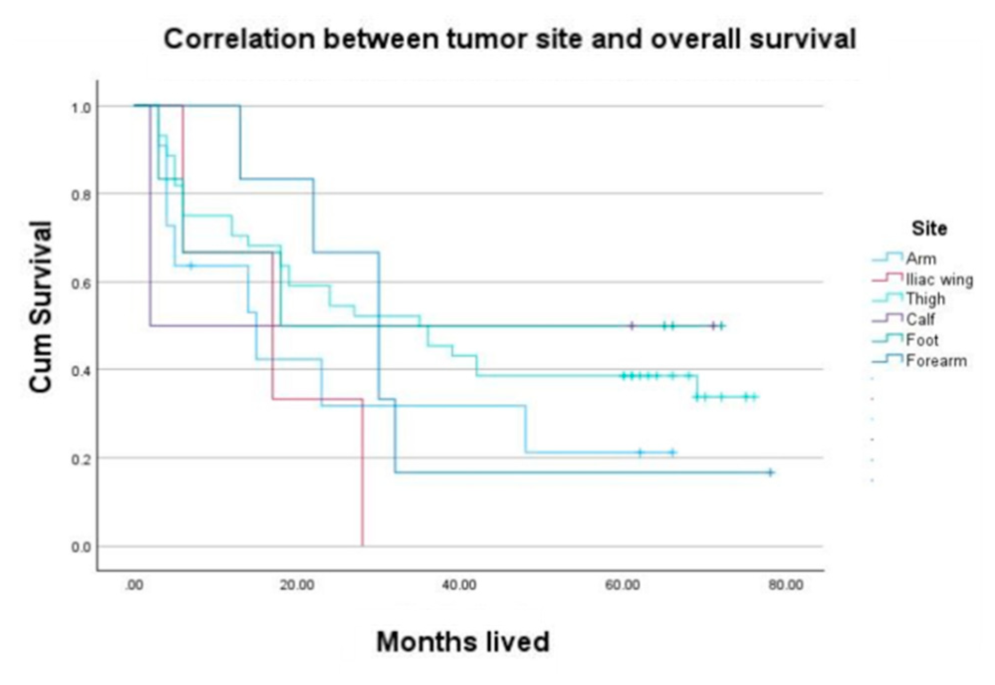
The distribution of the anatomical site of patients with STS, as well as the relationship between tumor location and the overall survival rate, are presented in
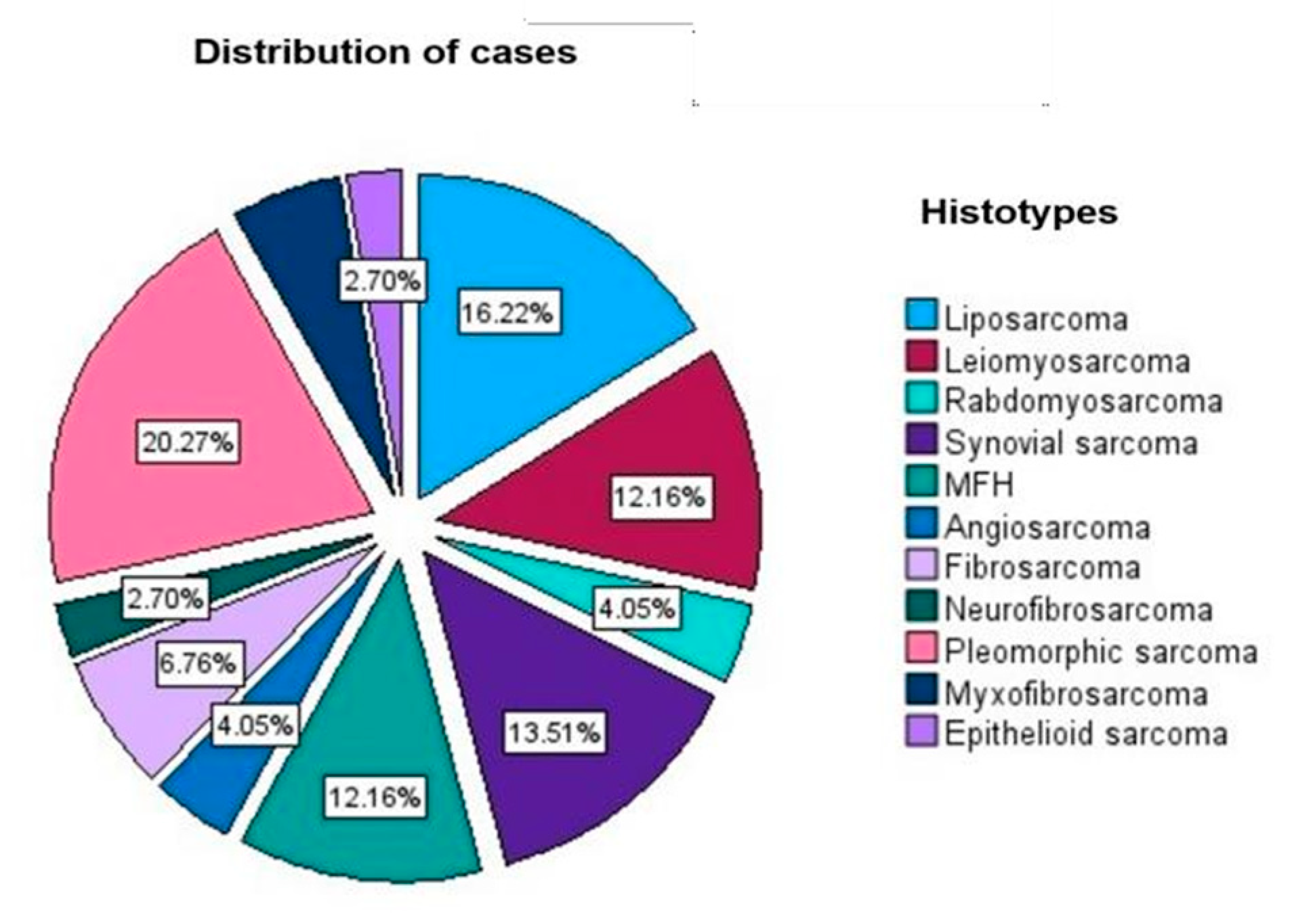
Figure 1. Primary tumors that developed in the extremities had a better prognosis than those with intraabdominal extension, corresponding to the overall comparison of data due to the possibility to perform a complete oncological excision with R0 margin. According to prior findings (American Cancer Society), undifferentiated pleomorphic sarcoma was the most common histopathogical type diagnosed in our group study (
Figure 2).
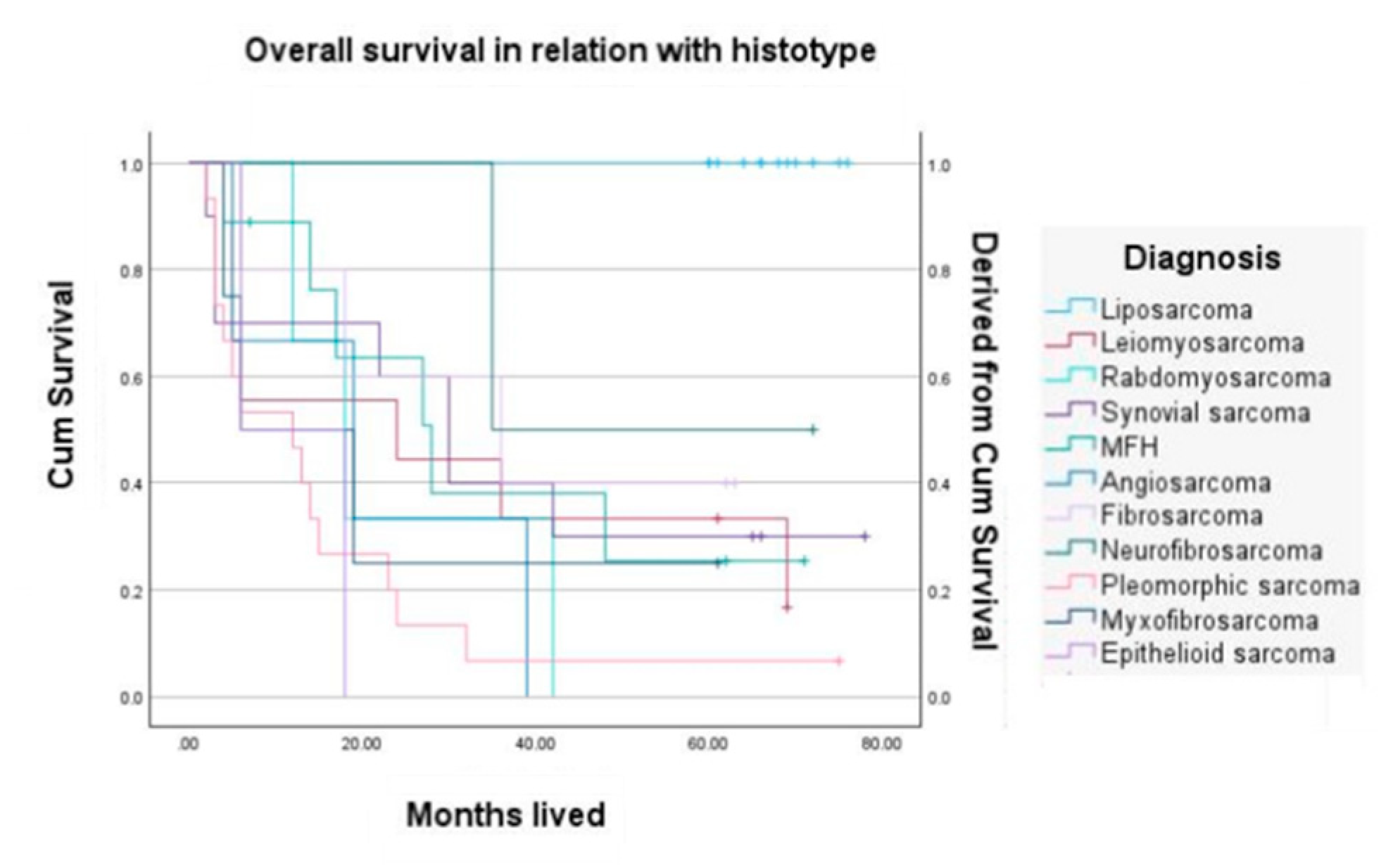
The histological (sub)type of the tumor appears to be one of the most noteworthy elements influencing the outcome. However, if prognostically favorable tumor (sub)types are examined alongside less favorable (sub)types, as is the case with most STS reports, the distinction between the two may become imperceptible. In our study the prognosis for liposarcoma was generally better than that of leiomyosarcoma, which was better than that of rabdomyosarcoma and synovial sarcoma sustained by significant statistical correlation with P value of p<0.001 as seen below in
Table 1. by applying Kaplan Maier survival curve (
Figure 3). However, the prognosis varies significantly throughout the different histological subtypes and grade of liposarcomas.
The correlation between tumor size and depth is widely established, and several researchers have observed comparable outcomes for individuals with superficial and deep tumors when they are categorized based on their grade and size [
9,
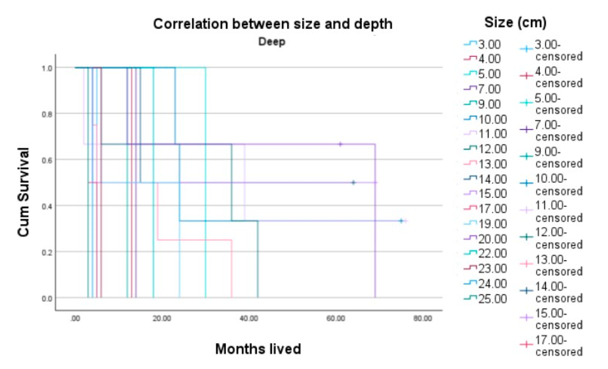
10]. The Kaplan-Meier approach used in our study's presented data reveals a decreased 5-year overall survival rate for patients with deep-localized tumors larger than 5 cm (as seen in
Figure 4) compared to patients that were diagnosed with superficial soft tissue sarcomas (
Figure 5).
The term "vascular invasion" refers to the occurrence of tumor cells in any area containing an endothelial lining. The clinicopathologic results and baseline demographics of our group of patients are typical of the literature's overall STS patient population. Even in a multivariate model that included the patient's age, score, histopathologic group, tumor grade, size, and depth, among other prognostic variables, we discovered that vascular invasion was associated with size and to a worse overall survival.
Data generated by the Illumina DRAGEN RNA pipeline that identified a significant number of variants across all samples, most of which were SNPs (95.4%), are presented in the
Table 2. The workflow also highlighted a number of homozygous and heterozygous indels (4.6%).
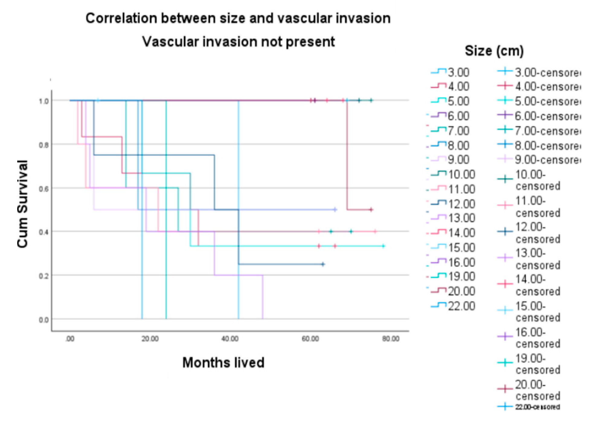
Figure 6 demonstrates a better outcome of the patients when vascular invasion is absent, compared to
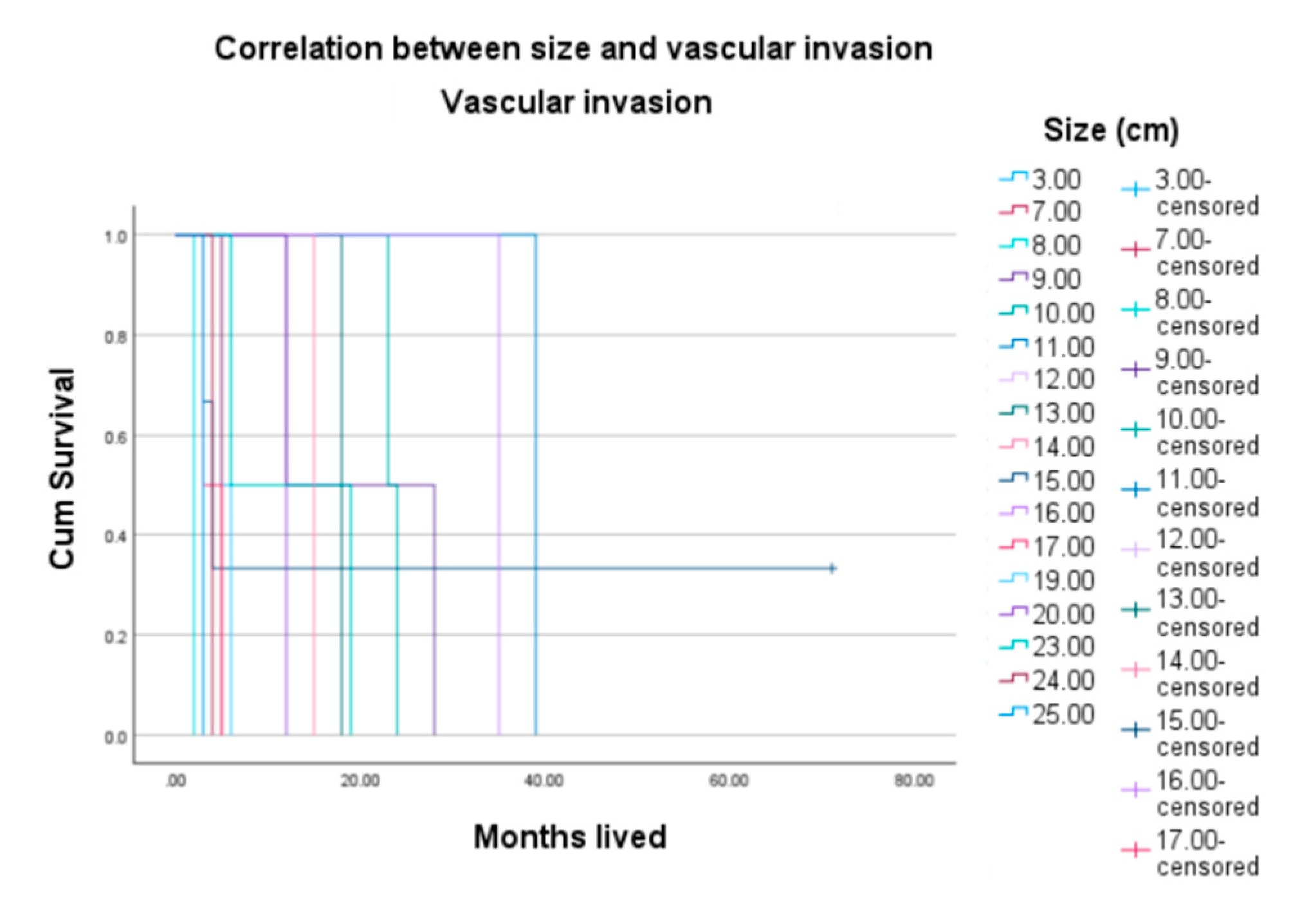
Figure 7 that indicates lower survival rates in patients where vascular invasion is present.
The Dragen Fusion Features Statistics table includes data produced by the Illumina Dragen RNA pipeline. The "Fusion Gene" column lists the parent genes involved in the fusion, ordered from 5' to 3' based on their transcripts.
In cases where a fusion breakpoint overlaps multiple genes, each gene is listed separately as a candidate in different rows. The "Score" column represents the confidence score of each fusion call (
Table 3).
While previous scientific literature has associated COL1A::PDGFB with dermatofibrosarcoma protuberans (DFSP), our findings reveal COL1A2::COL1A1 in a patient with desmoid-type fibromatosis. This discovery suggests a potential connection between different collagen- related gene fusions and distinct types of fibromatosis. We also noted the presence of SS18::SSX1 in a patient diagnosed with synovial sarcoma, consistent with previous findings. Tumor specimen was obtained from a 34 years old female who was diagnosed with a large (123/101/86mm) palpable mass located in the posterolateral compartment of the thigh. The HP and IHC examination revealed the presence of a biphasic synovial sarcoma for which the patient was admitted to Department of Oncology for neoadjuvant chemo and radiotherapy before surgical excision of the tumor. For the moment the patient is in our evidence for periodic follow-up with no evidence of local recurrence or distant metastasis.
Fusions
COL1A2::COL1A1,
SF3B1::NOM1 and
SS18::SSX1 were identified by both the DRAGEN and the RNA-Seq Alignment pipeline, suggesting high confidence in the detection of these fusion events. DRAGEN includes an RNA-seq (splicing-aware) aligner, as well as RNA specific analysis components for gene expression quantification and gene fusion detection (
Figure 8).
Discussions
Soft tissue sarcomas are defined as relatively rare, wide- ranging mesenchymal tumors that possess various tendencies to behave aggressively. The challenges arising from the overall rarity of this tumor family are compounded by the complicated interplay between anatomic site and histology in sarcomas, both of which are highly relevant from a therapeutic perspective.
The more precise and specialized the diagnosis, the more accurate the treatment and prognosis, despite the unpredictable nature of this pathology. Nonetheless, there are significant variations in clinical outcomes even among high risk localized sarcomas, with around half of patients experiencing a long-term remission and the other half relapsing within five years.
The majority of STSs exhibit high-grade metastatic behavior in multiple organs and are locally aggressive. The majority of patients with high-grade STS develop pulmonary metastases, usually within the first two years after the tumor's initial diagnosis. The lungs are the most common site of distant metastasis [
11]. The challenge of distant metastasis continues to exist today, despite the apparent improvement of the local recurrence issue. As much as 50% of patients pass away from their medical condition, and despite advances in local disease control, little progress has been made in terms of overall patient survival [
12]. This implies that surgery only affects local disease and that metastasis has already happened by the time of the initial diagnosis. In fact, after diagnosis, 10% of patients had metastatic disease [
13], and 25% of patients with localized cancer go on to develop distant dissemination [
14].
Radiation therapy, in combination with or without surgical resection of the main tumor with broad oncologic margins, lowers the probability of developing local recurrence but has no effect on the growth of distant metastases or patient survival [
15]. Systemic chemotherapy has restricted effects as well; response rates are minimal, estimated to be between 25 and 30 percent [
16]. Pulmonary metastases continue to be a leading cause of death in STS patients, although multimodal therapy - including surgery, chemotherapy, and radiation - has improved the survival rates in those individuals.
Sarcomas are rare tumors that are challenging to investigate. Reports of incidence, survival, and prevalence from a single institution are unlikely to be trustworthy; instead, representative data must be obtained through pooled data from population-based cancer registries.
This study focuses primarily with the prognosis of patients diagnosed with STS of the extremities. A prognosis is typically used to suggest a prediction of how a condition will progress when it first manifests. It can describe the disease's natural course, in which case no treatment is necessary, or its clinical course, in which case medical treatment will be needed. Due to the disease's uncommon and consequently small patient population, prognosis studies of patients diagnosed with STS are generally constrained.
Prognostic factors
An outcome of interest at a given time is estimated by a variable known as a prognostic factor. Prognostic factors influence not only therapy decisions, diagnostic techniques, and follow-up schedules, but also inform patients in terms of expected prognosis. The prognostic factors for local recurrence and mortality as well as diagnostic factors for non-metastasized STS in the extremities are the main focus of this study.
Prognostic factors are frequently classified into three categories: patient-related, tumor-related, and therapy- related factors. This allows for the separation of biological and therapeutic aspects. Factors that pertain to the patient comprise age, gender, duration of symptoms, and diagnostic year. Anatomical location, depth, size, compartmentalization, and grade are among the variables associated with tumors. Unplanned surgery, the type of surgery, the surgical margin, radiation, and chemotherapy are some treatment-related factors.
Malignancy grade
To enhance the prediction of local aggressiveness and metastatic potential, malignancy grading is applied to most STS cases. When determining the malignancy grade, a pathologist will typically assess intratumoural necrosis, rate of mitosis, and level of architectural and cellular differentiation. The FNCLCC [
17] and the NCI system [
18] are the two most popular systems. Within the category of tumors with high malignancy grade, there are differences in the probability of metastasis; hence, prognostication algorithms that combine many tumor parameters have been created more recently. For non- metastatic high-grade STS, Engellau et al. developed a stepwise model of [
19] that defined two risk groups and predicted the risk of metastatic relapse. Vascular invasion or at least two of the following three characteristics— tumor size >8 cm, tumor necrosis, and peripheral infiltrative growth pattern—were present in high-risk tumors. At five years, the high-risk group's cumulative incidence of metastasis was 51%, whereas the low-risk group's was 5%.
Histotype
Certain histotypes of sarcoma exhibit consistent behavior, making them unaffected by methods used for prognostication and malignancy grading. Lipoma-like liposarcomas (which behave in a generally benign manner), while pleomorphic liposarcoma or the round cell variant of liposarcoma have a high risk of metastasis. High- risk tumors include rhabdomyosarcoma, synovial sarcoma.
Site and size
The rate of metastatic extracompartmental tumors is nearly five times higher than the rate of metastatic subcutaneous tumors, and double that of intramuscular tumors [
20].
Extremity localization indicates a better prognosis since the tumor can be completely resectable. An independent prognostic factor cited by the literature is the size of the tumor [
17]. Size over 5 cm is correlated with a poorer metastasis free survival. Few studies indicated that the chance of metastasis could throughout the intermediate period of five years for high-grade but small tumors, and these should be observed to over five years to rule out the chance of a delayed manifestation of metastasis [
21].
Depth
A greater rate of metastasis has been linked to tumors that originate deep within the deep fascia. When examining entire tumor sections, soft-tissue sarcomas exhibiting a microscopically "infiltrative" development pattern are found to be substantially more likely to recur locally and systemically than having development patterns indicative of "pushing" lesions [
19].
Vascular invasion
Vascular invasion seems to be a significant harmful pathologic component in STS of the extremities. Vascular invasion was associated with worse OS even after taking into consideration other known prognostic variables. Understanding a patient's status can aid in risk stratification and management planning; hence, it should be taken into account in future nomograms and prognostic classification systems.
Molecular characterization of soft-tissue sarcoma
Molecular pathology is becoming an increasingly popular diagnostic tool, surpassing the constraints of light microscopy. Genetic signatures have predictive as well as diagnostic value signs. A multitude of chromosomal translocations, some specific to the tumor type have been identified in soft-tissue sarcomas.
In terms of molecular biology, soft tissue sarcomas can be divided into two main groups [
22]:
Sarcomas with non-specific genetic alterations and complex, with imbalanced karyotypes (such as leiomyosarcoma)
Sarcomas with specific genetic alterations and relatively simple karyotypes such as reciprocal chromosomal translocations and specific oncogenic mutations.
Next-generation sequencing (NGS) is a cutting-edge method that produces hundreds of thousands to hundreds of millions of short "reads" in a single run, allowing for the massively parallel, cost-effective sequencing of genomic regions. The test detects most kinds of alterations, including SNV and indels, using "deep" sequencing. Messenger RNA (mRNA) analysis through next- generation sequencing (NGS) provides complex data on gene expression levels, the existence of sequence and structural variants, as well as aberrant RNA phenotypes. Fusion genes are already integrated into the molecular classification of sarcomas, but adult sarcomas do not often present such abnormalities [
23,
24].
Conclusions
The current study's survival statistics are on accordance with the most reliable published findings. There was a significant advancement toward more limb-saving surgery over the research period. Early diagnosis, histologic type, superficial and small size of the tumor, were found to be favorable outcome variables by prognostic factor analyses.
Understanding a patient's prognostic factors may be helpful in assessing risk as well as treatment planning.
These findings suggest that mutation-specific clinical trials may be able to be accessed by patients through the use of targeted NGS. To ascertain whether these mutations are clinically significant therapeutic targets in sarcoma, more research is necessary.
