Next Article in Journal
Affective Temperaments and Personality Traits in Couple Well-Being
Previous Article in Journal
Medical and Psychological Management of Patients with a History of Surgical Treatment for Pancreatic Diseases
 
 
Journal of Mind and Medical Sciences is published by MDPI from Volume 12 Issue 1 (2025). Previous articles were published by another publisher in Open Access under a CC-BY (or CC-BY-NC-ND) licence, and they are hosted by MDPI on mdpi.com as a courtesy and upon agreement with Valparaiso University (ValpoScholar).
Article

Correlation between Molecular Prognostic Factors and Bevacizumab Therapeutic Resistance in Patients with Metastatic Colorectal Cancer; The AVAMET Study

by
Adrian Paul Suceveanu
1,
Dragos Serban
2,*,
Andreea Daniela Caloian
3,
Georgeta Camelia Cozaru
4,
Anca Chisoi
4,
Anca Antonela Nicolau
4,
Ioan Sergiu Micu
5 and
Andra Iulia Suceveanu
1
1
Department of Gastroenterology, Faculty of Medicine, Ovidius University of Constanta, Constanta, Romania
2
4th Department of General Surgery, Emergency University Hospital Bucharest, Carol Davila University of Medicine and Pharmacy, Bucharest, Romania
3
St. Apostol Andrew Emergency Clinical Hospital, Department of Oncology, Faculty of Medicine, Ovidius University of Constanta, Constanta, Romania
4
Center for Research and Development of the Morphological and Genetic Studies of Malignant Pathology, Faculty of Medicine, Ovidius University of Constanta, Constanta, Romania
5
Doctoral School of Medicine, Faculty of Medicine, Ovidius University of Constanta, Constanta, Romania
*
Author to whom correspondence should be addressed.
J. Mind Med. Sci. 2024, 11(1), 175-182; https://doi.org/10.22543/2392-7674.1477
Submission received: 12 December 2023 / Revised: 12 January 2024 / Accepted: 16 February 2024 / Published: 30 April 2024

Abstract

:
Objectives. Colorectal cancer is responsible for more than two million cases diagnosed annually. Despite early diagnosis through screening programs and adequate treatment, 25% of cases are diagnosed with metastatic disease while up to 50% of patients diagnosed early progress to metastatic disease. Materials and Methods. The study of 40 patients aims to identify the role of placental growth factor and heparin-binding growth factor as prognostic tools for current treatments involving resistance to bevacizumab in metastatic colorectal cancer. Results. Our results suggested that overall survival was influenced by serum fibroblast growth factor 1 levels. A cut-off value of 260.71 pg/mL was considered predictive of a disease-free survival/DFS of 6 months to 1 year, while a cut-off value of 117.51 pg/mL was considered predictive of a better DFS. Regarding placental growth factor involvement, a serum level of 13.75 pg/mL as a first determination and 11.48 pg/mL after 8 months of chemotherapy and anti-vascular endothelial growth factor therapy were considered cut-off values for 1- to 3-year overall survival/OS, while 6.45 pg/mL and 8.12 pg/mL levels were considered cut-off levels for 3- to 5-year OS. Conclusions. The results of the current study detected cutoff levels that may better predict treatment resistance in advanced-stage colorectal cancer and poor OS and DFS rates. Serum levels of placental growth factor and fibroblast growth factor 1 at diagnosis become important prognostic factors predicting resistance to bevacizumab in metastatic colorectal cancer.

Introduction

Colorectal cancer (CRC) represents an important health problem in the world, being the most common digestive cancer disease, with more than 2 million cases diagnosed annually that lead to a significant overload of health systems. Despite early diagnosis through screening programs, it is still the third leading cause of cancer death in both men and women [1,2]. Current curative options are still limited in tumors diagnosed in advanced stages, with 25% of cases being diagnosed with already metastatic disease. Furthermore, even in cases diagnosed early studies have shown that 40 to 50% of patients will progress to metastatic disease despite oncologic treatment [3,4]. Angiogenesis is an essential mechanism for normal tissue function, but is also an important process involved in tumor growth and distant dissemination, with malignant cells requiring new vessels to support their growth and metastasis. Several factors/molecules have been attributed to be involved in tumor angiogenesis, such as: vascular endothelial growth factor (VEGF), platelet-derived growth factor (PDGF), fibroblastic growth factor (FGF), placental growth factor (PlGF) and angiopoetin, the last being currently the most investigated [5,6,7]. VEGF is considered the most important regulator of the angiogenic process, being very well expressed especially in advanced CR tumors. PlGF is a member of the VEGF family and has an important role in tumor angiogenesis, being well correlated with the density of micro-vessels within the tumor and cancer progression. FGFs represent a distinct class of growth factors associated with the proangiogenic phenotype of CRC [8,9,10]. Current limited knowledge implies new studies to discover biomarkers capable of providing a better understanding of cancer biology, treatment resistance and prognosis/survival in metastatic disease [11,12]. Our study aims to identify the role of ELISA-detected PlGF and FGF1 factors as prognostic tools for resistance to bevacizumab treatment in metastatic CRC, literature data being quite limited at this time.

Materials and Methods

Participants, Data Collection and Ethical Statement

A sample of 40 patients with CRC were dynamically tested for the serum levels of PlGF and FGF1, during chemotherapy based on bevacizumab. A control set consisting of 38 non-cancerous patients from the Gastroenterology Clinic of St. Apostol Andrei County Emergency Hospital was also tested for PlGF and FGF1 factors. All samples were collected from patients referred to the Sf. Apostol Andrei County Emergency Hospital for CRC screening and treatment in the Gastroenterology clinic, being followed up and treated in the Oncology Clinic with bevacizumab and anti-VEGF according to the diagnosis and location/extension of the disease. The medical history was obtained retrospectively from the medical records of the hospital.
After collection of serum analyzes and demographic data, samples were tested for serum levels of PlGF and FGF1. The obtained data were then analyzed by correlation with data on overall survival (OS) with and without symptoms (DFS), treatment, the presence of metastases and other parameters of interest in CRC disease, using statistical methods. The study was approved by the Ethics Committee of Emergency Clinical St Apostle Andrew Hospital Constanta and by the “Ovidius” University of Constanta, in respect to all current low regulations. Participation of subjects was entirely voluntary and written consent was obtained.

Specimen Collection

PlGF serum testing. Blood samples were collected in tubes by venipuncture from 40 patients with colorectal cancer treated with chemotherapy, to determine the concentration of PlGF (Placental Growth Factor) by the ELISA method. Serum from blood samples was extracted by centrifugation at 3000× g for 30 min using SL16R Thermo Scientific centrifuge and tested for PlGF (Fine Test, produced by Wuhan Fine Biological Technology Co. Ltd., with sensitivity < 9.375 pg/mL for PlGF and no significant cross-reactivity or interference between PlGF and analogs) using an enzyme-linked immunosorbent assay (ELISA) according to the kits manufacturer’s instructions. The Fine Test kit was intended for detecting specific antibodies in a sample by ELISA method—(i.e., a solid phase coated with specific antigen-antibody from the analyzed sample—labeled antibody). The labeled antibody (conjugate) is an animal immunoglobulin fraction of human IgG conjugated with horseradish peroxidase. Peroxidase activity is determined in the test by a substrate containing TMB. Positivity is indicated when the blue color appears; after stopping the solution has been added, the blue changed to yellow. The yellow color intensity was measured by a photometer at 450 nm, and it was proportional to the concentration of specific IgG antibodies in the sample.
FGF1 serum testing. Another set of 40 blood samples were collected from the same patients for FGF1 concentration determination (Heparin-binding growth factor 1) using the ELISA method. Serum was extracted from blood samples by the previous explained method. Subsequent test for FGF1 (Fine Test, produced by Wuhan Fine Biological Technology Co., Ltd. with sensitivity < 18.75 pg/mL for FGF1 and no significant cross-reactivity or interference between FGF1 or analogs was observed) were performed. The method used the current recommendations from the specialized literature on this topic [13].

Statistical Analyses

Statistical analyses were computed using SPSS Statistics (IBM SPSS Statistics for Windows, Version 23.0). The descriptive statistics (mean, range and percentage) were computed for continuous variables. Correlations were nonparametric (Spearman analysis). Also, we conducted a Kaplan-Meier analysis to compare the survival probability over a definite timescale between two groups of patients. The log rank statistics test was used to compare factor levels. Further, the hazard ratio was estimated by using a Cox regression model with 95% confidence intervals.

Results

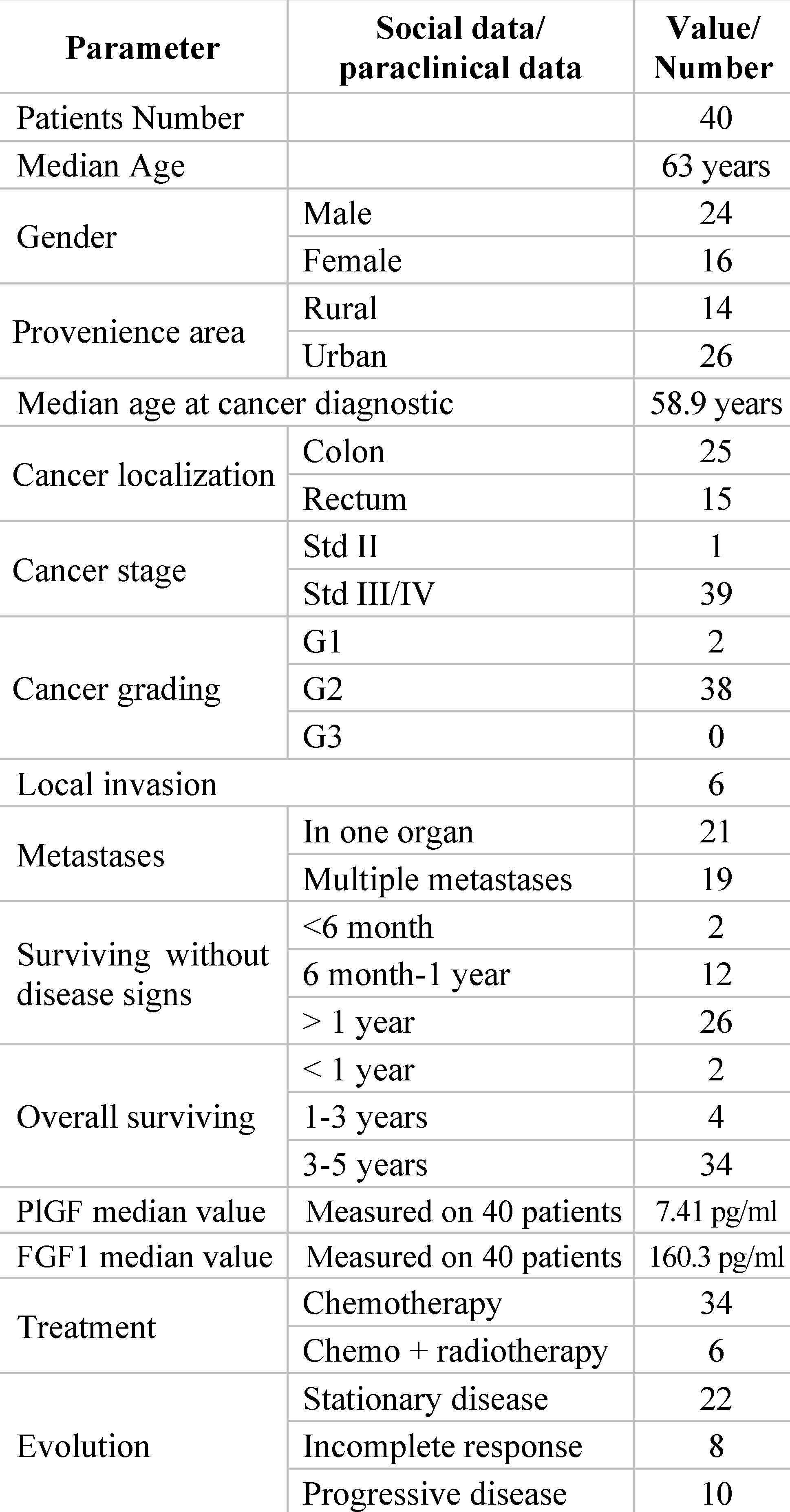
Socio-Demographic and Clinical Data of the Patients Included in the Study

The main patient’s demographic characteristics and tumor samples are presented in Table 1. Of the total of 40 enrolled patients, 24 were men and 16 were women, with a gender ratio of 1.5 in favor of men.
The median age was 63 years, with no significant differences between men (median age 62.83 years) and women (median age 63.25 years) (p > 0.5, ns). Demographics showed interesting results. From the rural area 14 patients were enrolled, 12 patients being men and 2 patients being women, while of the 26 patients enrolled from the urban environment, 12 were men and 14 were women, the statistics revealing a significant number of women diagnosed in the environment urban compared to rural (p = 0.0231, ss). The age of the patients from rural was younger (57.86 years) compared to the age of patients from the urban area (65.77 years), also having statistical significance (p = 0.0111, ss) (Table 1). In the control group (38 cancer-free patients) were enrolled 12 man and 26 women. Median age of the patients’ group was 58.97 years, while the median age for patients in control group was 55.88 years. All patients from the control group were from urban areas.
Of 40 patients with CRC, 25 had colonic cancer (CC) and 15 rectal cancer (RC) (Table no 1). According to gender, from the total of 24 male patients, 14 patients had CC, and 10 patients had RC. Of 16 females, 11 cases had CC and 5 cases had RC. The median age of patients at the moment of CRC diagnosis was 58.69 years for male patients and 59.06 years for female patients. For patients from rural areas, the median age at cancer diagnosis was 54.14 years and for patients from urban area median age at cancer diagnosis was 61.46 years, with a significant difference (p = 0.0033, ss). At the time of diagnosis, 39 out of 40 patients in the study group had stage III/IV of cancer (Table 1). Only one male patient was staged II, enrolled due to tumor progression during the study period. In the stage III/IV group there were 23 male and 16 female patients. The histopathological exam identified all cancers as ADK (adenocarcinoma). There was no case described as a poorly differentiated tumor. The majority of cases (38 patients) were described as moderately differentiated and only 2 cases were described as well differentiated. All cases of well differentiated cancer were female. In the moderately differentiated category (38 patients) 24 were male, and 14 female patients; the histopathological examination identified local invasion in 6 cases from 40 patients. Of those 6 cases, 3 had serous, perineural, and vascular invasion, and 3 of them had only serous invasion. Of 3 patients with multiple local invasion (serous, perineural and vascular) one was male and 2 were female. At the moment of diagnosis, all patients had lymph node or distant metastases. 19 patients had multiple distant determinations (Table 1). Most patients had hepatic metastases (31 cases) followed by pulmonary metastases (20 cases). Other localizations were peritoneal (9 cases), bone (2 cases), lymph nodes (3 cases) and nervous system (1 case).

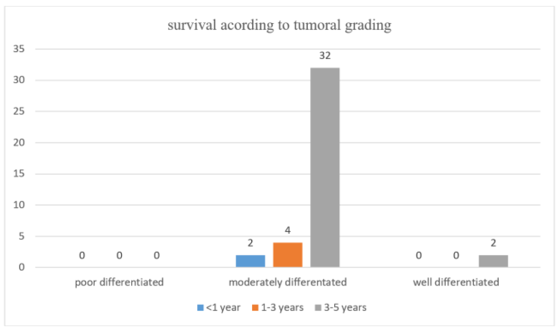
Survival Data During Anti-VEGF Treatment Overall Survival (OS) Rates

At the end of the study follow-up period, 17 of the group of patients with multiple metastases survived more than 3 years (3–5-year survival group), and 2 patients survived about 2 years (1–3-year survival group). No patient with multiple metastases died in the first year after diagnosis. From the entire patient group, 2 patients with liver metastasis died in less than 1 year from diagnosis. All patients with well differentiated tumors survived 3–5 years from diagnosis. In moderate differentiated cancer category, the majority of the patients survived 3–5 years from diagnosis (32 patients), other 4 patients survived 1–3 years, and only 2 patients died in the first year from diagnosis (Figure 1).

Disease-Free Survival (DFS) Rates

Disease-free survival followed the OS trend. The majority of patients with moderately differentiated tumors survived without signs of disease more than 1 year, 12 survived without disease 6 month to 1 year and only 2 survived without disease signs less than 6 month. In the case of well differentiated tumors, both patients survived without disease signs more than 1 year. All patients were under treatment at the onset of the study. 34 patients received chemotherapy in the form of FOLFOX or FOLFIRI regimens plus bevacizumab, while 6 patients received chemo-radio associated therapy (Table 1). During the study period, 22 patients had stationary disease, 8 had incomplete response, and 10 of them had progressive disease. The test for equality of survival distributions showed a significant difference (X2 (1, 40) = 6.586, p = 0.010) between the group of patients with rectal cancer receiving chemo-radiotherapy plus bevacizumab as treatment (67% probability of survival after 24 months) and the group only receiving chemotherapy plus bevacizumab (9% probability of survival after 24 months). Thus, the patients with rectal cancer benefiting from the complex treatment had a survival probability with 58% higher (Figure 2).
The Cox regression indicated that the presence of multiple metastases did not predict the hazard for death (B = −0.462, SE = 0.427, p = 0.279), and neither did the age (B = −0.033, SE = 0.021, p = 0.125). Thus, the only significant predictor is the treatment (B = 1.907, SE = 0.799, p = 0.017).

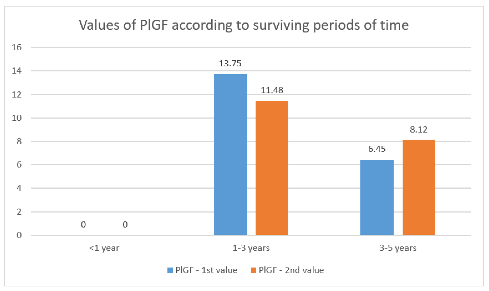
PlGF Values Data and Dynamics

The test group had 40 patients enrolled. All patients had PlGF measured at baseline. After 8 months, PlGF was measured again for a dynamic evolution of this parameter. At this time, only 23 patients had a second PlGF measurement. At the time of first determination, the median value for PlGF was 7.41 pg/mL. The median value was calculated on the entire lot of 40 patients. The control lot had 38 patients and only one measurement of PlGF. Median value was 4.02 pg/mL. As observed, value of PlGF in the test group was almost double compared to that of control group. At second measurement, on 23 patients, the median value of PlGF was 8.71 pg/mL. The median value of PlGF at the first measurement, calculated on 23 patients, was 7.71 pg/mL. As described, value of PlGF was rising from the first to the second measurement. Between the two determinations of PlGF, there was a positive correlation, but no statistically significant correlation (r (48) = 0.400, p = 0.058) (Table 2).
Patients had metastases from the beginning of treatment. This increasing value of PlGF may be due to the development of metastases, despite the treatment that clearly slowed the progression of the disease.
All patients who underwent two PlGF measurements survived more than 1 year. In the group of patients with overall survival (OS) 1–3 years, PlGF values decreased from the first to second measurement. The median value at first measurement was 13.75 pg/mL, while the second measurement was 11.48 pg/mL. In the group of patients with OS more than 3 years (3–5 years), PlGF values raised from first to second determination. At the first measurement PlGF was 6.45 pg/mL and at the second measurement, the median value was 8.12 pg/mL. Despite these observations, the values of PlGF in patients surviving 1–3 years were higher than those of patients surviving 3–5 years (p = 0.044, ss, respectively p = 0.029, ss) (Figure 3).
When analyzing data for patients’ disease-free survival (DFS), we noticed that patients that had two PlGF measurements survived more than 6 months without signs of disease. In terms of DFS, patients were split in two categories: DFS of disease 6-month–1 year and DFS more than 1 year. The values of serum PlGF raised from the first to second measurement in both groups. For category with DFS 6 month–1 year, the median value of PlGF was 10.00 pg/mL and the second was 10.63 pg/mL. In the category of DFS longer than 1 year, the median value for PlGF was at first measurement 6.49 pg/mL and at second measurement 7.68 pg/mL.

Variation over Time of FGF1 Values

Patients enrolled (40) had first FGF1 measurement at the initiation of the treatment. After 8-month FGF1 was measured again (for dynamic evolution). Only 23 patients undergo the second FGF1 measurement. At first determination median value for FGF1 was 160.36 pg/mL. Median value was calculated on entire lot of 40 patients. The control lot had 38 patients and only one measurement of FGF1. The median value was 30.19 pg/mL. Comparing the test group with the control group, we can observe that the FGF1 value was 5 times higher in the test group. At second measurement, on 23 patients, the median value of FGF1 was 175.67 pg/mL. The median value of FGF1 at first measurement, calculated on 23 patients, was 167.32 pg/mL. As seen, value of FGF1 had an increase from the first to second measurement. Between the two determinations of FGF1 was identified a statistically significant positive correlation (r (48) = 0.635, p = 0.001) (Table 3).
All patients who underwent two FGF1 measurements survived more than 1 year. In the group of patients with overall survival 1–3 years, FGF1 values decreased from the first to second measurement. Median value at first measurement was 224.77 pg/mL, and at second measurement it was 165.11 pg/mL (p = 0.0021, ss). In the group of patients surviving more than 3 years (3–5 years), the FGF1 values raised from the first to second determination. At the first measurement FGF1 was 155.22 pg/mL and at the second measurement median value was 177.90 pg/mL (p = 0.058, ns) (Figure 4). We detected a significant difference between the serum median values in both determinations, between the two groups with different OS, meaning 1–3 years, respectively 3–5 years (p = 0.0039, ss, respectively, p = 0.011, ss).
When analyzing data for DFS we can notice that patients having two FGF1 measurements survived more than 6 months without signs of disease. In both categories (surviving without signs of disease 6 months–1 year category and more than 1 year category) median values of FGF1 raised from the first to second measurement. For the category with DFS ranging from 6 months–1 year, the median value of the first determination of FGF1 was 260.71 pg/mL and the second was 282.58 pg/mL, with a small increase (p > 0.05, ns). In the category of DFS longer than 1 year, the median value for FGF was at first measurement 117.51 pg/mL and at second measurement was 118.66 pg/mL, with no significant value (p > 0.05, ns). Comparing the results according to the length of DFS, we detected significant differences between the first and second determination in our two groups of patients (p = 0.0144, ss for the first median serum value, respectively 0.006, ss for the second median serum value).

Discussions

Despite tremendous efforts to decrease the incidence and mortality rates of CRC, it is still the third leading cause of cancer for both men and women, with 2 million cases diagnosed in 2020 [14,15]. The screening strategies only improved the discovery of precancerous CR adenomatous polyps or tumors in curative stages [16,17,18,19]. The advanced CRC still remains a challenge for worldwide physicians, The International Agency for Research on Cancer (IARC) estimates a pessimistic outlook, the global incidence of CRC raising with 56% between 2020 and 2040, reaching around 3 million new cases per year. The mortality rates were estimated to increase even more, with 69% until 2040, this meaning about 1.6 million deaths worldwide [20,21,22]. Most of this increase is expected to occur in developed countries, with a high Development Activity Index, where independent risk factors for CRC occurrence, as alcohol and tobacco consumption, diabetes mellites or obesity are dramatically increasing [23,24]. On this setting, research is focused on detecting prognostic factors to avoid the resistance to treatment, to improve current therapeutic strategies and to open new perspectives for targeted treatments. Even 10 years after the FDA approval of the first anti-VEGF, bevacizumab, resistance to anti-VEGF treatments still remains a challenge for physicians. The mechanisms of resistance are not yet completely understood. The involvement of PlGF in CRC progression was already studied and published by several authors. Using immunohistochemistry analysis of tumor tissue samples, researchers demonstrated that PlGF over-expression was more frequently present in lymph node and distant metastatic disease, being correlated with a poor prognosis [25,26,27]. Other authors demonstrated that the serum PlGF levels may serve as prognostic tool in non-metastatic CRC, too [28]. Moreover, the specialized literature lacks data regarding the dynamic prognostic values of serum PlGF levels during anti-VEGF therapy with bevacizumab.
The results of our study showed that serum levels of PlGF (measured by ELISA technique) in patients with OS of 1–3 years were significantly higher than those of patients with OS of 3–5 years (p = 0.044, ss, respectively p = 0.029, ss). A serum level of 13.75 pg/mL as a first determination and 11.48 pg/mL after 8 months of chemo and anti-VEGF therapy were considered cut-off values for OS of 1 to 3 years in advanced CRC, while 6.45 pg/mL and 8.12 pg/mL levels were considered cut-off levels for OS of 3 to 5 years. Despite the fact that the second determination revealed an increase of the PlGF serum value in the group of patients with prolonged survival over 3 years, these results had no impact on OS in our study group. In addition, for the category of 6 month 1-year DFS, first median value of PlGF was 10.00 pg/mL and second was 10.63 pg/mL, while in category of DFS more than 1 year, the median value for PlGF was at first measurement 6.49 pg/mL and at second measurement 7.68 pg/mL; such data were considered cut-off values for better prognosis during complex treatment in advanced CRC disease. By these results, our study confirms the prognostic value of serum PlGF level in advanced CR tumors and becomes a facile tool to monitor the response to anti-VEGF treatment. Moreover, serum PlGF level combined with FOBT tests used in screening strategies seems to be a reasonable alternative for those subjects who are reluctant to stool-based screening methods and who have been tested as FOBT negative. This biomarker is useful not only as an indicator in the evolution of unfavorable prognosis, but also as a tool for early diagnosis of the disease [29,30,31].
Referring to the aberrant expression of FGF-1 in CRC, studies suggest that overexpression of the FGFR-1 gene is correlated with the occurrence of liver metastases. As a consequence, the overexpression of this gene might be used as a predictor of liver metastasis in patients with CRC [32,33]. Looking further, our study demonstrated that the serum level of FGF-1 becomes an easier and easier tool to predict the worse course and the occurrence of metastases in CRC. The ELISA method performed in the serum of our CRC patients revealed interesting results. Thus, all CRC patients treated with chemotherapy in combination with anti-VEGF and who underwent two FGF1 measurements survived more than 1 year. In the group of patients with OS 1-3 years, FGF1 values decreased significantly (p = 0.0021, ss). In the group of patients with OS more than 3 years (3–5 years), FGF1 values raised from first to second determination, but with a low statistical significance (0.058, ns). Our results suggested that OS was influenced by serum FGF-1 overload in both circumstances, that is, at diagnosis and during chemo and anti-VEGF treatment (p = 0.0039, ss, respectively, p = 0.011, ss). A cut-off value of 155.22 pg/mL was considered predictive for a OS of 3 to 5 years, before anti-VEGF treatment. In the same time, a cut-off value of 177.90 pg/mL after 8 months of chemo and anti-VEGF therapy was considered enough to provide a OS of 3–5 years.
Median values higher than 224.77 pg/mL, as we obtained in the group with OS of 1–3 years, were considered for a worse prognosis even if during treatment the value was significantly improved. Regarding DFS, results showed that FGF1 values increased from the first to the second measurement in both categories of patients. Comparing the outcome according to DFS duration, we detected significant differences between the first and second determination between the two groups of patients (p = 0.0144, ss for the first median serum value, respectively 0.006, ss, the second median serum value). The cut-off value of 260.71 pg/mL was considered predictive for a DFS of 6 months to 1 year, while a cut-off value of 117.51 pg/mL was considered predictive for a better DFS. A better understanding of the predictive factors for the success of anti-VEGF treatment in metastatic colorectal cancer may therefore help to optimize therapeutic management and increase patient safety [34,35,36].

Conclusions

Both evaluated serum parameters, namely PlGF and FGF-1, were useful in confirming the proposed objectives for this study. The results of the current study detected cutoff levels that may better predict treatment resistance in advanced-stage CRC and poor OS and DFS rates. Serum levels of PlGF and FGF-1 at the time of diagnosis become important prognostic factors predicting resistance to bevacizumab (AVASTIN) in advanced/metastatic CRC.

Highlights

Prognostic Biomarkers in Metastatic CRC: This study focuses on the under-investigated prognostic roles of placental growth factor (PlGF) and fibroblast growth factor 1 (FGF1) identified by ELISA techniques in patients with metastatic colorectal cancer (CRC) undergoing treatment with bevacizumab.
Insights for Improved Outcomes: By highlighting the roles of PlGF and FGF1, the data obtained in this study provide valuable information on disease prognosis, treatment resistance and overall survival in advanced CRC. Such an exploration of new biomarkers seems to be a promising perspective for the further development of treatment strategies able to improve the therapeutic outcomes of patients with metastatic CRC.

Informed Consent Statement

Any aspect of the work covered in this manuscript has been conducted with the ethical approval of all relevant bodies and that such approvals are acknowledged within the manuscript. Informed consent was obtained from all subjects involved in the study.

Informed Consent Statement

There are no known conflicts of interest in the publication of this article. The manuscript was read and approved by all authors.

Acknowledgments

The study was conducted in Center for Research and Development of the Morphological and Genetic Studies of Malignant Pathology (CEDMOG), an affiliated institution of Ovidius University of Constanta, and established as a grant research program following the competition organized by these institutions.

Abbreviations

CRC: Colorectal Cancer, VEGF: Vascular Endothelial Growth Factor, PDGF: Platelet-Derived Growth Factor, FGF: Fibroblastic Growth Factor, PlGF: Placental Growth Factor, TMB: 3,3′,5,5′-Tetramethylbenzidine (used in ELISA for colorimetric detection), CC: Colonic Cancer, RC: Rectal Cancer, CR: Colorectal, DFS: Disease-Free Survival, OS: Overall Survival, IARC: International Agency for Research on Cancer, FDA: U.S. Food and Drug Administration, ELISA: Enzyme-Linked Immunosorbent Assay, OS: Overall Survival, FOBT: Fecal Occult Blood Test, FGFR-1: Fibroblast Growth Factor Receptor 1, CEDMOG: Center for Research and Development of the Morphological and Genetic Studies of Malignant Pathology

References

  1. Bretthauer, M.; Løberg, M.; Wieszczy, P.; Kalager, M.; Emilsson, L.; Garborg, K.; Rupinski, M.; Dekker, E.; Spaander, M.; Bugajski, M.; et al. Effect of Colonoscopy Screening on Risks of Colorectal Cancer and Related Death. N. Engl. J. Med. 2022, 387, 1547–1556. [Google Scholar] [CrossRef] [PubMed]
  2. Chan, S.C.H.; Liang, J.Q. Advances in tests for colorectal cancer screening and diagnosis. Expert Rev. Mol. Diagn. 2022, 22, 449–460. [Google Scholar] [CrossRef]
  3. Ohhara, Y.; Fukuda, N.; Takeuchi, S.; Honma, R.; Shimizu, Y.; Kinoshita, I.; Dosaka-Akita, H. Role of targeted therapy in metastatic colorectal cancer. World J. Gastrointest. Oncol. 2016, 8, 642–655. [Google Scholar] [CrossRef] [PubMed]
  4. An, S.-X.; Yu, Z.-J.; Fu, C.; Wei, M.-J.; Shen, L.-H. Biological factors driving colorectal cancer metastasis. World J. Gastrointest. Oncol. 2024, 16, 259–272. [Google Scholar] [CrossRef] [PubMed]
  5. Rodriguez-Pascual, J.; Cubillo, A. Dynamic Biomarkers of Response to Antiangiogenic Therapies in Colorectal Cancer: A Review. Curr. Pharmacogenom. Pers. Med. 2018, 15, 81–85. [Google Scholar] [CrossRef]
  6. Macarulla, T.; Montagut, C.; Sánchez-Martin, F.J.; Granja, M.; Verdaguer, H.; Sastre, J.; Tabernero, J. The role of PIGF blockade in the treatment of colorectal cancer: Overcoming the pitfalls. Expert Opin. Biol. Ther. 2019, 20, 15–22. [Google Scholar] [CrossRef]
  7. Henrich, L.M.; Greimelmaier, K.; Wessolly, M.; Klopp, N.A.; Mairinger, E.; Krause, Y.; Berger, S.; Wohlschlaeger, J.; Schildhaus, H.-U.; Baba, H.A.; et al. The Impact of Cancer-Associated Fibroblasts on the Biology and Progression of Colorectal Carcinomas. Genes 2024, 15, 209. [Google Scholar] [CrossRef]
  8. Saif, W. Anti-VEGF agents in metastatic colorectal cancer (mCRC): Are they all alike? Cancer Manag. Res. 2013, 5, 103–115. [Google Scholar] [CrossRef]
  9. Rejali, L.; Nazemalhosseini-Mojarad, E.; Valle, L.; Maghsoudloo, M.; Aghdaei, H.A.; Mohammadpoor, H.; Zali, M.R.; Khanabadi, B.; Entezari, M.; Hushmandi, K.; et al. Identification of antisense and sense RNAs of intracrine fibroblast growth factor components as novel biomarkers in colorectal cancer and in silico studies for drug and nanodrug repurposing. Environ. Res. 2023, 239, 117117. [Google Scholar] [CrossRef]
  10. Zawawi, S.S.A.; Azram, N.A.S.M.; Sulong, S.; Zakaria, A.D.; Lee, Y.Y.; Jalil, N.A.C.; Musa, M. Identification of AOC3 and LRRC17 as Colonic Fibroblast Activation Markers and Their Potential Roles in Colorectal Cancer Progression. Asian Pac. J. Cancer Prev. 2023, 24, 3099–3107. [Google Scholar] [CrossRef]
  11. Wolf, D.; Salcher, S.; Pircher, A. The multivisceral landscape of colorectal cancer metastasis: Ifmplications for targeted therapies. J. Clin. Investig. 2024, 134. [Google Scholar] [CrossRef] [PubMed]
  12. Motofei, I.G. Biology of cancer; from cellular and molecular mechanisms to developmental processes and adaptation. Semin. Cancer Biol. 2021, 86, 600–615. [Google Scholar] [CrossRef] [PubMed]
  13. Cambrea, S.C.; Aschie, M.; Resul, G.; Mitroi, A.F.; Chisoi, A.; Nicolau, A.A.; Baltatescu, G.I.; Cretu, A.M.; Lupasteanu, G.; Serbanescu, L.; et al. HPV and HIV Coinfection in Women from a Southeast Region of Romania—PICOPIV Study. Medicina 2022, 58, 760. [Google Scholar] [CrossRef] [PubMed]
  14. Savlovschi, C.; Serban, D.; Andreescu, C.; Dascalu, A.; Pantu, H. Economic analysis of medical management applied for left co-lostomy. Chirurgia 2013, 108, 666–669. [Google Scholar]
  15. Zheng, L.; Li, B.; Lei, L.; Wang, L.-J.; Zeng, Z.-P.; Yang, J.-D. Effect of screening colonoscopy frequency on colorectal cancer mortality in patients with a family history of colorectal cancer. World J. Gastrointest. Oncol. 2024, 16, 354–363. [Google Scholar] [CrossRef]
  16. Suceveanu, A.I.; Suceveanu, A.; Voinea, F.; Mazilu, L.; Mixici, F.; Adam, T. Introduction of cytogenetic tests in colorectal cancer screening. J. Gastrointestin. Liver Dis. 2009, 18, 33–38. [Google Scholar]
  17. Savlovschi, C.; Serban, D.; Trotea, T.; Borcan, R.; Dumitrescu, D. Post-surgery morbidity and mortality in colorectal cancer in elderly subjects. Chirurgia 2013, 108, 177–9. [Google Scholar]
  18. Mazilu, L.; Ciufu, N.; Gălan, M.; Suceveanu, A.I.; Parepa, I.R.; Tofolean, D. Postherapeutic follow-up of colorectal cancer patients treated with curative intent. Chirurgia 2012, 107, 55–58. [Google Scholar]
  19. Şavlovschi, C.; Comandaşu, M.; Şerban, D. Specifics of diagnosis and treatment in synchronous colorectal cancers (SCC). Chirurgia 2013, 108, 43–45. [Google Scholar]
  20. Ciuhu, A.N.; Rahnea-Niță, R.A.; Popescu, M.; Badiu, C.D.; Pantea Stoian, A.M.; Lupuliasa, D.; Gherghiceanu, F.; Diaconu, C.C.; Rahnea-Niță, G. Evidence of strong opioid therapy for palliation of breathlessness in cancer patients. Farmacia 2017, 65, 173–178. [Google Scholar]
  21. Soerjomataram, I.; Bray, F. Planning for tomorrow: global cancer incidence and the role of prevention 2020–2070. Nat. Rev. Clin. Oncol. 2021, 18, 663–672. [Google Scholar] [CrossRef] [PubMed]
  22. Ginghină, O.; Negrei, C.; Hudiță, A.; Ioana-Lavric, V.; Gălățeanu, B.; Dragomir, S.; Burcea Dragomiroiu, G.T.A.; Bârcă, M.; Nițipir, C.; Diaconu, C.C.; et al. In vitro impact of some natural compounds on HT-29 colorectal adenocarcinoma cells. Farmacia 2017, 65, 947–953. [Google Scholar]
  23. Bray, F.; Jemal, A.; Grey, N.; Ferlay, J.; Forman, D. Global cancer transitions according to the Human Development Index (2008–2030): A population-based study. Lancet Oncol. 2012, 13, 790–801. [Google Scholar] [CrossRef] [PubMed]
  24. Tulin, A.; Slavu, I.; Tulin, R.; Alecu, L.; Jecan, R.; Orlov, C.; Iaciu, C.; Stanculeanu, D.; Hainarosie, R.; Pituru, S.; et al. Does sex of the patient play a role in survival for MSI colorectal cancer? J. Mind Med Sci. 2018, 5, 101–108. [Google Scholar] [CrossRef]
  25. Van Cutsem, E.; Paccard, C.; Chiron, M.; Tabernero, J. Impact of Prior Bevacizumab Treatment on VEGF-A and PlGF Levels and Outcome Following Second-Line Aflibercept Treatment: Biomarker Post Hoc Analysis of the VELOUR Trial. Clin. Cancer Res. 2020, 26, 717–725. [Google Scholar] [CrossRef] [PubMed]
  26. Caraban, B.M.; Matei, E.; Cozaru, G.C.; Aşchie, M.; Deacu, M.; Enciu, M.; Bălţătescu, G.I.; Chisoi, A.; Dobrin, N.; Petcu, L.; et al. PD-L1, CD4+, and CD8+ Tumor-Infiltrating Lymphocytes (TILs) Expression Profiles in Melanoma Tumor Microenvironment Cells. J. Pers. Med. 2023, 13, 221. [Google Scholar] [CrossRef]
  27. Constantin, V.D.; Silaghi, A.; Epistatu, D.; Dumitriu, A.S.; Paunica, S.; Bălan, D.G.; Socea, B. Diagnosis and management of colon cancer patients presenting in advanced stages of complications. J. Mind Med. Sci. 2023, 10, 51–65. [Google Scholar] [CrossRef]
  28. Schölch, S.; Bogner, A.; Bork, U.; Rahbari, M.; Győrffy, B.; Schneider, M.; Reissfelder, C.; Weitz, J.; Rahbari, N.N. Serum PlGF and EGF are independent prognostic markers in non-metastatic colorectal cancer. Sci. Rep. 2019, 9, 1–12. [Google Scholar] [CrossRef]
  29. Wei, S.; Tsao, P.; Wang, Y.; Lin, B.; Wu, D.; Tsai, W.; Chen, J.; Wong, J. Using serum placenta growth factor could improve the sensitivity of colorectal cancer screening in fecal occult blood negative population: A multicenter with independent cohort validation study. Cancer Med. 2019, 8, 3583–3591. [Google Scholar] [CrossRef]
  30. Suceveanu, A.I.; Suceveanu, A.; Dumitru, E.; Alexandrescu, L.; Voinea, F. The feasibility of FOBT tests in colorectal cancer screening in Dobrogea. Rom. J. Gastroenterol. 2005, 14, 213–7. [Google Scholar]
  31. Tobi, M.; Antaki, F.; Rambus, M.A.; Yang, Y.-X.; Kaplan, D.; Rodriguez, R.; Maliakkal, B.; Majumdar, A.; Demian, E.; Tobi, Y.Y.; et al. The Non-Invasive Prediction of Colorectal Neoplasia (NIPCON) Study 1995–2022: A Comparison of Guaiac-Based Fecal Occult Blood Test (FOBT) and an Anti-Adenoma Antibody, Adnab-9. Int. J. Mol. Sci. 2023, 24, 17257. [Google Scholar] [CrossRef] [PubMed]
  32. Sato, T.; Oshima, T.; Yoshihara, K.; Yamamoto, N.; Yamada, R.; Nagano, Y.; Fujii, S.; Kunisaki, C.; Shiozawa, M. Overexpression of the fibroblast growth factor receptor-1 gene correlates with liver metastasis in colorectal cancer. Oncol. Rep. 1994, 21, 211–216. [Google Scholar] [CrossRef] [PubMed]
  33. Lim, S.H.; Cho, H.J.; Kim, K.-M.; Lim, H.Y.; Kang, W.K.; Lee, J.; Park, Y.S.; Kim, H.C.; Kim, S.T. Comprehensive molecular analysis to predict the efficacy of chemotherapy containing bevacizumab in patients with metastatic colorectal cancer. Oncol. Res. Featur. Preclin. Clin. Cancer Ther. 2023, 31, 855–866. [Google Scholar] [CrossRef]
  34. Serban, D.; Smarandache, A.; Cristian, D.; Tudor, C.; Duta, L.; Dascalu, A. Medical errors and patient safety culture—Shifting the healthcare paradigm in Romanian hospitals. Romanian J. Leg. Med. 2020, 28, 195–201. [Google Scholar] [CrossRef]
  35. Cheng, X.; Li, X.; Yang, X.; Fang, S.; Wang, Z.; Liu, T.; Zheng, M.; Zhai, M.; Yang, Z.; Shen, T.; et al. Successful Treatment of pMMR MSS IVB Colorectal Cancer Using Anti-VEGF and Anti-PD-1 Therapy in Combination of Gut Microbiota Transplantation: A Case Report. Cureus 2023, 15, e42347. [Google Scholar] [CrossRef]
  36. Hashimoto, T.; Otsu, S.; Hironaka, S.; Takashima, A.; Mizusawa, J.; Kataoka, T.; Fukuda, H.; Tsukamoto, S.; Hamaguchi, T.; Kanemitsu, Y. Phase II biomarker identification study of anti-VEGF agents with FOLFIRI for pretreated metastatic colorectal cancer. Futur. Oncol. 2023, 19, 1593–1600. [Google Scholar] [CrossRef]
Figure 1. Survival according to tumoral grading.
Jmms 11 00022 g001
Figure 2. Survival curve evaluated on a 24 months period for the RC patients that benefited from two different treatments: chemotherapy and a combination between chemotherapy and radiotherapy.
Jmms 11 00022 g002
Figure 3. PlGF values according to survival time periods.
Jmms 11 00022 g003
Figure 4. PlGF values according to survival time periods.
Jmms 11 00022 g004
Table 1. Patient data and tumor characteristics at the time of CRC diagnosis.
Jmms 11 00022 i001
Table 2. Spearman’s correlations between first and second PlGF measurements.
Jmms 11 00022 i002
Table 3. Spearman’s correlations between first and second FGF1 measurements.
Jmms 11 00022 i003

