Contemporary 0.55 T MRI for Lung Disease Assessment in Children and Young Adults with Cystic Fibrosis: A Proof-of-Concept Study
by
 , , , , ,
Nadine Bayerl
1,* ,
,
Maximilian Hinsen
1,
Stephan Ellmann
1,2 ,
,
Armin M. Nagel
1,
Lisa C. Adams
3,
Teresa Gerhalter
1,4,
Rafael Heiss
1,
Tobias Bäuerle
1,5,
Sabina Schmitt-Grohé
6,
Michael Uder
1,
Oliver Rompel
1,† and
Alexander Schnell
6,† 1
Institute of Radiology, Uniklinikum Erlangen, Friedrich-Alexander-Universität Erlangen-Nürnberg, Maximiliansplatz 3, 91054 Erlangen, Germany
2
RNZ—Radiologisch-Nuklearmedizinisches Zentrum, Martin-Richter-Str. 43, 90489 Nürnberg, Germany
3
Department of Diagnostic and Interventional Radiology, Technical University of Munich, Ismaninger Str. 22, 81675 Munich, Germany
4
Department of Neurology, Medical University of Graz, Auenbruggerplatz 22, 8036 Graz, Austria
5
Department of Diagnostic and Interventional Radiology, University Medical Center of the Johannes Gutenberg University Mainz, Langenbeckstr. 1, 55131 Mainz, Germany
6
Department of Pediatrics and Adolescent Medicine, Uniklinikum Erlangen, Friedrich-Alexander-Universität Erlangen-Nürnberg, Loschgestr. 15, 91054 Erlangen, Germany
*
Author to whom correspondence should be addressed.
†
These authors contributed equally to this work.
Tomography 2026, 12(9), 126; https://doi.org/10.3390/tomography12090126
Submission received: 7 July 2026
/
Revised: 24 August 2026
/
Accepted: 25 August 2026
/
Published: 31 August 2026
Simple Summary
Cystic fibrosis is an inherited disease that gradually damages the lungs, so patients need repeated imaging throughout their lives. Chest X-rays are widely used, but they involve radiation that accumulates over time, which is a particular concern for children and young adults. This study evaluated first experiences with a modern low-field MRI scanner as a radiation-free alternative. In 28 examinations of 22 young patients, MRI produced higher lung disease scores than chest X-rays and more consistent results between readers. Because the two methods were graded with different scoring systems and no CT scan was available for comparison, the higher MRI scores alone do not prove that MRI detects more disease. These findings suggest low-field MRI may represent a reliable monitoring option, encouraging larger studies.
Abstract
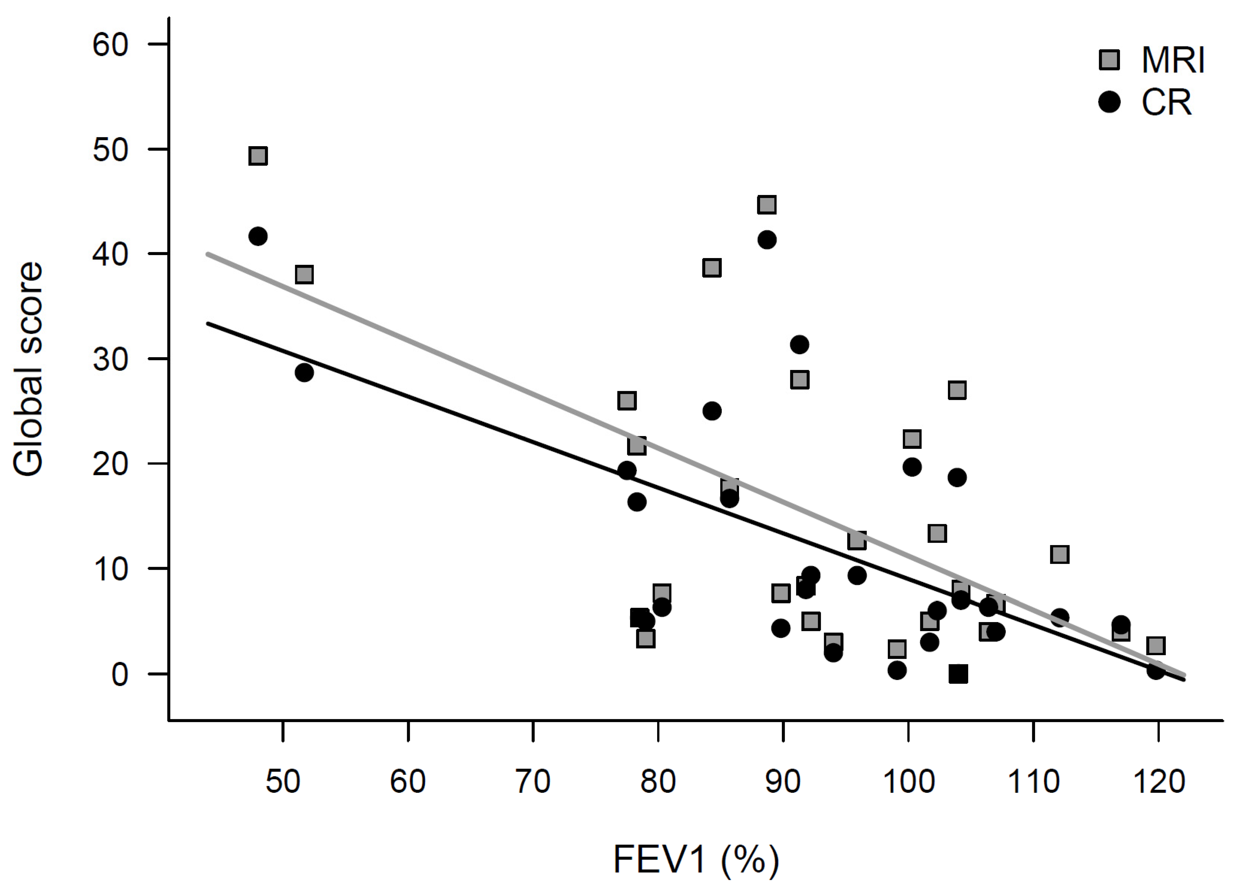
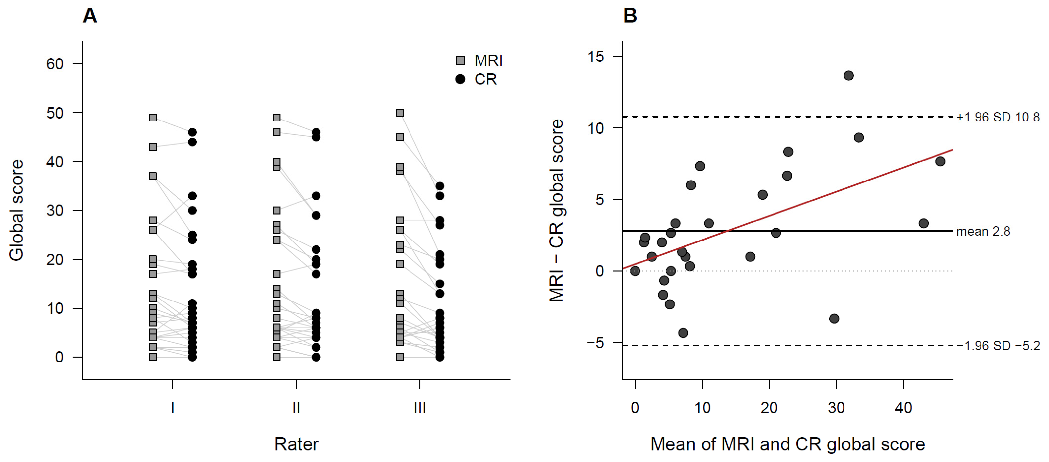
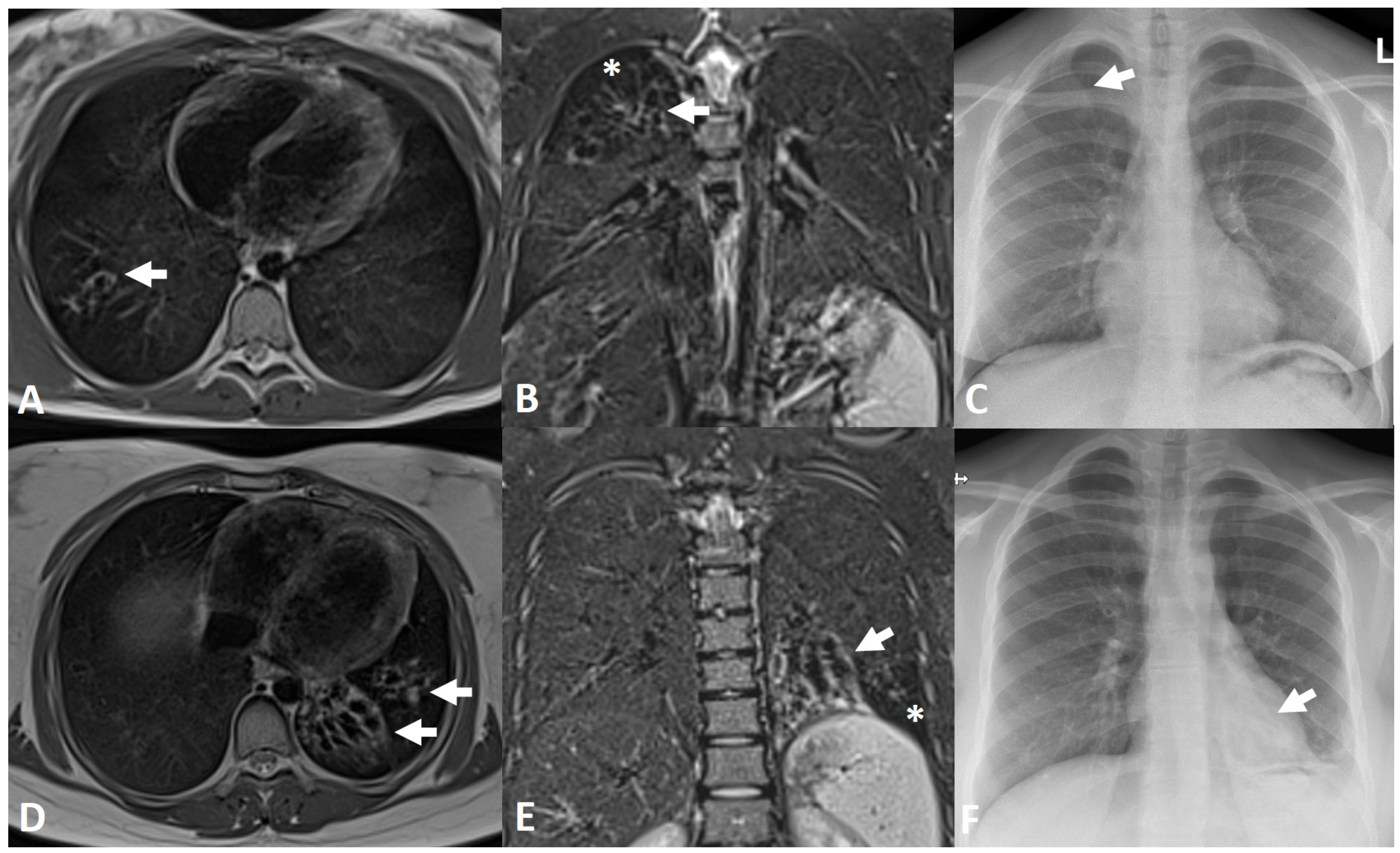
Background: Lung surveillance in cystic fibrosis (CF) relies on chest radiography (CR) despite cumulative radiation. Low-field MRI may be a radiation-free alternative for children and young adults. Methods: We prospectively compared 0.55 T MRI and CR in 28 same-day examinations of 22 people with CF (mean age 13 ± 5 years). Three raters scored the disease with the MRI CF score and a modified Chrispin–Norman score, expanded from two to three zones per hemithorax to match the six MRI lobes. Analyses used per-examination values averaged across the raters (Wilcoxon tests, intraclass correlation coefficients, ICC). Results: MRI yielded higher global scores (median 8.2 vs. 6.7, p = 0.002) and higher centrilobular opacity scores (p = 0.005, adjusted p = 0.03). Air trapping was also higher on MRI (p = 0.025) but not after correction for multiple comparisons (adjusted p = 0.12). The other categories did not differ. Both correlated inversely with FEV1 (p < 0.001), without a significant modality-by-FEV1 interaction (p = 0.08). Interobserver agreement was higher for MRI (ICC = 0.93) than CR (0.82), difference 0.12 (95% bootstrap CI 0.06 to 0.18). The findings held in a per-participant sensitivity analysis. Conclusions: 0.55 T MRI is a feasible, radiation-free alternative to CR in CF, yielding higher modality-specific scores and more consistent interobserver agreement. Because the instruments were only category-mapped and no CT reference was available, higher scores do not establish superior lesion detection.