
Understanding the Dermoscopic Patterns of Basal Cell Carcinoma Using Line-Field Confocal Tomography

 , ,
, ,  , , , , ,
Abstract
1. Introduction
2. Materials and Methods
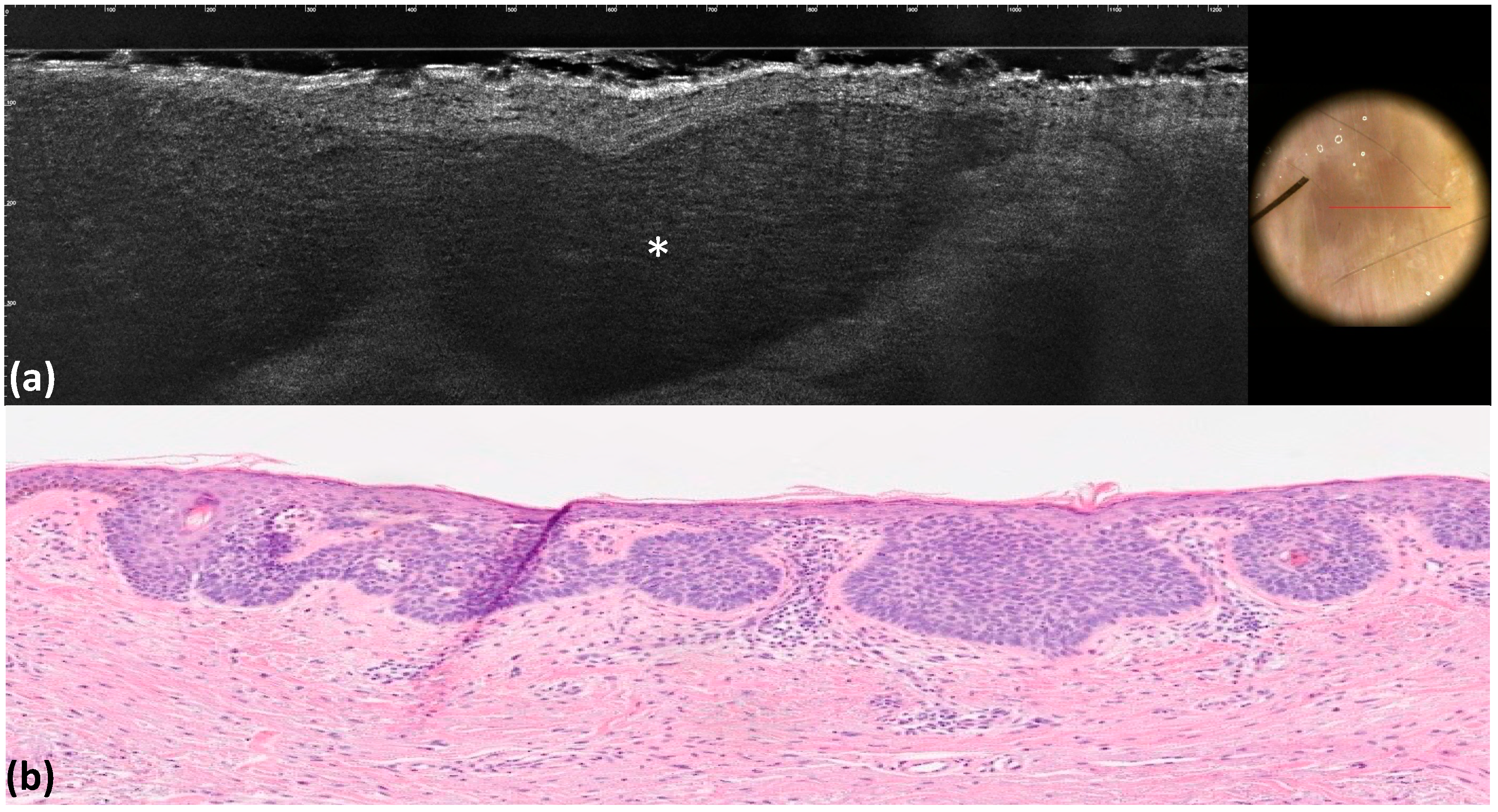
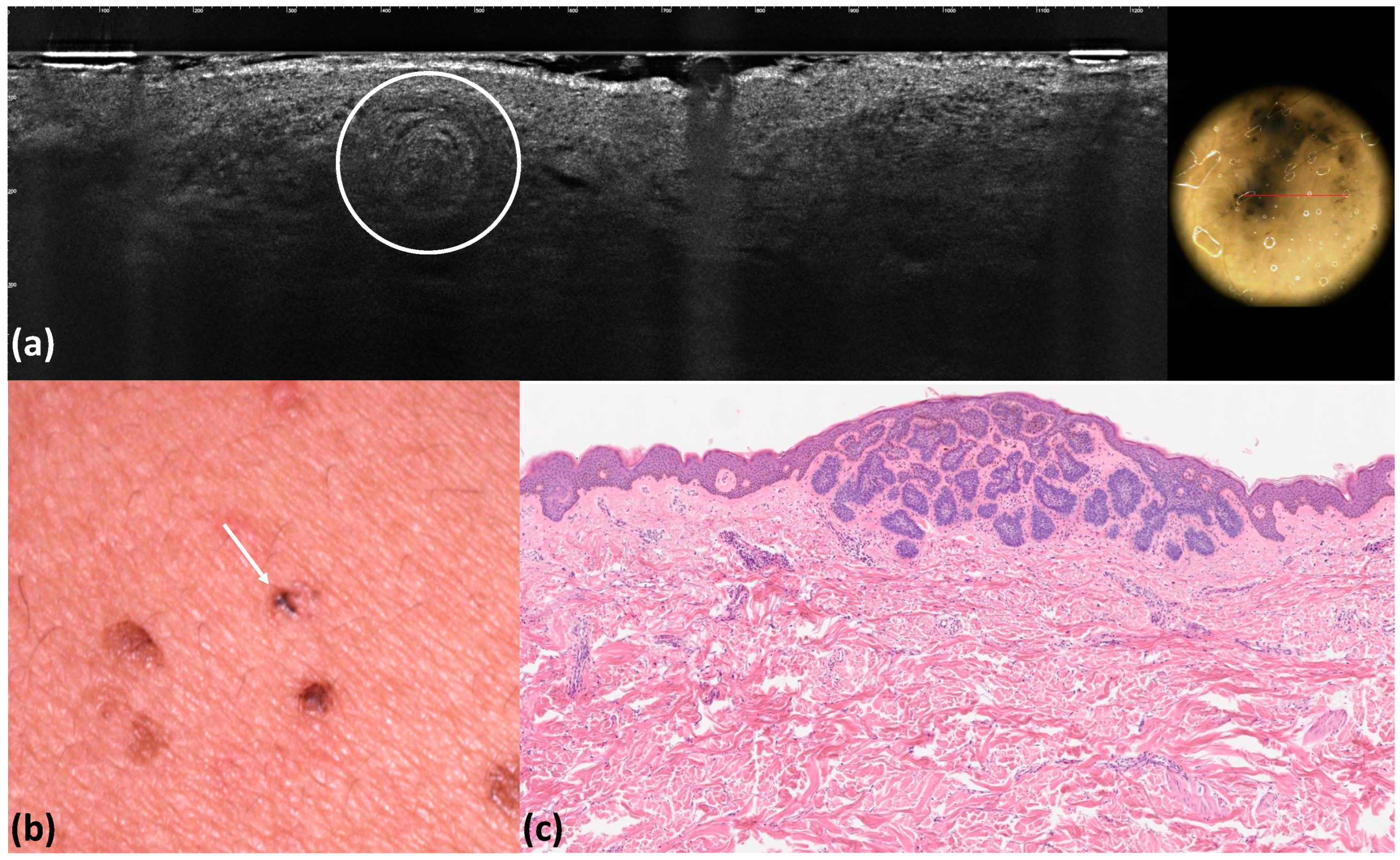
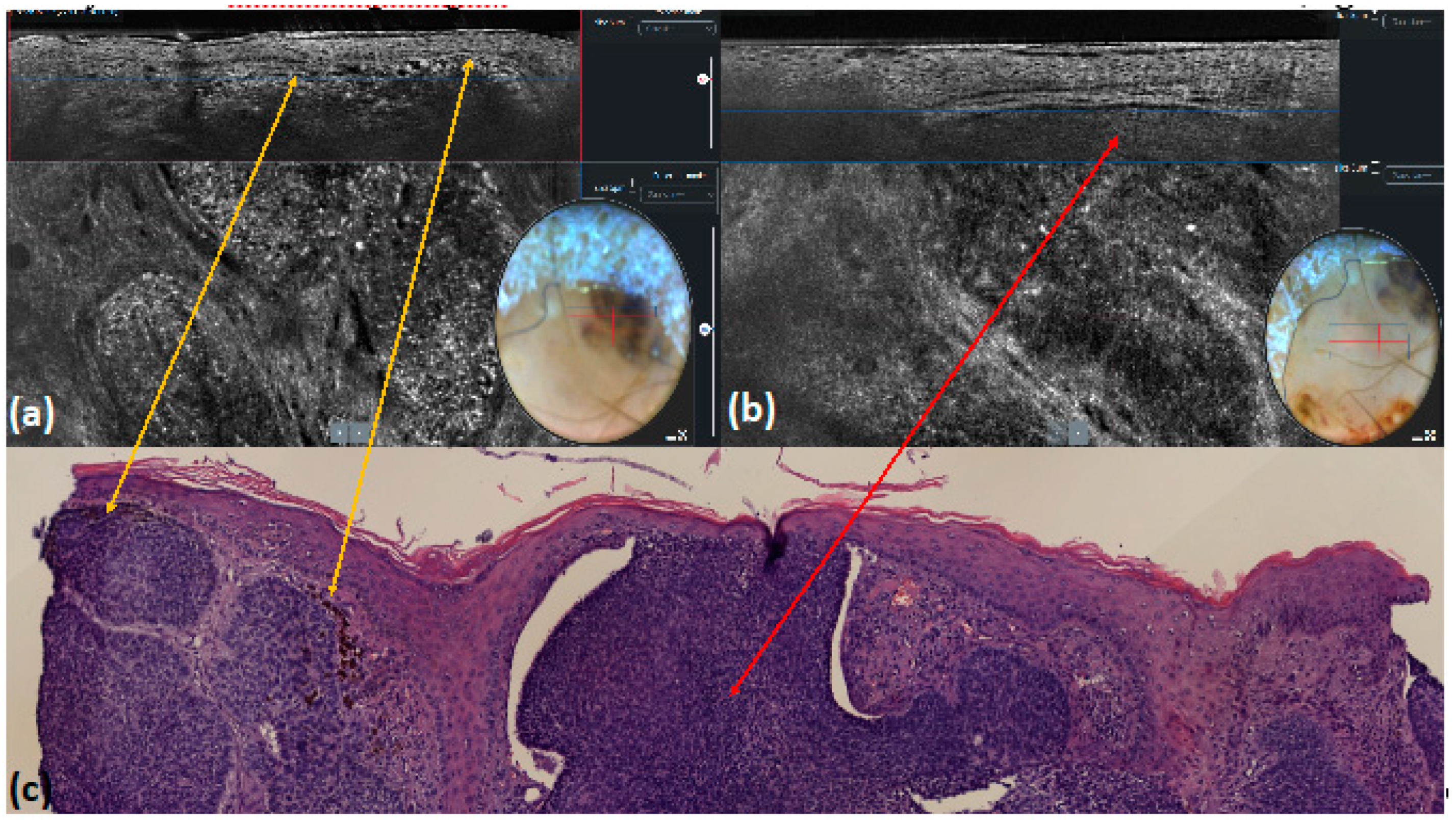
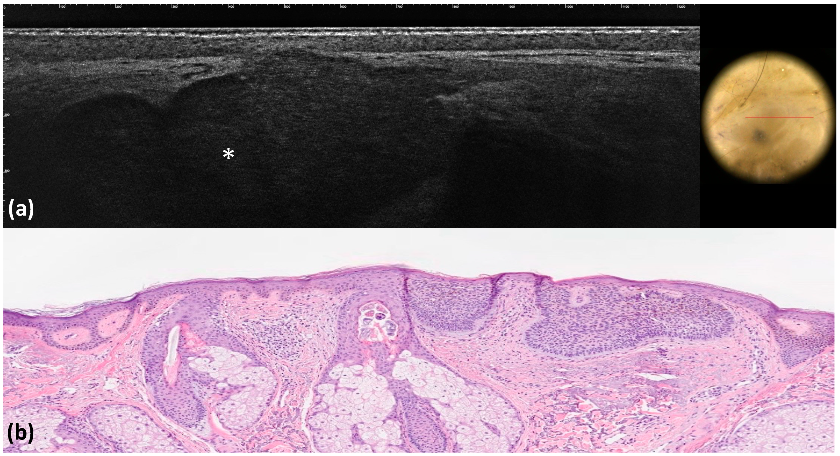
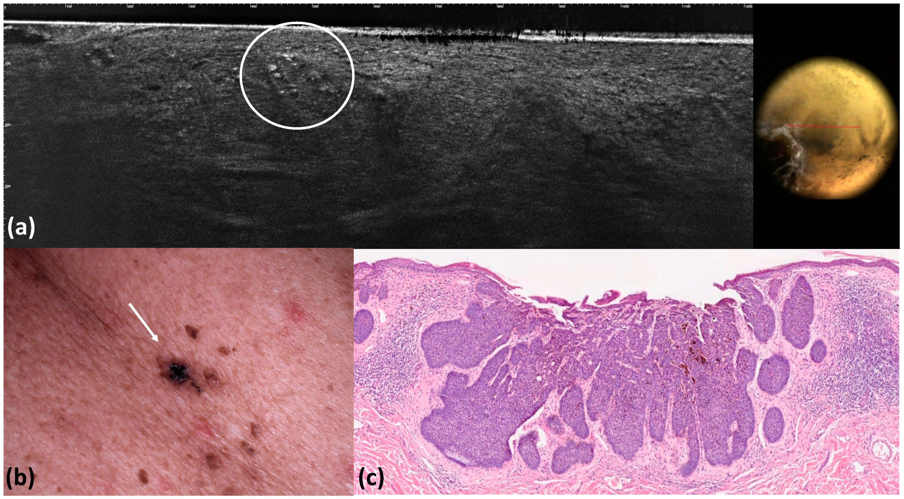
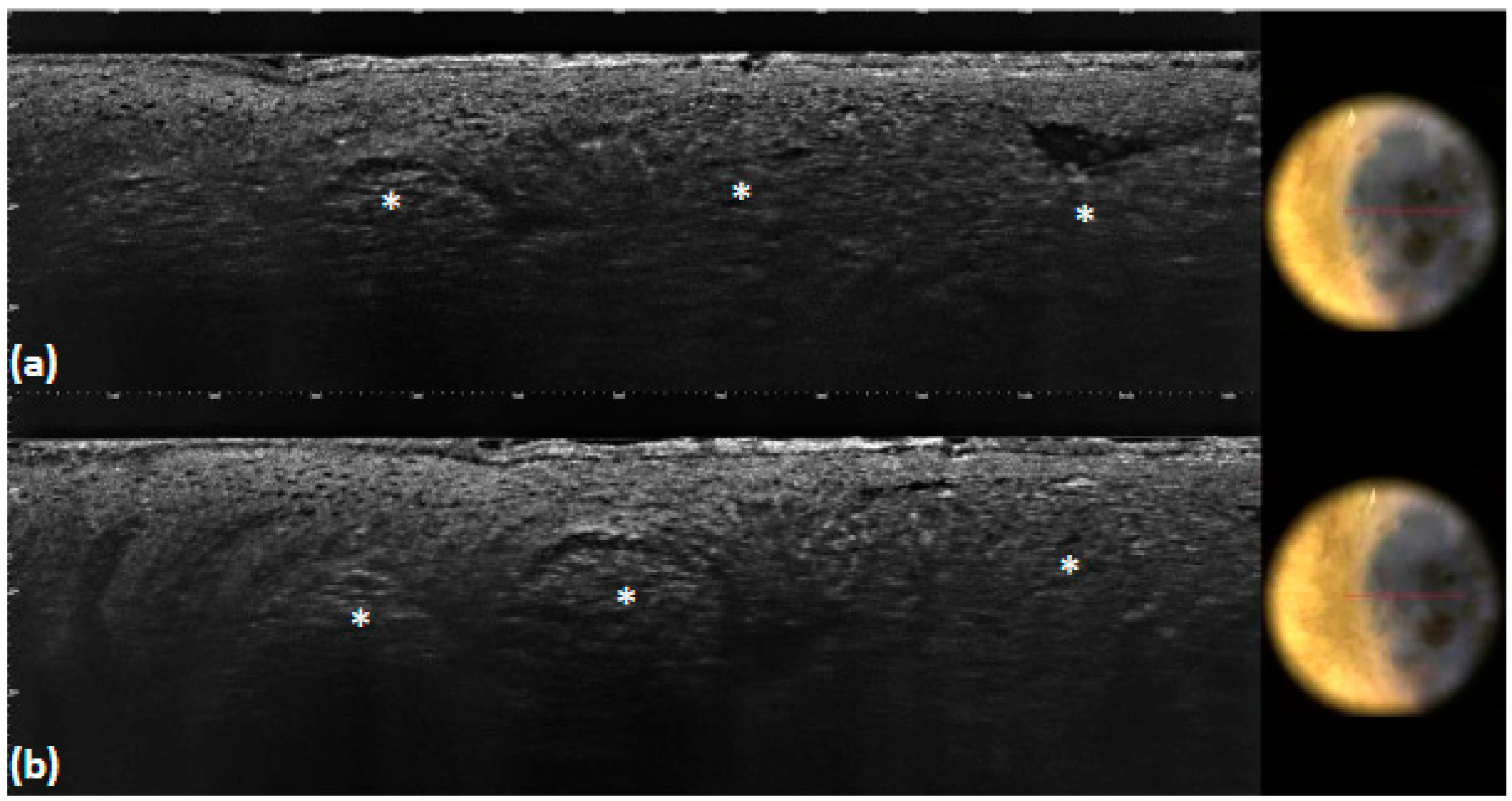
3. Results
4. Discussion
5. Conclusions
Author Contributions
Funding
Institutional Review Board Statement
Informed Consent Statement
Data Availability Statement
Conflicts of Interest
References
- Rubin, A.I.; Chen, E.H.; Ratner, D. Basal-cell carcinoma. N. Engl. J. Med. 2005, 353, 2262–2269. [Google Scholar] [CrossRef]
- Devine, C.; Srinivasan, B.; Sayan, A.; Ilankovan, V. Epidemiology of basal cell carcinoma: A 10-year comparative study. Br. J. Oral Maxillofac. Surg. 2018, 56, 101–106. [Google Scholar] [CrossRef] [PubMed]
- Cameron, M.C.; Lee, E.; Hibler, B.P.; Barker, C.A.; Mori, S.; Cordova, M.; Nehal, K.S.; Rossi, A.M. Basal cell carcinoma: Epidemiology; pathophysiology; clinical and histological subtypes; and disease associations. J. Am. Acad. Dermatol. 2019, 80, 303–317. [Google Scholar] [CrossRef]
- Marzuka, A.G.; Book, S.E. Basal cell carcinoma: Pathogenesis, epidemiology, clinical features, diagnosis, histopathology, and management. Yale J. Biol. Med. 2015, 88, 167–179. [Google Scholar] [PubMed]
- Reiter, O.; Mimouni, I.; Gdalevich, M.; Marghoob, A.A.; Levi, A.; Hodak, E.; Leshem, Y.A. The diagnostic accuracy of dermoscopy for basal cell carcinoma: A systematic review and meta-analysis. J. Am. Acad. Dermatol. 2019, 80, 1380–1388. [Google Scholar] [CrossRef] [PubMed]
- Menzies, S.W. Dermoscopy of pigmented basal cell carcinoma. Clin. Dermatol. 2002, 20, 268–269. [Google Scholar] [CrossRef]
- Puig, S.; Cecilia, N.; Malvehy, J. Dermoscopic criteria and basal cell carcinoma. G. Ital. Dermatol. Venereol. 2012, 147, 135–140. [Google Scholar]
- Altamura, D.; Menzies, S.W.; Argenziano, G.; Zalaudek, I.; Soyer, H.P.; Sera, F.; Avramidis, M.; DeAmbrosis, K.; Fargnoli, M.C.; Peris, K. Dermatoscopy of basal cell carcinoma: Morphologic variability of global and local features and accuracy of diagnosis. J. Am. Acad. Dermatol. 2010, 62, 67–75. [Google Scholar] [CrossRef]
- Cameron, M.C.; Lee, E.; Hibler, B.P.; Giordano, C.N.; Barker, C.A.; Mori, S.; Cordova, M.; Nehal, K.S.; Rossi, A.M. Basal cell carcinoma: Contemporary approaches to diagnosis, treatment, and prevention. J. Am. Acad. Dermatol. 2019, 80, 321–339. [Google Scholar] [CrossRef]
- Heibel, H.D.; Hooey, L.; Cockerell, C.J. A Review of Noninvasive Techniques for Skin Cancer Detection in Dermatology. Am. J. Clin. Dermatol. 2020, 21, 513–524. [Google Scholar] [CrossRef]
- Ruini, C.; Schuh, S.; Sattler, E.; Welzel, J. Line-field confocal optical coherence tomography-Practical applications in dermatology and comparison with established imaging methods. Ski. Res. Technol. 2021, 27, 340–352. [Google Scholar] [CrossRef]
- Verzi, A.E.; Micali, G.; Lacarrubba, F. Line-Field Confocal Optical Coherence Tomography May Enhance Monitoring of Superficial Basal Cell Carcinoma Treated with Imiquimod 5% Cream: A Pilot Study. Cancers 2021, 13, 4913. [Google Scholar] [CrossRef]
- Chauvel-Picard, J.; Chauvel-Picard, J.; Bérot, V.; Tognetti, L.; Orte Cano, C.; Fontaine, M.; Lenoir, C.; Pérez-Anker, J.; Puig, S.; Dubois, A.; et al. Line-field confocal optical coherence tomography as a tool for three-dimensional in vivo quantification of healthy epidermis: A pilot study. J. Biophotonics 2022, 15, e202100236. [Google Scholar] [CrossRef]
- Ruini, C.; Schuh, S.; Gust, C.; Kendziora, B.; Frommherz, L.; French, L.E.; Hartmann, D.; Welzel, J.; Sattler, E. Line-field optical coherence tomography: In vivo diagnosis of basal cell carcinoma subtypes compared with histopathology. Clin. Exp. Dermatol. 2021, 46, 1471–1481. [Google Scholar] [CrossRef] [PubMed]
- Gwet, K.L. Computing inter-rater reliability and its variance in the presence of high agreement. Br. J. Math. Stat. Psychol. 2008, 61 Pt 1, 29–48. [Google Scholar] [CrossRef] [PubMed]
- Brereton, R.G. Contingency tables, confusion matrices, classifiers and quality of prediction. J. Chemom. 2021, 35, e3331. [Google Scholar] [CrossRef]
- Lallas, A.; Apalla, Z.; Ioannides, D.; Argenziano, G.; Castagnetti, F.; Moscarella, E.; Longo, C.; Palmieri, T.; Ramundo, D.; Zalaudek, I. Dermoscopy in the diagnosis and management of basal cell carcinoma. Future Oncol. 2015, 11, 2975–2984. [Google Scholar] [CrossRef]
- Lallas, A.; Apalla, Z.; Argenziano, G.; Longo, C.; Moscarella, E.; Specchio, F.; Raucci, M.; Zalaudek, I. The dermatoscopic universe of basal cell carcinoma. Dermatol. Pract. Concept. 2014, 4, 11–24. [Google Scholar] [CrossRef] [PubMed]
- Yelamos, O.; Braun, R.P.; Liopyris, K.; Wolner, Z.J.; Kerl, K.; Gerami, P.; Marghoob, A.A. Dermoscopy and dermatopathology correlates of cutaneous neoplasms. J. Am. Acad. Dermatol. 2019, 80, 341–363. [Google Scholar] [CrossRef]
- Tabanlioglu Onan, D.; Sahin, S.; Gököz, O.; Erkin, G.; Cakır, B.; Elçin, G.; Kayıkçıoğlu, A. Correlation between the dermatoscopic and histopathological features of pigmented basal cell carcinoma. J. Eur. Acad. Dermatol. Venereol. 2010, 24, 1317–1325. [Google Scholar] [CrossRef]
- Perino, F.; Suarez, R.; Perez-Anker, J.; Carrera, C.; Rezze, G.G.; Primiero, C.A.; Alos, L.L.; Díaz, A.; Barreiro, A.; Puig, S. Concordance of in vivo reflectance confocal microscopy and horizontal-sectioning histology in skin tumours. J. Eur. Acad. Dermatol. Venereol. 2023, 38, 124–135. [Google Scholar] [CrossRef] [PubMed]
- Murgia, G.; Denaro, N.; Boggio, F.; Nazzaro, G.; Benzecry, V.; Bortoluzzi, P.; Passoni, E.; Garrone, O.; Marzano, A. Basosquamous Carcinoma: Comprehensive Clinical and Histopathological Aspects, Novel Imaging Tools, and Therapeutic Approaches. Cells 2023, 12, 2737. [Google Scholar] [CrossRef] [PubMed]
- Gust, C.; Schuh, S.; Welzel, J.; Daxenberger, F.; Hartmann, D.; French, L.E.; Ruini, C.; Sattler, E.C. Line-Field Confocal Optical Coherence Tomography Increases the Diagnostic Accuracy and Confidence for Basal Cell Carcinoma in Equivocal Lesions: A Prospective Study. Cancers 2022, 14, 1082. [Google Scholar] [CrossRef] [PubMed]
- Cinotti, E.; Brunetti, T.; Cartocci, A.; Tognetti, L.; Suppa, M.; Malvehy, J.; Perez-Anker, J.; Puig, S.; Perrot, J.L.; Rubegni, P. Diagnostic Accuracy of Line-Field Confocal Optical Coherence Tomography for the Diagnosis of Skin Carcinomas. Diagnostics 2023, 13, 361. [Google Scholar] [CrossRef]
- Donelli, C.; Suppa, M.; Tognetti, L.; Perrot, J.L.; Calabrese, L.; Pérez-Anker, J.; Malvehy, J.; Rubegni, P.; Cinotti, E. Line-Field Confocal Optical Coherence Tomography for the Diagnosis of Skin Carcinomas: Real-Life Data over Three Years. Curr. Oncol. 2023, 30, 8853–8864. [Google Scholar] [CrossRef] [PubMed]
- Suppa, M.; Fontaine, M.; Dejonckheere, G.; Cinotti, E.; Yélamos, O.; Diet, G.; Tognetti, L.; Miyamoto, M.; Orte Cano, C.; Perez-Anker, J.; et al. Line-field confocal optical coherence tomography of basal cell carcinoma: A descriptive study. J. Eur. Acad. Dermatol. Venereol. 2021, 35, 1099–1110. [Google Scholar] [CrossRef] [PubMed]
- Palmisano, G.; Orte Cano, C.; Fontaine, M.; Lenoir, C.; Cinotti, E.; Tognetti, L.; Rubegni, P.; Perez-Anker, J.; Puig, S.; Malvehy, J.; et al. Dermoscopic criteria explained by LC-OCT: Negative maple leaf-like areas. J. Eur. Acad. Dermatol. Venereol. 2023, 38, e271–e273. [Google Scholar] [CrossRef] [PubMed]
- Giacomel, J.; Zalaudek, I. Dermoscopy of superficial basal cell carcinoma. Dermatol. Surg. 2005, 31, 1710–1713. [Google Scholar] [PubMed]
- Micantonio, T.; Gulia, A.; Altobelli, E.; Di Cesare, A.; Fidanza, R.; Riitano, A.; Fargnoli, M.C.; Peris, K. Vascular patterns in basal cell carcinoma. J. Eur. Acad. Dermatol. Venereol. 2011, 25, 358–361. [Google Scholar] [CrossRef]
- Minagawa, A. Dermoscopy-pathology relationship in seborrheic keratosis. J. Dermatol. 2017, 44, 518–524. [Google Scholar] [CrossRef]
- Reiter, O.; Mimouni, I.; Dusza, S.; Halpern, A.C.; Leshem, Y.A.; Marghoob, A.A. Dermoscopic features of basal cell carcinoma and its subtypes: A systematic review. J. Am. Acad. Dermatol. 2021, 85, 653–664. [Google Scholar] [CrossRef] [PubMed]
- Lallas, A.; Argenziano, G.; Kyrgidis, A.; Apalla, Z.; Moscarella, E.; Longo, C.; Ferrara, G.; Piana, S.; Benati, E.; Zendri, E.; et al. Dermoscopy uncovers clinically undetectable pigmentation in basal cell carcinoma. Br. J. Dermatol. 2014, 170, 192–195. [Google Scholar] [CrossRef] [PubMed]


| Overall, n (%) (n = 100) | |
|---|---|
| Male, n (%) | 53 (53) |
| Age at diagnosis, mean (SD) | 65.46 (13.36) |
| BCC localization | |
| Trunk, n (%) | 25 (25) |
| Inferior extremities, n (%) | 12 (12) |
| Superior extremities, n (%) | 6 (6) |
| Head and neck, n (%) | 57 (57) |
| Dermoscopic pigmented BCC, n (%) | 59 (59) |
| BCC histological subtypes, n (%) | |
| Superficial, n (%) | 54 (54) |
| Nodular, n (%) | 20 (20) |
| Infiltrating, n (%) | 16 (16) |
| Micronodular, n (%) | 5 (5) |
| Basosquamous, n (%) | 5 (5) |
| Overall (n = 100) | |||||||
|---|---|---|---|---|---|---|---|
| Dermoscopy Criteria | LC-OCT Criteria | Dermoscopy | LC-OCT | P | CCR | Concordance | p Concordance |
| Ulceration or erosion | Ulceration or erosion | 7 (7.0) | 7 (7.0) | 1 | 1 | 1 | <0.001 |
| Maple-leaf figures | Medium-reflective lobules with maple-leaf shapes attached to the epidermis | 12 (12.0) | 12 (12.0) | 1 | 1 | 1 | <0.001 |
| Blue–gray globules | Roundish medium-reflective tumor lobules detached from the epidermis with variable hyper-reflective spots | 40 (40.0) | 36 (36.0) | 0.662 | 0.96 | 0.92 | <0.001 |
| Blue–gray ovoid nests | Medium-reflective tumor lobules detached from the epidermis with variable hyper-reflective spots | 10 (10.0) | 9 (9.0) | 1 | 0.99 | 0.99 | <0.001 |
| Telangiectasia or arborizing vessels | Telangiectasia or arborizing vessels | 51 (51.0) | 39 (39.0) | 0.118 | 0.88 | 0.76 | <0.001 |
| Multiple blue–gray dots | Hyper-reflective roundish small areas inside tumor lobules | 18 (18.0) | 13 (13.0) | 0.435 | 0.95 | 0.93 | <0.001 |
| Pink–white areas | Medium-reflective lobules | 10 (10.0) | 8 (8.0) | 0.805 | 0.98 | 0.98 | <0.001 |
| Blue-whitish veils | Hyper-reflective lobules in the entire dermis | 9 (9.0) | 9 (9.0) | 1 | 1 | 1 | <0.001 |
| Milia-like cysts | Milia-like cysts | 9(9.0) | 5 (5.0) | 0.406 | 0.90 | 0.89 | <0.001 |
Disclaimer/Publisher’s Note: The statements, opinions and data contained in all publications are solely those of the individual author(s) and contributor(s) and not of MDPI and/or the editor(s). MDPI and/or the editor(s) disclaim responsibility for any injury to people or property resulting from any ideas, methods, instructions or products referred to in the content. |
© 2024 by the authors. Licensee MDPI, Basel, Switzerland. This article is an open access article distributed under the terms and conditions of the Creative Commons Attribution (CC BY) license (https://creativecommons.org/licenses/by/4.0/).
Share and Cite
Barbarossa, L.; D’Onghia, M.; Cartocci, A.; Suppa, M.; Tognetti, L.; Cappilli, S.; Peris, K.; Perez-Anker, J.; Malvehy, J.; Baldino, G.; et al. Understanding the Dermoscopic Patterns of Basal Cell Carcinoma Using Line-Field Confocal Tomography. Tomography 2024, 10, 826-838. https://doi.org/10.3390/tomography10060063
Barbarossa L, D’Onghia M, Cartocci A, Suppa M, Tognetti L, Cappilli S, Peris K, Perez-Anker J, Malvehy J, Baldino G, et al. Understanding the Dermoscopic Patterns of Basal Cell Carcinoma Using Line-Field Confocal Tomography. Tomography. 2024; 10(6):826-838. https://doi.org/10.3390/tomography10060063
Chicago/Turabian StyleBarbarossa, Lorenzo, Martina D’Onghia, Alessandra Cartocci, Mariano Suppa, Linda Tognetti, Simone Cappilli, Ketty Peris, Javiera Perez-Anker, Josep Malvehy, Gennaro Baldino, and et al. 2024. "Understanding the Dermoscopic Patterns of Basal Cell Carcinoma Using Line-Field Confocal Tomography" Tomography 10, no. 6: 826-838. https://doi.org/10.3390/tomography10060063
APA StyleBarbarossa, L., D’Onghia, M., Cartocci, A., Suppa, M., Tognetti, L., Cappilli, S., Peris, K., Perez-Anker, J., Malvehy, J., Baldino, G., Militello, C., Perrot, J. L., Rubegni, P., & Cinotti, E. (2024). Understanding the Dermoscopic Patterns of Basal Cell Carcinoma Using Line-Field Confocal Tomography. Tomography, 10(6), 826-838. https://doi.org/10.3390/tomography10060063