Investigation of the Effect of Nanocrystalline Calcium Carbonate-Substituted Hydroxyapatite and L-Lysine and L-Arginine Surface Interactions on the Molecular Properties of Dental Biomimetic Composites
Abstract
:1. Introduction
2. Materials and Methods
2.1. Methodology for Obtaining Samples
2.2. Methods of Structural and Spectroscopic Analysis of the Samples
2.2.1. X-ray Structural Analysis
2.2.2. Optical Microscopy
2.2.3. Transmission Electron Microscopy (TEM)
2.2.4. Fourier Transform Infrared (FTIR) Spectroscopy
2.2.5. Information about Diagnostic Methods Used in Work, Provided for Comparison
3. Results
3.1. X-ray Diffractometry
3.2. Microscopy
3.3. FTIR Spectroscopy
4. Discussion
5. Conclusions
Author Contributions
Funding
Institutional Review Board Statement
Informed Consent Statement
Data Availability Statement
Acknowledgments
Conflicts of Interest
References
- Chun, H.J.; Park, K.; Kim, C.-H.; Khang, G. Novel Biomaterials for Regenerative Medicine; Springer: Berlin/Heidelberg, Germany, 2018; ISBN 9789811309472. [Google Scholar]
- Upadhyay, A.; Pillai, S.; Khayambashi, P.; Sabri, H.; Lee, K.T.; Tarar, M.; Zhou, S.; Harb, I.; Tran, S.D. Biomimetic aspects of oral and dentofacial regeneration. Biomimetics 2020, 5, 51. [Google Scholar] [CrossRef] [PubMed]
- Mousavi, S.M.; Yousefi, K.; Hashemi, S.A.; Afsa, M.; Bahrani, S.; Gholami, A.; Ghahramani, Y.; Alizadeh, A.; Chiang, W.-H. Renewable carbon nano-materials: Novel resources for dental tissue engineering. Nanomaterials 2021, 11, 2800. [Google Scholar] [CrossRef] [PubMed]
- Katti, D.R.; Sharma, A.; Ambre, A.H.; Katti, K.S. Molecular interactions in biomineralized hydroxyapatite amino acid modified nanoclay: In silico design of bone biomaterials. Mater. Sci. Eng. C 2015, 46, 207–217. [Google Scholar] [CrossRef] [PubMed]
- Seredin, P.V.; Goloshchapov, D.L.; Prutskij, T.; Ippolitov, Y.u.A. Fabrication and characterisation of composites materials similar optically and in composition to native dental tissues. Results Phys. 2017, 7, 1086–1094. [Google Scholar] [CrossRef]
- Pina, S.; Oliveira, J.M.; Reis, R.L. Biomimetic strategies to engineer mineralized human tissues. In Handbook of Bioceramics and Biocomposites; Antoniac, I.V., Ed.; Springer International Publishing: Cham, Switzerland, 2016; pp. 503–519. ISBN 978-3-319-12460-5. [Google Scholar]
- Turon, P.; del Valle, L.; Alemán, C.; Puiggalí, J. Biodegradable and biocompatible systems based on hydroxyapatite nanoparticles. Appl. Sci. 2017, 7, 60. [Google Scholar] [CrossRef] [Green Version]
- Nemati, E.; Gholami, A. Nano bacterial cellulose for biomedical applications: A mini review focus on tissue engineering. Adv. Appl. NanoBio Technol. 2021, 2, 93–101. [Google Scholar]
- Gholami, A.; Hashemi, S.A.; Yousefi, K.; Mousavi, S.M.; Chiang, W.-H.; Ramakrishna, S.; Mazraedoost, S.; Alizadeh, A.; Omidifar, N.; Behbudi, G.; et al. 3D nanostructures for tissue engineering, cancer therapy, and gene delivery. J. Nanomater. 2020, 2020, e1852946. [Google Scholar] [CrossRef]
- Memarpour, M.; Shafiei, F.; Rafiee, A.; Soltani, M.; Dashti, M.H. Effect of hydroxyapatite nanoparticles on enamel remineralization and estimation of fissure sealant bond strength to remineralized tooth surfaces: An in vitro study. BMC Oral Health 2019, 19, 92. [Google Scholar] [CrossRef] [PubMed]
- Alipour, A. Virus decorated nanobiomaterials as scaffolds for tissue engineering. Adv. Appl. NanoBio Technol. 2021, 2, 79–85. [Google Scholar]
- Barot, T.; Rawtani, D.; Kulkarni, P. Nanotechnology-based materials as emerging trends for dental applications. Rev. Adv. Mater. Sci. 2021, 60, 173–189. [Google Scholar] [CrossRef]
- Zafar, M.S.; Amin, F.; Fareed, M.A.; Ghabbani, H.; Riaz, S.; Khurshid, Z.; Kumar, N. Biomimetic aspects of restorative dentistry biomaterials. Biomimetics 2020, 5, 34. [Google Scholar] [CrossRef] [PubMed]
- Wang, J.; Liu, Z.; Ren, B.; Wang, Q.; Wu, J.; Yang, N.; Sui, X.; Li, L.; Li, M.; Zhang, X.; et al. Biomimetic mineralisation systems for in situ enamel restoration inspired by amelogenesis. J. Mater. Sci. Mater. Med. 2021, 32, 1–17. [Google Scholar] [CrossRef]
- Dorozhkin, S.V. Hydroxyapatite and Other Calcium Orthophosphates: Bioceramics, Coatings and Dental Applications [Hardcover]; Nova Science Publishers, Inc.: New York, NY, USA, 2017; ISBN 978-1-5361-1897-1. [Google Scholar]
- Comeau, P.; Willett, T. Impact of side chain polarity on non-stoichiometric nano-hydroxyapatite surface functionalization with amino acids. Sci. Rep. 2018, 8, 1–11. [Google Scholar] [CrossRef] [PubMed] [Green Version]
- Palazzo, B.; Walsh, D.; Iafisco, M.; Foresti, E.; Bertinetti, L.; Martra, G.; Bianchi, C.L.; Cappelletti, G.; Roveri, N. Amino acid synergetic effect on structure, morphology and surface properties of biomimetic apatite nanocrystals. Acta Biomater. 2009, 5, 1241–1252. [Google Scholar] [CrossRef] [PubMed]
- Tavafoghi, M.; Cerruti, M. The role of amino acids in hydroxyapatite mineralization. J. R. Soc. Interface 2016, 13, 20160462. [Google Scholar] [CrossRef] [Green Version]
- Lagazzo, A.; Barberis, F.; Carbone, C.; Ramis, G.; Finocchio, E. Molecular level interactions in brushite-aminoacids composites. Mater. Sci. Eng. C Mater. Biol. Appl. 2017, 70, 721–727. [Google Scholar] [CrossRef]
- El Rhilassi, A.; Mourabet, M.; Bennani-Ziatni, M.; El Hamri, R.; Taitai, A. Interaction of some essential amino acids with synthesized poorly crystalline hydroxyapatite. J. Saudi Chem. Soc. 2016, 20 (Suppl. S1), S632–S640. [Google Scholar] [CrossRef] [Green Version]
- Tavafoghi Jahromi, M.; Cerruti, M. Amino acid/ion aggregate formation and their role in hydroxyapatite precipitation. Cryst. Growth Des. 2015, 15, 1096–1104. [Google Scholar] [CrossRef]
- Lin, Z.; Hu, R.; Zhou, J.; Ye, Y.; Xu, Z.; Lin, C. A further insight into the adsorption mechanism of protein on hydroxyapatite by FTIR-ATR spectrometry. Spectrochim. Acta Part A Mol. Biomol. Spectrosc. 2017, 173, 527–531. [Google Scholar] [CrossRef] [PubMed]
- Gómez-Morales, J.; Delgado-López, J.M.; Iafisco, M.; Hernández-Hernández, A.; Prat, M. Amino acidic control of calcium phosphate precipitation by using the vapor diffusion method in microdroplets. Cryst. Growth Des. 2011, 11, 4802–4809. [Google Scholar] [CrossRef]
- Rimola, A.; Corno, M.; Garza, J.; Ugliengo, P. Ab initio modelling of protein–biomaterial interactions: Influence of amino acid polar side chains on adsorption at hydroxyapatite surfaces. Phil. Trans. R. Soc. A 2012, 370, 1478–1498. [Google Scholar] [CrossRef] [Green Version]
- Katti, K.S.; Ambre, A.H.; Peterka, N.; Katti, D.R. Use of unnatural amino acids for design of novel organomodified clays as components of nanocomposite biomaterials. Philos. Trans. R. Soc. Lond. A Math. Phys. Eng. Sci. 2010, 368, 1963–1980. [Google Scholar] [CrossRef] [PubMed]
- Otsuka, Y.; Ito, A.; Takeuchi, M.; Tanaka, H. Effect of amino acid on calcium phosphate phase transformation: Attenuated total reflectance-infrared spectroscopy and chemometrics. Colloid Polym. Sci. 2019, 297, 155–163. [Google Scholar] [CrossRef]
- da Silva, C.M.F.; de Menezes Costa, A.F.; Costa, A.R.; Neves, J.G.; de Godói, A.P.T.; de Góes, V.F.F. Influence of different acid etching times on the shear bond strength of brackets bonded to bovine enamel. Saudi Dent. J. 2021, 33, 474–480. [Google Scholar] [CrossRef]
- Erceg, I.; Maltar-Strmečki, N.; Jurašin, D.D.; Strasser, V.; Ćurlin, M.; Lyons, D.M.; Radatović, B.; Mlinarić, N.M.; Kralj, D.; Sikirić, M.D. Comparison of the effect of the amino acids on spontaneous formation and transformation of calcium phosphates. Crystals 2021, 11, 792. [Google Scholar] [CrossRef]
- Mukherjee, K.; Ruan, Q.; Nutt, S.; Tao, J.; De Yoreo, J.J.; Moradian-Oldak, J. Peptide-based bioinspired approach to regrowing multilayered aprismatic enamel. ACS Omega 2018, 3, 2546–2557. [Google Scholar] [CrossRef] [Green Version]
- Ding, L.; Han, S.; Wang, K.; Zheng, S.; Zheng, W.; Peng, X.; Niu, Y.; Li, W.; Zhang, L. Remineralization of enamel caries by an amelogenin-derived peptide and fluoride in vitro. Regen. Biomater. 2020, 7, 283–292. [Google Scholar] [CrossRef] [PubMed] [Green Version]
- Sullivan, R.; Rege, A.; Corby, P.; Klaczany, G.; Allen, K.; Hershkowitz, D.; Goldder, B.; Wolff, M. Evaluation of a dentifrice containing 8% arginine, calcium carbonate, and sodium monofluorophosphate to repair acid-softened enamel using an intra-oral remineralization model. J. Clin. Dent. 2014, 25, A14–A19. [Google Scholar]
- Mousavi, S.M.; Hashemi, S.A.; Salahi, S.; Hosseini, M.; Amani, A.; Babapoor, A. Development of Clay Nanoparticles Toward Bio and Medical Applications; IntechOpen: London, UK; Rijeka, Croatia, 2018; pp. 167–191. ISBN 978-1-78923-728-3. [Google Scholar]
- Mousavi, S.M.; Zarei, M.; Hashemi, S.A.; Ramakrishna, S.; Lai, C.W.; Chiang, W.-H.; Gholami, A.; Omidifar, N.; Shokripour, M. Asymmetric membranes: A potential scaffold for wound healing applications. Symmetry 2020, 12, 1100. [Google Scholar] [CrossRef]
- Mousavi, S.-M.; Nejad, Z.M.; Hashemi, S.A.; Salari, M.; Gholami, A.; Ramakrishna, S.; Chiang, W.-H.; Lai, C.W. Bioactive agent-loaded electrospun nanofiber membranes for accelerating healing process: A review. Membranes 2021, 11, 702. [Google Scholar] [CrossRef]
- Ashoori, Y.; Mohkam, M.; Heidari, R.; Abootalebi, S.N.; Mousavi, S.M.; Hashemi, S.A.; Golkar, N.; Gholami, A. Development and in vivo characterization of probiotic lysate-treated chitosan nanogel as a novel biocompatible formulation for wound healing. BioMed Res. Int. 2020, 2020, e8868618. [Google Scholar] [CrossRef]
- Combes, C.; Cazalbou, S.; Rey, C. Apatite biominerals. Minerals 2016, 6, 34. [Google Scholar] [CrossRef] [Green Version]
- Goloshchapov, D.L.; Lenshin, A.S.; Savchenko, D.V.; Seredin, P.V. Importance of defect nanocrystalline calcium hydroxyapatite characteristics for developing the dental biomimetic composites. Results Phys. 2019, 13, 102158. [Google Scholar] [CrossRef]
- Seredin, P.; Goloshchapov, D.; Kashkarov, V.; Ippolitov, Y.; Bambery, K. The investigations of changes in mineral–organic and carbon–phosphate ratios in the mixed saliva by synchrotron infrared spectroscopy. Results Phys. 2016, 6, 315–321. [Google Scholar] [CrossRef] [Green Version]
- Seredin, P.; Goloshchapov, D.; Ippolitov, Y.; Vongsvivut, J. Development of a new approach to diagnosis of the early fluorosis forms by means of FTIR and Raman microspectroscopy. Sci. Rep. 2020, 10, 20891. [Google Scholar] [CrossRef] [PubMed]
- Seredin, P.; Goloshchapov, D.; Ippolitov, Y.; Vongsvivut, J. Engineering of a biomimetic interface between a native dental tissue and restorative composite and its study using synchrotron FTIR microscopic mapping. Int. J. Mol. Sci. 2021, 22, 6510. [Google Scholar] [CrossRef] [PubMed]
- Hernández, B.; Pflüger, F.; Derbel, N.; De Coninck, J.; Ghomi, M. Vibrational analysis of amino acids and short peptides in hydrated media. VI. amino acids with positively charged side chains: L-Lysine and L-Arginine. J. Phys. Chem. B 2010, 114, 1077–1088. [Google Scholar] [CrossRef]
- Brasinika, D.; Tsigkou, O.; Tsetsekou, A.; Missirlis, Y.F. Bioinspired synthesis of hydroxyapatite nanocrystals in the presence of collagen and L-Arginine: Candidates for bone regeneration. J. Biomed. Mater. Res. 2015. [Google Scholar] [CrossRef]
- Pazderka, T.; Kopecký, V. Drop coating deposition Raman spectroscopy of proteinogenic amino acids compared with their solution and crystalline state. Spectrochim. Acta Part A Mol. Biomol. Spectrosc. 2017, 185, 207–216. [Google Scholar] [CrossRef]
- Seredin, P.; Goloshchapov, D.; Ippolitov, Y. Pimm vongsvivut pathology-specific molecular profiles of saliva in patients with multiple dental caries—potential application for predictive, preventive and personalised medical services. EPMA J. 2018, 9, 195–203. [Google Scholar] [CrossRef] [PubMed]
- Barth, A. The infrared absorption of amino acid side chains. Prog. Biophys. Mol. Biol. 2000, 74, 141–173. [Google Scholar] [CrossRef]
- Wolpert, M.; Hellwig, P. Infrared spectra and molar absorption coefficients of the 20 alpha amino acids in aqueous solutions in the spectral range from 1800 to 500 cm−1. Spectrochim. Acta Part A Mol. Biomol. Spectrosc. 2006, 64, 987–1001. [Google Scholar] [CrossRef] [PubMed]
- Paiva, F.M.; Batista, J.C.; Rêgo, F.S.C.; Lima, J.A.; Freire, P.T.C.; Melo, F.E.A.; Mendes Filho, J.; de Menezes, A.S.; Nogueira, C.E.S. Infrared and Raman spectroscopy and DFT calculations of DL amino acids: Valine and lysine hydrochloride. J. Mol. Struct. 2017, 1127, 419–426. [Google Scholar] [CrossRef]
- Ye, Q.; Spencer, P. Analyses of material-tissue interfaces by Fourier transform infrared, Raman spectroscopy, and chemometrics. In Material-Tissue Interfacial Phenomena; Elsevier: Amsterdam, The Netherlands, 2017; pp. 231–251. ISBN 978-0-08-100330-5. [Google Scholar]
- Seredin, P.; Goloshchapov, D.; Kashkarov, V.; Ippolitov, Y.; Ippolitov, I.; Vongsvivut, J. To the question on the use of multivariate analysis and 2D visualisation of synchrotron ATR-FTIR chemical imaging spectral data in the diagnostics of biomimetic sound dentin/dental composite interface. Diagnostics 2021, 11, 1294. [Google Scholar] [CrossRef] [PubMed]
- Garrido, C.; Aguayo, T.; Clavijo, E.; Gómez-Jeria, J.S.; Campos-Vallette, M.M. The effect of the PH on the interaction of L-Arginine with colloidal silver nanoparticles. A Raman and SERS study: Effect of PH on interaction of Arg with colloidal AgNps. J. Raman Spectrosc. 2013, 44, 1105–1110. [Google Scholar] [CrossRef]
- Tao, M.; Zhu, M.; Wu, C.; He, Z. Degradation kinetic study of lysine in lysine hydrochloride solutions for injection by determining its main degradation product. Asian J. Pharm. Sci. 2015, 10, 57–63. [Google Scholar] [CrossRef] [Green Version]
- Rozenberg, M.; Shoham, G. FTIR spectra of solid poly-L-Lysine in the stretching NH mode range. Biophys. Chem. 2007, 125, 166–171. [Google Scholar] [CrossRef] [PubMed]
- Boeckx, B.; Maes, G. Experimental and theoretical observation of different intramolecular H-bonds in lysine conformations. J. Phys. Chem. B 2012, 116, 12441–12449. [Google Scholar] [CrossRef]
- Drouet, C. Apatite formation: Why it may not work as planned, and how to conclusively identify apatite compounds. BioMed Res. Int. 2013, 2013, 490946. [Google Scholar] [CrossRef] [PubMed] [Green Version]
- Rey, C.; Marsan, O.; Combes, C.; Drouet, C.; Grossin, D.; Sarda, S. Characterization of calcium phosphates using vibrational spectroscopies. In Advances in Calcium Phosphate Biomaterials; Springer Series in Biomaterials Science and Engineering; Springer: Berlin/Heidelberg, Germany, 2014; pp. 229–266. ISBN 978-3-642-53979-4. [Google Scholar]
- Aliaga, A.E.; Garrido, C.; Leyton, P.; Diaz, F.G.; Gomez-Jeria, J.S.; Aguayo, T.; Clavijo, E.; Campos-Vallette, M.M.; Sanchez-Cortes, S. SERS and theoretical studies of arginine. Spectrochim. Acta Part A Mol. Biomol. Spectrosc. 2010, 76, 458–463. [Google Scholar] [CrossRef] [PubMed]
- Matei, A.; Drichko, N.; Gompf, B.; Dressel, M. Far-infrared spectra of amino acids. Chem. Phys. 2005, 316, 61–71. [Google Scholar] [CrossRef]
- Sunatkari, A.L.; Talwatkar, S.S.; Tamgadge, Y.S.; Muley, G.G. Synthesis, characterization and optical properties of L-Arginine stabilized gold nanocolloids. Nanosci. Nanotechnol. 2015, 5, 30–35. [Google Scholar]
- Ji, Z.; Santamaria, R.; Garzón, I.L. Vibrational circular dichroism and IR absorption spectra of amino acids: A density functional study. J. Phys. Chem. A 2010, 114, 3591–3601. [Google Scholar] [CrossRef] [PubMed]
- Antoniac, I.V. Handbook of Bioceramics and Biocomposites; Antoniac, I.V., Ed.; Springer International Publishing: Cham, Switzerland, 2016; ISBN 978-3-319-12459-9. [Google Scholar]
- O’Brien, W.J. (Ed.) Dental Materials and Their Selection, 4th ed.; Quintessence Pub. Co.: Hanover Park, IL, USA, 2008; ISBN 978-0-86715-437-5. [Google Scholar]
- El Gezawi, M.; Wölfle, U.C.; Haridy, R.; Fliefel, R.; Kaisarly, D. Remineralization, regeneration, and repair of natural tooth structure: Influences on the future of restorative dentistry practice. ACS Biomater. Sci. Eng. 2019, 5, 4899–4919. [Google Scholar] [CrossRef] [PubMed]
- Goloshchapov, D.; Buylov, N.; Emelyanova, A.; Ippolitov, I.; Ippolitov, Y.; Kashkarov, V.; Khudyakov, Y.; Nikitkov, K.; Seredin, P. Raman and XANES spectroscopic study of the influence of coordination atomic and molecular environments in biomimetic composite materials integrated with dental tissue. Nanomaterials 2021, 11, 3099. [Google Scholar] [CrossRef] [PubMed]
- Yanyan, S.; Guangxin, W.; Guoqing, S.; Yaming, W.; Wuhui, L.; Osaka, A. Effects of amino acids on conversion of calcium carbonate to hydroxyapatite. RSC Adv. 2020, 10, 37005–37013. [Google Scholar] [CrossRef]
- Saranya, S.; Samuel Justin, S.J.; Vijay Solomon, R.; Wilson, P. L-Arginine directed and ultrasonically aided growth of nanocrystalline hydroxyapatite particles with tunable morphology. Colloids Surf. A Physicochem. Eng. Asp. 2018, 538, 270–279. [Google Scholar] [CrossRef]
- Markovic, M.; Fowler, B.O.; Tung, M.S. Preparation and comprehensive characterization of a calcium hydroxyapatite reference material. J. Res. Natl. Inst. Stand. Technol. 2004, 109, 553. [Google Scholar] [CrossRef]
- Cacciotti, I. Multisubstituted hydroxyapatite powders and coatings: The influence of the codoping on the hydroxyapatite performances. Int. J. Appl. Ceram. Technol. 2019, 16, 1864–1884. [Google Scholar] [CrossRef]
- Zhang, Z.; Pan, H.; Tang, R. Molecular dynamics simulations of the adsorption of amino acids on the hydroxyapatite {100}-water interface. Front. Mater. Sci. China 2008, 2, 239–245. [Google Scholar] [CrossRef]


| Detection Features | Techniques | ||
|---|---|---|---|
| X-ray Structural Analysis | Synchrotron Fourier Transform Infrared Spectroscopy | Transmission Electron Microscopy | |
| Identification | Crystalline structure | Molecular and chemical bonds | Morphology of compounds, surface geometry |
| Source for characterisation | X-rays | Synchrotron radiation in Infrared range of spectrum | Electrons |
| Resolution | Angstrom | cm−1 | nm |
| Description | Providing information on structures, phases, preferred crystal orientations (texture), and other structural parameters of the dried solution samples | Identify or characterize organic/bio materials through creating a spectrum that shows molecular vibrations | Imagining and characterization of nanoparticles with high spatial resolution |
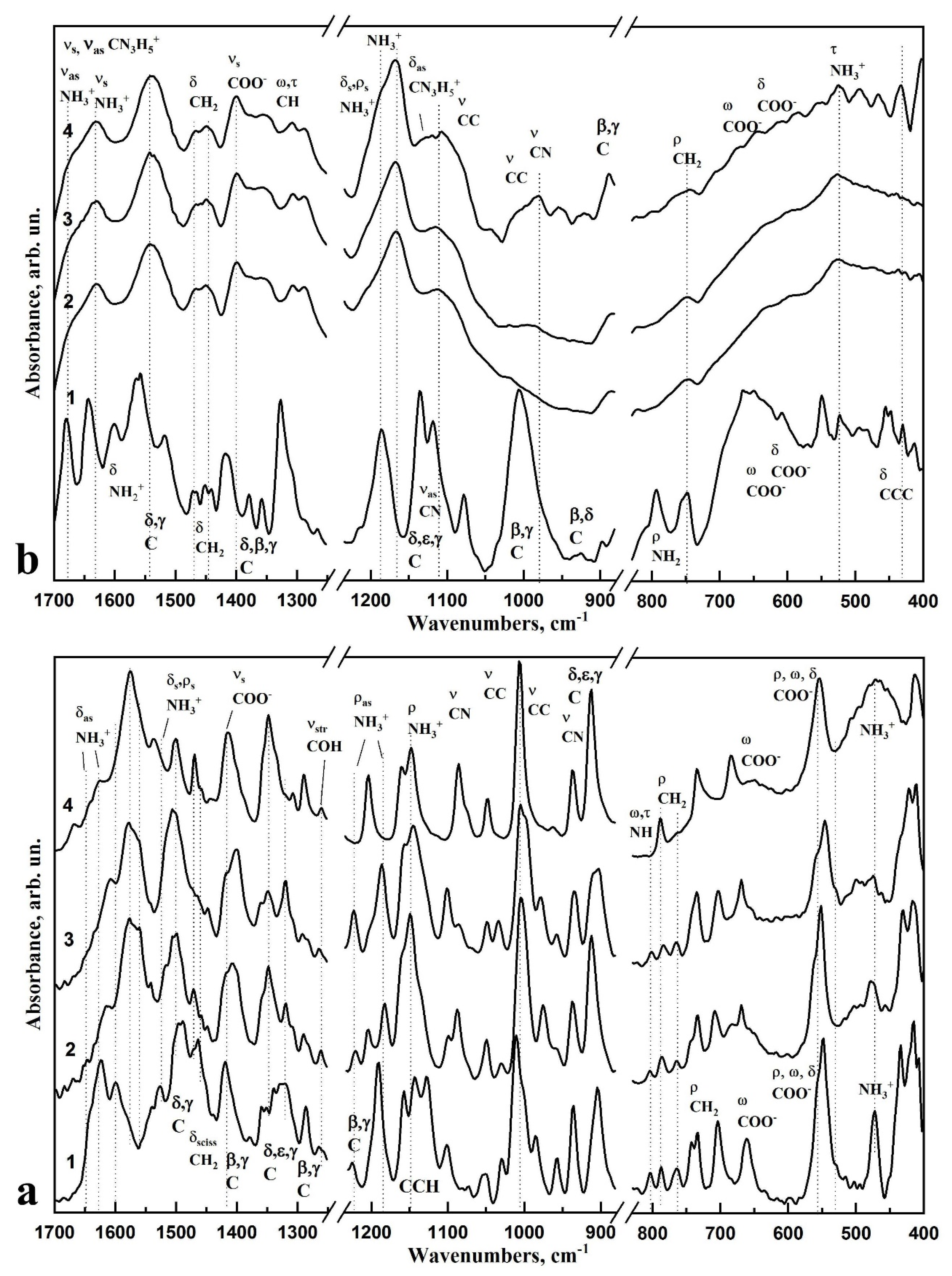
| Active Modes | Frequency of the Absorption Bands in the FTIR Spectra of the Samples, cm−1 | Ref.: Study Reference Number | |||||
|---|---|---|---|---|---|---|---|
| L-LysHCl | n-cHAp/L-LysHCl Composites | ||||||
| pH ≤ 5 | pH ≥ 7.5 | pH ≥ 11.2 | pH ≤ 5 | pH ≥ 7.5 | pH ≥ 11.2 | ||
| δas NH3+ | 1637 1629 1610 | 1635 1621 1612 | 1639 1627 1616 | 1642 1624 1612 | 1645 1628 1610 | 1651 1622 1609 | [41,46,47,51,52] |
| δ CH32+ | 1470 | 1472 | 1468 | 1469 1459 | 1465 1460 | 1465 1459 | [41,46,47,51] |
| vs COO− | 1415 | 1416 | 1414 | 1413 | 1417 | 1415 | [41,46,53] |
| ω Cγ, τ Cδ, τ Cε | 1348 | 1349 | 1348 | 1347 | 1349 | 1348 | [41,46,47] |
| ρas NH3+, τ Cβ, Cβ-Cα-Hα, ω Cδ | 1220 | 1222 | - | - | 1222 | 1222 | [41,46,47] |
| ρas NH3+, ρ Cε | 1182 | 1186 | - | - | 1185 | 1186 | [41,46,47] |
| ρas NH3+, C-Cα-Hα | 1148 | 1145 | 1148 | 1161 | 1155 | 1155 | [41,46,47] |
| PO43−, v3 | - - | - - | - - | 1089 1027 | 1091 1024 | 1099 1024 | [37,39,54,55] |
| PO43−,v1 | - | - | - | 970 | 966 | 974 | [37,39,54,55] |
| v C-C, v C-N ω Cγ, τ Cδ, τ Cε, | 1002 975 937 911 | 1003 978 935 907 | 1007 979 937 912 | 1003 961 935 912 | 1003 961 935 908 | 1003 978 935 905 | [41,46,47,51,53] |
| τ,ω NH | 805 | 802 | - | - | 803 | 803 | [41,46] |
| ρ CH2 | 785 764 | 782 764 | 788 - | 787 - | 782 - | 782 764 | |
| ρ CH2, v C-C, δ COO | 704 | - | 708 | 702 | 704 | - | [47,52,56] |
| OH (n-cHap) | - | - | - | 632 | 630 | 631 | [37,39,54,55] |
| PO43−,v4 | - | - | - | 602 558 | 602 559 | 601 560 | [37,39,54,55] |
| ρ, ω, δ, COO− | 560 551 | 562 555 | 560 553 | 554 | 569 550 | 570 547 | [51,57] |
| NH3+ torsion | 477 | 477–480 | 460–480 | 464–478 | 473 | 474 | [57] |
| Active Modes | Frequency of the Absorption Bands in the FTIR Spectra of the Samples, cm−1 | Ref.: Study Reference Number | |||||
|---|---|---|---|---|---|---|---|
| L-ArgHCl | Composites n-cHAp/L-ArgHCl | ||||||
| pH ≤ 5 | pH ≥ 7.5 | pH ≥ 11.2 | pH ≤ 5 | pH ≥ 7.5 | pH ≥ 11.2 | ||
| vs CN3H5+, vas NH3+ | 1678 1666 | 1678 1669 | 1675 1667 | 1680 1667 | 1678 1669 | 1680 1669 | [41,56,58,59] |
| vas CN3H5+, vs NH3+ | 1629 1620 | 1630 1621 | 1631 1621 | 1632 1620 | 1630 1617 | 1632 1619 | [41,56,58,59] |
| vs NH3+ | 1543 | 1543 | 1543 | 1543 | 1541 | 1541 | [41,56,58,59] |
| δ CH2 | 1467 1449 | 1467 1449 | 1467 1449 | 1466 1454 | 1472 1456 | 1469 1449 | [41,56,58,59] |
| vs COO− | 1401 | 1401 | 1401 | 1402 | 1398 | 1400 | [41,56,58,59] |
| N-Cα-Hα, Cβ-Cα-Hα | 1356 | 1362 | 1356 | 1360 | 1361 | 1361 | [41,56,58,59] |
| Cβ twisting, Cγ rocking, Cβ-Cα-Hα | 1307 | 1307 | 1307 | 1307 | 1309 | 1308 | [41,56,58,59] |
| δ, ρ NH3+ | 1204 1188 1167 | 1205 1185 1168 | 1207 1185 1168 | 1207 1193 1153 | 1212 1198 1167 | 1208 1195 1169 | [41,56,58,59] |
| vas CN3H5+ | 1127 | 1123 | 1120 | - | - | - | [41,56,58,59] |
| v3 PO43− | - - | - - | - - | 1089 1027 | 1091 1024 | 1099 1024 | [37,39,54,55] |
| ρ CH2 | - | 985 | 983 | - | - | - | [41,56,58,59] |
| v C-C | 885 | 886 | 890 | - | - | - | [41,56,58,59] |
| v1 PO43− | - | - | - | 970 | 966 | 974 | [37,39,54,55] |
| ω, δ COO− | 630 593 | 632 594 | 639 588 | 573 | 573 | 574 | [46] |
| OH | - | - | - | 629 | 631 | 632 | [37,39,54,55] |
| v4 PO43− | - | - | - | 600 559 | 600 558 | 599 558 | [37,39,54,55] |
| τ NH3+ | 526 | 525 | 524 | 530 | 526 | 524 | [41,56,58,59] |
Publisher’s Note: MDPI stays neutral with regard to jurisdictional claims in published maps and institutional affiliations. |
© 2021 by the authors. Licensee MDPI, Basel, Switzerland. This article is an open access article distributed under the terms and conditions of the Creative Commons Attribution (CC BY) license (https://creativecommons.org/licenses/by/4.0/).
Share and Cite
Goloshchapov, D.; Kashkarov, V.; Nikitkov, K.; Seredin, P. Investigation of the Effect of Nanocrystalline Calcium Carbonate-Substituted Hydroxyapatite and L-Lysine and L-Arginine Surface Interactions on the Molecular Properties of Dental Biomimetic Composites. Biomimetics 2021, 6, 70. https://doi.org/10.3390/biomimetics6040070
Goloshchapov D, Kashkarov V, Nikitkov K, Seredin P. Investigation of the Effect of Nanocrystalline Calcium Carbonate-Substituted Hydroxyapatite and L-Lysine and L-Arginine Surface Interactions on the Molecular Properties of Dental Biomimetic Composites. Biomimetics. 2021; 6(4):70. https://doi.org/10.3390/biomimetics6040070
Chicago/Turabian StyleGoloshchapov, Dmitry, Vladimir Kashkarov, Kirill Nikitkov, and Pavel Seredin. 2021. "Investigation of the Effect of Nanocrystalline Calcium Carbonate-Substituted Hydroxyapatite and L-Lysine and L-Arginine Surface Interactions on the Molecular Properties of Dental Biomimetic Composites" Biomimetics 6, no. 4: 70. https://doi.org/10.3390/biomimetics6040070
APA StyleGoloshchapov, D., Kashkarov, V., Nikitkov, K., & Seredin, P. (2021). Investigation of the Effect of Nanocrystalline Calcium Carbonate-Substituted Hydroxyapatite and L-Lysine and L-Arginine Surface Interactions on the Molecular Properties of Dental Biomimetic Composites. Biomimetics, 6(4), 70. https://doi.org/10.3390/biomimetics6040070


