Review

Research Progress and Challenges in 3D Printing of Bioceramics and Bioceramic Matrix Composites

College of Mechanical and Electrical Engineering, Shaanxi University of Science and Technology, Xi’an 710021, China
*
Author to whom correspondence should be addressed.
Biomimetics 2025, 10(7), 428; https://doi.org/10.3390/biomimetics10070428
Submission received: 8 May 2025 / Revised: 19 June 2025 / Accepted: 23 June 2025 / Published: 1 July 2025

Abstract

Three-dimensional printing techniques can prepare complex bioceramic parts and scaffolds with high precision and accuracy, low cost, and customized geometry, which greatly broadens their application of 3D-printed bioceramics and bioceramic matrix composites in the clinical field. Nevertheless, the inadequate mechanical properties of 3D-printed bioceramic scaffolds, such as compressive strength, wear resistance, flexural strength, fracture toughness, and other properties, are a bottleneck problem and severely limit their application, which are overcome by introducing reinforcements. Three-dimensional printing techniques and the mechanical property of bioceramics and bioceramic matrix composites with different reinforcements, as well as their potential applications for bone tissue engineering, are discussed. In addition, the biological performance of 3D-printed bioceramics and scaffolds and their applications are presented. To address the challenges of insufficient mechanical strength and mismatched biological performance in bioceramic scaffolds, we summarize current solutions, including the advantages and strengthening effects of fiber, particle, whisker, and ion doping. The effectiveness of these methods is analyzed. Finally, the limitations and challenges in 3D printing of bioceramics and bioceramic matrix composites are discussed to encourage future research in this field. Our work offers a helpful guide to research and medical applications, especially application in the tissue engineering fields of bioceramics and bioceramic matrix composites.

1. Introduction

Three-dimensional (3D) printing, also known as additive manufacturing, is used to prepare 3D parts for diverse applications [1]. The principle of 3D printing is to utilize a computer to control materials that print parts in a layer-by-layer fashion [2], which was first proposed in 1986 [3]. Compared to the conventional technique, 3D printing can fabricate complex and customized composite parts with high precision and accurate size and geometry [4]. Bioceramics, such as hydroxyapatite (HA) [5], calcium phosphate (CaP) [6], β-tricalcium phosphate (β-TCP), α-tricalcium phosphate (α-TCP), biphasic calcium phosphate (BCP), alumina (Al2O3), zirconium dioxide (ZrO2) bioactive glass (BG) [7], calcium silicate (CaSiO3) [8], bioactive borate glass (BBG) [9], and so on, usually have unique properties, including excellent biocompatibility and osteoconductive properties [10]. They play a pivotal role in bone tissue engineering [11] and also lay the foundation for soft tissue repair and regeneration [12]. However, the limitations of bioceramics are their inadequate mechanical properties, especially fracture toughness, and so it is commonly applied for non-load-bearing conditions [13]. Among bioceramics, HA has excellent biological characteristics owing to its composition being similar to the primary inorganic component found in natural bone [14], which has been widely used as a repair and replacement material for bone grafts [15]. Unfortunately, HA cannot satisfy load-bearing applications like fracture fixation or spinal fusion due to inadequate mechanical properties [16].
Usually, bioceramic parts with complex shapes and personalized designs can hardly be fabricated through traditional techniques because the fabrication process has been limited by printing molds or manual technology [17]. A biomaterial scaffold is a 3D mesh-type component made of various types of biocompatible, bioactive, and biodegradable material, which helps to support cell adhesion and proliferation in tissue engineering [18]. Bioceramic parts produced by 3D printing can be tailored more precisely to the human body, offering numerous benefits in various applications [19]. Furthermore, 3D printing technology ensures the accurate replication of intricate details, such as the internal structures of bones or teeth. By mimicking the natural structures of the human body, bioceramics can integrate with surrounding tissues, promoting faster healing and preventing rejection reactions. The variety of bioceramic materials available for 3D printing allows for tailored compositions to meet specific tissue engineering needs. For instance, bioceramics reinforced with reinforcements can be created to provide better support for load-bearing applications like bone replacements. This customization capability ensures optimal performance and longevity of implants.
This review provides an introduction to the fabrication of bioceramics using different 3D printing techniques and highlights the enhancement of the mechanical properties of bioceramics and scaffolds. In most cases, reinforcements such as fibers, particles, whiskers, and ionic doping are incorporated in bioceramics and scaffolds, which are characterized by high mechanical properties and excellent bioactivity. These functional bioceramic composites and scaffolds have significant promise for the application of medical devices and implants, such as artificial joints, dental implants, and bone defect repair.

2. Three-Dimensional Printing of Bioceramics

Bioceramic parts with a similar structure to the natural bone tissue and reproducible properties can be manufactured by 3D printing techniques.

2.1. Direct Ink Writing (DIW)

Direct ink writing (DIW), also called robocasting, automated casting, or direct printing, uses the shear-thinning behavior of ceramic-loaded inks, which flow when being pressed but hold their shape after extrusion [20]. By moving a nozzle, it deposits these high-solid-loading inks layer by layer to build 3D scaffolds. The ink must retain its 3D shape without collapsing after being extruded. Among 3D printing techniques, DIW is capable of producing free-standing 3D components with walls of a superior aspect ratio and is characterized by low manufacturing costs, high molding efficiency, and simplicity [21,22,23]. In the process of DIW, ink flows through a syringe barrel and nozzle, is expelled from the nozzle, and then is deposited onto the underlying printed layers [24]. Compared to powder-based additive manufacturing technologies like binder-jetting, scaffolds with better mechanical properties can be printed with DIW. Additionally, the technique uses a low content of organic binders, and the composites can be easily produced. These make DIW a common and attractive technique to obtain scaffolds with macropores for biomedical applications. Lewis et al. [25] have introduced the capability of inks and provided examples of 3D ceramic structures with droplet- and filament-based DIW techniques.
The ink features can influence the printing of the ceramics by DIW. A viscoelastic ink can exhibit both viscosity and elasticity when subjected to external force, which is favorable to stable extrusion of the ink and the formation of a uniform filament in the scaffold. The ink viscosity decreases when the ink is subjected to shear forces (shear-thinning). The shear-thinning promotes the ink to be extruded smoothly through a nozzle during a DIW process, reducing the risk of nozzle blockage and filament breakage. For a bioceramic ink, a proper yield stress (the minimum stress required for the ink to start flowing) can ensure that the ink is neither too viscous to extrude nor too thin to form a molded rod in the scaffold. Moreover, the porosity and mechanical properties of bioceramic scaffolds are determined by processing parameters of DIW. Zhang et al. [26] employed DIW 3D printing to fabricate TCP bioceramic scaffolds. By adjusting the filament diameter (100–500 μm) and interlayer overlap (10–50%), they achieved a controllable porosity range of 40% to 70%. The results indicated that as porosity increased, the Young’s modulus of the scaffolds decreased exponentially. The compressive strength and modulus of the HA scaffolds through DIW were comparable to those of cancellous bone. The slurry characteristics, including rheology, particle size distribution, morphology, and wetting properties, are very important in the DIW technique. Somers et al. [27] fabricated microporous scaffolds with the doped β-TCP in ink. Compared with the undoped scaffolds, the compressive strength and density of the doped scaffolds were significantly improved via DIW. Based on optimized slurry characteristics in DIW, on-demand customized bone substitutes were developed via DIW and can be implanted in a load-bearing site.
Yang et al. [28] prepared the Al2O3 green body impeller via DIW and thermally induced solidification. When the appropriate content of carrageenan was introduced to the ceramic slurry, the viscosity of the slurry increased at 55 °C. An optimized paste with 0.4 wt% carrageenan could solidify rapidly. The intricate-shaped engine blade samples with a nearly pore-free structure would not collapse. Al2O3 ceramic parts with fine and complicated features and inclined planes can be successfully prepared with DIW. Carloni et al. [29] fabricated transparent Al2O3 ceramic parts by 3D printing. The 3D-printed samples had nearly full density and transparency through post-processing steps such as debinding, vacuum sintering, and polishing. The density of Al2O3 ceramics can be improved after two-step vacuum sintering, and the highest-quality 3D-printed transparent ceramics were obtained. Li et al. [30] fabricated ZrO2 biological scaffolds using DIW with a 70 wt% water-based ink. The as-printed scaffolds showed a compressive strength of 8 MPa, and the sintered scaffolds achieved 63% porosity, making them suitable for tissue engineering applications. The density and mechanical properties of printed bioceramic parts were improved by vacuum sintering, laser sintering, two-step microwave sintering, and spark plasma sintering to a certain extent [31,32,33,34]. The addition of sintering additives can significantly improve the densification of bioceramic parts and osseointegration properties. It has been reported that incorporating additives can significantly enhance the sintering process and effectively reduce the sintering temperature [35]. Al2O3 ceramics with the addition of TiO2 effectively reduced the sintering time and energy consumption during the process. The densification and strength of Al2O3 ceramic parts were also improved by adding TiO2 [36]. Dense structures with high mechanical properties and complex shapes can be obtained by the post-processing of the printed bioceramics. The biological TiO2 scaffolds were fabricated by direct writing, offering the advantage of smaller feature sizes, simpler operation, and greater flexibility [37]. TiO2 scaffolds exhibited excellent biocompatibility and bioactivity, which were conducive to cell growth and adhesion.
Although there are many advantages of DIW, such as low manufacturing costs, flexibility of manufacturing, and equipment requirements that can be easily obtained, the parts manufactured by DIW generally need to be subsequently sintered and cured. Some bioceramics cannot be printed with DIW because of the high sintering temperature and extended debinding time in the process of the post-treatment after DIW. Additionally, the cracking and collapse of bioceramic parts can occur, which is caused by the decomposition and release of organic components during processing [38]. Porosity and cracks with heat treatments also influence the densification and mechanical performance of bioceramics [39] and can be reduced by increasing solid loading in an ink. Unfortunately, it is difficult to print bioceramics with high solid loading due to a lack of proper slurry with a low viscosity and high solid content for fabricating bioceramic green bodies in the direct writing molding process. The influence of morphology and structure of ceramic powder on slurry characteristics, forming, and printing need to be systematically investigated to obtain bioceramic scaffolds with high solid loading.

2.2. Fused Deposition Modeling (FDM)

Fused Deposition Modeling (FDM) is one of the most widely used 3D printing technologies, where a material in filament form is melted by a heater and extruded through a nozzle. Common materials for FDM include ABS, PC, PLA, PCL, and PPSF. This technique can also be used to process bioceramics and bioceramic matrix composite scaffolds. Bioceramic scaffolds comprising bioactive glasses or calcium phosphates exhibit a notable level of biocompatibility and possess the ability to induce bone growth and facilitate bone regeneration. The compressive strength of bioceramic scaffolds using FDM and slip casting achieved 16–18 MPa, which was higher than human cancellous bone [40]. The optimum compressive strength can be achieved when porosity was controlled within a certain range, which can promote the ingrowth of bone cells [41]. Song et al. [42] produced hierarchical scaffolds featuring customized macro/microporous architectures by FDM. The scaffolds, incorporating macropores (100–800 μm) and micropores (2–10 μm), were successfully created through the technique, which laid a good foundation for application in bone tissue regeneration. Glass-infiltrated Al2O3 scaffolds exhibited good translucency, and the mechanical properties were close to pure alumina through FDM. In addition, FDM printers also offer advantages such as being an energy-efficient process, a low-cost process with minimal material waste, and an easy and economical way to prepare customized dental ceramic restorations with various types so that it can be applied for bone restoration [43].
There are some challenges to consider when printing bioceramics using FDM [44]. The selection of suitable bioceramic materials, binders, and sacrificial materials is crucial to ensuring biocompatibility and obtaining excellent mechanical properties. The temperature and speed of printing must be carefully optimized to avoid material degradation or warping. The use of FDM may introduce certain polymers, such as ABS, PC, and so on, potentially leading to reduced bioactivity in bioceramics. The printed bioceramics often exhibit problems such as interlayer interfaces, voids, and surface roughness, which have an adverse impact on their overall quality and performance.

2.3. Digital Light Processing (DLP)

In recent years, bioceramic parts have been manufactured using stereolithography-based additive manufacturing due to its low cost, high accuracy, and short cycle time [45]. There are several technological variants (DLP, stereolithography (SLA), and others) of vat photopolymerization that can be used to print bioceramics [39]. The schematic diagram and principles of DLP are shown in Figure 1a,b [46,47].
In comparison with the conventional stereolithography point-line-layer scanning process, bioceramic components can be printed with high efficiency via DLP [48]. Furthermore, the size of the horizontal section of the bioceramic parts or the complexity of the structure had little impact on the molding speed of DLP. The ultrathin bioceramic scaffolds with high-size precision pores can also be developed. The schematic illustration is shown in Figure 1c. The 1.5 mm thick scaffolds with more than 60% porosity were constructed by DLP technology [49]. The scaffolds had significant osteogenic activity and specific shapes for bone repair.
HA, one of the most studied calcium phosphate bioceramics in bone tissue engineering, positively influences osteoblast adhesion and proliferation due to its chemical similarity to natural bone mineral [50]. This property enables the design of HA with tailored surface structures and charge to optimize material behavior in biological environments [51]. After HA scaffolds prepared through DLP are implanted, they will gradually degrade, and new tissues will form as the healing process progresses. SEM morphology illustrates that the distribution of cells increased with increasing culture days (1, 3, and 5 days) [52]. The cells formed connections with and covered the scaffold. Moreover, cells grown on the porous scaffold displayed an increased amount of calcium deposition. The surface of the sample exhibited faster cell proliferation. The precision of porous scaffold structures can be efficiently regulated with the help of DLP. Another common calcium phosphate ceramic is TCP, which is widely studied due to its rapid degradation rate and ability to form strong bone-ceramic bonds. TCP ceramics have demonstrated higher biodegradability than other biomaterial implants. The primary mechanism of TCP bioactivity involves the partial dissolution and release of calcium (Ca) and phosphate ions both in vitro and in vivo, leading to bioapatite precipitation on the bioceramic scaffold surface. β-TCP is the most advantageous form for TCP scaffolds due to its mechanical strength and chemical stability. β-TCP scaffolds with regulated pores were fabricated via DLP. The fabrication process of a patient-specific β-TCP scaffold is shown in Figure 2a [53]. The compressive strength (20–30 MPa) and elastic modulus (2–4 GPa) were increased compared with those (1.60 MPa and 513 MPa) in the previous studies [54]. The personalized β-TCP bioceramic scaffolds can be applied to treat large-scale mandible and crania defects in patients (Figure 2b,c). Green bodies with cured and dense pore struts exhibited highly interconnected pores and had few deformations or other defects via DLP printing (Figure 2d). The β-TCP bone scaffold with excellent biocompatibility was fabricated by DLP technology, which can promote the proliferation, adhesion, and differentiation of osteoblasts [55]. The viability of the bone marrow mesenchymal stem cells (BMSCs) remained above 90% from day 1 to day 3, which indicated that the scaffold exhibited minimal cytotoxicity. The confocal laser microscopy images of cells and scaffold show that the number of cells adhered and proliferated along the pore structure increased with the extended culture time. The α-TCP bioceramic scaffolds with pore sizes (300 μm–1 mm) demonstrated high fidelity and accuracy. They can be constructed and applied in bone tissue regeneration (Figure 2e,f) [56,57]. In addition, CaP bioceramic scaffolds with custom-made precisive structures were manufactured with high-resolution DLP technology and can be used for remote isolation bone regeneration [58]. Recently, Liu and colleagues used akermanite (Ca2MgSi2O7) as an alkaline bone implant for treating osteoporosis and achieved promising results. Their findings showed that akermanite performed better in bone regeneration during osteoporotic bone defect healing compared to β-TCP and Hardystone [59]. Liu et al. [60] compared the proliferation and osteogenic differentiation capabilities of stem cells on akermanite and β-TCP. The study indicated that human adipose-derived stem cells (hASCs) attached to and proliferated on akermanite ceramics in a similar way to β-TCP. Akermanite ceramics exhibit good in vitro osteogenic induction bioactivity, making them a potential scaffold for bone regeneration in tissue engineering applications. Hydroxyapatite-akermanite-Fe3O4 bioceramic scaffolds with controllable photothermal performance can be successfully fabricated [61]. The silica-doped HA ceramics prepared by DLP possessed a structure similar to trabecular bone (Figure 3a) [62]. The scaffolds are biodegradable and do not have potential cytotoxicity to cells.
In addition to calcium phosphate bioceramic scaffolds and calcium silicate bioceramic scaffolds, other bioceramic scaffolds by DLP also possess excellent biological properties. For example, the biological activity and mechanical properties of porous ZrO2 scaffolds were enhanced with the addition of calcium silicate and HA [63]. The incorporation of additives also influences the bioactivity of bioceramics. Zinc-containing silicocarnotite (Zn-CPS) scaffolds exhibited good antibacterial capacity, osteogenesis, and degradation [64]. The mechanism of Zn-CPS scaffolds stimulating antibacterial properties and bioadaptability is shown in Figure 3b. The protein absorption of CPS can be promoted by adding ZnO, and the antibacterial and osteogenesis ability of Zn-CPS can be enhanced when the content of ZnO increases. The macroporous dome-like meshes with varying mass ratios of CSi/CSi-Mg6 were prepared by DLP using wollastonite bioceramic without and with 6% magnesium addition (CSi, CSi-Mg6) [65]. The biodegradation process and the new bone growth of the CSi/CSi-Mg6 scaffolds and the titanium mesh are shown in Figure 3c. HA bone grafts degraded slowly when extending the implantation time, providing a certain spatial maintenance for further bone growth, while the degradability of bioceramic scaffolds containing 16% CSi can be significantly improved, and the scaffolds can accelerate the growth of new bone. The biodegradability of bioceramic scaffolds can be greatly improved with the addition of baghdadite [66].
As one of the most common bioceramics, ZrO2 ceramics possess high bending strength, toughness, chemical resistance, and biocompatibility [67]. The sintered ZrO2 all-ceramic teeth had a dense structure (Figure 1d) [68]. Their Vickers hardness was 12.6 GPa, and the fracture toughness was 5.25 MPa∙m1/2, which were similar to the one fabricated by dry pressing [69]. There are no obvious internal processing defects or cracks on the surface of the ZrO2 ceramics.
Post-processing after printing is necessary for bioceramic scaffolds and components after DLP printing. The density and strength of bioceramic parts increased after degreasing and sintering [45,70]. The porous akermanite bioceramics with over 80% porosity had improved compressive strength (more than 2 MPa) after being sintered at 1100 °C [71]. The compressive strength of sintered scaffolds with 60% elongation was 11.51 ± 1.75 MPa, which was higher than that of those with 0, 20%, and 40% elongation. Although the mechanical performance and density of bioceramics improve after post-processing, their mechanical properties are still poor and can only satisfy the requirement of repairing cancellous bone defects.
Bioceramic scaffolds produced through DLP methods display favorable mechanical properties and bioactivity, which include effective bone-inducing and blood vessel-forming capabilities. However, the inherent bioactivity of bioceramics can be reduced by additives such as dispersants and photopolymerizable resins, preventing the materials from achieving their optimal biological performance. These challenges emphasize the necessity of improving material compositions and manufacturing processes, through which key factors (including printability, structural integrity, and biological functionality) in DLP-based bioceramic scaffold production can be better balanced.
Figure 1. (a) Diagrammatic sketch [46] and (b) principles of DLP [47], (c) bioceramic scaffold via DLP for in situ orbital reconstruction [49], (d) ZrO2 teeth before and after sintering [68].
Biomimetics 10 00428 g001
Figure 2. (a) The fabrication of the patient-specific β-TCP bioceramic bone scaffold and the customized β-TCP bioceramic bone scaffold for the repair of (b) a large-scale defect in the mandible and (c) a crania defect, (d) β-TCP scaffold parts with distinct pore configurations prepared by DLP [53], (e) the design and fabrication of alveolar cleft porous scaffold [56], and (f) the flow chart for a patient with a left alveolar cleft [57].
Biomimetics 10 00428 g002
Figure 3. (a) The fabrication process of the trabecular mimicking silica-doped scaffolds by DLP [62], (b) the mechanism of Zn-CPS bioceramics stimulating bioadaptability [64], (c) a schematic representation of the biodegradation process and new bone growth in CSi/CSi-Mg6 scaffolds compared to titanium mesh. Blue, bone grafts; yellow, new bone; grey, scaffolds; pink, original bone [65].
Biomimetics 10 00428 g003

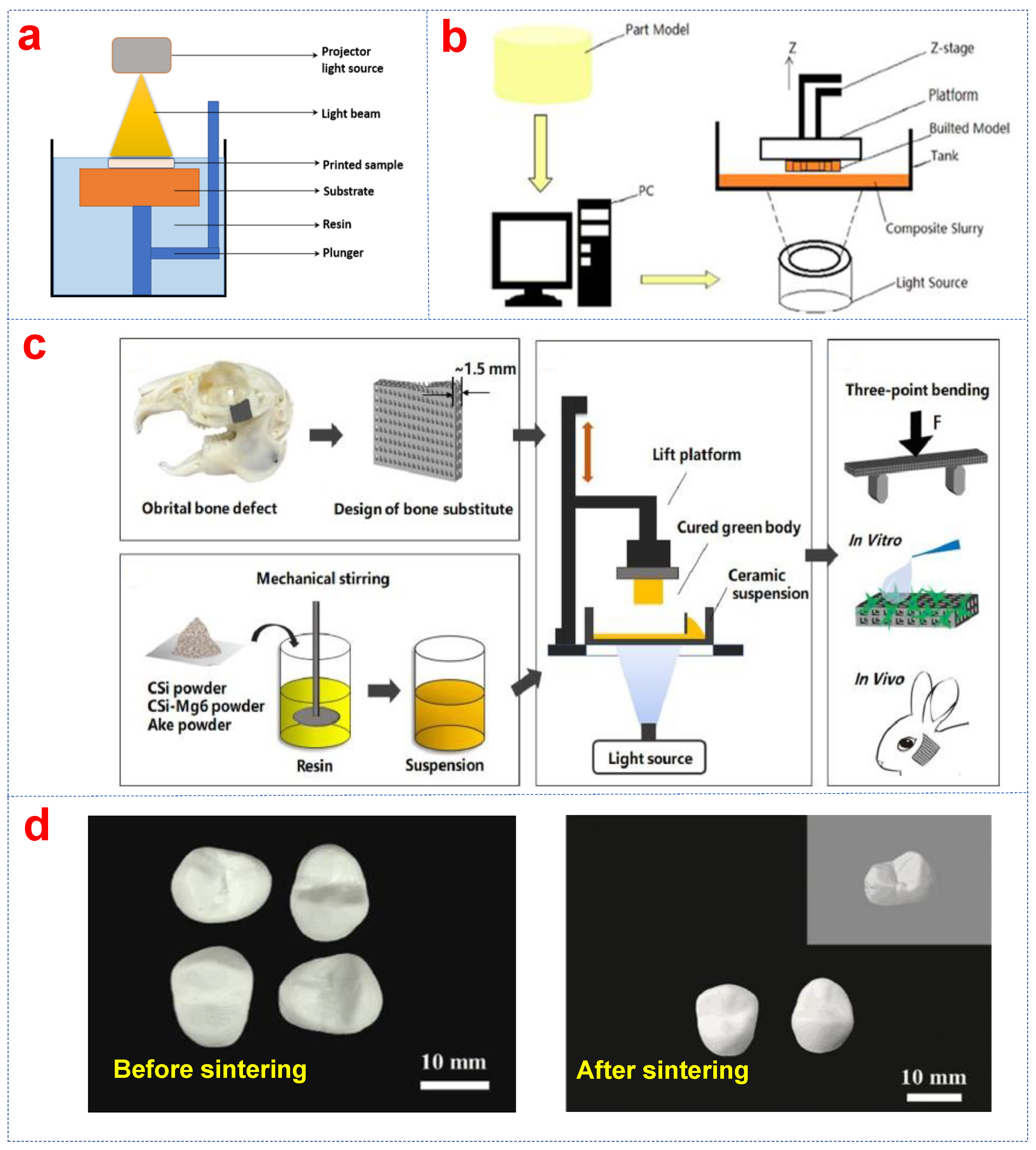
2.4. Stereolithography (SLA)

As a kind of photocuring and the most mature technique of additive manufacturing, SLA printing of bioceramics includes the fabrication of the slurry, printing, debinding, and sintering processes. The osteoid ceramic design and the SLA 3D printing process are shown in Figure 4a,b [72]. Compared with traditional methods, SLA can print components with large size, complex structure, high precision, and fine size, which has advantages for the flexibility of printing ceramic parts and the easy control of geometry [73]. HA bioceramic scaffolds with personalized, large, and osteoid structures were printed via SLA, which laid the foundation for blood vessel growth and large bone defect repair [74]. An optimized SLA printing process was successfully utilized to fabricate HA osteoid ceramics with approximately 54 mm in length and 30 mm in width that accurately simulate the structure of the human femur (Figure 4c). SLA printed complex parts such as a precise and compact school emblem, a cage without natural bone grafting, and a customized HA mandible (Figure 4d). Similarly, porous scaffolds with complexity and good interconnectivity were prepared via SLA, which allows for human umbilical vein endothelial cells to be evenly distributed and multiplied on scaffolds [75].
Al2O3-ZrO2 bioceramics are non-cytotoxic and have outstanding mechanical properties and biocompatibility. The surface of Al2O3-ZrO2 dental implants prepared through SLA was dense and smooth and free of cracks and delamination, as shown in Figure 4e [76]. Liquid precursor infiltration, in situ precipitation, and SLA were integrated to introduce the second phase and decrease the sintering temperature to obtain Al2O3-ZrO2 composites with an elastic modulus of 188–318 GPa [77,78]. Most bioceramic scaffolds have strength in the range of natural skull bone. The study by Lu et al. [79] showed that the pore geometry of calcium silicate scaffolds can be designed through SLA and influenced the mechanical and structural stability of the scaffolds. CaSiO3 bioceramics have better osteoinductivity, biological activity, and osseointegration capabilities compared with CaP bioceramics. They can release calcium (Ca2+) and silicon (Si4+) ions in body fluids, which induce the deposition of hydroxyapatite (HA) and form chemical bonds with the host bone, thereby promoting bone repair. The bioactivity, degradability, and bone regeneration potential of calcium silicate-based ceramics highlight their potential as third-generation biomaterials [8], which can promote regenerative responses in natural human tissues, such as the ability to induce osteogenesis, similar to hydroxyapatite [80]. Similarly, β-TCP bioceramic scaffolds with gyroid structures similar to cancellous bone were produced through SLA. The scaffolds exhibited excellent mechanical strength and cell proliferation in comparison to grid-like bioceramic scaffolds [81].
Liu et al. [82] prepared the HA scaffold with different pore sizes of 400, 500, and 600 μm through SLA. The HA scaffold had an enhanced compressive strength as the pore size increased. BCP bioceramics can significantly promote the proliferation of MC3T3-E1 cells through the release of Ca and PO3 ions while demonstrating excellent bioactivity [83]. HA is integrated with different calcium phosphate compositions to regulate mechanical and biological properties, further improving its bone regeneration performance [84]. BCP ceramics, which combine HA and β-TCP, play a vital role in bone tissue regeneration due to their biocompatibility, bioactivity, and osteoconductivity [85]. Compared to pure HA and pure β-TCP scaffolds, BCP scaffolds exhibit controlled degradation rates, better biocompatibility, and enhanced bone regeneration capacity [86]. The compressive strength of the BCP scaffolds was similar to that of cortical bone after sintering at 1250 °C [87]. The structure and size of the scaffold can be precisely controlled with SLA, and surgical procedures such as implant placements and complex surgeries can be accelerated and improved [83]. SLA can print HA parts with high precision using aqueous HA suspension with low viscosity, high curing depth, and small curing width. The chemical composition of HA with a complex structure was similar to that of the original HA powder [57].
Post-processing, such as the cleaning, curing, and finishing of printed parts, can heavily influence the performance of bioceramics produced with SLA. The residual tensile stress of ZrO2 ceramics was effectively regulated by the sintering temperature, holding time, and heat rate of the degreasing–sintering process [88]. The mechanical properties and density of bioceramics can also improve by changing sintering parameters. Densification and the density of bioceramics are affected by the degree of particle packing, solid content of the initial ink, and sintering atmosphere in post processing [89]. In addition, the phase of scaffolds can also influence mechanical properties of bioceramics.

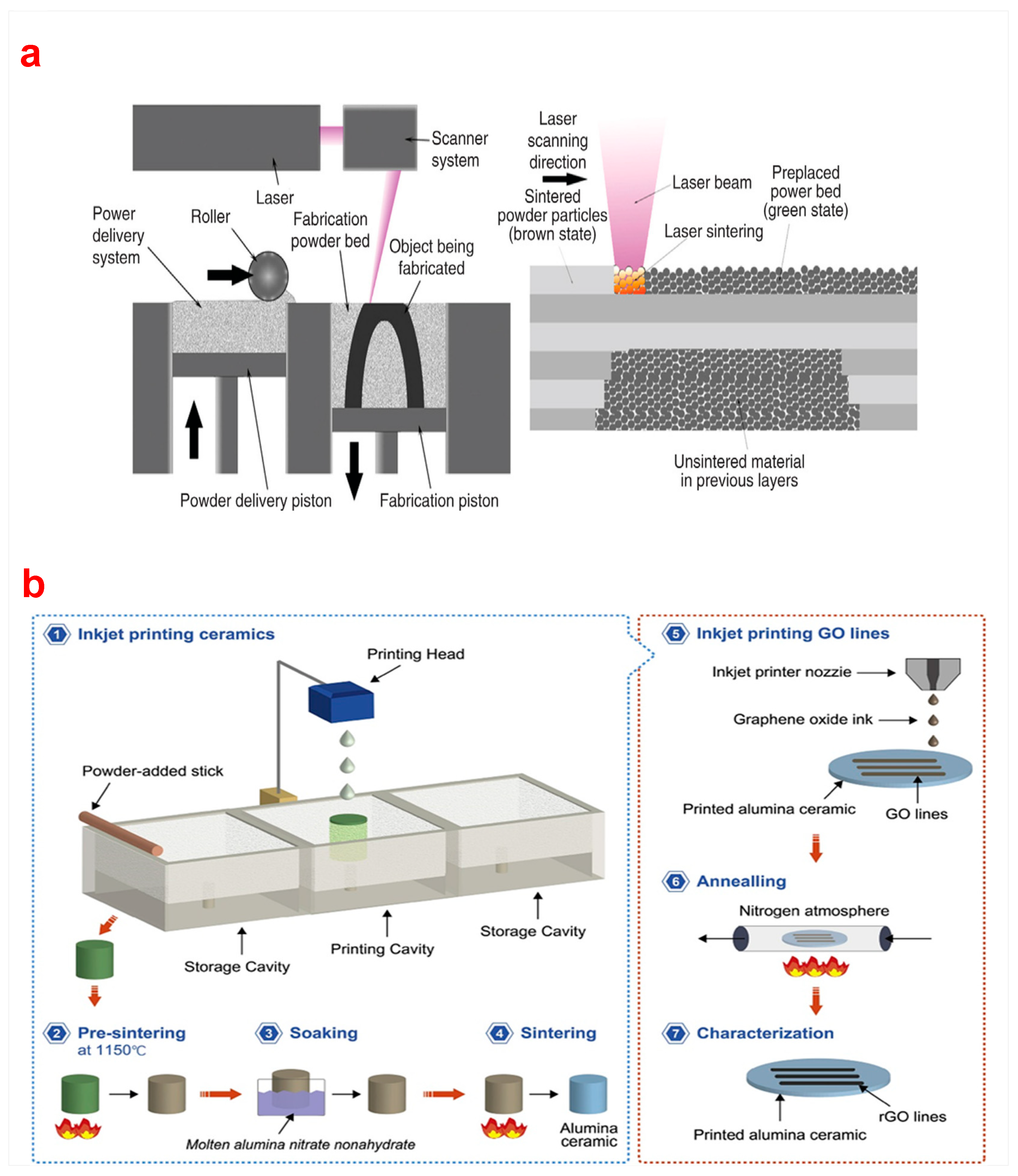
2.5. Selective Laser Sintering (SLS)

Bioceramics and scaffolds printed via SLS have the advantages of being self-supporting, having high precision, and being in customized shapes and sizes such as lattice, honeycomb, corrugated, and sandwich structures [9]. The working principle of SLS printing is shown in Figure 5a [90]. Bioactive borate glass (BBG) has bioactivity similar to 45S5 bioactive glass, and its surface can rapidly form apatite in simulated body fluid (SBF) within 6 h [91]. The bioactive borate glass/polycaprolactone composite scaffolds with biodegradability prepared by SLS exhibited excellent in vivo biological properties due to the printed internal porous structure [92]. Shuai et al. [93] prepared the porous CaP scaffolds with various weight ratios of TCP/HA powders through SLS. The compressive strength of the scaffolds with a ratio of 30/70 was 18.35 MPa. Additionally, high-strength Al2O3 parts were fabricated via slurry-based SLS of polymer-coated ceramic powders [94]. Bulina et al. [32] fabricated HA bioceramic scaffolds via SLS. The spot size, laser power, and scanning speed of HA powder under laser radiation were 0.2 mm, 4 W, and 640 mm/s, respectively, in the process of sintering. A dense layer of HA can be formed with different compositions of apatite powder. The high brittleness of bioceramics was also overcome by SLS, which has a great improvement over traditional technologies such as fiber bonding, melt casting, and freeze drying [95].
In addition, post-processing of the printed parts is usually required to improve their surface quality and mechanical properties after SLS. This may involve additional steps such as sanding, polishing, or applying coatings, which can be time consuming and add to the overall production cost.

2.6. Selective Laser Melting (SLM)

SLM is a 3D technology developed to prepare scaffolds with complex structures and large sizes and offers a promising improvement for bone implant applications [7]. Alumina and silica are commonly used bioceramic materials in SLM processes. Liu et al. [96] prepared highly dense Al2O3/GdAlO3/ZrO2 ternary eutectic ceramic parts with complex shapes through SLM. The thermal bubble inkjet method and high-temperature sintering were used to print Al2O3 and graphene circuits-on-ceramics [97]. The bioceramic printing process via thermal bubble inkjet technology is shown in Figure 5b, which can economically and efficiently print Al2O3 ceramics.
In considering these advancements in scaffold fabrication, the mechanical property of the printed bioceramics or scaffolds is one of the primary concerns in order to provide enough support at an implanted site. Different printing techniques and post-processing (such as debinding and sintering) have a significant impact on the properties of the printed bioceramics and scaffolds. The bioceramic scaffolds with different sizes were manufactured through DLP; the Al2O3-ZrO2 composite scaffold exhibits a high flexural resistance and elastic modulus [78]. The structural and mechanical stability of porous bioceramics can be achieved by optimizing the pore size and geometry. The compressive strength (0.6–16.8 MPa) of β-TCP bioceramic scaffolds were tuned to match the needs for various locations of cancellous bone [82]. In addition, it was found that the compressive strength of HA scaffolds decreased with the increase of pore size; the scaffold with ~600 μm pores had a promising application prospect. The HA and ZrO2 scaffolds printed by DLP showed different compressive strength, Vickers hardness, and fracture toughness. Sintering temperature is also one of the main factors that affect the mechanical properties of bioceramics. The compressive strength (149.54 ± 20.38 MPa) of BCP bioceramics sintered at 1250 °C was significantly lower than that at 1300 °C (370.03 ± 41.24 MPa) [84].
Figure 5. (a) Selective Laser Sintering [90], (b) printing process of bioceramics via thermal bubble inkjet technology [97].
Biomimetics 10 00428 g005
Bioceramic scaffolds fabricated through 3D printing exhibit the following advantages: (1) 3D printing provides precise control over both internal and external architectures, including pore geometry, porosity, and interconnectivity, enables high structural complexity, design flexibility, and patient-specific requirements [98]. (2) Optimized 3D-printed scaffolds can provide growth-guiding structures to support cell migration and proliferation, thereby facilitating regenerative bone tissue formation [99]. (3) The 3DP process allows for rapid fabrication while minimizing experimental errors [100]. Based on ISO/ASTM 52900:2021 and widely used 3D printing methods for bioceramics in the literature, the advantages and disadvantages for 3D printing of bioceramics are shown in Table 1.
As shown in Table 1, DIW and SLA have been widely used in the printing of bioceramics and bioceramic matrix composite scaffolds. Although DIW needs highly controlled printing slurries and the printed parts have low accuracy, it excels in the printing of bioceramics and bioceramic matrix composite scaffolds with biomimetic structures [102]. Continuous fiber-reinforced bioceramic matrix composite scaffolds can be produced to improve the mechanical property by DIW. DLP and SLA can fabricate high-precision bioceramic scaffolds [106,116], but the addition of photosensitive resins may reduce their bioactivity, which will influence the biocompatibility of the bioceramic scaffolds. Although SLS and SLM can produce diverse bioceramic scaffolds [119] and bioactive glass components [108] without introducing additives, their limited material compatibility poses a challenge. Additionally, SLM usually requires additional support materials when it creates bioceramic scaffolds. The 3D printing method exhibits distinct advantages and should be appropriately selected to fabricate various implants of bioceramics and bioceramic matrix composite scaffolds. These different 3D printing methods would provide great potential for bone repair, bone tissue regeneration, and biomedical applications, particularly for some complex-shaped pathological bone defect conditions [120]. Additionally, the primary factors, including the 3D printing process, material powder composition and morphology, and post-processing techniques, can possibly influence the microstructure formation of scaffolds. Nevertheless, 3D-printed bioceramic scaffolds with proper in vivo degradability and excellent mechanical properties remain a challenge.

3. Three-Dimensional Printing of Bioceramic Matrix Composites

Due to the inadequate mechanical properties or biological properties of printed bioceramics or scaffolds by 3D printing, reinforcements (fibers, particles, whiskers, ionic doping) are introduced to improve their mechanical properties and biological activity. The mechanical properties of the composite stent should be matched to the bone at the implant site to avoid the stress shielding effect or the lack of performance of the implant. In addition, the biodegradation rates of composite scaffolds can be matched with the appropriate rate for the gradual transfer of loads from implants to newly developed bone tissues and can eliminate the need for additional surgeries to remove the scaffold, reducing the risk of complications and improving patient outcomes.

3.1. Fiber Reinforcements

As a reinforcing material, the addition of fibers in bioceramics can enhance their strength and toughness, thus increasing their load-bearing capacity and resistance to fracture. This can be attributed to the improved microstructural integrity and the efficient stress transfer by the added fibers. Carbon fiber (CF) has great potential to reinforce bioceramics due to its biocompatibility and high strength and modulus. CF-reinforced bioceramics have been widely used for the fabrication of bioceramic components with excellent mechanical properties [121]. Continuous fiber-reinforced bioceramics can be printed on the condition that the precursors have excellent printability [120]. Continuous carbon fiber-reinforced polymer composites were fabricated by vat photopolymerization. Zhao et al. [122] used a self-designed 3D printer to fabricate continuous CF-reinforced HA composite (CF/HA) scaffolds with simultaneous improvement in strength and toughness. Although carbon fiber reinforcement improves the strength and toughness of bioceramics, its bioinert nature remains unavoidable and can significantly reduce the biological activity of scaffolds [123]. Thus, developing novel degradable fibers to enhance bioceramics poses a critical challenge in current research.

3.2. Particle Reinforcements

Glass is one of the particle reinforcement materials that is used to reinforce bioceramics, and silicate bioactive glass has been widely used for biomedical applications. Bioceramic glass has unique bone-bonding properties compared to other bioceramics [124]. Bioactive glass (BG) is primarily composed of Na2O, CaO, SiO2, and P2O5, exhibiting significant potential for healing and regenerating bone defects [125]. Its osteoconductive and osteogenic properties enhanced the proliferation and differentiation of progenitor cells [126]. Additionally, BG served as a viable alternative to other scaffold biomaterials due to its ability to promote angiogenesis under vascular endothelial growth factor (VEGF) stimulation [127]. BG also helped to form carbonated hydroxyapatite and bioactive surface layers, enabling interfacial bonding with surrounding tissues [90]. Bioactive ceramics and glasses can be combined with polymers to form composites with improved mechanical properties for bone regeneration. Three-dimensional biopolymer–glass composites with introduced cells have been developed to guide cell growth, proliferation, and differentiation [128]. Hence, BG scaffolds act as an excellent porous template with outstanding mechanical performance in the fields of tissue engineering and regenerative medicine [129]. Bose et al. [130] utilized a binder jetting 3D printing method to produce bioactive glass (45S5 BG)-reinforced TCP scaffolds. The HA apatite layer was enhanced with the addition of BG in 8 weeks in vitro. In addition, Kolan et al. [131] fabricated borate-based BG scaffolds with various porosities and architectures (cubic, spherical, crossed, gyroid, and diamond) using indirect SLS. In particular, after being implanted in a rat calvarial defect, scaffolds with a diamond structure and 50% porosity exhibited faster bone formation. Porous 45S5 bioactive glass–ceramic scaffolds prepared through SLS exhibited higher density, higher crystallinity, and fracture toughness [132]. By rapidly heating and cooling, the desirable crystallization phase Na2Ca2Si3O9 was formed. The β-TCP ceramic scaffolds had excellent biocompatibility to be applied for bone regeneration. Porous scaffolds of 58S BG/β-tricalcium phosphate (β-TCP/BG) manufactured through DLP had an increased compressive strength with the increase in solid loading [133]. The β-TCP/BG composite scaffolds can promote the attachment and osteogenic differentiation of bone marrow stem cells.
In addition to BG, MgO has also been considered as a particle reinforcement material for bioceramic scaffolds due to its exceptional biocompatibility and antibacterial properties. The biodegradation of biphasic calcium phosphate (BCP) in simulated body fluid (SBF) was effectively improved when 1 wt% MgO was introduced [134]. The macroporous dome-like meshes with varying mass ratios of CSi/CSi-Mg6 were prepared through DLP using wollastonite bioceramic without and with 6% magnesium addition (CSi, CSi-Mg6) [65]. The biodegradation process and the new bone growth of the CSi/CSi-Mg6 scaffolds and the titanium mesh are shown in Figure 3c. HA bone grafts degraded slowly when extending the implantation time, providing a certain spatial maintenance for further bone growth, while the degradability of bioceramic scaffolds containing 16% CSi can be significantly improved, and the scaffolds can accelerate the growth of new bone. The biodegradability of bioceramic scaffolds can be greatly improved with the addition of baghdadite [66]. Zhao et al. [135] improved 3D-printed hydroxyapatite (HA) scaffolds by integrating cerium-containing mesoporous bioactive glass nanoparticles (Ce-MBGNs). The results showed that incorporating Ce-MBGNs significantly enhanced the HA scaffolds’ mechanical strength, mineralization capacity, antioxidant activity, and osteogenic potential. Micro-CT results showed the impact of scaffold implantation on bone growth at the defect site at 4 and 12 weeks post-implantation. Compared with other groups, 5CeMBG displayed the most significant bone formation, particularly within interconnected macropores (Figure 6b) [136]. This strategy demonstrates promising potential for developing personalized and more effective bone defect repair solutions, particularly in challenging clinical cases involving inflammatory conditions.

3.3. Whisker Reinforcements

Whisker reinforcements such as HA and SiC whiskers are easily mixed with bioceramics and are widely used to enhance the properties of HA, Al2O3, ZrO2, and Si3N4 [137,138,139]. As a strengthening agent, the whiskers prevent catastrophic failure by obstructing crack deflection and dispersing the stress concentration. Using the 3D printing method, the mixture of HA powder and whiskers (5%, 10%, and 15%) was used to prepare a porous bone embryo scaffold that had the highest compressive strength with 10% HA whiskers [140]. Feng et al. [141] used HA whiskers to reinforce a calcium silicate ceramic and fabricated the porous calcium silicate scaffolds through SLS, which can overcome insufficient mechanical strength and resistance [142]. Similarly, a SiC whisker (SiCw) with exceptional comprehensive properties, including high strength, chemical stability, and biological properties, was utilized to reinforce nHA composites [143,144]. Xing et al. [145] prepared SiC-toughened Al2O3 (SiCw/Al2O3) ceramic parts with complex shapes by using SLA. The composite ceramics demonstrated a combination of intergranular and transgranular fracture modes. The fracture toughness was enhanced owing to SiC whisker pull out and crack deflection.

3.4. Ionic Doping Reinforcements

Ionic doping can modulate the crystalline structure of bioceramics and is regarded as one of the effective strategies to enhance their mechanical and functional properties. In terms of biological performance, ionic doping may promote cell proliferation, differentiation, and antibacterial efficacy [146]. In the rabbit femoral bone defect repair model, three-dimensional CT reconstruction and histological observations demonstrated that the CSiMg4@CSiMg10-p scaffold exhibited significantly higher osteogenic capability compared to other scaffolds after 12 weeks of implantation (Figure 6a). The biodegradable characteristic of bioceramic scaffolds is crucial for their successful application in tissue engineering, which promotes natural healing processes and eliminates the need for scaffold removal surgeries. After 4 and 12 weeks of implantation in rabbit skulls, Mg-doped calcium silicate (CSM) bioceramic scaffolds and titanium alloy scaffolds demonstrated differences in bone tissue ingrowth. At the 4th week, more bone tissue cells adhered to the inner wall and penetrated the pores within the scaffold compared with the titanium alloy scaffold [147]. Figure 6c displays histological sections stained with VG at 4 and 12 weeks. The CSM bioceramic scaffold exhibited gradual degradation with numerous bone tissue cells aligning along the inner wall of the scaffold. At the 12th week, new bone tissue had extended along the inner wall and into the interior. The scaffolds exhibited excellent osseointegration ability by fusing with bone tissue to create a robust interface between the scaffold and bone. However, the compressive strength of the CSM bioceramic scaffold decreased rapidly with in vivo degradation, which was not conducive to repairing load-bearing bone defects.
Ion-doped bioceramic scaffolds exhibit significant potential in immune modulation by releasing specific bioactive ions to enhance bone regeneration [148]. However, the synergistic effects between ions and their underlying interaction mechanisms require further investigation, while the precision and stability of ion doping ratios and distribution need improvement.
A hierarchical structure serves as a framework defining the structure–function relationships in natural bone, including cortical bone and cancellous bone. Various mechanical, biological, and chemical functions are carried out by macro-, micron-, and nanoscale structures. A stabilizing and supportive role is provided by cortical bone with notable mechanical strength, while cancellous bone with 50% to 90% porosity provides a proper environment for bone metabolism and hematopoietic function [149]. Inspired by the structure and composition of natural bone, bone substitute and repair materials should have a hierarchical and anisotropic structure and composition similar to natural bone to achieve desirable biological and mechanical performances. Such scaffolds for bone tissue engineering should contain multilevel pores from the micro- to the macro-scale, which facilitate bone regeneration and vascular ingrowth and can be applied for both non-load-bearing and load-bearing bone defects. Implanted bioceramics can interact with the surrounding tissues and stimulate specific cellular responses. This interaction can create a chemical bond between the implant and the tissue, promoting integration and long-term stability [150]. An interconnected pore structure in a scaffold can allow oxygen, nutrients, and waste to be delivered through the scaffold. A scaffold with high porosity has a large surface area, which will improve cell adhesion, cell migration, and tissue–scaffold interaction. Additionally, an ideal scaffold should have similar mechanical properties (including strength, stiffness, and toughness) to natural bone. It can provide a certain mechanical support and deliver it to the cell under physiological conditions, which can stimulate cell differentiation from osteogenic lineages into undesirable forms. It has been reported that some scaffolds achieved comparable mechanical properties to those of cancellous bone, which will provide enough mechanical support in repairing non-load-bearing bone defects. Nevertheless, few scaffolds possess similar strength, stiffness, and toughness to that of cortical bone. Their mechanical weaknesses, including non-ideal strength and toughness, restrict them from repairing load-bearing bones. Continuous monofilament fiber-reinforced bioceramics can obtain satisfying microstructure and mechanical properties for both the cancellous and cortical bone. However, 3D printer and printing technology can hardly prepare a continuous fiber-reinforced bioceramic scaffold with fine structure and mechanical properties because monofilament is not easily centered on, adjusted with, fed into, and bonded with the matrix in a nozzle during the process of printing. The adhesion performance between the continuous fiber and bioceramic matrix is weak, which seriously reduces the mechanical properties of the composite scaffold. This limits the clinical application of monofilament fiber-reinforced bioceramics mimicking natural bone in both microstructure and mechanical properties. Moreover, besides the common reinforcing materials, such as carbon fiber and glass fibers, few kinds of degradable fiber can be used to reinforce degradable and absorbable bioceramics for clinical application.
Figure 6. (a) Reconstruction micro-CT images showing the scaffolds of the HAs, 5MBGs, and 5Ce-MBGs groups implanted in rat calvarial defects at 4 and 12 weeks post-implantation [135]. (b) The behavior of magnesium-doped SiC scaffolds with different magnesium content after implantation [136]. (c) Histological sections of VG staining at 4 and 12 weeks following the implantation of various stents into a rabbit skull. The stents are shown in black, new bone in red, and fibrous tissue in blue. The length of the ruler is 300 μm [147].
Biomimetics 10 00428 g006

4. Summary and Outlook

Three-dimensional printing technology has been used to print hearts, kidneys, and artificial bone skulls, which can be used to replace bone skulls with damage and defect according to the specific conditions of patients. Three-dimensional-printed bioceramic scaffolds are especially highly suitable for bone repair applications owing to excellent biocompatibility, excellent osseointegration, excellent osteoconduction, excellent bioactivity, excellent personalized customization, and high accuracy. Direct ink writing technology with its unique advantages, such as good biocompatibility, material-saving properties, ease of operation, and fast-forming properties, has become one of the most promising 3D printing technologies in the biomedical field. Unfortunately, it is difficult to print bioceramics with high solid loading due to the lack of an excellent slurry with a low viscosity and high solid content for fabricating bioceramic green bodies in the direct writing molding process. The influence of the morphology and structure of ceramic powder on slurry characteristics, forming, and printing needs to be systematically investigated to obtain bioceramic scaffolds with high solid loading.
Furthermore, bone substitute and repair materials should have a hierarchical and anisotropic structure and composition similar to natural bone to achieve desirable biological and mechanical performance. For bone tissue engineering, scaffolds should contain multilevel pores from the micro- to the macro-scale and have satisfactory mechanical properties (including strength, stiffness, and toughness) for non-load-bearing and load-bearing bone repair applications. So far, some scaffolds achieved the mechanical properties comparable to that of cancellous bone, which were applied in repairing non-load-bearing bone defects. Nevertheless, few scaffolds possess similar strength, stiffness, and toughness to that of cortical bone. The implanted bioceramic components can be significantly enhanced by introducing different reinforcements. Continuous monofilament fiber-reinforced bioceramics can obtain satisfying microstructure and mechanical properties for both the cancellous and cortical bone. However, 3D printer and printing technology can hardly prepare a continuous fiber-reinforced bioceramic scaffold with fine structure and ideal mechanical properties because monofilament is not easily centered on, adjusted to, fed into, and bonded with the matrix in a nozzle during the printing process. The weak adhesion performance between the continuous fiber and bioceramic matrix seriously reduces the mechanical properties of the composite scaffold. Moreover, besides the common reinforcing materials such as carbon fiber and glass fiber, there are few kinds of degradable fibers for reinforcing degradable and absorbable bioceramics in clinical applications. Monofilament fiber-reinforced bioceramics mimicking natural bone in both microstructure and mechanical properties can be a solution in load-bearing applications, which urgently need research and development of the printer and novel technology for monofilament fiber-reinforced bioceramics. The monofilament fiber-reinforced bioceramic scaffolds with proper in vivo degradability will promote natural healing processes and eliminate the need for scaffold removal surgeries, which remain a challenge.
Although existing research has significantly improved the performance of traditional scaffolds, 3D-printed bioceramic scaffolds remain far from clinical application due to persistent challenges. First, current 3D printing technologies struggle to fabricate bioceramic scaffolds with both high strength and toughness. While high-temperature sintering is commonly used to produce high-strength ceramic scaffolds, their purely ceramic composition leads to insufficient toughness and fracture susceptibility in practical use. These limitations prevent 3D-printed bioceramic scaffolds from adapting to the unique mechanical demands of load-bearing skeletal environments, thereby restricting their applicability compared to metal implants. Second, the degradation rate of existing bioceramics often fails to align with new bone formation rates, hindering optimal bone-scaffold integration. Finally, current 3D-printed bioceramic scaffolds cannot accurately replicate the highly complex and ordered microstructure of natural bone tissue. These issues represent critical challenges in 3D printing of bioceramics. To address these challenges, future research should focus on refining existing 3D printing technologies to enhance precision for fabricating finer and more intricate structures, improving control over scaffold microarchitecture to mimic the composition and structure of natural high-strength/toughness materials (e.g., bone, nacre), and developing multi-material composite bioceramic scaffolds with superior mechanical properties and tunable degradation rates that match bone regeneration kinetics. Although 3D-printed bioceramic scaffolds are still largely confined to laboratory research, they present both significant opportunities and formidable challenges for clinical translation. This will be a significant opportunity and formidable challenge for clinical application.

Author Contributions

Conceptualization, X.Z.; methodology, X.Z.; software, X.Z.; validation, X.Z.; formal analysis, X.Z.; investigation, X.Z., J.L. and L.L.; resources, X.Z.; data curation, J.L. and L.L.; writing—original draft preparation, X.Z., J.L. and L.L.; writing—review and editing, X.Z. and J.L.; visualization, X.Z.; supervision, X.Z.; project administration, X.Z.; funding acquisition, X.Z. All authors have read and agreed to the published version of the manuscript.

Funding

This research was funded by the National Natural Science Foundation of China (52472285, 51772179, and 51072107), Science and Technology Innovation Team of Shaanxi Province, China (No. 2023-CX-TD-16); the Key Research and Development Program of Shaanxi Province, China (No. 2024GX-YBXM-171); the Youth Innovation Team of Shaanxi Universities (2019); and the Science Research Plan Program of Youth Innovation Team Construction of Education Department of Shaanxi Province (No. 21JP018). And the APC was funded by the Key Research and Development Program of Shaanxi Province, China (No. 2024GX-YBXM-171).

Acknowledgments

This study was supported by the National Natural Science Foundation of China (52472285, 51772179, and 51072107), Science and Technology Innovation Team of Shaanxi Province, China (No. 2023-CX-TD-16); the Key Research and Development Program of Shaanxi Province, China (No. 2024GX-YBXM-171); the Youth Innovation Team of Shaanxi Universities (2019); and the Science Research Plan Program of Youth Innovation Team Construction of Education Department of Shaanxi Province (No. 21JP018).

Conflicts of Interest

The authors declare no competing financial interests.

References

  1. Ghodbane, S.A.; Murthy, N.S.; Dunn, M.G.; Kohn, J. Achieving molecular orientation in thermally extruded 3D printed objects. Biofabrication. 2019, 11, 045004. [Google Scholar] [CrossRef] [PubMed]
  2. Shahrubudin, N.; Te Chuan, L.; Ramlan, R. An overview of critical success factors for implementing 3D printing technology in manufacturing firms. J. Appl. Eng. Sci. 2019, 17, 379–385. [Google Scholar] [CrossRef]
  3. Hull, C.W. Apparatus for Production of Three-Dimensional Objects by Stereolithography. U.S. Patent US4575330A, 8 August 1984. [Google Scholar]
  4. Wang, X.; Jiang, M.; Zhou, Z.; Gou, J.; Hui, D. 3D printing of polymer matrix composites: A review and prospective. Compos. Part B-Eng. 2017, 110, 442–458. [Google Scholar] [CrossRef]
  5. Leukers, B.; Gülkan, H.; Irsen, S.H.; Milz, S.; Tille, C.; Schieker, M.; Seitz, H. Hydroxyapatite scaffolds for bone tissue engineering made by 3D printing. J. Mater. Sci. Mater. Med. 2005, 16, 1121–1124. [Google Scholar] [CrossRef] [PubMed]
  6. Zhang, B.; Sun, H.; Wu, L.; Ma, L.; Xing, F.; Kong, Q.; Fan, Y.; Zhou, C.; Zhang, X. 3D printing of calcium phosphate bioceramic with tailored biodegradation rate for skull bone tissue reconstruction. Bio-Des. Manuf. 2019, 2, 161–171. [Google Scholar] [CrossRef]
  7. Rodrigo-Vázquez, C.S.; Kamboj, N.; Aghayan, M.; Sáez, A.; De Aza, A.H.; Rodríguez, M.A.; Hussainova, I. Manufacturing of silicon—Bioactive glass scaffolds by selective laser melting for bone tissue engineering. Ceram. Int. 2020, 46, 26936–26944. [Google Scholar] [CrossRef]
  8. Song, W.; Sun, W.; Chen, L.; Yuan, Z. In vivo Biocompatibility and Bioactivity of Calcium Silicate-Based Bioceramics in Endodontics. Front. Bioeng. Biotechnol. 2020, 8, 580954. [Google Scholar] [CrossRef]
  9. Dhinasekaran, D.; Jagannathan, M.; Rajendran, A.R.; Purushothaman, B. Microwave-assisted fabrication of nanostructured borate bioactive glass and its bioactivity. Biomater. Sci. 2024, 12, 4727–4734. [Google Scholar] [CrossRef]
  10. Deng, C.; Chang, J.; Wu, C. Bioactive scaffolds for osteochondral regeneration. J. Orthop. Translat. 2019, 17, 15–25. [Google Scholar] [CrossRef]
  11. Vallet-Regí, M.; Izquierdo-Barba, I.; Colilla, M. Structure and functionalization of mesoporous bioceramics for bone tissue regeneration and local drug delivery. Philos. Trans. A Math. Phys. Eng. Sci. 2012, 370, 1400–1421. [Google Scholar] [CrossRef]
  12. Kargozar, S.; Singh, R.K.; Kim, H.-W.; Baino, F. “Hard” ceramics for “Soft” tissue engineering: Paradox or opportunity? Acta Biomater. 2020, 115, 1–28. [Google Scholar] [CrossRef] [PubMed]
  13. Lopes, M.A.; Monteiro, F.J.; Santos, J.D. Glass-reinforced hydroxyapatite composites: Fracture toughness and hardness dependence on microstructural characteristics. Biomaterials 1999, 20, 2085–2090. [Google Scholar] [CrossRef] [PubMed]
  14. Chen, L.; Zhu, W.-M.; Fei, Z.-Q.; Chen, J.-L.; Xiong, J.-Y.; Zhang, J.-F.; Duan, L.; Huang, J.; Liu, Z.; Wang, D. The study on biocompatibility of porous nHA/PLGA composite scaffolds for tissue engineering with rabbit chondrocytes in vitro. BioMed Res. Int. 2013, 2013, 412745. [Google Scholar] [CrossRef]
  15. Gatti, A.; Zaffe, D.; Poli, G. Behaviour of tricalcium phosphate and hydroxyapatite granules in sheep bone defects. Biomaterials 1990, 11, 513–517. [Google Scholar] [CrossRef]
  16. Böstman, O.; Hirensalo, E.; Vainionpää, S.; Mäkelä, A.; Vihtonen, K.; Törmälä, P.; Rokkanen, P. Ankle fractures treated using biodegradable internal fixation. Clin. Orthop. Relat. Res. 1989, 238, 195–203. [Google Scholar] [CrossRef]
  17. Chia, H.N.; Wu, B.M. Recent advances in 3D printing of biomaterials. J. Biol. Eng. 2015, 9, 4. [Google Scholar] [CrossRef] [PubMed]
  18. Ma, H.; Feng, C.; Chang, J.; Wu, C. 3D-printed bioceramic scaffolds: From bone tissue engineering to tumor therapy. Acta Biomater. 2018, 79, 37–59. [Google Scholar] [CrossRef]
  19. Song, P.; Hu, C.; Pei, X.; Sun, J.; Sun, H.; Wu, L.; Jiang, Q.; Fan, H.; Yang, B.; Zhou, C. Dual modulation of crystallinity and macro-/microstructures of 3D printed porous titanium implants to enhance stability and osseointegration. J. Mater. Chem. B. 2019, 7, 2865–2877. [Google Scholar] [CrossRef]
  20. Yang, G.; Sun, Y.; Li, M.; Ou, K.; Fang, J.; Fu, Q. Direct-ink-writing (DIW) 3D printing functional composite materials based on supra-molecular interaction. Compos. Sci. Technol. 2021, 215, 109013. [Google Scholar] [CrossRef]
  21. Ahlfeld, T.; Köhler, T.; Czichy, C.; Lode, A.; Gelinsky, M. A methylcellulose hydrogel as support for 3D plotting of complex shaped calcium phosphate scaffolds. Gels 2018, 4, 68. [Google Scholar] [CrossRef]
  22. Tagliaferri, S.; Panagiotopoulos, A.; Mattevi, C. Direct ink writing of energy materials. Mater. Adv. 2021, 2, 540–563. [Google Scholar] [CrossRef]
  23. Li, Z.; Li, J.; Luo, H.; Yuan, X.; Wang, X.; Xiong, H.; Zhang, D. Direct ink writing of 3D piezoelectric ceramics with complex unsupported structures. Eur. Ceram. Soc. 2022, 42, 3841–3847. [Google Scholar] [CrossRef]
  24. Lewis, J.A.; Gratson, G.M. Direct writing in three dimensions. Mater. Today 2004, 7, 32–39. [Google Scholar] [CrossRef]
  25. Lewis, J.A.; Smay, J.E.; Stuecker, J.; Cesarano, J. Direct ink writing of three-dimensional ceramic structures. J. Am. Ceram. Soc. 2006, 89, 3599–3609. [Google Scholar] [CrossRef]
  26. Zhang, B.; Guo, L.; Chen, H.; Ventikos, Y.; Narayan, R.J.; Huang, J. Finite element evaluations of the mechanical properties of polycaprolactone/hydroxyapatite scaffolds by direct ink writing: Effects of pore geometry. J. Mech. Behav. Biomed. Mater. 2020, 104, 103665. [Google Scholar] [CrossRef]
  27. Somers, N.; Jean, F.; Lasgorceix, M.; Preux, N.; Delmotte, C.; Boilet, L.; Petit, F.; Leriche, A. Fabrication of doped β-tricalcium phosphate bioceramics by Direct Ink Writing for bone repair applications. Eur. Ceram. Soc. 2023, 43, 629–638. [Google Scholar] [CrossRef]
  28. Yang, L.; Zeng, X.; Ditta, A.; Feng, B.; Su, L.; Zhang, Y. Preliminary 3D printing of large inclined-shaped alumina ceramic parts by direct ink writing. J. Adv. Ceram. 2020, 9, 312–319. [Google Scholar] [CrossRef]
  29. Carloni, D.; Zhang, G.; Wu, Y. Transparent alumina ceramics fabricated by 3D printing and vacuum sintering. Eur. Ceram. Soc. 2021, 41, 781–791. [Google Scholar] [CrossRef]
  30. Li, Y.-y.; Li, L.-t.; Li, B. Direct write printing of three-dimensional ZrO2 biological scaffolds. Mater. Des. 2015, 72, 16–20. [Google Scholar] [CrossRef]
  31. Hossain, S.S.; Lu, K. Recent progress of alumina ceramics by direct ink writing: Ink design, printing and post-processing. Ceram. Int. 2023, 49, 10199–10212. [Google Scholar] [CrossRef]
  32. Bulina, N.V.; Titkov, A.I.; Baev, S.G.; Makarova, S.V.; Khusnutdinov, V.R.; Bessmeltsev, V.P.; Lyakhov, N.Z. Laser sintering of hydroxyapatite for potential fabrication of bioceramic scaffolds. Mater. Today Proc. 2021, 37, 4022–4026. [Google Scholar] [CrossRef]
  33. Veljovic, D.; Palcevskis, E.; Zalite, I.; Petrovic, R.; Janackovic, D. Two-step microwave sintering—A promising technique for the processing of nanostructured bioceramics. Mater. Lett. 2013, 93, 251–253. [Google Scholar] [CrossRef]
  34. Fu, L.; Engqvist, H.; Xia, W. Spark plasma sintering of biodegradable Si3N4 bioceramic with Sr, Mg and Si as sintering additives for spinal fusion. Eur. Ceram. Soc. 2018, 38, 2110–2119. [Google Scholar] [CrossRef]
  35. Wang, C.-J.; Huang, C.-Y. Effect of TiO2 addition on the sintering behavior, hardness and fracture toughness of an ultrafine alumina. Mater. Sci. Eng. A 2008, 492, 306–310. [Google Scholar] [CrossRef]
  36. Chen, G.; Zu, Y.; Luo, J.; Fu, X.; Zhou, W. Microstructure and superplastic behavior of TiO2-doped Al2O3–ZrO2 (3Y) composite ceramics. Mater. Sci. Eng. A 2012, 554, 6–11. [Google Scholar] [CrossRef]
  37. Wang, R.; Zhu, P.; Yang, W.; Gao, S.; Li, B.; Li, Q. Direct-writing of 3D periodic TiO2 bio-ceramic scaffolds with a sol-gel ink for in vitro cell growth. Mater. Des. 2018, 144, 304–309. [Google Scholar] [CrossRef]
  38. Liu, N.; Sun, X.; Chen, Z.; Xu, Z.; Dai, N.; Shi, G.; Cai, S.; Lv, X.; Zheng, C. Direct ink writing of dense alumina ceramics prepared by rapid sintering. Ceram. Int. 2022, 48, 30767–30778. [Google Scholar] [CrossRef]
  39. Lakhdar, Y.; Tuck, C.; Binner, J.; Terry, A.; Goodridge, R. Additive manufacturing of advanced ceramic materials. Prog. Mater. Sci. 2021, 116, 100736. [Google Scholar] [CrossRef]
  40. Esslinger, S.; Gadow, R. Additive manufacturing of bioceramic scaffolds by combination of FDM and slip casting. J. Eur. Ceram. Soc. 2020, 40, 3707–3713. [Google Scholar] [CrossRef]
  41. Kalita, S.J.; Bose, S.; Hosick, H.L.; Bandyopadhyay, A. Development of controlled porosity polymer-ceramic composite scaffolds via fused deposition modeling. Mater. Sci. Eng. C 2003, 23, 611–620. [Google Scholar] [CrossRef]
  42. Song, P.; Zhou, C.; Fan, H.; Zhang, B.; Pei, X.; Fan, Y.; Jiang, Q.; Bao, R.; Yang, Q.; Dong, Z. Novel 3D porous biocomposite scaffolds fabricated by fused deposition modeling and gas foaming combined technology. Compos. Part B-Eng. 2018, 152, 151–159. [Google Scholar] [CrossRef]
  43. Arnesano, A.; Padmanabhan, S.K.; Notarangelo, A.; Montagna, F.; Licciulli, A. Fused deposition modeling shaping of glass infiltrated alumina for dental restoration. Ceram. Int. 2020, 46, 2206–2212. [Google Scholar] [CrossRef]
  44. Singh, D.; Singh, R.; Boparai, K.S. Development and surface improvement of FDM pattern based investment casting of biomedical implants: A state of art review. J. Manuf. Process. 2018, 31, 80–95. [Google Scholar] [CrossRef]
  45. Feng, C.; Zhang, K.; He, R.; Ding, G.; Xia, M.; Jin, X.; Xie, C. Additive manufacturing of hydroxyapatite bioceramic scaffolds: Dispersion, digital light processing, sintering, mechanical properties, and biocompatibility. J. Adv. Ceram. 2020, 9, 360–373. [Google Scholar] [CrossRef]
  46. Mahendran, T.; Renold, E.S. Review of Physical, Mechanical, and Biological Characteristics of 3D-Printed Bioceramic Scaffolds for Bone Tissue Engineering Applications. ACS Biomater. Sci. Eng. 2022, 8, 5060–5093. [Google Scholar] [CrossRef]
  47. He, R.; Liu, W.; Wu, Z.; An, D.; Huang, M.; Wu, H.; Jiang, Q.; Ji, X.; Wu, S.; Xie, Z. Fabrication of complex-shaped zirconia ceramic parts via a DLP-stereolithography-based 3D printing method. Ceram. Int. 2018, 44, 3412–3416. [Google Scholar] [CrossRef]
  48. Chen, Z.; Li, Z.; Li, J.; Liu, C.; Lao, C.; Fu, Y.; Liu, C.; Li, Y.; Wang, P.; He, Y. 3D printing of ceramics: A review. Eur. Ceram. Soc. 2019, 39, 661–687. [Google Scholar] [CrossRef]
  49. Wang, J.; Dai, X.; Peng, Y.; Liu, M.; Lu, F.; Yang, X.; Gou, Z.; Ye, J. Digital light processing strength-strong ultra-thin bioceramic scaffolds for challengeable orbital bone regeneration and repair in Situ. Appl. Mater. Today. 2021, 22, 100889. [Google Scholar] [CrossRef]
  50. Huang, J.; Best, S.; Bonfield, W.; Brooks, R.; Rushton, N.; Jayasinghe, S.; Edirisinghe, M. In vitro assessment of the biological response to nano-sized hydroxyapatite. J. Mater. Sci. Mater. Med. 2004, 15, 441–445. [Google Scholar] [CrossRef]
  51. Salinas, A.J.; Esbrit, P.; Vallet-Regí, M. A tissue engineering approach based on the use of bioceramics for bone repair. Biomater. Sci. 2013, 1, 40–51. [Google Scholar] [CrossRef]
  52. Liu, Z.; Liang, H.; Shi, T.; Xie, D.; Chen, R.; Han, X.; Shen, L.; Wang, C.; Tian, Z. Additive manufacturing of hydroxyapatite bone scaffolds via digital light processing and in vitro compatibility. Ceram. Int. 2019, 45, 11079–11086. [Google Scholar] [CrossRef]
  53. Zhang, F.; Yang, J.; Zuo, Y.; Li, K.; Mao, Z.; Jin, X.; Zhang, S.; Gao, H.; Cui, Y. Digital light processing of β-tricalcium phosphate bioceramic scaffolds with controllable porous structures for patient specific craniomaxillofacial bone reconstruction. Mater. Des. 2022, 216, 110558. [Google Scholar] [CrossRef]
  54. Baino, F.; Magnaterra, G.; Fiume, E.; Schiavi, A.; Tofan, L.P.; Schwentenwein, M.; Verné, E. Digital light processing stereolithography of hydroxyapatite scaffolds with bone-like architecture, permeability, and mechanical properties. J. Am. Ceram. Soc. 2022, 105, 1648–1657. [Google Scholar] [CrossRef]
  55. Liu, S.; Mo, L.; Bi, G.; Chen, S.; Yan, D.; Yang, J.; Jia, Y.-G.; Ren, L. DLP 3D printing porous β-tricalcium phosphate scaffold by the use of acrylate/ceramic composite slurry. Ceram. Int. 2021, 47, 21108–21116. [Google Scholar] [CrossRef]
  56. Yin, X.; Li, Q.; Hong, Y.; Yu, X.; Yang, X.; Bao, Z.; Yu, M.; Yang, H.; Gou, Z.; Zhang, B. Customized reconstruction of alveolar cleft by high mechanically stable bioactive ceramic scaffolds fabricated by digital light processing. Mater. Des. 2022, 218, 110659. [Google Scholar] [CrossRef]
  57. Germaini, M.-M.; Belhabib, S.; Guessasma, S.; Deterre, R.; Corre, P.; Weiss, P. Additive manufacturing of biomaterials for bone tissue engineering–A critical review of the state of the art and new concepts. Prog. Mater. Sci. 2022, 130, 100963. [Google Scholar] [CrossRef]
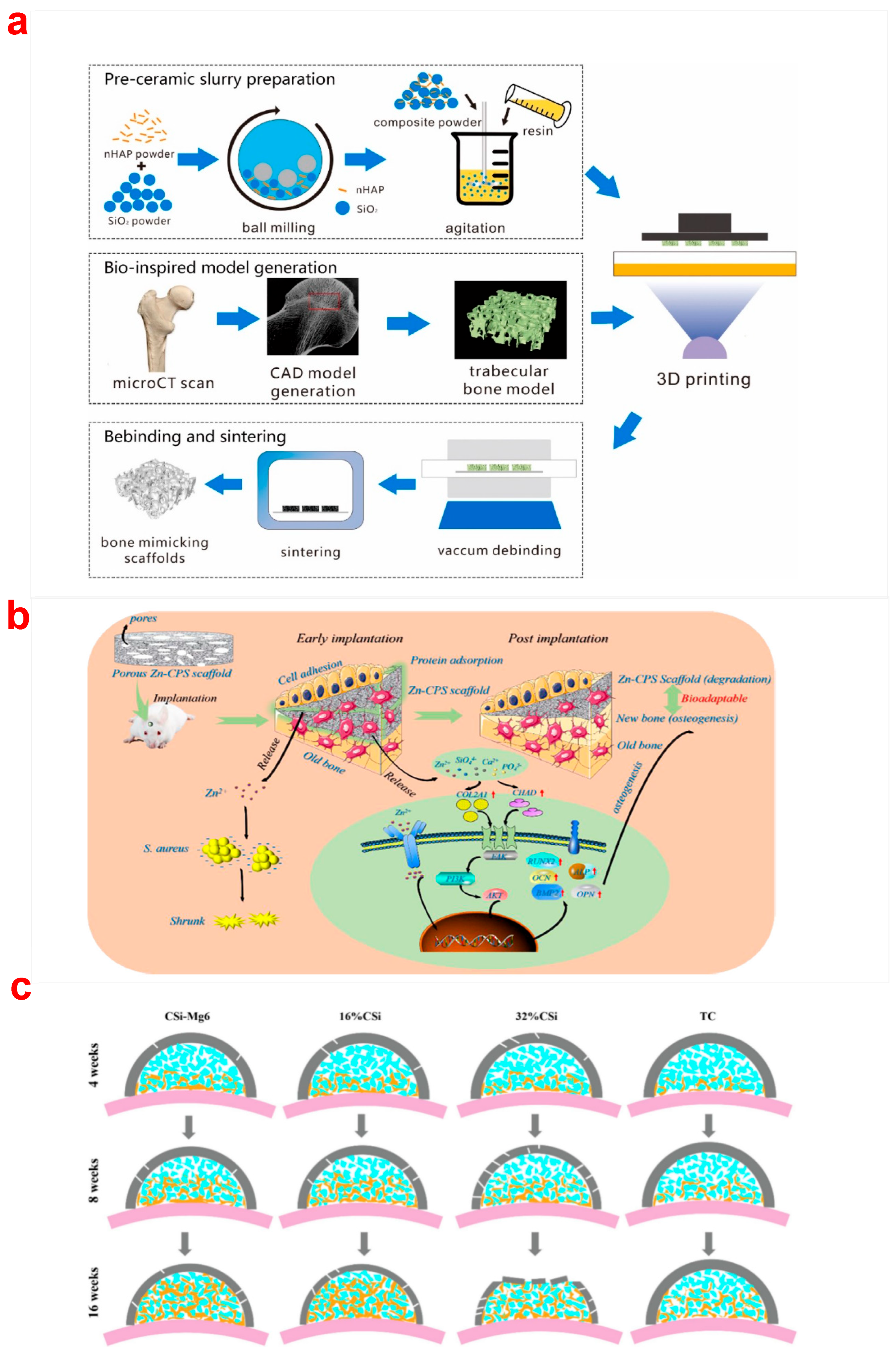
  58. Zhang, B.; Xing, F.; Chen, L.; Zhou, C.; Gui, X.; Su, Z.; Fan, S.; Zhou, Z.; Jiang, Q.; Zhao, L. DLP fabrication of customized porous bioceramics with osteoinduction ability for remote isolation bone regeneration. Mat. Sci. Eng. C-Mater. 2023, 145, 213261. [Google Scholar] [CrossRef]
  59. Liu, W.; Wang, T.; Zhao, X.; Dan, X.; William, W.; Pan, H. Akermanite used as an alkaline biodegradable implants for the treatment of osteoporotic bone defect. Bioact. Mater. 2016, 1, 151–159. [Google Scholar] [CrossRef]
  60. Liu, Q.; Lian, C.; Shuo, Y.; Chen, L.; Liu, G.; Chang, J.; Cuia, L. A comparative study of proliferation and osteogenic differentiation of adipose-derived stem cells on akermanite and β-TCP ceramics. Biomaterials 2008, 29, 4792–4799. [Google Scholar] [CrossRef]
  61. Pan, M.-Z.; Hua, S.-B.; Wu, J.-M.; Su, J.-J.; Zhang, X.-Y.; Shi, Y.-S. Composite bioceramic scaffolds with controllable photothermal performance fabricated by digital light processing and vacuum infiltration. Eur. Ceram. Soc. 2023, 43, 2245–2252. [Google Scholar] [CrossRef]
  62. Zhang, C.; Yuan, Y.; Zeng, Y.; Chen, J. DLP 3D printed silica-doped HAp ceramic scaffolds inspired by the trabecular bone structure. Ceram. Int. 2022, 48, 27765–27773. [Google Scholar] [CrossRef]
  63. Zhang, H.; Jiao, C.; He, Z.; Ge, M.; Tian, Z.; Wang, C.; Wei, Z.; Shen, L.; Liang, H. Fabrication and properties of 3D printed zirconia scaffold coated with calcium silicate/hydroxyapatite. Ceram. Int. 2021, 47, 27032–27041. [Google Scholar] [CrossRef]
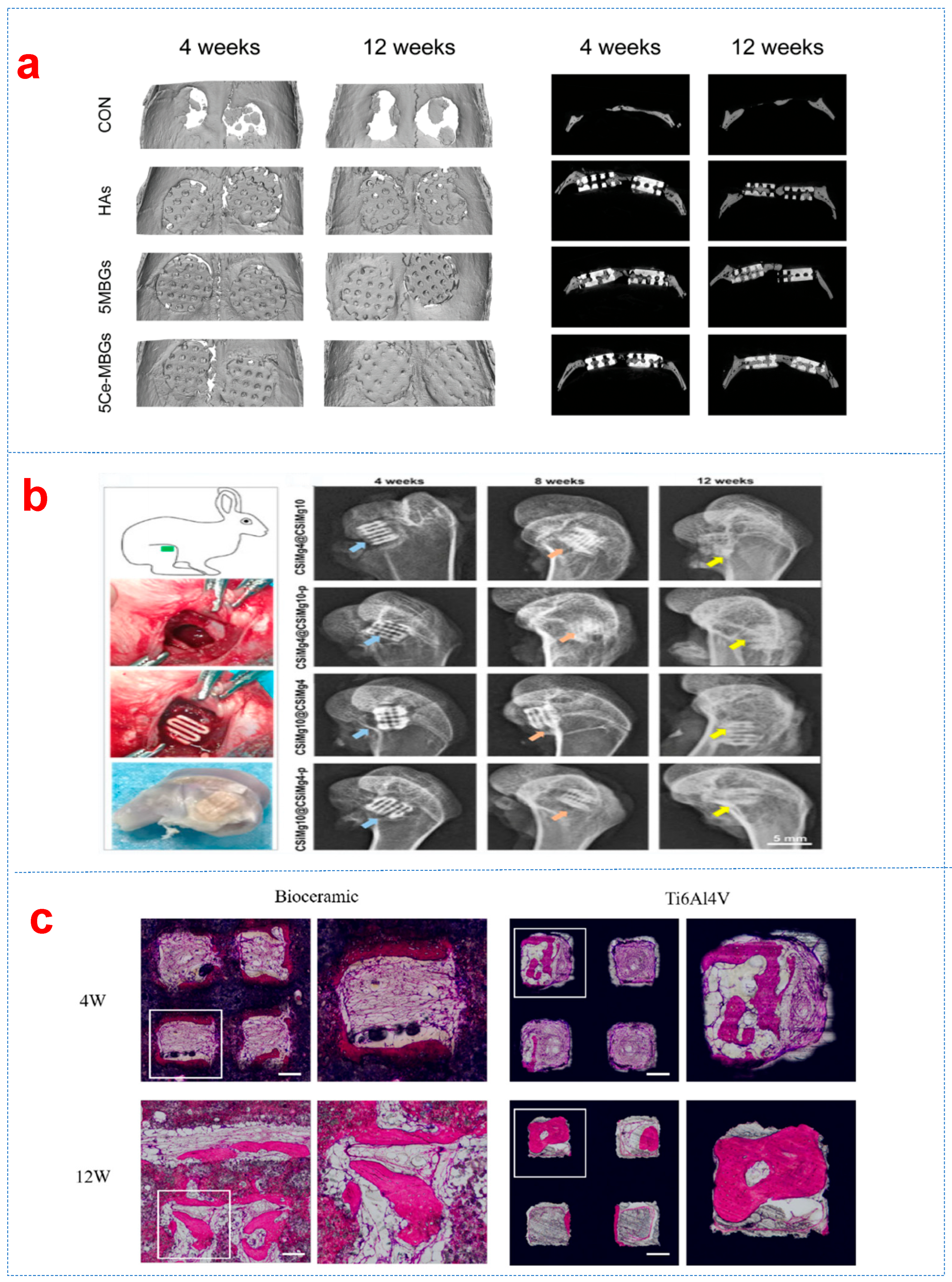
  64. Deng, F.; Bu, Z.; Hu, H.; Huang, X.; Liu, Z.; Ning, C. Bioadaptable bone regeneration of Zn-containing silicocarnotite bioceramics with moderate biodegradation and antibacterial activity. Appl. Mater. Today. 2022, 27, 101433. [Google Scholar] [CrossRef]
  65. Xu, C.; Wu, F.; Yang, J.; Wang, H.; Jiang, J.; Bao, Z.; Yang, X.; Yang, G.; Gou, Z.; He, F. 3D printed long-term structurally stable bioceramic dome scaffolds with controllable biodegradation favorable for guided bone regeneration. Chem. Eng. J. 2022, 450, 138003. [Google Scholar] [CrossRef]
  66. Jodati, H.; Evis, Z.; Tezcaner, A.; Alshemary, A.Z.; Motameni, A. 3D porous bioceramic based boron-doped hydroxyapatite/baghdadite composite scaffolds for bone tissue engineering. J. Mech. Behav. Biomed. Mater. 2023, 140, 105722. [Google Scholar] [CrossRef]
  67. Komissarenko, D.; Roland, S.; Seeber, B.S.; Graule, T.; Blugan, G. DLP 3D printing of high strength semi-translucent zirconia ceramics with relatively low-loaded UV-curable formulations. Ceram. Int. 2023, 49, 21008–21016. [Google Scholar] [CrossRef]
  68. Chen, F.; Zhu, H.; Wu, J.-M.; Chen, S.; Cheng, L.-J.; Shi, Y.-S.; Mo, Y.-C.; Li, C.-H.; Xiao, J. Preparation and biological evaluation of ZrO2 all-ceramic teeth by DLP technology. Ceram. Int. 2020, 46, 11268–11274. [Google Scholar] [CrossRef]
  69. Zhang, F.; Li, L.-f.; Wang, E.-z. Effect of micro-alumina content on mechanical properties of Al2O3/3Y-TZP composites. Ceram. Int. 2015, 41, 12417–12425. [Google Scholar] [CrossRef]
  70. Zhao, H.; Xing, H.; Lai, Q.; Zhao, Y.; Chen, Q.; Zou, B. Additive manufacturing of graphene oxide/hydroxyapatite bioceramic scaffolds with reinforced osteoinductivity based on digital light processing technology. Mater. Des. 2022, 223, 111231. [Google Scholar] [CrossRef]
  71. Hua, S.-B.; Yuan, X.; Wu, J.-M.; Su, J.; Cheng, L.-J.; Zheng, W.; Pan, M.-Z.; Xiao, J.; Shi, Y.-S. Digital light processing porous TPMS structural HA & akermanite bioceramics with optimized performance for cancellous bone repair. Ceram. Int. 2022, 48, 3020–3029. [Google Scholar] [CrossRef]
  72. Moritz, T.; Maleksaeedi, S. Additive manufacturing of ceramic components. In Additive Manufacturing—Materials, Processes, Quantifications and Applications; Elsevier: Amsterdam, The Netherlands, 2018; pp. 105–161. [Google Scholar]
  73. Hundley, J.M.; Eckel, Z.C.; Schueller, E.; Cante, K.; Biesboer, S.M.; Yahata, B.D.; Schaedler, T.A. Geometric characterization of additively manufactured polymer derived ceramics. Addit. Manuf. 2017, 18, 95–102. [Google Scholar] [CrossRef]
  74. Liu, K.; Wu, X.; Liu, J.; Yang, H.; Li, M.; Qiu, T.; Dai, H. Design and manufacture of a customized, large-size and high-strength bioactive HA osteoid composite ceramic by stereolithography. Ceram. Int. 2023, 49, 11630–11640. [Google Scholar] [CrossRef]
  75. Gauvin, R.; Chen, Y.-C.; Lee, J.W.; Soman, P.; Zorlutuna, P.; Nichol, J.W.; Bae, H.; Chen, S.; Khademhosseini, A. Microfabrication of complex porous tissue engineering scaffolds using 3D projection stereolithography. Biomaterials 2012, 33, 3824–3834. [Google Scholar] [CrossRef] [PubMed]
  76. Chen, F.; Wu, Y.-R.; Wu, J.-M.; Zhu, H.; Chen, S.; Hua, S.-B.; He, Z.-X.; Liu, C.-Y.; Xiao, J.; Shi, Y.-S. Preparation and characterization of ZrO2-Al2O3 bioceramics by stereolithography technology for dental restorations. Addit. Manuf. 2021, 44, 102055. [Google Scholar] [CrossRef]
  77. Wu, H.; Liu, W.; Huang, R.; He, R.; Huang, M.; An, D.; Li, H.; Jiang, Q.; Tian, Z.; Ji, X. Fabrication of high-performance Al2O3-ZrO2 composite by a novel approach that integrates stereolithography-based 3D printing and liquid precursor infiltration. Mater. Chem. Phys. 2018, 209, 31–37. [Google Scholar] [CrossRef]
  78. Coppola, B.; Lacondemine, T.; Tardivat, C.; Montanaro, L.; Palmero, P. Designing alumina-zirconia composites by DLP-based stereolithography: Microstructural tailoring and mechanical performances. Ceram. Int. 2021, 47, 13457–13468. [Google Scholar] [CrossRef]
  79. Lu, F.; Wu, R.; Shen, M.; Xie, L.; Liu, M.; Li, Y.; Xu, S.; Wan, L.; Yang, X.; Gao, C. Rational design of bioceramic scaffolds with tuning pore geometry by stereolithography: Microstructure evaluation and mechanical evolution. Eur. Ceram. Soc. 2021, 41, 1672–1682. [Google Scholar] [CrossRef]
  80. Raghavendra, S.S.; Jadhav, G.R.; Gathani, K.M.; Kotadia, P. Bioceramics in endodontics—A review. J. Istanb. Univ. Fac. Dent. 2017, 51, S128–S137. [Google Scholar] [CrossRef] [PubMed]
  81. Zhang, Y.; Zhang, Q.; He, F.; Zuo, F.; Shi, X. Fabrication of cancellous-bone-mimicking β-tricalcium phosphate bioceramic scaffolds with tunable architecture and mechanical strength by stereolithography 3D printing. Eur. Ceram. Soc. 2022, 42, 6713–6720. [Google Scholar] [CrossRef]
  82. Liu, R.; Ma, L.; Liu, H.; Xu, B.; Feng, C.; He, R. Effects of pore size on the mechanical and biological properties of stereolithographic 3D printed HAp bioceramic scaffold. Ceram. Int. 2021, 47, 28924–28931. [Google Scholar] [CrossRef]
  83. Dong, D.; Su, H.; Li, X.; Fan, G.; Zhao, D.; Shen, Z.; Liu, Y.; Guo, Y.; Yang, C.; Liu, L. Microstructures and mechanical properties of biphasic calcium phosphate bioceramics fabricated by SLA 3D printing. J. Manuf. Process. 2022, 81, 433–443. [Google Scholar] [CrossRef]
  84. Witek, L.; Smay, J.; Silva, N.R.; Guda, T.; Ong, J.L.; Coelho, P.G. Sintering effects on chemical and physical properties of bioactive ceramics. J. Adv. Ceram. 2013, 2, 274–284. [Google Scholar] [CrossRef]
  85. Le Nihouannen, D.; Duval, L.; Lecomte, A.; Julien, M.; Guicheux, J.; Daculsi, G.; Layrolle, P. Interactions of total bone marrow cells with increasing quantities of macroporous calcium phosphate ceramic granules. J. Mater. Sci. Mater. Med. 2007, 18, 1983–1990. [Google Scholar] [CrossRef]
  86. Wu, S.-C.; Hsu, H.-C.; Hsu, S.-K.; Wang, W.-H.; Ho, W.-F. Preparation and characterization of four different compositions of calcium phosphate scaffolds for bone tissue engineering. Mater. Charact. 2011, 62, 526–534. [Google Scholar] [CrossRef]
  87. Melchels, F.P.; Feijen, J.; Grijpma, D.W. A review on stereolithography and its applications in biomedical engineering. Biomaterials 2010, 31, 6121–6130. [Google Scholar] [CrossRef]
  88. Zhao, L.; Jiang, Z.; Zhang, C.; Guo, W. Regulation of residual stress in stereolithography printing of ZrO2 ceramics. Ceram. Int. 2023, 49, 30801–30810. [Google Scholar] [CrossRef]
  89. Wang, H.; Shen, F.; Li, Z.; Zhou, B.; Zhao, P.; Wang, W.; Cheng, B.; Yang, J.; Li, B.; Wang, X. Preparation of high-performance ZrO2 bio-ceramics by stereolithography for dental restorations. Ceram. Int. 2023, 49, 28048–28061. [Google Scholar] [CrossRef]
  90. Molitch-Hou, M. Overview of additive manufacturing process. In Additive Manufacturing—Materials, Processes, Quantifications and Applications; Elsevier: Amsterdam, The Netherlands, 2018; pp. 1–38. [Google Scholar]
  91. Sun, S.; Fei, G.; Wang, X.; Xie, M.; Guo, Q.; Fu, D.; Wang, Z.; Wang, H.; Luo, G.; Xia, H. Covalent adaptable networks of polydimethylsiloxane elastomer for selective laser sintering 3D printing. Chem. Eng. J. 2021, 412, 128675. [Google Scholar] [CrossRef]
  92. Han, J.; Wu, J.; Xiang, X.; Xie, L.; Chen, R.; Li, L.; Ma, K.; Sun, Q.; Yang, R.; Huang, T. Biodegradable BBG/PCL composite scaffolds fabricated by selective laser sintering for directed regeneration of critical-sized bone defects. Mater. Des. 2023, 225, 111543. [Google Scholar] [CrossRef]
  93. Shuai, C.; Li, P.; Liu, J.; Peng, S. Optimization of TCP/HAP ratio for better properties of calcium phosphate scaffold via selective laser sintering. Mater. Charact. 2013, 77, 23–31. [Google Scholar] [CrossRef]
  94. Tang, H.-H.; Chiu, M.-L.; Yen, H.-C. Slurry-based selective laser sintering of polymer-coated ceramic powders to fabricate high strength alumina parts. Eur. Ceram. Soc. 2011, 31, 1383–1388. [Google Scholar] [CrossRef]
  95. Shuai, C.; Nie, Y.; Gao, C.; Feng, P.; Zhuang, J.; Zhou, Y.; Peng, S. The microstructure evolution of nanohydroxapatite powder sintered for bone tissue engineering. J. Exp. Nanosci. 2013, 8, 762–773. [Google Scholar] [CrossRef]
  96. Liu, H.; Su, H.; Shen, Z.; Wang, E.; Zhao, D.; Guo, M.; Zhang, J.; Liu, L.; Fu, H. Direct formation of Al2O3/GdAlO3/ZrO2 ternary eutectic ceramics by selective laser melting: Microstructure evolutions. Eur. Ceram. Soc. 2018, 38, 5144–5152. [Google Scholar] [CrossRef]
  97. Huang, Z.; Tang, Y.; Guo, H.; Feng, X.; Zhang, T.; Li, P.; Qian, B.; Xie, Y. 3D printing of ceramics and graphene circuits-on-ceramics by thermal bubble inkjet technology and high temperature sintering. Ceram. Int. 2020, 46, 10096–10104. [Google Scholar] [CrossRef]
  98. Woo Lee, J.; Cho, D.-W. 3D printing technology over a drug delivery for tissue engineering. Curr. Pharm. Des. 2015, 21, 1606–1617. [Google Scholar] [CrossRef] [PubMed]
  99. Vaezi, M.; Seitz, H.; Yang, S. A review on 3D micro-additive manufacturing technologies. Int. J. Adv. Manuf. Technol. 2013, 67, 1721–1754. [Google Scholar] [CrossRef]
  100. Siamak, E.; Azadeh, M.; Pedro, M.; Antonia, P.; Alexandra, L.; José, M.F. Robocasting of 45S5 bioactive glass scaffolds for bone tissue engineering. J. Eur. Ceram. Soc. 2014, 34, 107–118. [Google Scholar] [CrossRef]
  101. Liu, X.; Miao, Y.; Liang, H.; Diao, J.; Hao, L.; Shi, Z.; Zhao, N.; Wang, Y. 3D-printed bioactive ceramic scaffolds with biomimetic micro/nano-HAp surfaces mediated cell fate and promoted bone augmentation of the bone–implant interface in vivo. Bioact. Mater. 2022, 12, 120–132. [Google Scholar] [CrossRef]
  102. Shao, H.; Shi, J.; Huang, Z.; Yang, W.; Wang, H. 3D-Printed Bioceramic Scaffolds with High Strength and High Precision. Crystals 2023, 13, 1061. [Google Scholar] [CrossRef]
  103. Luo, D.; Su, J.; Zou, Y.; Hua, S.; Cheng, L.; Qi, D.; Yuan, X.; Zhu, H.; Liu, C.; Shi, Y.; et al. 3D-printed triply periodic minimal surface bioceramic scaffold for bone defect treatment with tunable structure and mechanical properties. Ceram. Int. 2024, 50, 39020–39031. [Google Scholar] [CrossRef]
  104. Li, Q.; An, X.; Liang, J.; Liu, Y.; Hu, K.; Lu, Z.; Yue, X.; Li, J.; Zhou, Y.; Sun, X. Balancing flexural strength and porosity in DLP-3D printing Al2O3 cores for hollow turbine blades. J. Mater. Sci. Technol. 2022, 104, 19–32. [Google Scholar] [CrossRef]
  105. Shan, Y.; Bai, Y.; Yang, S.; Zhou, Q.; Wang, G.; Zhu, B.; Zhou, Y.; Fang, W.; Wen, N.; He, R.; et al. 3D-printed strontium-incorporated β-TCP bioceramic triply periodic minimal surface scaffolds with simultaneous high porosity, enhanced strength, and excellent bioactivity. J. Adv. Ceram. 2023, 12, 1671–1684. [Google Scholar] [CrossRef]
  106. Hua, S.; Su, J.; Deng, Z.; Wu, J.; Cheng, L.; Yuan, X.; Chen, F.; Zhu, H.; Qi, D.; Xiao, X.; et al. Microstructures and properties of 45S5 bioglass® & BCP bioceramic scaffolds fabricated by digital light processing. Addit. Manuf. 2021, 45, 102074. [Google Scholar] [CrossRef]
  107. Dmytro, S. Simulation of the selective laser sintering/melting process of bioactive glass 45S5. Simul. Model. Pract. Theory 2024, 136, 103009. [Google Scholar] [CrossRef]
  108. Nikhil, K.; Miguel, A.R.; Ramin, R.; Konda, G.; Irina, H. Bioceramic scaffolds by additive manufacturing for controlled delivery of the antibiotic vancomycin. Proc. Natl. Acad. Sci. USA 2019, 68, 185–190. [Google Scholar] [CrossRef]
  109. Kim, C.G.; Han, K.S.; Lee, S.; Kim, M.C.; Kim, S.Y.; Nah, J. Fabrication of Biocompatible Polycaprolactone–Hydroxyapatite Composite Filaments for the FDM 3D Printing of Bone Scaffolds. Appl. Sci. 2021, 11, 6351. [Google Scholar] [CrossRef]
  110. Carola, E.C.; Francesca, S.; Francesca, G.; Francesco, M.; Alessandro, S.; Alfonso, M. One-step solvent-free process for the fabrication of high loaded PLA/HA composite filament for 3D printing. J. Therm. Anal. 2018, 134, 575–582. [Google Scholar] [CrossRef]
  111. Bruyas, A.; Lou, F.; Stahl, A.M.; Gardner, M.; Maloney, W.; Goodman, S.; Yang, Y.P. Systematic characterization of 3D-printed PCL/β-TCP scaffolds for biomedical devices and bone tissue engineering: Influence of composition and porosity. J. Mater. Res. 2018, 33, 1948–1959. [Google Scholar] [CrossRef]
  112. Drummer, D.; Cifuentes-Cuéllar, S.; Rietzel, D. Suitability of PLA/TCP for fused deposition modeling. Rapid Prototyp. J. 2012, 18, 500–507. [Google Scholar] [CrossRef]
  113. Krishna, C.R.K.; Ming, C.L.; Gregory, E.H.; Mariano, V. Effect of material, process parameters, and simulated body fluids on mechanical properties of 13-93 bioactive glass porous constructs made by selective laser sintering. J. Mech. Behav. Biomed. 2012, 13, 14–24. [Google Scholar] [CrossRef]
  114. Liu, J.; Gao, C.; Feng, P.; Peng, S.; Shuai, C. Selective laser sintering of β-TCP/nano-58S composite scaffolds with improved mechanical properties. Mater. Des. 2015, 84, 395–401. [Google Scholar] [CrossRef]
  115. Gao, C.; Yang, B.; Hu, H.; Liu, J.; Shuai, C.; Peng, S. Enhanced sintering ability of biphasic calcium phosphate by polymers used for bone scaffold fabrication. Mater. Sci. Eng. 2013, 33, 53491–53501. [Google Scholar] [CrossRef] [PubMed]
  116. Passakorn, T.; Ruth, F.; Simon, G.; Robert, L.; Ian, T.; Aldo, R.B.; Jürgen, S. Processing of 45S5 Bioglass® by lithography-based additive manufacturing. Mater. Lett. 2012, 74, 81–84. [Google Scholar] [CrossRef]
  117. Chugunov, S.; Adams, N.A.; Akhatov, I. Evolution of SLA-Based Al2O3 Microstructure During Additive Manufacturing Process. Materials 2020, 13, 3928. [Google Scholar] [CrossRef]
  118. Liu, X.; Zou, B.; Xing, H.; Huang, C. The preparation of ZrO2-Al2O3 composite ceramic by SLA-3D printing and sintering processing. Ceram. Int. 2020, 46, 937–944. [Google Scholar] [CrossRef]
  119. Ahmad, M.; Rodziah, A. HA-SLA: A Hierarchical Autonomic SLA Model for SLA Monitoring in Cloud Computing. J. Softw. Eng. Appl. 2013, 6, 114–117. [Google Scholar] [CrossRef]
  120. Zhang, Y.; Tan, S.; Yin, Y. C-fibre reinforced hydroxyapatite bioceramics. Ceram. Int. 2003, 29, 113–116. [Google Scholar] [CrossRef]
  121. Mei, H.; Yan, Y.; Feng, L.; Dassios, K.G.; Zhang, H.; Cheng, L. First printing of continuous fibers into ceramics. J. Am. Ceram. Soc. 2019, 102, 3244–3255. [Google Scholar] [CrossRef]
  122. Zhao, X.; Liu, A.; Zhou, L.; Yang, Z.; Wei, S.; Zhao, Z.; Fan, Q.; Ma, L. 3D printing of bioactive macro/microporous continuous carbon fibre reinforced hydroxyapatite composite scaffolds with synchronously enhanced strength and toughness. Eur. Ceram. Soc. 2022, 42, 4396–4409. [Google Scholar] [CrossRef]
  123. Inagaki, M.; Kameyama, T. Phase transformation of plasma-sprayed hydroxyapatite coating with preferred crystalline orientation. Biomaterials 2007, 28, 2923–2931. [Google Scholar] [CrossRef]
  124. Lacroix, J.; Jallot, E.; Lao, J. Gelatin-bioactive glass composites scaffolds with controlled macroporosity. Chem. Eng. J. 2014, 256, 9–13. [Google Scholar] [CrossRef]
  125. Wang, X.; Zhang, L.; Ke, X.; Wang, J.; Yang, G.; Yang, X.; He, D.; Shao, H.; He, Y.; Fu, J. 45S5 Bioglass analogue reinforced akermanite ceramic favorable for additive manufacturing mechanically strong scaffolds. RSC Adv. 2015, 5, 102727–102735. [Google Scholar] [CrossRef]
  126. Rainer, A.; Giannitelli, S.M.; Abbruzzese, F.; Traversa, E.; Licoccia, S.; Trombetta, M. Fabrication of bioactive glass–ceramic foams mimicking human bone portions for regenerative medicine. Acta Biomater. 2008, 4, 362–369. [Google Scholar] [CrossRef]
  127. Leach, J.K.; Kaigler, D.; Wang, Z.; Krebsbach, P.H.; Mooney, D.J. Coating of VEGF-releasing scaffolds with bioactive glass for angiogenesis and bone regeneration. Biomaterials 2006, 27, 3249–3255. [Google Scholar] [CrossRef] [PubMed]
  128. Hernandez-Tenorio, F.; Giraldo-Estrada, C. Characterization and chemical modification of pullulan produced from a submerged culture of Aureobasidium pullulans ATCC 15233. Polym. Test. 2022, 114, 107686. [Google Scholar] [CrossRef]
  129. Simorgh, S.; Alasvand, N.; Khodadadi, M.; Ghobadi, F.; Kebria, M.M.; Milan, P.B.; Kargozar, S.; Baino, F.; Mobasheri, A.; Mozafari, M. Additive manufacturing of bioactive glass biomaterials. Methods 2022, 208, 75–91. [Google Scholar] [CrossRef]
  130. Bose, S.; Bhattacharjee, A.; Banerjee, D.; Boccaccini, A.R.; Bandyopadhyay, A. Influence of random and designed porosities on 3D printed tricalcium phosphate-bioactive glass scaffolds. Addit. Manuf. 2021, 40, 101895. [Google Scholar] [CrossRef] [PubMed]
  131. Kolan, K.C.R.; Huang, Y.-W.; Semon, J.A.; Leu, M.C. 3D-printed biomimetic bioactive glass scaffolds for bone regeneration in rat calvarial defects. IJB 2020, 6, 274. [Google Scholar] [CrossRef]
  132. Liu, J.; Hu, H.; Li, P.; Shuai, C.; Peng, S. Fabrication and characterization of porous 45S5 glass scaffolds via direct selective laser sintering. Mater. Manuf. Process. 2013, 28, 610–615. [Google Scholar] [CrossRef]
  133. Li, X.; Zhang, H.; Shen, Y.; Xiong, Y.; Dong, L.; Zheng, J.; Zhao, S. Fabrication of porous β-TCP/58S bioglass scaffolds via top-down DLP printing with high solid loading ceramic-resin slurry. Mater. Chem. Phys. 2021, 267, 124587. [Google Scholar] [CrossRef]
  134. Xia, X.; Yang, Y.; Hou, Z.; Cao, Y.; Dai, W.; Li, B.; Dong, T.; SAAD, O.; Ling, S.; Xue, W. Enhanced Osteogenic Potential of Magnesium Oxide-Doped Biphasic Calcium Phosphate Bioceramics Prepared by Stereolithography. Ceram. Int. 2025, 51, 17955–17967. [Google Scholar] [CrossRef]
  135. Zhao, Z.; Chen, X.; Chen, H.; Zhang, J.; Du, L.; Jiang, F.; Zheng, K. Cerium-Containing Mesoporous Bioactive Glass Nanoparticles Reinforced 3D-Printed Bioceramic Scaffolds toward Enhanced Mechanical, Antioxidant, and Osteogenic Activities for Bone Regeneration. Adv. Healthc. Mater. 2025, 14, 2404346. [Google Scholar] [CrossRef] [PubMed]
  136. Chen, Y.; Huang, J.; Liu, J.; Wei, Y.; Yang, X.; Lei, L.; Chen, L.; Wu, Y.; Gou, Z. Tuning filament composition and microstructure of 3D-printed bioceramic scaffolds facilitate bone defect regeneration and repair. Regen. Biomater. 2021, 8, rbab007. [Google Scholar] [CrossRef] [PubMed]
  137. Zhang, H.; Darvell, B.W. Synthesis and characterization of hydroxyapatite whiskers by hydrothermal homogeneous precipitation using acetamide. Acta Biomater. 2010, 6, 3216–3222. [Google Scholar] [CrossRef] [PubMed]
  138. Wu, H.; Gao, M.; Zhu, D.; Zhang, S.; Pan, Y.; Pan, H.; Liu, Y.; Oliveira, F.J.; Vieira, J.M. SiC whisker reinforced multi-carbides composites prepared from B4C and pyrolyzed rice husks via reactive infiltration. Ceram. Int. 2012, 38, 3519–3527. [Google Scholar] [CrossRef]
  139. Tanimoto, Y.; Teshima, M.; Nishiyama, N.; Yamaguchi, M.; Hirayama, S.; Shibata, Y.; Miyazaki, T. Tape-cast and sintered β-tricalcium phosphate laminates for biomedical applications: Effect of milled Al2O3 fiber additives on microstructural and mechanical properties. J. Biomed. Mater. Res. B 2012, 100, 2261–2268. [Google Scholar] [CrossRef]
  140. Ma, C.; Zeng, Q.; Yu, L.; Yu, S.; Song, J.; Ma, Y.; Dong, X. Preparation and characterization of 3D printed hydroxyapatite-whisker-strengthened hydroxyapatite scaffold coated with biphasic calcium phosphate. Chin. J. Mech. Eng. 2023, 2, 100097. [Google Scholar] [CrossRef]
  141. Feng, P.; Wei, P.; Li, P.; Gao, C.; Shuai, C.; Peng, S. Calcium silicate ceramic scaffolds toughened with hydroxyapatite whiskers for bone tissue engineering. Mater. Charact. 2014, 97, 47–56. [Google Scholar] [CrossRef]
  142. Wu, C.; Ramaswamy, Y.; Boughton, P.; Zreiqat, H. Improvement of mechanical and biological properties of porous CaSiO3 scaffolds by poly (D, L-lactic acid) modification. Acta Biomater. 2008, 4, 343–353. [Google Scholar] [CrossRef]
  143. Farhadi, K.; Namini, A.S.; Asl, M.S.; Mohammadzadeh, A.; Kakroudi, M.G. Characterization of hot pressed SiC whisker reinforced TiB2 based composites. Int. J. Refract. Met. Hard Mater. 2016, 61, 84–90. [Google Scholar] [CrossRef]
  144. Zhao, X.; Yang, J.; Xin, H.; Wang, X.; Zhang, L.; He, F.; Liu, Q.; Zhang, W. Improved dispersion of SiC whisker in nano hydroxyapatite and effect of atmospheres on sintering of the SiC whisker reinforced nano hydroxyapatite composites. Mater. Sci. Eng. C 2018, 91, 135–145. [Google Scholar] [CrossRef] [PubMed]
  145. Xing, H.; Zou, B.; Wang, X.; Hu, Y.; Huang, C.; Xue, K. Fabrication and characterization of SiC whiskers toughened Al2O3 paste for stereolithography 3D printing applications. J. Alloys Compd. 2020, 828, 154347. [Google Scholar] [CrossRef]
  146. Li, C.; Zhang, W.; Nie, Y.; Du, X.; Huang, C.; Li, L.; Long, J.; Wang, X.; Tong, W.; Qin, L. Time-sequential and multi-functional 3D printed MgO2/PLGA scaffold developed as a novel biodegradable and bioactive bone substitute for challenging postsurgical osteosarcoma treatment. Adv. Mater. 2024, 36, 2308875. [Google Scholar] [CrossRef]
  147. Wang, S.; Huang, Z.; Liu, L.; Shi, Z.a.; Liu, J.; Li, Z.; Hao, Y. Design and study of in vivo bone formation characteristics of biodegradable bioceramic. Mater. Des. 2021, 212, 110242. [Google Scholar] [CrossRef]
  148. Qi, H.; Zhang, B.; Lian, F. 3D-printed bioceramic scaffolds for bone defect repair: Bone aging and immune regulation. Front. Bioeng. Biotechnol. 2025, 13, 1557203. [Google Scholar] [CrossRef]
  149. Zhao, X.; Zhang, Y.; Wang, P.; Guan, J.; Zhang, D. Construction of multileveled and oriented micro/nano channels in Mg doped hydroxyapitite bioceramics and their effect on mimicking mechanical property of cortical bone and biological performance of cancellous bone. Biomater. Adv. 2024, 161, 213871. [Google Scholar] [CrossRef]
  150. Filion, T.M.; Li, X.; Mason-Savas, A.; Kreider, J.M.; Goldstein, S.A.; Ayers, D.C.; Song, J. Elastomeric osteoconductive synthetic scaffolds with acquired osteoinductivity expedite the repair of critical femoral defects in rats. Tissue Eng. Part A 2011, 17, 503–511. [Google Scholar] [CrossRef]
Figure 4. (a) The SLA process of an osteoid ceramic, (b) the stereolithography technique [72], (c) an osteoid HA bioceramic of large segment with different orientation views, (d) a customized HA mandible [74], and (e) Al2O3-ZrO2 dental implants with SLA [76].
Biomimetics 10 00428 g004
Table 1. Advantages and disadvantages for 3D printing of bioceramics.
3D Printing MethodsAdvantagesDisadvantagesMaterials
Direct Ink Writing (DIW)Suitable for high solids slurries;
Flexible structural design;
Low cost
Slow printing speed;
The rheological properties of the slurry are required
Bioactive glass [100]
HAp [101]
CSi-Mg [102]
β-TCP [27]
Digital Light Processing (DLP)Fast printing speed;
High precision;
Add photosensitive resin;
Limited materials; Post-processing is required
HA [103]
Al2O3 [104]
ZrO2 [68]
β-TCP [105]
Bioactive glass [106]
Selective Laser Melting (SLM)High molding density;
Excellent mechanical properties
Expensive equipment;
Only suitable for metals or ceramics with high melting points
Bioactive glass [107]
CaO3Si [108]
Fused Deposition Manufacturing (FDM)Equipment of low cost;
Simple operation;
Multi-material printing
Low accuracy;
Weak inter-layer adhesion
PCL/HA [109]
PLA/HA [110]
PCL/β-TCP [111]
PLA/β-TCP [112]
Selective Laser Sintering (SLS)No support structure;
Suitable for porous and complex geometries;
High utilization
The high surface roughness; Post-treatment; High-temperature sintering lead to grain coarseningBioactive glass [113]
β-TCP [114]
HA/β-TCP [115]
Stereolithography (SLA)High resolution;
Suitable for fine structures
High cost;
Material selection is restricted;
Post-processing is required
Bioactive glass [116]
Al2O3 [117]
ZrO2 [118]
HA [119]
β-TCP [82]
Disclaimer/Publisher’s Note: The statements, opinions and data contained in all publications are solely those of the individual author(s) and contributor(s) and not of MDPI and/or the editor(s). MDPI and/or the editor(s) disclaim responsibility for any injury to people or property resulting from any ideas, methods, instructions or products referred to in the content.

