Review

Allergic Food Sensitization and Disease Manifestation in the Fetus and Infant: A Perspective

by
Russell J. Hopp
1,*,
Peggy Salazar
2 and
Muhammad Asghar Pasha
2
1
Department of Pediatrics, University of Nebraska Medical Center and Children’s Hospital and Medical Center, Omaha, NE 68114, USA
2
Division of Allergy and Immunology, Albany Medical College, 176 Washington Avenue Extension, Suite 102, Albany, NY 12203, USA
*
Author to whom correspondence should be addressed.
Allergies 2021, 1(2), 115-122; https://doi.org/10.3390/allergies1020009
Submission received: 17 March 2021 / Revised: 15 April 2021 / Accepted: 26 April 2021 / Published: 11 May 2021
(This article belongs to the Section Food Allergy)

Abstract

:
Even though allergic disease is identified in the first year of life, it is often in a less forward fashion, with elements of a wait and see approach. If the infant does not have an anaphylactic food reaction, other less dramatic allergic phenomenon is often under-emphasized, waiting for additional concerns. We approached this with a conception to first conduct birthday surveys, attempting to link intrauterine and peri-birth circumstances to affect better allergy recognition in young infants.

1. Introduction

The conceived fetus has many promising outcomes, but becoming allergically sensitized, and to have an allergic disease develop within the first year of birth, is an unwelcome outcome. A recent manuscript thoroughly discusses food allergic anaphylactic reactions in infants, and the clinical management during and after [1]. We review here the information on how that infant was set up to have a food allergic reaction, including intrauterine immunology, intrauterine IgE sensitization, the presence of IgE sensitization at birth, breast feeding sensitization, transcutaneous sensitization, and the allergic diseases that typically result from food allergic sensitization, including anaphylaxis, within the first year.

2. Genetic and Epigenetic Potentials

In the broad sense, the immediate family presence of an allergic disease provides familial, presumably genetic [2], pressure to have the same, or related, allergic disease eventually develop in the infant’s antenatal life. In the case of asthma, recent studies are showing strong perinatal influences [3,4,5,6,7].
Atopic dermatitis in a parent seems to being stronger atopic dermatitis potential to their child [5,8]. Allergic rhinitis and food allergy in a parent provide allergic influence on the fetus, but less direct influence for those specific diseases. The genetics of an allergic disease has had remarkable breakthroughs, and many chromosome and gene areas have been identified using population and sib-sib analyses but without a single specific gene identified for a specific disease [9]. Very likely, multiple genes contribute to each disease, along with intrauterine epigenetic influences [5,10,11,12], and intrauterine and infant post-birth environmental pressures [5,11,13,14].

3. Fetal Immunity Development and the Production of IgE

Substantial information has accumulated on fetal immunity, largely building off mouse studies [5,10,14,15,16]. A recent review of human immunology was published in Science in 2020 [17]. We recapitulated it here briefly, focusing on the components of the immune system essential for development of allergy. Macrophages and tissue mast cells appear very early in the first trimester. Early mast cells do not have the gene for the alpha unit of IgE receptors, but IgE can be detected after 11 weeks as mature B cells will appear by 9 weeks. Naïve T cells appear about 8 weeks, as does the thymic tissue. Neutrophils and eosinophils are in the bone marrow by 20 weeks. Therefore, by 20 weeks the fetus is relatively armed, and needs antigen to proceed, but in their sterile world only a few bacteria or infectious by-products, and very certainly almost no allergens can invade. The multiple cells involved in the post-uterine life allergic process are present; based on the subsequent discussion, their ability to execute an allergic response scenario appears to be well dampened until after birth.
The TH2 (T2) immune system is the critical component of the newborn through adult life that drives the atopic response and contributes to the development of an allergic disease [18]. TH2 lymphocytes are characterized by the prostaglandin D2 receptor CRTH2 [11]. In the fetus, there is a skewing of the lymphocyte (CD4+) population to the TH2 phenotype [11,12]. The neonate, then, in the right (wrong) exposure environment has the functional elements to produce an intrauterine IgE response. Maternal IgG crosses the placenta, while IgE is (theoretically) an excluded [11,15,19]. The fetus receives food, and potentially other foreign antigens, delivered via the placenta interface. The maternal IgG may balance the fetal response to foreign antigen, but IgE can be produced by the fetus [14]. The subsequent discussion on post-uterine allergic disease will focus on the extreme uncommonness of allergic clinical responsivity in the first months of life. This would indirectly suggest that fetal production of sufficient IgE to an allergen, food for example, is primed and ready for post-uterine production and the production and release requires the non-maternal environment and possibly the additional (first) exposure(s) to induce the first post-uterine allergic reaction.
Fetal tissue can produce IgE in the late first–early second trimester [14]. It is largely bound, or is under-represented, as a B-cell class switching potential [14,15,17,20], and not overtly released into the direct fetal circulation for mast cell/basophil/eosinophil binding. It’s likely there are brakes to control intrauterine IgE levels to not endanger the developing fetus. The mere fact that fetuses of atopic/allergic mothers or fathers don’t have increased intrauterine demise is likely in-direct proof that the level of specific IgE in infants is low enough, and the volume of allergen food is low enough, to not induce an allergic intrauterine response, and likely other protective factors, such as diminished mast cell IgE binding, are in play [17].
Cord blood total IgE is low, as is specific IgE; studies have supported some specific IgE production, although this area has remained controversial [16]. The use of umbilical cord IgE was attempted as a predictor for future atopic disease or in epidemiological studies [16,21] but has not received any recent research direction. A recent search at Clinicaltrial.gov (accessed on 3 January 2021) showed two studies of cord blood IgE in Taiwan [22]. Older studies of cord blood IgE yielded data that was associative for future allergic disease [23] or presented maternal or environmental factors that influenced cord blood IgE [24].
A pertinent discussion to the topic of intrauterine IgE sensitization, and subsequent early life allergic reaction, was data from the PASTURE study published in 2008 [11]. The most frequent IgE in cord blood for foods was milk, eggs, hazelnut, wheat, and soy [11]. A study of ex-vivo transplacental transfer from 2000 reports studies document allergen specific T-cell proliferation (implying exposure and, at least, T-cell sensitization) for alpha-casein, beta-casein, kappa-casein, bovine serum albumin, and ovalbumin [25]. A recent study in mice suggested maternal IgE may cross the placenta [26]. If true in humans, the amount and degree of specific binding to mast cells is negligible. In most fetuses and early infants, the non-bound half-life of IgE allows for quick reduction (unless the infant themselves maintain extrauterine production) and no clinical consequences.
If a fetus can develop their own food specific IgE, then the food of interest must reach the developing fetus. A review of protein sources to the infants from 1990 largely references work from the 1970s–1980s [27]. The emphasis, at that point, was amino acids. T-cells, as mentioned, recognize larger amino-acid constructs [25], but the translation to B-cell message, class-switching, and IgE production in the fetus is largely unknown.
The exact format of maternally ingested protein, that is eaten, digested, absorbed, and passed to the fetus, is largely speculative [28]. A recent interesting case report of abdominal hives after wheat ingestion specifically suggests more rapid absorption of ingested protein can be locally absorbed, and due to enhanced maternal circulation, it could be more easily passed to the fetus [29]. The exact nature of fetal specific IgE promotion lineage, whether to linear or conformational epitopes, then becomes more relevant, based on the size of the protein transferred, intact conformational or shorter amino acid linear sequences. (linear). The available literature strongly supports amino acid transfer, but virtually no processes is suggested for larger components of food or allergen transfer, although it obviously could be based, at least, on cord blood specific IgE recovery.

4. Birth and Immediate Post-Birth Allergenic Experiences

The method of birth has influence on the future development of allergies and allergic disease in children [30]. Otherwise, any swallowed maternal blood containing maternal IgE only adds to the intrauterine IgE levels the infant is born with. The infant is now either specifically IgE sensitized (in theory) or not, and life happens (Figure 1).
Several reports have provided immediate allergy epidemiological data in infants. In a United States population study of food-induced anaphylaxis in an urban hospital, birth-age 18 years, 13% of the total (n = 357) were less than the age 12 months [31]. Eggs and cow’s milk were the two most common foods. In a European study, comparing infants (<12 months) to older 1–6 years, 59 of 375 children were <12 months, with a mean age of 6 months [32]. Cow’s milk and hen’s eggs were the two most common.
Cow’s milk IgE specific anaphylaxis, generated by infant milk-based formula, is an exceptionally rare occurrence in the first several months of life. It is conceivable that a rapid shift to a non-milk formula may (disguise) lessen the chance of a severe allergic reaction, although the first replacement is often a lactose-free cow’s milk product. In breast-fed infants, with maternal ingestion of milk protein, anaphylaxis is an equally non-existent occurrence [33]. Case reports of a fish allergic reaction to maternally transferred protein have been reported, but not until 4 months or later [34,35], suggesting an immunological brake to similar reactions to other breast-transferred food proteins early in life. The presence of food protein in breast milk has been recently reviewed, with much person-to-person variability, but evidence for, at least, milk, wheat, and egg protein components are detectable [33]. The ability to actually measure the allergenic protein may not be sensitive enough.

5. Advancing Post-Uterine IgE Sensitization

If the fetus initiated the development of IgE antibodies, or was already sensitized in-utero (in theory, based on recoverable specific IgE in cord blood), then advancement of further sensitization, especially to food, will presumably depend on continued exposure, awaiting the proper moment for clinical expression (Figure 1). The other possibility is that in-utero sensitization occurred, but post-uterine internal immunological control, or a major change in post-uterine exposure un-enhances further IgE production. The third possibility, currently undertaken as a major national and international approach, is to reduce or prevent post-uterine food IgE sensitization and subsequent allergic reactions, using early introduction to all foods [36]. A comprehensive report of further recommendations of this model of primary prevention is available [36].
IgE food sensitization is enhanced or initiated by exposure post-birth predominately via two methods. One, skin contact with food allergens still not eaten; two, exposure of food antigens/allergens via breast milk. Infant skin exposure to foods is nearly universal if the food is eaten by others in the home [33,37]. Genetic predispositions to being an atopic infant was of course, pre-programmed, and exposure becomes step 2. If the allergen already had intrauterine IgE priming, the re-exposure post-birth via skin contact is likely.
Intact skin is more resistant to food absorption, and the recommendation to apply a moisturizer from birth has received some success [38]. The presence of other skin abnormalities, especially common ones, could enhance absorption. In this regard, infantile seborrheic dermatitis [39] and/or genetic decreases in (pro)filaggrin production are culprits [40].
Either with food transcutaneous absorption [41], or air-borne exposure to food substance in the airstream of the house or care facility [42] or transfer of food protein via the breast milk [33], the infant either starts the class switch process that was under-developed in-utero or rapidly begins their de-novo extra-uterine IgE production.
The relative lack of anaphylaxis in the first 2–3 months suggests a more delayed production of IgE to the point of sufficiency of inducing anaphylaxis or contributing to atopic dermatitis [43,44].

6. Food-Associated IgE Mediated Allergic Diseases in Young Infants

As previously mentioned, IgE-mediated food anaphylaxis does occur prior to 12 months [31,32], and eggs and milk are the most common foods, followed by peanuts [31]. The concept of age-specific IgE-mediated disease in the first year of life has been recently discussed, including anaphylaxis [1,18] (Figure 1).
A second presentation of food allergies is within the context of atopic dermatitis. A perplexing clinical presentation in early childhood is an admixture of seborrhea and atopic dermatitis. If the child also has xerosis cutis with lowered filaggrin levels [45], a trifecta of skin permeability exists, which often get lumped into the term eczema and has been discussed as over-lap [46], and although the author was admixing atopic dermatitis and psoriasis [46], the concept works for other combinations. The clinician needs to separate the different disease conditions and provide counseling and therapeutic intervention for each [46].
IgE-mediated food allergens can play a role in the atopic dermatitis process, as the permeable skin (with xerosis and/or seborrhea) may have allowed for further transcutaneous sensitization. If a true IgE-mediated food allergy (anaphylaxis) has not occurred, the presence of positive IgE testing results, either specific IGE levels on venous samples or percutaneous allergy tests. the clinician must face the dilemma of avoidance or continuation of feeding the specific food implicated in testing. The recent review by Rajanai et al. suggests a clinical approach for suspected food allergen in breastfed or formula fed infants with atopic dermatitis [33]. A reasonable time of avoidance may assist with improving the AD; re-introduction of a specific avoided food can resume skin itching and AD exacerbation. If a specific food is avoided, or previously eaten, an oral challenge needs to be performed if avoided for 6–12 months due to the potential development of an anaphylactic reaction [47]. It’s also common the positive food has not been eaten by the infant and the only solution is to stop it in the mother’s diet and avoid stored breast milk.
The IgE-mediated food reaction in infants focused solely on the gastrointestinal tract is less common and requires a large differential diagnosis. Cow’s milk-induced acute proctocolitis can also include vomiting, but the IgE testing for milk is almost universally negative [48,49,50]. Isolated vomiting without hypotension may occur with milk, and the differential includes food protein enterocolitis (FPIES) [49] or isolated cow’s milk allergy [48,50]. The IgE test will be almost always be negative in FPIES [49], but positive if cow’s milk is the allergen in the later scenario and is a subset of anaphylaxis. The first ingestion of egg, wheat, or peanut may be the concern, and the presence of IgE antibodies helps direct the diagnosis, although all three are less common for FPIES [51] and extremely rare for proctocolitis [52,53]. FPIES has other foods of concern, including oats and rice [51]. A negative skin test is expected.
When chronic upper gastrointestinal tract symptoms several considerations for food-induced disease exists. Chronic FPIES, with milk or soy, but with negative allergy tests may be responsible, while eosinophilic esophagitis, or EGID can occur, more often in boys, and likely, but necessarily, accompanied by IgE-mediated positive tests [52]. An esophagogastroduodenoscopy may be necessary with required biopsies.
Isolated chronic diarrhea is less likely IgE-mediated, and allergy testing is a potential part of an evaluation, but diarrhea alone is unlikely a single or multiple IgE-mediated food allergen concern. An EGD with or without a colonoscopy may direct an eosinophilia-mediated concern.
Neither asthma or allergic rhinitis has specific IgE-mediated food concerns under most circumstances but has been reported and evaluated long-term [53].

7. Illustrative Case

We illustrate with a case seen by RJH as the manuscript was being written.
VFB presented to clinic at 7 months. She was a Caucasian female and a product of a full-term uneventful pregnancy. Mom drank cow’s milk and ate peanuts during pregnancy and continued after pregnancy. She was exclusively breast feeding and had xerosis and truncal eczema and had rashes on face and body when eating “new” foods. She breast-fed while eating or bottle fed with breast milk. She had aggressively refused cow’s milk formula. Both parents had xerosis and hyper-creased palms. Older brother had “eczema”. The baby was well developed and developmentally appropriate. She had patches of eczema, hyper-creased palms, and pictures on the mother’s phone confirmed hives, facially, and on trunk. Skin testing revealed 4+ reactions to peanuts and milk, 3+ to dog and HDM.

8. Summary

Infants can present with allergic diseases well before their first birthday, which must argue for earlier IgE sensitization than presentation of the disease (Figure 1). We attempted here to take a conception to early life approach to emphasize the pathways to IgE sensitization and the eventual associated allergic disease onset. Since young infants can present with allergic diseases, an understanding of the complicated course of events that must happen is critical and were examined here, using a broad base of clinical experience, punctuated by an exact illustrative case.

Author Contributions

Conceptualization, writing and editing: R.J.H. and M.A.P. Resources: P.S. All authors have read and agreed to the published version of the manuscript.

Funding

This research received no external funding.

Institutional Review Board Statement

Not applicable.

Informed Consent Statement

Not applicable.

Data Availability Statement

Not applicable.

Conflicts of Interest

The authors declare no conflict of interest.

References

  1. Tsuang, A.; Chan, E.S.; Wang, J. Food-induced anaphylaxis in infants: Can new evidence assist with implementation of food allergy prevention and treatment? J. Allergy Clin. Immunol. Pract. 2021, 9, 57–69. [Google Scholar] [CrossRef] [PubMed]
  2. Steinke, J.W.; Borish, L.; Rosenwasser, L.J. 5. Genetics of hypersensitivity. J. Allergy Clin. Immunol. 2003, 111, S495–S501. [Google Scholar] [CrossRef]
  3. Hallas, H.W.; Chawes, B.L.; Arianto, L.; Rasmussen, M.A.; Kunøe, A.; Stokholm, J.; Bønnelykke, K.; Bisgaard, H. Children with asthma have fixed airway obstruction through childhood unaffected by exacerbations. J. Allergy Clin. Immunol. Pract. 2020, 8, 1263–1271.e3. [Google Scholar] [CrossRef]
  4. Hallas, H.W.; Chawes, B.L.; Rasmussen, M.A.; Arianto, L.; Stokholm, J.; Bønnelykke, K.; Bisgaard, H. Airway obstruction and bronchial reactivity from age 1 month until 13 years in children with asthma: A prospective birth cohort study. PLoS Med. 2019, 16, e1002722. [Google Scholar] [CrossRef] [PubMed]
  5. Wu, C.-C.; Chen, R.-F.; Kuo, H.-C. Different implications of paternal and maternal atopy for perinatal IgE production and asthma development. Clin. Dev. Immunol. 2012, 2012. [Google Scholar] [CrossRef] [Green Version]
  6. Noakes, P.S.; Holt, P.G.; Prescott, S.L. Maternal smoking in pregnancy alters neonatal cytokine responses. Allergy 2003, 58, 1053–1058. [Google Scholar] [CrossRef]
  7. Guerra, S.; Lombardi, E.; Stern, D.A.; Sherrill, D.L.; Gilbertson-Dahdal, D.; Wheatley-Guy, C.M.; Snyder, E.M.; Wright, A.L.; Martinez, F.D.; Morgan, W.J. Fetal origins of asthma: A longitudinal study from birth to age 36 years. Am. J. Respir. Crit. Care Med. 2020, 202, 1646–1655. [Google Scholar] [CrossRef] [PubMed]
  8. Ravn, N.H.; Halling, A.S.; Berkowitz, A.G.; Rinnov, M.R.; Silverberg, J.I.; Egeberg, A.; Thyssen, J.P. How does parental history of atopic disease predict the risk of atopic dermatitis in a child? A systematic review and meta-analysis. J. Allergy Clin. Immunol. 2020, 145, 1182–1193. [Google Scholar] [CrossRef]
  9. Steinke, J.W. Genetics of Hypersensitivity. In Allergy Frontiers: Epigenetics, Allergens and Risk Factors; Springer: Berlin/Heidelberg, Germany, 2009; pp. 227–238. [Google Scholar]
  10. Thomsen, S.F. Atopic dermatitis: Natural history, diagnosis, and treatment. Int. Sch. Res. Notices 2014, 2014. [Google Scholar] [CrossRef] [Green Version]
  11. Pfefferle, P.I.; Sel, S.; Ege, M.J.; Büchele, G.; Blümer, N.; Krauss-Etschmann, S.; Herzum, I.; Albers, C.E.; Lauener, R.P.; Roponen, M.; et al. Cord blood allergen-specific IgE is associated with reduced IFN-γ production by cord blood cells: The Protection against Allergy—Study in Rural Environments (PASTURE) Study. J. Allergy Clin. Immunol. 2008, 122, 711–716. [Google Scholar] [CrossRef] [PubMed]
  12. Brunner, P.M.; Israel, A.; Zhang, N.; Leonard, A.; Wen, H.C.; Huynh, T.; Tran, G.; Lyon, S.; Rodriguez, G.; Immaneni, S.; et al. Early-onset pediatric atopic dermatitis is characterized by TH2/TH17/TH22-centered inflammation and lipid alterations. J. Allergy Clin. Immunol. 2018, 141, 2094–2106. [Google Scholar] [CrossRef] [Green Version]
  13. Peng, C.; Cardenas, A.; Rifas-Shiman, S.L.; Hivert, M.F.; Gold, D.R.; Platts-Mills, T.A.; Lin, X.; Oken, E.; Baccarelli, A.A.; Litonjua, A.A.; et al. Epigenome-wide association study of total serum immunoglobulin E in children: A life course approach. Clin. Epigenetics 2018, 10, 1–14. [Google Scholar] [CrossRef] [PubMed]
  14. Lima, J.O.; Zhang, L.; Atkinson, T.P.; Philips, J.; Dasanayake, A.P.; Schroeder, H.W., Jr. Early expression of Iϵ, CD23 (FcϵRII), IL-4Rα, and IgE in the human fetus. J. Allergy Clin. Immunol. 2000, 106, 911–917. [Google Scholar] [CrossRef]
  15. Ekström, E.S.; Nilsson, C.; Holmlund, U.; Ploeg, I.V.; Sandstedt, B.; Lilja, G.; Scheynius, A. IgE is expressed on, but not produced by, fetal cells in the human placenta irrespective of maternal atopy. Clin. Exp. Immunol. 2002, 127, 274–282. [Google Scholar] [CrossRef]
  16. Sybilski, A.J.; Doboszynska, A.; Samolinski, B. Total and antigen-specific IGE levels in umbilical cord blood. Eur. J. Med Res. 2009, 14, 1–4. [Google Scholar] [CrossRef] [Green Version]
  17. Park, J.-E.; Jardine, L.; Gottgens, B.; Teichmann, S.A.; Haniffa, M. Prenatal development of human immunity. Science 2020, 368, 600–603. [Google Scholar] [CrossRef]
  18. Hopp, R.J. Hypersensitivity reactions: An everyday occurrence in pediatric allergy clinics. Pediatric Allergy Immunol. Pulmonol. 2020, 33, 12–18. [Google Scholar] [CrossRef] [PubMed] [Green Version]
  19. Mimoun, A.; Delignat, S.; Peyron, I.; Daventure, V.; Lecerf, M.; Dimitrov, J.D.; Kaveri, S.V.; Bayry, J.; Lacroix-Desmazes, S. Relevance of the Materno-Fetal Interface for the Induction of Antigen-Specific Immune Tolerance. Front. Immunol. 2020, 11, 810. [Google Scholar] [CrossRef]
  20. McGovern, N.; Shin, A.; Low, G.; Low, D.; Duan, K.; Yao, L.J.; Msallam, R.; Low, I.; Shadan, N.B.; Sumatoh, H.R.; et al. Human fetal dendritic cells promote prenatal T-cell immune suppression through arginase-2. Nature 2017, 546, 662–666. [Google Scholar] [CrossRef] [PubMed]
  21. Arshad, S.H. Primary prevention of asthma and allergy. J. Allergy Clin. Immunol. 2005, 116, 3–14. [Google Scholar] [CrossRef]
  22. Chen, C.H. Predictors of Cord Blood Immunoglobulin (IgE) Levels. 2009. Available online: Clinicaltrials.gov (accessed on 3 January 2021).
  23. Szépfalusi, Z.; Loibichler, C.; Pichler, J.; Reisenberger, K.; Ebner, C.; Urbanek, R.; Sz, Z. Direct evidence for transplacental allergen transfer. Pediatric Res. 2000, 48, 404–407. [Google Scholar] [CrossRef] [PubMed] [Green Version]
  24. Msallam, R.; Balla, J.; Rathore, A.P.; Kared, H.; Malleret, B.; Saron, W.A.; Liu, Z.; Hang, J.W.; Dutertre, C.A.; Larbi, A.; et al. Fetal mast cells mediate postnatal allergic responses dependent on maternal IgE. Science 2020, 370, 941–950. [Google Scholar] [CrossRef] [PubMed]
  25. Fan, B. Nutrition during Pregnancy. Part 1: Weight Gain. Part 2: Nutrient Supplements; National Academy of Sciences, Institute of Medicine: Washington, DC, USA, 1990. [Google Scholar]
  26. Larqué, E.; Ruiz-Palacios, M.; Koletzko, B. Placental regulation of fetal nutrient supply. Curr. Opin. Clin. Nutr. Metab. Care 2013, 16, 292–297. [Google Scholar] [CrossRef] [PubMed]
  27. Vichyanond, P. Localized abdominal urticaria: A distinct clinical phenotype of wheat allergy in young children. J. Allergy Clin. Immunol. Pract. 2020, 8, 3650–3652.e1. [Google Scholar] [CrossRef]
  28. Renz-Polster, H.; David, M.R.; Buist, A.S.; Vollmer, W.M.; O’connor, E.A.; Frazier, E.A.; Wall, M.A. Caesarean section delivery and the risk of allergic disorders in childhood. Clin. Exp. Allergy 2005, 35, 1466–1472. [Google Scholar] [CrossRef]
  29. Samady, W.; Trainor, J.; Smith, B.; Gupta, R. Food-induced anaphylaxis in infants and children. Ann. Allergy Asthma Immunol. 2018, 121, 360–365. [Google Scholar] [CrossRef]
  30. Pouessel, G.; Beaudouin, E.; Tanno, L.K.; Drouet, M.; Deschildre, A.; Labreuche, J.; Renaudin, J.M. Network AV.Food-related anaphylaxis fatalities: Analysis of the Allergy Vigilance Network® database. Allergy 2019, 74, 1193–1196. [Google Scholar] [CrossRef]
  31. Rajani, P.S.; Martin, H.; Groetch, M.; Järvinen, K.M. Presentation and management of food allergy in breastfed infants and risks of maternal elimination diets. J. Allergy Clin. Immunol. Pract. 2020, 8, 52–67. [Google Scholar] [CrossRef]
  32. Arima, T.; Campos-Alberto, E.; Funakoshi, H.; Inoue, Y.; Tomiita, M.; Kohno, Y.; Shimojo, N. Immediate systemic allergic reaction in an infant to fish allergen ingested through breast milk. Asia Pac. Allergy 2016, 6, 257. [Google Scholar] [CrossRef] [Green Version]
  33. Monti, G.; Marinaro, L.; Libanore, V.; Peltran, A.; Muratore, M.C.; Silvestro, L. Anaphylaxis Due to Fish Hypersensitivity in an Exclusively Breastfed Infant; Università degli Studi di Torino: Torino, Italy, 2006. [Google Scholar]
  34. Fleischer, D.M.; Conover-Walker, M.K.; Christie, L.; Burks, A.W.; Wood, R.A. Peanut allergy: Recurrence and its management. J. Allergy Clin. Immunol. 2004, 114, 1195–1201. [Google Scholar] [CrossRef]
  35. Tan, B.M.; Sher, M.R.; Good, R.A.; Bahna, S.L. Severe food allergies by skin contact. Ann. Allergy Asthma Immunol. 2001, 86, 583–586. [Google Scholar] [CrossRef]
  36. Bawany, F.; Beck, L.A.; Järvinen, K.M. Halting the march: Primary prevention of atopic dermatitis and food allergies. J. Allergy Clin. Immunol. Pract. 2020, 8, 860–875. [Google Scholar] [CrossRef] [PubMed]
  37. Alexopoulos, A.; Kakourou, T.; Orfanou, I.; Xaidara, A.; Chrousos, G. Retrospective analysis of the relationship between infantile seborrheic dermatitis and atopic dermatitis. Pediatric Dermatol. 2014, 31, 125–130. [Google Scholar] [CrossRef] [PubMed]
  38. Elias, P.M.; Schmuth, M. Abnormal skin barrier in the etiopathogenesis of atopic dermatitis. Curr. Allergy Asthma Rep. 2009, 9, 265–272. [Google Scholar] [CrossRef] [PubMed]
  39. Satitsuksanoa, P.; van de Veen, W.; Akdis, M. B cells in food allergy. J. Allergy Clin. Immunol. 2021, 147, 49–51. [Google Scholar] [CrossRef]
  40. Brough, H.A.; Makinson, K.; Penagos, M.; Maleki, S.J.; Cheng, H.; Douiri, A.; Stephens, A.C.; Turcanu, V.; Lack, G. Distribution of peanut protein in the home environment. J. Allergy Clin. Immunol. 2013, 132, 623–629. [Google Scholar] [CrossRef]
  41. Irahara, M.; Shinahara, W.; Sugimoto, M.; Ogawa, Y.; Shitsukawa, K.; Kubota, K.; Yang, L.; Ohya, Y.; Saito, H.; Kagami, S.; et al. Trajectories of class–switching-related egg and cow’s milk allergen-specific immunoglobulin isotype formation and its modification by eczema with low-and high-affinity immunoglobulin E during early infancy. Immun. Inflamm. Dis. 2019, 7, 74–85. [Google Scholar] [CrossRef] [Green Version]
  42. Brunner, P.M. Early immunologic changes during the onset of atopic dermatitis. Ann. Allergy Asthma Immunol. 2019, 123, 152–157. [Google Scholar] [CrossRef] [Green Version]
  43. Silverberg, N.B. A practical overview of pediatric atopic dermatitis, part 1: Epidemiology and pathogenesis. Cutis 2016, 97, 267–271. [Google Scholar] [PubMed]
  44. Siegfried, E.C.; Hebert, A.A. Diagnosis of atopic dermatitis: Mimics, overlaps, and complications. J. Clin. Med. 2015, 4, 884–917. [Google Scholar] [CrossRef] [PubMed] [Green Version]
  45. Ho, H.-E.; Chehade, M. Development of IgE-mediated immediate hypersensitivity to a previously tolerated food following its avoidance for eosinophilic gastrointestinal diseases. J. Allergy Clin. Immunology. Pract. 2018, 6, 649–650. [Google Scholar] [CrossRef] [PubMed]
  46. Pensabene, L.; Salvatore, S.; D’Auria, E.; Parisi, F.; Concolino, D.; Borrelli, O.; Thapar, N.; Staiano, A.; Vandenplas, Y.; Saps, M. Cow’s milk protein allergy in infancy: A risk factor for functional gastrointestinal disorders in children? Nutrients 2018, 10, 1716. [Google Scholar] [CrossRef] [Green Version]
  47. Caubet, J.C.; Szajewska, H.; Shamir, R.; Nowak-Węgrzyn, A. Non-IgE-mediated gastrointestinal food allergies in children. Pediatric Allergy Immunol. 2017, 28, 6–17. [Google Scholar] [CrossRef] [PubMed]
  48. Venter, C.; Brown, T.; Meyer, R.; Walsh, J.; Shah, N.; Nowak-Węgrzyn, A.; Chen, T.X.; Fleischer, D.M.; Heine, R.G.; Levin, M.; et al. Better recognition, diagnosis and management of non-IgE-mediated cow’s milk allergy in infancy: iMAP—an international interpretation of the MAP (Milk Allergy in Primary Care) guideline. Clin. Transl. Allergy 2018, 8, 1–9. [Google Scholar] [CrossRef] [PubMed] [Green Version]
  49. Maciag, M.C.; Bartnikas, L.M.; Sicherer, S.H.; Herbert, L.J.; Young, M.C.; Matney, F.; Westcott-Chavez, A.A.; Petty, C.R.; Phipatanakul, W.; Bingemann, T.A. A Slice of Food Protein–Induced Enterocolitis Syndrome (FPIES): Insights from 441 Children with FPIES as Provided by Caregivers in the International FPIES Association. J. Allergy Clin. Immunol. Pract. 2020, 8, 1702–1709. [Google Scholar] [CrossRef] [PubMed]
  50. Martin, V.M.; Virkud, Y.V.; Seay, H.; Hickey, A.; Ndahayo, R.; Rosow, R.; Southwick, C.; Elkort, M.; Gupta, B.; Kramer, E.; et al. Prospective Assessment of Pediatrician-Diagnosed Food Protein–Induced Allergic Proctocolitis by Gross or Occult Blood. J. Allergy Clin. Immunol. Pract. 2020, 8, 1692–1699.e1. [Google Scholar] [CrossRef] [PubMed]
  51. Martin, V.M.; Virkud, Y.V.; Phadke, N.A.; Su, K.W.; Seay, H.; Atkins, M.R.; Keet, C.; Shreffler, W.G.; Yuan, Q. Increased IgE-Mediated Food Allergy With Food Protein-Induced Allergic Proctocolitis. Pediatrics 2020, 146. [Google Scholar] [CrossRef]
  52. Erwin, E.A.; James, H.R.; Gutekunst, H.M.; Russo, J.M.; Kelleher, K.J.; Platts-Mills, T.A. Serum IgE measurement and detection of food allergy in pediatric patients with eosinophilic esophagitis. Ann. Allergy Asthma Immunol. 2010, 104, 496–502. [Google Scholar] [CrossRef] [Green Version]
  53. Huang, S.W. Follow-up of children with rhinitis and cough associated with milk allergy. Pediatric Allergy Immunol. 2007, 18, 81–85. [Google Scholar] [CrossRef] [PubMed]
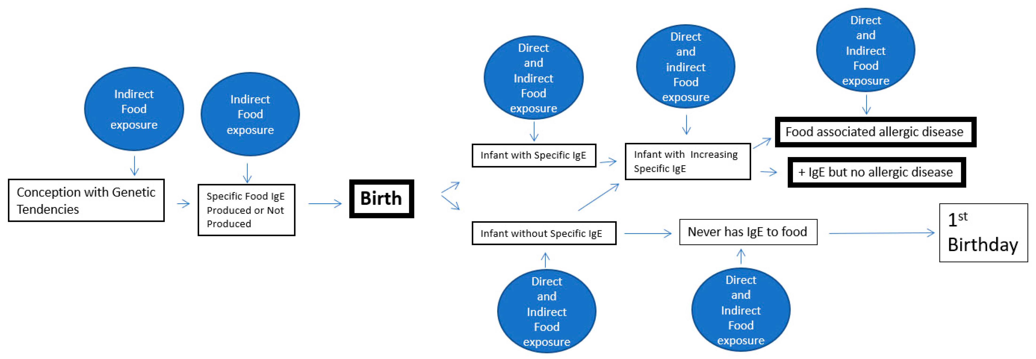
Figure 1. A schematic representation of the intrauterine chances to produce specific IgE and the subsequent events until the first birthday for clinical expression of an IgE mediated disease.
Allergies 01 00009 g001
Publisher’s Note: MDPI stays neutral with regard to jurisdictional claims in published maps and institutional affiliations.