Internal Morphology of Mandibular Second Premolars Using Micro-Computed Tomography
Abstract
:1. Introduction
1.1. Root Canal Morphology
1.2. Analysis of the Morphology of the Root Canal
1.3. Micro-CT Applications in Dental Research
2. Materials and Methods
2.1. Teeth Selection
2.2. Morphological Analysis by Micro-Computed Tomography
3. Results
4. Discussion
4.1. Methodology
4.1.1. Cone Beam Computed Tomography (CBCT)
4.1.2. Micro-Computed Tomography (Micro-CT)
4.2. Limitations
5. Conclusions
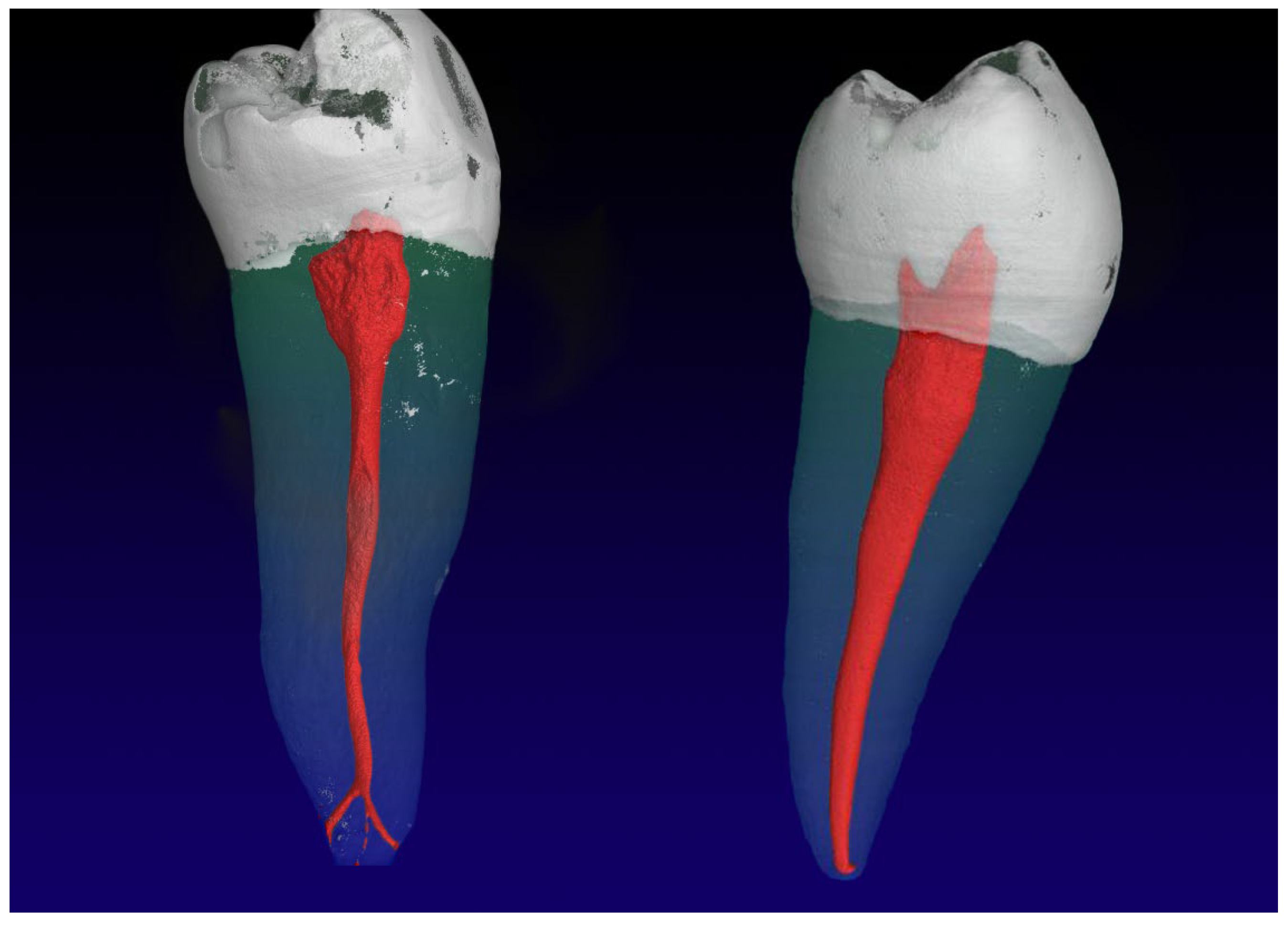
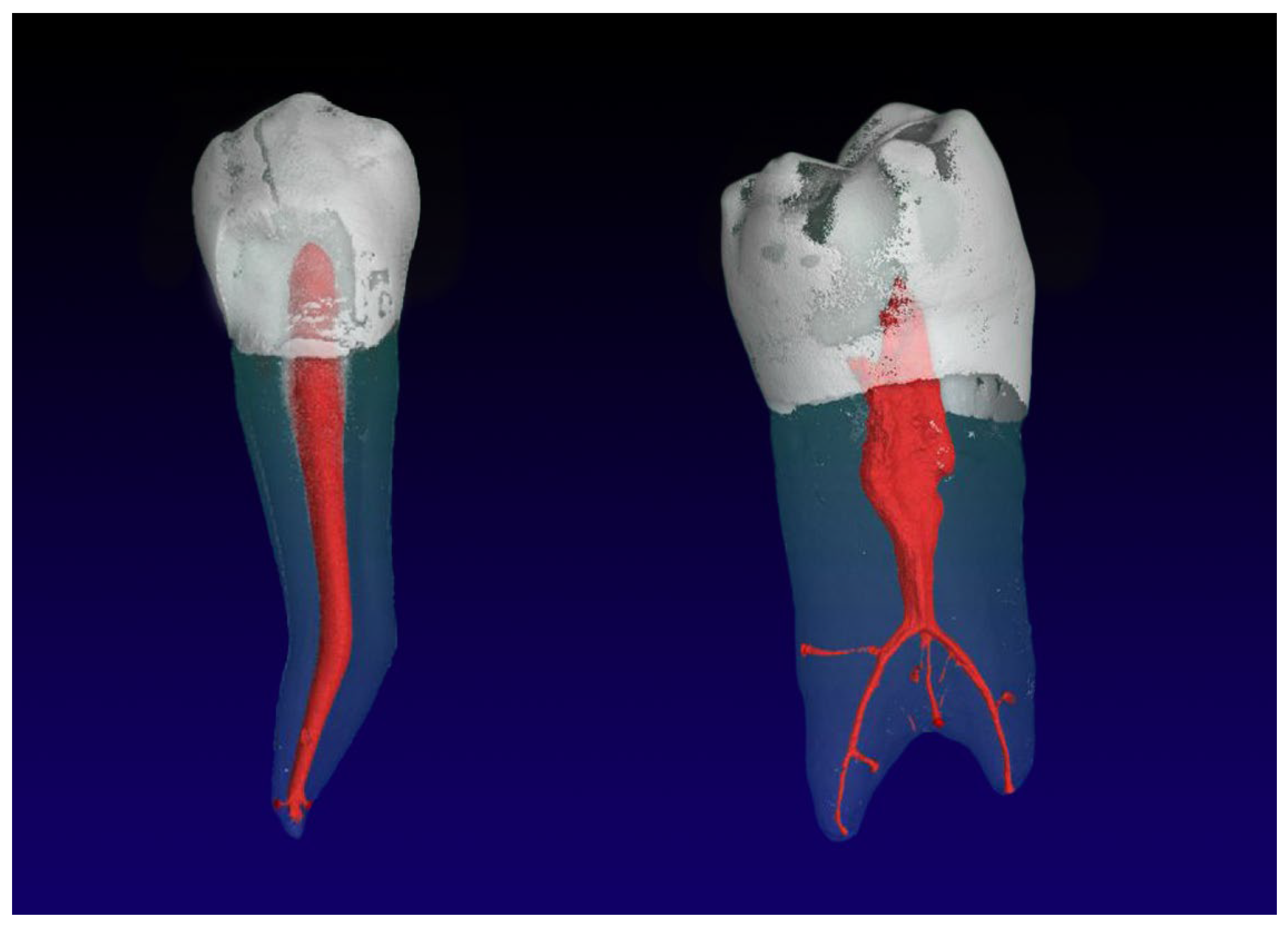
- The most frequently root canal configurations (RCC) observed mandibular second premolar (Mn2P) were 1-1-1/1 (54.9%), 1-1-1/2 (14.7%), 1-1-2/2 (10.8%), and another nine RCCs.
- The majority of Mn2Ps were single-rooted (98%) and presented with one (57.8%) or two physiological foramina (32.4%), almost one in ten teeth showed at least three main foramina.
- Almost two thirds showed accessory root canals, predominantly located in the apical third.
- The mainly single-rooted sample of Mn2Ps showed less frequent morphological diversifications than Mn1Ps.
Author Contributions
Funding
Institutional Review Board Statement
Informed Consent Statement
Data Availability Statement
Acknowledgments
Conflicts of Interest
References
- Cleghorn, B.M.; Christie, W.H.; Dong, C.C. The root and root canal morphology of the human mandibular first premolar: A literature review. J. Endod. 2007, 33, 509–516. [Google Scholar] [CrossRef] [PubMed]
- Wolf, T.G.; Anderegg, A.L.; Wierichs, R.J.; Campus, G. Root canal morphology of the mandibular second premolar: A systematic review and meta-analysis. BMC Oral Health 2021, 21, 309. [Google Scholar] [CrossRef]
- Green, D. Double canals in single roots. Oral Surg. Oral Med. Oral Pathol. 1973, 35, 689–696. [Google Scholar] [CrossRef] [PubMed]
- Ingle, J.I. A standardized endodontic technique utilizing newly designed instruments and filling materials. Oral Surg. Oral Med. Oral Pathol. 1961, 14, 83–91. [Google Scholar] [CrossRef] [PubMed]
- Pineda, F.; Kuttler, Y. Mesiodistal and buccolingual roentgenographic investigation of 7275 root canals. Oral Surg. Oral Med. Oral Pathol. 1972, 33, 101–110. [Google Scholar] [CrossRef] [PubMed]
- Awawdeh, L.; Abdullah, H.; Al-Qudah, A. Root form and canal morphology of Jordanian maxillary first premolars. J. Endod. 2008, 34, 956–961. [Google Scholar] [CrossRef]
- Sikri, V.K.; Sikri, P. Mandibular premolars: Aberrations in pulp space morphology. Indian J. Dent. Res. 1994, 5, 9–14. [Google Scholar]
- Hounsfield, G.N. Computerized transverse axial scanning (tomography). 1. Description of system. Br. J. Radiol. 1973, 46, 1016–1022. [Google Scholar] [CrossRef]
- Guldberg, R.E.; Lin, A.S.; Coleman, R.; Robertson, G.; Duvall, C. Microcomputed tomography imaging of skeletal development and growth. Birth Defects Res. C Embryo Today 2004, 72, 250–259. [Google Scholar] [CrossRef]
- Bentley, M.D.; Ortiz, M.C.; Ritman, E.L.; Romero, J.C. The use of microcomputed tomography to study microvasculature in small rodents. Am. J. Physiol. Regul. Integr. Comp. Physiol. 2002, 282, 1267–1279. [Google Scholar] [CrossRef]
- Rhodes, J.S.; Ford, T.R.; Lynch, J.A.; Liepins, P.J.; Curtis, R.V. Micro-computed tomography: A new tool for experimental endodontology. Int. Endod. J. 1999, 32, 165–170. [Google Scholar] [CrossRef]
- Swain, M.V.; Xue, J. State of the art of micro-CT applications in dental research. Int. J. Oral Sci. 2009, 1, 177–188. [Google Scholar] [CrossRef]
- Aksoy, U.; Küçük, M.; Versiani, M.A.; Orhan, K. Publication trends in micro-CT endodontic research: A bibliometric analysis over a 25-year period. Int. Endod. J. 2021, 54, 343–353. [Google Scholar] [CrossRef] [PubMed]
- Oi, T.; Saka, H.; Ide, Y. Three-dimensional observation of pulp cavities in the maxillary first premolar tooth using micro-CT. Int. Endod. J. 2004, 37, 46–51. [Google Scholar] [CrossRef] [PubMed]
- Lee, J.K.; Ha, B.H.; Choi, J.H.; Heo, S.M.; Perinpanayagam, H. Quantitative three-dimensional analysis of root canal curvature in maxillary first molars using micro-computed tomography. J. Endod. 2006, 32, 941–945. [Google Scholar] [CrossRef] [PubMed]
- Peters, O.A. Three-dimensional analysis of root canal geometry by high-resolution computed tomography. J. Dent. Res. 2000, 79, 1405–1409. [Google Scholar] [CrossRef] [PubMed]
- Bjorndal, L.; Carlsen, O.; Thuesen, G.; Darvann, T.; Kreiborg, S. External and internal macromorphology in 3D-reconstructed maxillary molars using computerized X-ray microtomography. Int. Endod. J. 1999, 32, 3–9. [Google Scholar] [CrossRef]
- Plotino, G.; Grande, N.M.; Pecci, R.; Bedini, R.; Pameijer, C.H.; Somma, F. Three-dimensional imaging using microcomputed tomography for studying tooth macromorphology. J. Am. Dent. Assoc. 2006, 137, 1555–1561. [Google Scholar] [CrossRef]
- Paes da Silva Ramos Fernandes, L.M.; Rice, D.; Ordinola-Zapata, R.; Alvares Capelozza, A.L.; Bramante, C.M.; Jaramillo, D.; Christensen, H. Detection of various anatomic patterns of root canals in mandibular incisors using digital periapical radiography, 3 cone-beam computed tomographic scanners, and micro-computed tomographic imaging. J. Endod. 2014, 40, 42–45. [Google Scholar] [CrossRef]
- Briseño-Marroquín, B.; Paqué, F.; Maier, K.; Willershausen, B.; Wolf, T.G. Root canal morphology and configuration of 179 maxillary first molars by means of micro-computed tomography: An ex vivo study. J. Endod. 2015, 41, 2008–2013. [Google Scholar] [CrossRef]
- Ahmed, H.M.A.; Dummer, P.M.H. A new system for classifying tooth, root and canal anomalies. Int. Endod. J. 2018, 51, 389–404. [Google Scholar] [CrossRef]
- Vertucci, F.J. Root canal anatomy of the human permanent teeth. Oral Surg. Oral Med. Oral Pathol. 1984, 58, 589–599. [Google Scholar] [CrossRef] [PubMed]
- Weine, F.S.; Healey, H.J.; Gerstein, H.; Evanson, L. Canal configuration in the mesiobuccal root of the maxillary first molar and its endodontic significance. Oral Surg. Oral Med. Oral Pathol. 1969, 28, 419–425. [Google Scholar] [CrossRef] [PubMed]
- Scheid, R.C.; Weiss, G. Woelfel’s Dental Anatomy, 8th ed.; Wolters Kluwer: Philadelphia, PA, USA, 2012. [Google Scholar]
- Paqué, F.; Ganahl, D.; Peters, O.A. Effects of root canal preparation on apical geometry assessed by micro-computed tomography. J. Endod. 2009, 35, 1056–1059. [Google Scholar] [CrossRef]
- Ok, E.; Altunsoy, M.; Nur, B.G.; Aglarci, O.S.; Çolak, M.; Güngör, E. A cone-beam computed tomography study of root canal morphology of maxillary and mandibular premolars in a Turkish population. Acta Odontol. Scand. 2014, 72, 701–706. [Google Scholar] [CrossRef] [PubMed]
- Park, J.W.; Lee, J.K.; Ha, B.H.; Choi, J.H.; Perinpanayagam, H. Three-dimensional analysis of maxillary first molar mesiobuccal root canal configuration and curvature using micro-computed tomography. Oral Surg. Oral Med. Oral Pathol. Oral Radiol. Endodontology 2009, 108, 437–442. [Google Scholar] [CrossRef]
- Bürklein, S.; Heck, R.; Schäfer, E. Evaluation of the root canal anatomy of maxillary and mandibular premolars in a selected German population using cone-beam computed tomographic data. J. Endod. 2017, 43, 1448–1452. [Google Scholar] [CrossRef]
- Martins, J.N.R.; Gu, Y.; Marques, D.; Francisco, H.; Caramês, J. Differences on the root and root canal morphologies between Asian and white ethnic groups analyzed by cone-beam computed tomography. J. Endod. 2018, 44, 1096–1104. [Google Scholar] [CrossRef]
- Rödig, T.; Hülsmann, M. Diagnosis and root canal treatment of a mandibular second premolar with three root canals. Int. Endod. J. 2003, 36, 912–919. [Google Scholar] [CrossRef]
- Macri, E.; Zmener, O. Five canals in a mandibular second premolar. J. Endod. 2000, 26, 304–305. [Google Scholar] [CrossRef]
- Hosseinpour, S.; Kharazifard, M.J.; Khayat, A.; Naseri, M. Root canal morphology of permanent mandibular premolars in Iranian population: A systematic review. Iran. Endod. J. 2016, 11, 150–156. [Google Scholar]
- Sert, S.; Bayirli, G.S. Evaluation of the root canal configurations of the mandibular and maxillary permanent teeth by gender in the Turkish population. J. Endod. 2004, 30, 391–398. [Google Scholar] [CrossRef]
- Rajakeerthi, R.; Nivedhitha, M.S.B. Use of cone beam computed tomography to identify the morphology of maxillary and mandibular premolars in Chennai population. Braz. Dent. Sci. 2019, 22, 55–62. [Google Scholar] [CrossRef]
- Patel, S.; Durack, C.; Abella, F.; Shemesh, H.; Roig, M.; Lemberg, K. Cone beam computed tomography in endodontics—A review. Int. Endod. J. 2015, 48, 3–15. [Google Scholar] [CrossRef]
- Venskutonis, T.; Plotino, G.; Juodzbalys, G.; Mickevičienė, L. The importance of cone-beam computed tomography in the management of endodontic problems: A review of the literature. J. Endod. 2014, 40, 1895–1901. [Google Scholar] [CrossRef] [PubMed]
- Matherne, R.P.; Angelopoulos, C.; Kulild, J.C.; Tira, D. Use of cone-beam computed tomography to identify root canal systems in vitro. J. Endod. 2008, 34, 87–89. [Google Scholar] [CrossRef] [PubMed]
- Abdinian, M.; Razavian, H.; Jenabi, N. In vitro comparison of cone beam computed tomography with digital periapical radiography for detection of vertical root fracture in posterior teeth. J. Dent. 2016, 17, 84–90. [Google Scholar]
- Estrela, C.; Reis Bueno, M.; Leles, C.R.; Azevedo, B.; Azevedo, J.R. Accuracy of cone beam computed tomography and panoramic and periapical radiography for detection of apical periodontitis. J. Endod. 2008, 34, 273–279. [Google Scholar] [CrossRef]
- Koivisto, J.; van Eijnatten, M.; Kiljunen, T.; Shi, X.-Q.; Wolff, J. Effective radiation dose in the wrist resulting from a radiographic device, two CBCT devices and one MSCT device: A comparative study. Radiat. Prot. Dosim. 2018, 179, 58–68. [Google Scholar] [CrossRef] [PubMed]
- Peters, O.A.; Paqué, F. Root canal preparation of maxillary molars with the self-adjusting file: A micro-computed tomography study. J. Endod. 2011, 37, 53–57. [Google Scholar] [CrossRef] [PubMed]
- Filpo-Perez, C.; Bramante, C.M.; Villas-Boas, M.H.; Húngaro Duarte, M.A.; Versiani, M.A.; Ordinola-Zapata, R. Micro-computed tomographic analysis of the root canal morphology of the distal root of mandibular first molar. J. Endod. 2015, 41, 231–236. [Google Scholar] [CrossRef]
- Wolf, T.G.; Paqué, F.; Zeller, M.; Willershausen, B.; Briseño-Marroquín, B. Root canal morphology and configuration of 118 mandibular first molars by means of micro-computed tomography: An ex vivo study. J. Endod. 2016, 42, 610–614. [Google Scholar] [CrossRef] [PubMed]
- Wolf, T.G.; Kozaczek, C.; Campus, G.; Paqué, F.; Wierirchs, R.J. Root canal morphology of 116 maxillary second premolars by micro-computed tomography in a mixed Swiss-German population with systematic review. J. Endod. 2020, 46, 1639–1647. [Google Scholar] [CrossRef] [PubMed]
- Peters, O.A.; Schönenberger, K.; Laib, A. Effects of four Ni-Ti preparation techniques on root canal geometry assessed by micro computed tomography. Int. Endod. J. 2001, 34, 221–230. [Google Scholar] [CrossRef] [PubMed]
- Paqué, F.; Peters, O.A. Micro-computed tomography evaluation of the preparation of long oval root canals in mandibular molars with the self-adjusting file. J. Endod. 2011, 37, 517–521. [Google Scholar] [CrossRef]


| Weine et al., 1969 [23] | Vertucci, 1984 [21] | Briseño Marroquín et al., 2015 [20] | Absolute (n) | Mean (%) |
|---|---|---|---|---|
| I | I | 1-1-1/1 | 56 | 54.9 |
| V | 1-1-1/2 | 15 | 14.7 | |
| 1-1-1/3 | 3 | 2.9 | ||
| V | 1-1-2/2 | 11 | 10.8 | |
| 1-1-2/3 | 1 | 1.0 | ||
| V | 1-2-2/2 | 5 | 4.9 | |
| VII | 1-2-1/2 | 1 | 1.0 | |
| II | 2-1-1/1 | 3 | 2.9 | |
| VI | 2-1-2/2 | 1 | 1.0 | |
| 1-1-3/3 | 4 | 3.9 | ||
| 1-1-2/5 | 1 | 1.0 | ||
| 1-1-1/4 | 1 | 1.0 |
| Foramina | ||
|---|---|---|
| Main Apical Foramina | n | % |
| /1 | 59 | 57.8 |
| /2 | 33 | 32.4 |
| /3 | 8 | 7.8 |
| /4 | 1 | 1.0 |
| /5 | 1 | 1.0 |
| Accessory Foramina | n | % |
| 0 | 36 | 35.3 |
| 1 | 36 | 35.3 |
| 2 | 22 | 21.6 |
| 3 | 3 | 2.9 |
| 4 | 3 | 2.9 |
| 5 | 2 | 2.0 |
| Canals | ||
|---|---|---|
| Connecting Canals | n | % |
| None | 92 | 90.2 |
| Co | 0 | 0.0 |
| Mi | 3 | 2.9 |
| Ap | 7 | 6.9 |
| Accessory Canals | n | % |
| None | 36 | 35.3 |
| Co | 0 | 0.0 |
| Mi | 9 | 8.8 |
| Api | 57 | 55.9 |
Disclaimer/Publisher’s Note: The statements, opinions and data contained in all publications are solely those of the individual author(s) and contributor(s) and not of MDPI and/or the editor(s). MDPI and/or the editor(s) disclaim responsibility for any injury to people or property resulting from any ideas, methods, instructions or products referred to in the content. |
© 2023 by the authors. Licensee MDPI, Basel, Switzerland. This article is an open access article distributed under the terms and conditions of the Creative Commons Attribution (CC BY) license (https://creativecommons.org/licenses/by/4.0/).
Share and Cite
Wolf, T.G.; Basmaci, S.; Schumann, S.; Waber, A.L. Internal Morphology of Mandibular Second Premolars Using Micro-Computed Tomography. J. Imaging 2023, 9, 257. https://doi.org/10.3390/jimaging9120257
Wolf TG, Basmaci S, Schumann S, Waber AL. Internal Morphology of Mandibular Second Premolars Using Micro-Computed Tomography. Journal of Imaging. 2023; 9(12):257. https://doi.org/10.3390/jimaging9120257
Chicago/Turabian StyleWolf, Thomas Gerhard, Samuel Basmaci, Sven Schumann, and Andrea Lisa Waber. 2023. "Internal Morphology of Mandibular Second Premolars Using Micro-Computed Tomography" Journal of Imaging 9, no. 12: 257. https://doi.org/10.3390/jimaging9120257
APA StyleWolf, T. G., Basmaci, S., Schumann, S., & Waber, A. L. (2023). Internal Morphology of Mandibular Second Premolars Using Micro-Computed Tomography. Journal of Imaging, 9(12), 257. https://doi.org/10.3390/jimaging9120257


