Article

Efficient Recovery of Gadolinium from Contaminated Waters Using Manganese Ferrite Nanoparticles

1
Department of Chemistry, LAQV-REQUIMTE—Associated Laboratory for Green Chemistry, University of Aveiro, 3810-193 Aveiro, Portugal
2
Department of Biology, CESAM—Centre for Environmental and Marine Studies, University of Aveiro, 3810-193 Aveiro, Portugal
3
LCA—Central Laboratory of Analysis, University of Aveiro, 3810-193 Aveiro, Portugal
4
Department of Chemistry, CICECO—Centre for Research in Ceramics and Composite Materials, University of Aveiro, 3810-193 Aveiro, Portugal
*
Author to whom correspondence should be addressed.
Recycling 2025, 10(2), 57; https://doi.org/10.3390/recycling10020057
Submission received: 4 March 2025 / Revised: 23 March 2025 / Accepted: 28 March 2025 / Published: 1 April 2025

Abstract

:
The widespread use of gadolinium (Gd) in medical and industrial applications, especially as a contrast agent in magnetic resonance imaging (MRI), has led to its increasing presence in surface waters, disrupting natural geochemical cycles and posing risks to aquatic ecosystems. Addressing this challenge, recent studies have explored the potential of magnetic materials, such as spinel ferrite nanoparticles, in the removal of Gd from contaminated water sources. The present study specifically focused on the use of MnFe2O4 nanoparticles to remove Gd from contaminated solutions, employing response surface methodology (RSM) to optimize sorption conditions. Key variables evaluated included salinity (0–30 g/L), initial Gd concentration (1–5 μmol/L), and sorbent dose (20–180 mg/L), at a fixed pH of 6. The results revealed that salinity had a minimal impact on Gd sorption, likely due to the high sorbent mass used. Optimal conditions were identified as a sorbent dose of 165 mg/L, an initial Gd concentration of 1.3 μmol/L, and a salinity level of 13.4 g/L, at pH 6. The process was efficient and rapid, achieving over 90% Gd removal within 1 h in both freshwater and saline conditions, and over 75% removal in mineral water within 3 h. The high efficiency and celerity of this method suggest that MnFe2O4 nanoparticles are a promising solution for treating Gd-contaminated hospital effluents. Future research should focus on validating these results in real-world effluent matrices and addressing the environmental and economic aspects of large-scale implementation, thereby contributing to sustainable water remediation strategies.

Graphical Abstract

1. Introduction

Gadolinium (Gd), a rare earth element from the lanthanide series, has become indispensable in modern healthcare due to its critical role as a contrast agent in magnetic resonance imaging (MRI) [1,2]. The paramagnetic properties of gadolinium ions (Gd3+) enhance the quality of MRI scans, enabling better diagnosis and treatment of various medical conditions [3]. Since their introduction in the 1980s, gadolinium-based contrast agents (GBCAs) have been widely used in hospitals and clinics worldwide, revolutionizing the field of medical imaging [4]. Beyond healthcare, Gd is also used in industries such as electronics, nuclear reactors, and the production of phosphors [2]. However, the extensive use of GBCAs has raised significant environmental and health concerns [5]. Once administered, GBCAs are excreted by patients and enter wastewater systems, eventually reaching natural water bodies [1,6]. This has led to the classification of gadolinium as an emerging contaminant, posing potential risks to human health and aquatic ecosystems [2,5].
The contamination of natural waters with Gd is primarily attributed to effluents from wastewater treatment plants (WWTPs) [1,5]. These facilities are typically not designed to remove complex compounds like GBCAs, leading to the discharge of Gd into rivers, lakes, and coastal waters [1,6,7,8]. In uncontaminated systems, Gd concentrations typically range from 1 to 4 ng/L in rivers and up to 181 ng/L in surface waters. However, in contaminated streams, concentrations can reach up to 207 μg/L. Near WWTP effluent discharge points, reported concentrations are even higher, ranging from 200 to 1100 μg/L [2], highlighting the significant anthropogenic impact on surface water contamination. The release of Gd from WWTP into previously uncontaminated systems often results in concentrations far exceeding natural background levels, indicating a persistent and widespread environmental concern [8]. Upon entering aquatic systems, Gd can bioaccumulate and biomagnify, leading to adverse effects on aquatic organisms. Studies have shown that Gd can induce biochemical alterations in exposed fauna, with its toxicity primarily linked to interference with calcium-dependent biological processes [9]. Due to the similar ionic radii of Gd3+ and Ca2+ [10], Gd can block calcium channels, disrupting essential physiological functions, including muscle contraction, neurotransmission, and enzyme activity [9]. In humans, the use of GBCAs has been linked to nephrogenic systemic fibrosis (NSF), a rare but severe condition that affects the skin and internal organs, particularly in individuals with impaired kidney function [11,12,13]. Recent studies have also raised concerns about Gd accumulation in the brain, bones, and other tissues, even in people with normal renal function [13,14]. In aquatic environments, Gd poses significant risks due to its potential to bioaccumulate in living organisms, leading to toxic effects that can disrupt entire ecosystems [15]. For example, marine mussels like Mytilus galloprovincialis have exhibited neurotoxicity, metabolic dysfunction, and oxidative stress following Gd exposure [16]. Similar effects have been observed in other bivalve species, including the clams Corbicula fluminea and Spisula solida, and the mussels Dreissena rostriformis bugensis and Dreissena polymorpha, which showed altered oxidative stress responses after Gd accumulation [17,18,19]. Additionally, Gd has been shown to interfere with the embryonic development of sea urchins, including species such as Paracentrotus lividus, Arbacia lixula, Heliocidaris tuberculata, and Centrostephanus rodgersii [20].
Growing concerns over Gd contamination have prompted the exploration of various methods for its removal from water [16]. Among the commonly employed water treatments (chemical precipitation, ion exchange, liquid–liquid extraction, and sorption) [21], sorption stands out as one of the most promising due to its simplicity, cost-effectiveness, wide availability, and high removal efficiency [22,23]. Sorption works by adhering Gd ions to the surface of solid materials, which can then be removed from the water through magnetic separation or other separation techniques. Various materials have been tested as sorbents for Gd, including clays [24], zeolites [25], covalent organic frameworks [26], chitosan-based materials [27], and nanomaterials [28,29,30]. Recent advancements in nanotechnology, in particular, have opened new avenues for enhancing the efficiency and effectiveness of sorption processes [21].
Nanomaterials, characterized by their high surface area and enhanced reactivity, offer significant advantages over traditional sorbents in the removal of Gd from contaminated waters. These materials provide more active sites for sorption, leading to higher capacities and faster removal rates. Depending on their size, composition, and properties, nanomaterials are available in various forms, including nanoparticles, nanotubes, nanowires, nanofilms or nanolayers, nanocomposites, and quantum dots [28]. They can be classified by their magnetic or non-magnetic properties. Among these, spinel ferrites, a class of magnetic nanoparticles with the chemical formula MFe2O4 (where M represents transition metals like Fe, Cd, Ni, Co, Mn, Zn, Cu, or Mg), stand out as particularly effective [22,30]. Over the past decade, spinel ferrites have proven highly effective in water decontamination, successfully removing toxic metals such as arsenic [31,32], cadmium [33], mercury [34], and lead [35,36]. In addition to their strong magnetic properties, which facilitate their easy separation in aqueous media, spinel ferrites are valued for their low eco-toxicity and cost-effectiveness, making them an attractive option for environmental remediation [37,38].
Manganese ferrite nanoparticles (MnFe2O4), a type of spinel ferrite, possess unique magnetic properties that make them highly effective for a wide range of applications, including water treatment. These nanoparticles have shown high efficiency in removing various contaminants, including rare earth elements (REEs) and other critical pollutants. Recent studies have highlighted their potential in the removal of REEs, such as lanthanum (La3+) and cerium (Ce3+), from contaminated solutions. For example, Ghobadi et al. [22] reported maximum sorption capacities of 785 mg/g for La3+ and 770 mg/g for Ce3+ using MnFe2O4 nanoparticles under optimal conditions (pH 7 and an initial REE concentration of 500 mg/L). Similarly, Liu et al. [39] achieved maximum sorption capacities of 757 mg/g for La3+ and 751 mg/g for Ce3+ under the same conditions. Both studies also evaluated the performance of modified MnFe2O4 nanoparticles, concluding that they outperformed their unmodified counterparts. However, the higher costs associated with nanoparticle modification highlight the importance of more sustainable alternatives. While unmodified MnFe2O4 nanoparticles may be less efficient, they offer a more practical balance between cost, efficiency, and environmental impact. Their rapid sorption capabilities, high efficiency, stability, and ease of magnetic separation make them particularly promising for industrial applications.
Given the increasing use of Gd in healthcare and its growing presence in the environment, this study investigates the removal capacity of Gd ions using manganese ferrite nanoparticles. The study seeks to evaluate the impact of key parameters, including pH, element concentration, salinity, and contact time, on the sorption process, along with analyzing its kinetics. The overarching goal is to assess the potential of nanomaterial-based sorption techniques for treating a variety of aquatic environments contaminated by hospital effluents, contributing to the development of sustainable strategies to safeguard water resources and public health from the rising threat of Gd pollution.

2. Results

2.1. Characterization of Manganese Ferrite Nanoparticles

A thorough characterization of MnFe2O4 nanoparticles was reported in our previous work [40]. This detailed analysis revealed that the nanoparticles are spherical in shape, with an average diameter of 75 ± 15 nm, a pore volume of 0.11 cm3/g, and a BET surface area of 39.2 m2/g. Zeta potential measurements indicated values of 16.6 mV at pH 4 and −23.3 ± 0.9 mV at pH 6 (n = 5), suggesting that in a solution at pH 6, these nanoparticles exhibit a negatively charged surface, in contrast to the positively charged surface observed at pH 4. Furthermore, magnetic hysteresis analysis confirmed the ferrimagnetic nature of the nanoparticles, with a specific saturation magnetization of 48.1 emu/g. The X-ray diffraction (XRD) patterns of the MnFe2O4 nanoparticles revealed distinct peaks that are characteristic of the crystalline spinel structure of MnFe2O4, as identified by the JCPDS—International Centre for Diffraction Data (PDF card 01-071-4919). The FT-IR spectrum showed a band at 537 cm−1 corresponding to metal–O stretching vibrations within the lattice, while the peak at 1095 cm−1 is associated with metal –OH and metal –OH2 stretching vibrations, which suggest the presence of water adsorbed on the oxide surface [32]. The band at 1643 cm−1 is attributed to H–O–H bending vibrations, resulting from molecular water adsorbed or integrated into the crystalline structure. The broader band at 3369 cm−1 is linked to O–H stretching modes of chemisorbed water.

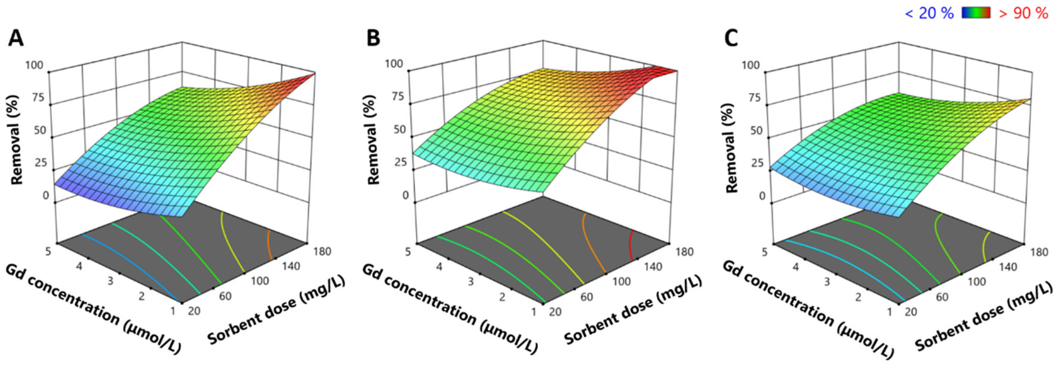
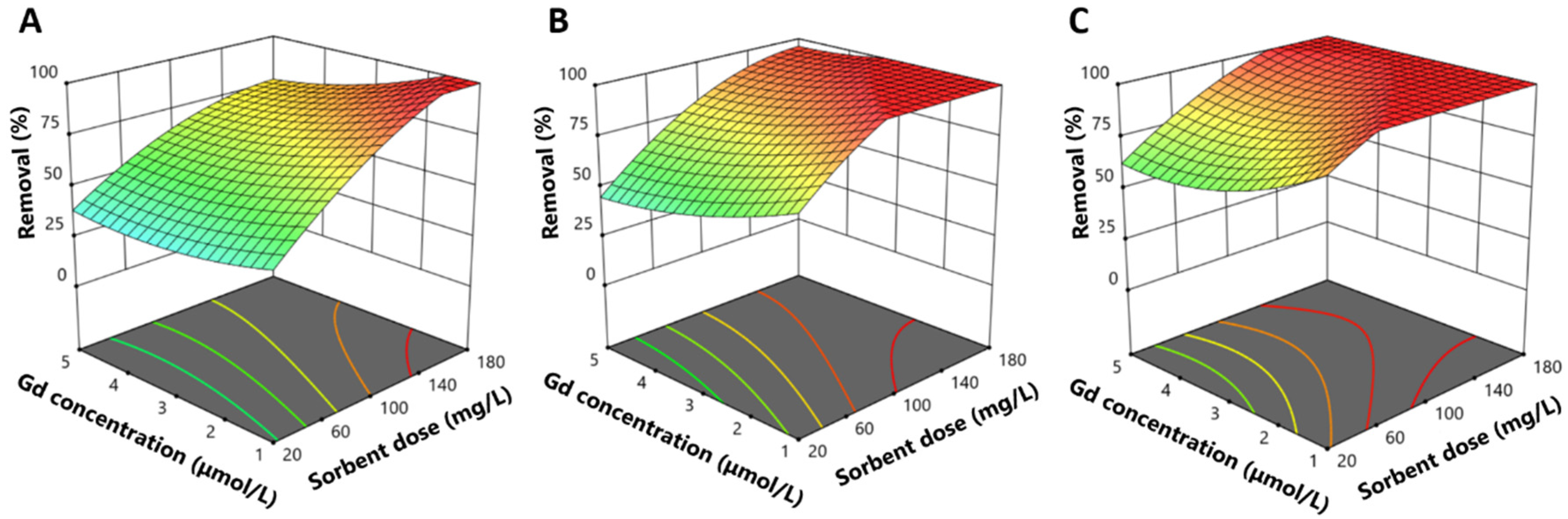
2.2. Response Surface Experiments: Evaluation of the Influence of Salinity, Gadolinium Concentration, and Sorbent Dose

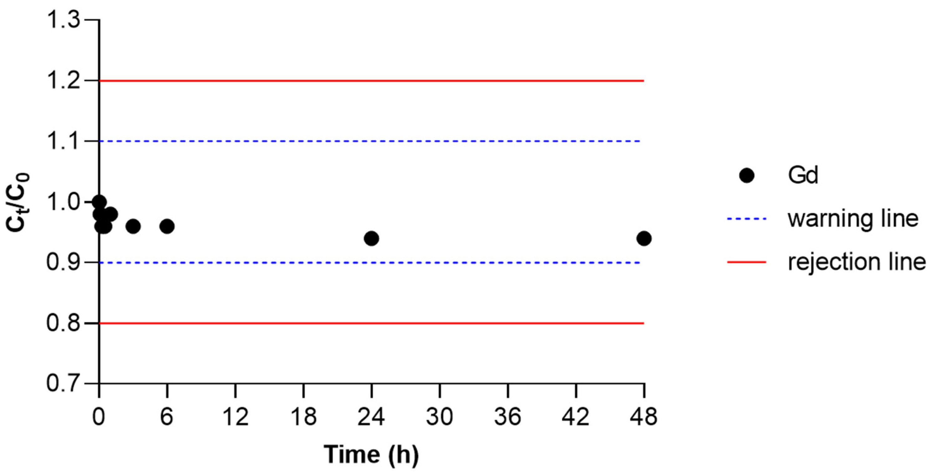
2.2.1. Influence of Ionic Strength

Figure 1 shows the control chart of the normalized Gd concentration over time. Control charts are graphical tools featuring warning and rejection lines to aid in determining which values meet quality standards. Ideally, the experimental control values should remain within the warning lines (10% error) or, at the very least, within the rejection lines (20% error). If the values fall outside the rejection lines, this indicates a loss of concentration or contamination, and such experiments are not accepted and must be repeated. Therefore, Figure 1 demonstrates that the Ct/C0 values lie within the warning lines and are close to 1, with maximum and minimum values of 1.0 and 0.94, respectively.
The influence of ionic strength (salinity) on Gd removal efficiency is illustrated in Figure 2, which represents the response surfaces that highlight the effects of Gd concentration and sorbent dosage across varying salinity levels (0, 15, and 30 g/L) after 1 h of contact time. The model’s R2 value of 0.8934 indicates a good fit, suggesting the strong predictive capacity of the data and satisfactory correspondence between the observed and estimated values [41]. These results reveal a clear relationship between salinity and Gd removal efficiency. At salinity zero (0 g/L), a maximum removal efficiency of 98.6% was achieved, with a sorbent dose of 180 mg/L and an initial Gd concentration of 1 μmol/L. At a salinity of 15 g/L, complete removal (100%) was attained, with nanoparticle dosages of between 140 and 180 mg/L and an initial Gd concentration of 1 to 1.75 μmol/L. In contrast, at a salinity of 30 g/L, the removal efficiency decreased to a maximum of 80.4%, with a nanoparticle dose of 180 mg/L and an initial Gd concentration of 1 μmol/L. The minimum removal efficiencies and the removals achieved under the central point condition are presented in Tables S1 and S2 of the Supplementary Material. Additionally, applying response surface methodology allows for outcome optimization through mathematical modeling, facilitating the prediction of ideal operational conditions. Through this optimization, the ideal conditions identified for complete Gd removal after 1 h of contact time were a nanoparticle dose of 165 mg/L, an initial Gd concentration of 1.3 μmol/L, and a salinity of 13.4 g/L at pH 6.

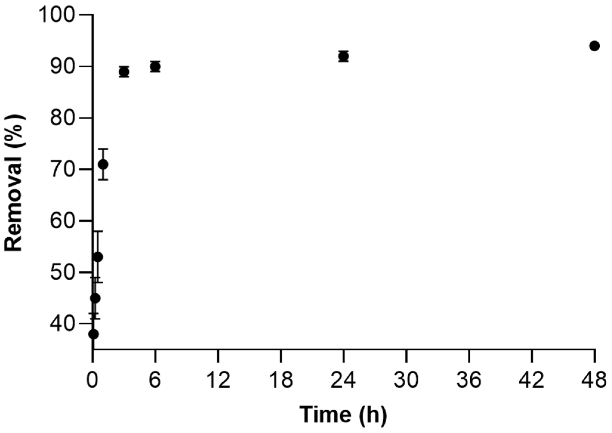
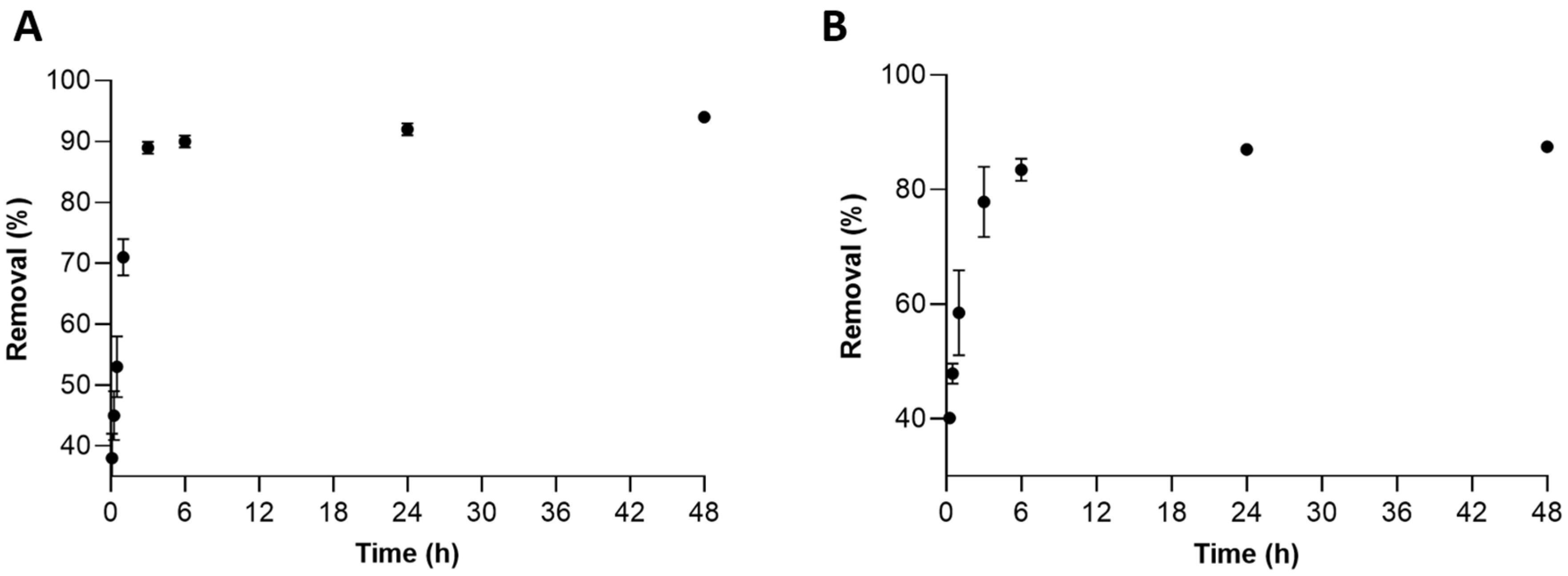
2.2.2. Influence of Contact Time

Figure 3 illustrates the influence of contact time on the percentage of Gd removal at a salinity level of 15 g/L, following 1, 3, and 24 h of contact time. Under conditions with a sorbent dose of 180 mg/L and an initial Gd concentration of 1 μmol/L, complete Gd removal was achieved at all tested contact times. Notably, as contact time increased, the range of sorbent dosages and initial Gd concentrations that allowed for 100% removal also broadened. Specifically, full removal was observed with sorbent doses of 140–180 mg/L and initial Gd concentrations of 1–1.75 μmol/L after 1 h, 60–180 mg/L of sorbent and 1–3.75 μmol/L of Gd concentration after 3 h, and 40–180 mg/L of sorbent and 1–5 μmol/L of Gd concentration after 24 h. Similarly, lower removal rates also increased over time. The minimum removal efficiencies are provided in Table S3 of the Supplementary Material.

2.2.3. Sorption Kinetics Studies

The Box–Behnken model is a mathematical approach that requires the replication of central points to ensure data consistency and reproducibility. Figure 4 illustrates the percentages of Gd removal obtained under the central point conditions: nanoparticle dose of 100 mg/L, initial Gd concentration of 3 μmol/L, salinity of 15 g/L, and pH 6. Water samples were collected after contact times of 0, 5, 15, and 30 min, and 1, 3, 6, 24, and 48 h. Each point includes the corresponding standard deviations, which show minimal error bars, with a maximum deviation of 10%, indicating high precision and confirming the robustness of the experimental design. Larger standard deviations observed during the first hour were attributed to variability in the rapid magnetic separation process, which was expedited to avoid disruption of the kinetic analysis. These variations diminished over time, as the separation process could be performed more slowly. Gd removal reached 89% after 3 h, with a slight increase to 94% after 48 h.
The results presented in Figure 4 were fitted to three kinetic models to determine the contact time required for sorption between the sorbate and sorbent, as well as to better understand the Gd sorption mechanism. The kinetic models used were pseudo-first order (PFO), pseudo-second order (PSO), and Elovich, as depicted in Figure 5. Table 1 summarizes the fitting parameters for all models, with the R2 value being a key parameter indicating the quality of the model fit—the closer R2 is to 1, the better the model reflects the experimental data.

2.2.4. Influence of Matrix Complexity

To assess the influence of matrix complexity, a new assay examined the efficacy of MnFe2O4 nanoparticles in removing Gd from more complex systems, given that hospital effluents may contain a variety of ions. This experiment used a mixture of nine REEs (Y, La, Ce, Pr, Nd, Eu, Gd, Tb, and Dy) in mineral water with a total mineralization of 32 ± 2 mg/L (Si (9.0 ± 0.4 mg/L, Cl (9.6 ± 0.4 mg/L), HCO3 (2.9 ± 0.5 mg/L), SO42− (1.4 ± 0.2 mg/L), NO3− (1.9 ± 0.2 mg/L), Na+ (5.6 ± 0.4 mg/L), Mg2+ (1.0 ± 0.2 mg/L), and Ca2+ (0.6 ± 0.2 mg/L) [42]). Figure 6 represents the Gd removal percentages achieved using MnFe2O4 nanoparticles during a contact time of 48 h under two experimental conditions. In this figure, graph A depicts removal kinetics over time for a simple matrix (ultrapure water) with a salinity of 15 g/L, while graph B shows the kinetics for a more complex matrix (bottled mineral water) containing multiple elements. As illustrated, high Gd removal rates were achieved in both cases, with 89% and 90% removal after 3 and 6 h, respectively, in condition A, and 78% and 83.5% removal after 3 and 6 h, respectively, in condition B. In graph A, Gd removal plateaued after 3 h, whereas in graph B, a plateau was reached after 6 h, with no significant changes up to 48 h. At 48 h, final Gd removal percentages were 94% and 87.5% for conditions A and B, respectively.

3. Discussion

3.1. Gd Removal Efficiency by MnFe2O4

The results presented throughout this study enable a critical analysis of the efficacy of MnFe2O4 nanoparticles in removing Gd from contaminated solutions, examining the effects of variables such as ionic strength, contact time, and matrix complexity. Sorption was initially tested in single-element Gd solutions, followed by multi-element solutions with matrices of varying complexity. These tests demonstrated that Gd removal efficiency is sensitive to sorbent dosage, salinity, and contact time, indicating a promising potential for application in hospital wastewater and saline environments.
The pH of hospital effluents resulting from magnetic resonance imaging (MRI) procedures, particularly those containing gadolinium-based contrast agents (GdCAs), generally ranges from neutral to slightly acidic, similar to common wastewater, and can be influenced by water treatments or variations in hospital chemicals [43]. The pH conditions used in this study are aligned with those found in hospital effluents. Moreover, the high removal rates observed at pH 6 can be attributed to the negatively charged surface of MnFe2O4 nanoparticles at this pH, which enhances interactions with the positively charged Gd cationic species (REE3+ ions and carbonate complexes). For pH > 6, these interactions are strong, facilitating the removal process. However, these interactions can be inhibited at a lower pH, as the nanoparticles become positively charged with decreasing pH, thus repelling the cationic Gd species [22].
Under simpler conditions (single-element Gd solution), MnFe2O4 nanoparticles proved to be effective in removing Gd, showing a clear dependency on sorbent mass, salinity, and contact time. Control solutions (Figure 1) exhibited minimal changes in Gd concentration, confirming no loss or contamination. A crucial factor in sorption studies is the evaluation of ionic strength (salinity), which can significantly influence sorbent efficiency. It is well established that an increase in ionic strength can significantly hinder sorption processes. In this context, the present study investigated the efficiency of MnFe2O4 nanoparticles under varying ionic strengths, with salinities of 0, 15, and 30 (Figure 2). The results show that ionic strength had a negligible effect on the sorption capacity of Gd. The reduction in removal percentage under high salinity conditions may be attributed to the higher concentration of dissolved ions in seawater. These ions can directly compete with Gd ions for available sorption sites on the surface of MnFe2O4 nanoparticles. This phenomenon aligns with the principle of ionic competition, where more abundant ions in the solution preferentially occupy the sorbent’s active sites, reducing the capacity to remove less abundant ions (in this case, Gd). Additionally, at elevated salinities, the presence of high NaCl levels means that Na+ ions not only compete for sorption sites but also act as a barrier for larger ions like REE3+ [44]. Similar behavior is observed in other saline solutions, such as seawater filtrates, as noted in this study. Generally, as ionic strength increases, the activity of the sorbent’s active sites and the metal ions diminish, leading to reduced sorption capacity [45]. Additionally, the influence of contact time was evaluated in this study. From the results presented in Figure 3, for a salinity of 15 g/L, it is evident that shorter contact times require higher sorbent doses and lower Gd concentrations for complete removal, whereas longer contact times allow for complete removal across a wider range of Gd concentrations, both low and high, with adequate sorbent doses. Previous studies have shown that spinel-type ferrites, such as MnFe2O4 and CuFe2O4 nanoparticles, exhibit minimal reduction in sorption efficiency even in highly saline environments. For example, Pinto et al. [29] observed that increasing NaCl concentrations up to 30 g/L had little or no effect on the removal of rare earth elements (REEs) by MnFe2O4 nanoparticles in aqueous solutions. Similarly, Tu et al. [46] and Tu and Johnston et al. [47] reported that the sorption of Pb, La, Ce, and Nd by ferrite nanoparticles remained nearly unchanged at NaCl concentrations up to 58 g/L. These results suggest that manganese ferrites may exhibit high efficiency in removing Gd not only from hospital effluents, as evidenced by their intrinsic resistance to high ionic strengths, but also from contaminated aquatic environments, such as seawater or estuarine waters.

3.2. Analysis of Sorption Mechanisms

Kinetic studies (Figure 5) demonstrated that the Elovich model provided the best fit to the data, with the highest R2 value (0.9623) and lowest standard deviation (Sx/y = 0.2347). This suggests that the adsorption process follows a heterogeneous sorption mechanism, which is characteristic of systems where surface activation energy varies and chemisorption plays a dominant role. The Elovich model, which is particularly suited for materials with heterogeneous sorption surfaces such as MnFe₂O₄ nanoparticles, accounts for variations in adsorption energy and the availability of sorption sites throughout the process. However, its accuracy was lower during the initial phase of sorption, particularly within the first few minutes, when rapid adsorption occurs. This discrepancy is likely due to the simultaneous contribution of multiple mechanisms, including surface adsorption and intraparticle diffusion, which are not fully captured by the model. Despite this limitation, the Elovich model provided the best overall fit across the entire sorption period, making it the most suitable model for describing the adsorption kinetics in this study. These findings are consistent with previous studies employing MnFe₂O₄ nanoparticles for metal removal, such as the adsorption of Nd from contaminated solutions [40], where the Elovich model also demonstrated superior alignment with experimental data. This supports the consensus that metal sorption onto magnetic ferrites typically involves a combination of electrostatic interactions and chemisorption processes, such as ion exchange and complexation [22,32,48,49]. Furthermore, the low standard deviation (Sx/y) of the Elovich model suggests minimal variability in residuals, reinforcing its reliability in representing the experimental data. Based on the observed kinetic behavior and experimental conditions, complexation and ion exchange are likely the dominant mechanisms governing Gd removal by MnFe₂O₄ nanoparticles. Ion exchange should be particularly considered due to the influence of salinity, which can enhance ionic exchange interactions between Gd ions and cations such as Mn and Fe within the nanoparticle structure. Further investigations are necessary to elucidate the dominant sorption mechanism in this system. Future studies should include monitoring Gd concentration over time and analyzing the products formed during the adsorption process. Additionally, desorption studies, temperature-dependent experiments, and equilibrium analysis would provide valuable complementary insights. Moreover, understanding the type of interaction between Gd and the nanoparticles could help evaluate their potential application in real-world scenarios, such as treating wastewater at hospital effluent stations.

3.3. Solution in Complex Matrices

Lastly, the results in Figure 6 demonstrate that sorption efficiency remained high in more complex matrices (Figure 6B), contrasting with the expected reduction due to the increased presence of ions and matrix complexity. In contrast, mineral water, rich in Ca2+, represents a more competitive system for ion sorption. As mentioned in the introduction, Gd substitutes Ca2+ in the body due to their similar chemical properties and ionic sizes. This parallel may extend to the chemical behavior of Gd in the matrix: Ca2+, present in higher concentrations, could directly compete for sorption sites on MnFe2O4 nanoparticles, marginally reducing removal efficiency. This competition suggests that the nanoparticles’ relative affinity for Gd partially overcomes Ca2+ competition but still affects efficiency in more complex matrices. Additionally, although the reduction from 89% to 78% (or 83.5% after 6 h) may seem minor in percentage terms, it is significant in practical contexts. In real-world scenarios, such as treating wastewater containing Gd, even small efficiency variations can have substantial implications, particularly in processes where residual Gd concentrations must be minimized to meet stringent environmental standards. This reduction could be attributed not only to direct Ca2+ competition but also to other ions present, collectively impacting electrostatic interaction and ion exchange mechanisms. The more complex matrix (mineral water) contains not just Ca2+ but also a range of other ions, such as Mg2+, Na+, and K+, which could occupy sorption sites or alter the local ionic equilibrium. Additionally, anions such as carbonate ions may promote the formation of complexes and modify the speciation of Gd in solutions [29]. This interaction highlights the robustness of the material, as removal efficiency still exceeds 75% after 3 h under competitive conditions. Thus, these results emphasize MnFe2O4’s promise for applications in complex waters, such as hospital wastewater or even natural water sources. However, the slight efficiency reduction in complex matrices indicates that operational adjustments, such as higher sorbent doses or longer contact times, might be necessary to ensure efficient removal on a large scale.

3.4. Comparison with Other Sorbents and Environmental Applications

To contextualize the efficiency of MnFe2O4 nanoparticles in this study, a comparative analysis with previously reported sorbents is presented in Table 2. Most literature reports investigate sorption at considerably higher initial element concentrations (500 mg/L) and with greater sorbent dosages (0.3 g/L) than those used in this study (0.204–0.472 mg/L and 0.165 g/L, respectively). These concentrations were not meant to simulate a contamination scenario and are unlikely to be found in a realistic contamination scenario. Additionally, previous studies often assess removal efficiency in ultrapure or deionized water, which lacks the ionic competition present in real-world aqueous environments. In contrast, our experiments were conducted in saline water (13.4 salinity) and mineral bottled water, providing a more realistic assessment of sorption performance under environmentally relevant conditions. While functionalized MnFe2O4 variants, such as MnFe2O4–GO, MnFe2O4–Al2O4, and MnFe2O4@SiO2–chitosan, have demonstrated enhanced sorption capacities, their increased synthesis complexity and cost may limit their large-scale applicability. The results of this study demonstrate that non-functionalized MnFe2O4 nanoparticles achieve high Gd removal efficiencies even in complex matrices, suggesting that functionalization may not always be necessary for effective rare earth element removal in realistic environmental scenarios. These findings reinforce the importance of evaluating sorbent performance under realistic contamination scenarios, where concentrations are typically much lower than those tested in laboratory conditions.

3.5. Limitations and Future Directions

The findings of this study highlight MnFe2O4 nanoparticles as an effective sorbent for Gd removal across various matrices, even under conditions of high ionic strength. However, the cyclic regeneration performance of the material was not evaluated in the present study. Nonetheless, similar experiments have previously been conducted within our research group. In these studies, 180 mg/L of MnFe2O4 nanoparticles were exposed to an ultrapure water solution spiked with 3 μM of a mixture of nine rare earth elements (REEs) (Y, La, Ce, Pr, Nd, Eu, Gd, Tb, Dy) at pH 8 [29]. Desorption was performed using a 0.1 M HNO3 solution for 30 min, followed by rinsing with ultrapure water to remove any residual desorption solution before reusing subsequent sorption cycles. A total of five sorption/desorption cycles were carried out. Moreover, the simplicity of the process and the low cost of the material make MnFe2O4 nanoparticles a promising option for the treatment of Gd-contaminated effluents, such as hospital wastewater. However, to minimize the environmental impact of Gd residues from MRI procedures, further studies are required to assess the feasibility of large-scale applications. This includes evaluating wastewater treatment efficiency, economic viability, and potential integration into hospital infrastructure or treatment facilities.

4. Materials and Methods

4.1. Materials and Reagents

Mono-elemental commercial standard REE solutions used were sourced from Alfa Aesar and Inorganic VenturesTM: Y (1001 ± 4 mg/L), La (1000 ± 5 mg/L), Ce (1001 ± 7 mg/L), Pr (1000 ± 4 mg/L), Nd (1001 ± 3 mg/L), Eu (1003 ± 4 mg/L), Gd (1000 ± 2 mg/L), Tb (1000 ± 4 mg/L), and Dy (1001 ± 4 mg/L), presented as REE3+ in a 2–7% (v/v) HNO3 solution, while HNO3 (65% v/v) and NaOH (>98% purity) for pH adjustments were supplied by Merck. All reagents were obtained from certified suppliers and used as received, without further purification. Ultrapure water (Milli-Q, 18 MΩ cm−1) was produced using a Millipore Integral 10 system, and the mineral bottled water used was from Penacova®, Portugal [42]. Saline water was prepared by diluting filtered seawater with ultrapure water, which was collected from Ria de Aveiro (Portugal, 40°38′39″ N, 8°44′43″ W) during high tide. Water salinity was measured using a WTW-series 720 multiparameter device. All glassware was adequately washed with ultrapure water, soaked in 25% (v/v) HNO3 solution for at least 24 h, and then rinsed with ultrapure water for later use.

4.2. Synthesis and Characterization of MnFe2O4 Nanoparticles

The MnFe2O4 nanoparticles used in this study were synthesized following the method described by Tavares et al. [32], as were the reagents and procedures used for the synthesis. The synthesis involved dissolving KOH (34 mmol) and KNO3 (15 mmol) in 25 mL of ultrapure water, previously purged with nitrogen gas. This solution was then heated to 60 °C under a nitrogen atmosphere with continuous mechanical stirring at 500 rpm. Once the salts were fully dissolved, a solution containing 10 mL of MnSO4⋅H2O (6 mmol) and 15 mL of FeSO4⋅7H2O (11 mmol) was gradually added, and the stirring speed was increased to 700 rpm. After maintaining this stirring speed for 30 min, the mixture was heated to 90 °C in an oil bath and left under a nitrogen atmosphere without stirring for 4 h. The resulting black product was magnetically separated, rinsed with ultrapure water and ethanol, and then dried at 40 °C.
The nanoparticles underwent comprehensive characterization using several techniques. Transmission electron microscopy (TEM) was employed to analyze their morphology and particle size, using a Hitachi H-9000 TEM microscope operated at 300 kV. Brunauer–Emmett–Teller (BET) analysis via nitrogen adsorption/desorption was used to determine surface area, using a Gemini V2.0 Micromeritics instrument. The crystalline structure of the nanoparticles was characterized by X-ray powder diffraction (XRD) analysis. Powdered samples were measured with a Philips Analytical PW 3050/60 X’Pert PRO diffractometer in θ/2θ configuration, featuring an X’Celerator detector. Data collection was automated using the X’Pert Data Collector v2.0b software, with monochromatized Cu Kα radiation (wavelength λ = 1.54056 Å) applied at 45 kV and 40 mA. Fourier transform infrared (FT-IR) spectra were obtained using a Mattson 7000 spectrometer at a resolution of 4 cm−1, employing a horizontal attenuated total reflectance (ATR) cell. Magnetization and zeta potential measurements were also performed. Direct current (dc) magnetization susceptibility was measured between 10 and 300 K, under an applied field of Happ = 50 Oe, after initial cooling in both the presence and absence of the field. Furthermore, magnetization as a function of the applied field was measured at 300 K. These measurements were conducted using an MPMS 5s (by Quantum Design, California, United States) magnetometer with a reciprocal sample measurement system. Further details regarding these methodologies, including TEM images, magnetization graphs, XRD patterns, and FTIR spectra, can be found in previous studies [40].

4.3. Response Surface Experiments: Evaluation of the Influence of Salinity, Gadolinium Concentration, and Sorbent Dose

To evaluate the potential of MnFe2O4 nanoparticles for treating water contaminated by hospital effluents, sorption experiments were conducted to assess the efficiency of these nanoparticles in removing Gd from spiked ultrapure water. Working solutions were prepared by adding a specific volume of Gd stock solution to ultrapure water, followed by the addition of MnFe2O4 nanoparticles in 500 mL Schott flasks. These experiments were subjected to continuous mechanical stirring with a glass rod at room temperature (20 °C). The effect of salinity was also examined by preparing solutions with salinities of 15 and 30, achieved by diluting filtered seawater with ultrapure water. After a brief ultrasonic treatment to disperse the nanoparticles, the sorption process was initiated (t0). Parallel control experiments, using Gd-contaminated water without sorbent, were conducted to account for possible contamination or Gd loss. Water samples were collected over 48 h, the nanoparticles were magnetically separated from aqueous solution using an external magnetic field, and samples were acidified to pH < 2 and stored at 4 °C for later analysis.
To optimize Gd removal, experimental conditions were designed using a Box–Behnken design, a three-level factorial method with three variables, encoded as −1, 0, and +1, where −1 and +1 represent the minimum and maximum conditions, respectively, and 0 represents the central point. This experimental design has been previously applied to rare earth element sorption studies [40]. In this study, the three variables considered were salinity (0, 15, 30 g/L), initial Gd concentration (1, 3, 5 µmol/L), and sorbent dose (20, 100, 180 mg/L) at pH 6, with three replicates performed at the central point to improve model accuracy. The experimental conditions are outlined in Table 3.
Surface response methodology analysis was conducted using Design-Expert V13 software (StatEase®) to analyze the data and predict the optimal process conditions. This approach applies mathematical and statistical methods to evaluate the relationship between quantitative variables and one or more responses, expressed through a second-order polynomial function. This function was used to assess the linear, quadratic, and interaction effects of the variables, as shown in Equation (1):
Y = β 0 + i = 1 k β i X i + i = 1 k β i i X i 2 + i < j k β i j X i X j
where Y represents the response variable, β 0 is the constant term, and β i represents the linear coefficients, β i i the quadratic coefficients, and β i j the interaction terms. The independent variables X i , X j , , X k are the coded values, calculated using Equation (2):
X k = x k x 0 x k
where X k is the coded value, x k is the actual value, x 0 is the central value, and x k is the difference between levels of the variable. ANOVA was employed to determine the significance of each effect, with only those effects with a p-value of less than 0.05 being included in the final model (Equation (1)). Response surfaces were then calculated to estimate Gd removal percentages (R, %) after 1 and 3 h, based on Equation (3), with removal efficiency as the ratio of the initial concentration (C0) to the concentration at time t (Ct):
R % = C 0 C t C 0 × 100
Given that the Gd was removed by sorption onto the nanoparticles, the amount of Gd per unit mass of sorbent (qt, mg/g) was calculated using Equation (4), where m (g) is the sorbent mass, V (L) is the solution volume, and the other variables retain their previously defined meanings:
q t = C 0 C t m × V

Sorption Kinetics Studies

A further series of experiments was performed to explore the kinetics of Gd sorption. These investigations adhered to the central point parameters outlined in the Box–Behnken design, specifically incorporating an initial Gd concentration of 3 μmol/L, a salinity level of 15 g/L, and a sorbent dosage of 100 mg/L. The methodology utilized was consistent with previously established procedures, including the implementation of control solutions alongside the experimental setups to validate the accuracy of the results. Samples were taken at predetermined intervals (0 min, 5 min, 15 min, 30 min, 1 h, 3 h, 6 h, 24 h, and 48 h) to observe the dynamics of the sorption process over time. The resulting data were subsequently analyzed using three distinct kinetic models: the pseudo-first-order (PFO) model (Equation (5)) [50], the pseudo-second-order (PSO) model (Equation (6)) [51], and the Elovich model (Equation (7)) [52]. The PFO model, which is grounded in Lagergren’s theory, posits that the rate of sorption is directly proportional to the number of accessible sorption sites, making it particularly relevant for systems where site availability is a constraining factor. Conversely, the PSO model is based on the assumption that chemisorption, involving chemical interactions between sorbent and sorbate, controls the sorption process, making it more suitable for systems where chemical bonding predominates. The Elovich model, while also focused on chemisorption, incorporates the concept of site heterogeneity, suggesting that different sorption sites possess varying activation energies, thus accounting for non-uniformity in sorption rates across the surface. Each of these models offers distinct insights into the sorption mechanisms, providing a comprehensive understanding of both the physical and the chemical processes involved. Within each of these models, qt indicates the Gd concentration at time t, qe (mg/g) denotes the concentration at equilibrium, and k1 (1/h) and k2 (g/μg h) represent the kinetic constants for the PFO and PSO models, respectively. Moreover, α (μg h/g) specifies the sorption rate, while β (g/μg) describes the desorption behavior in the Elovich model.
q t = q e 1 e k 1 t
q t = q e 2 k 2 t 1 + q e k 2 t
q t = 1 β ln 1 + α β t

4.4. Comparison Between Mono-Elemental and Multi-Elemental Sorption of REEs in Different Matrices by MnFe2O4

To assess the potential of MnFe2O4 nanoparticles in a more complex matrix, a new sorption assay was conducted to evaluate the efficiency of these nanoparticles in removing Gd from a solution containing a mixture of nine rare earth elements (REEs: Y, La, Ce, Pr, Nd, Eu, Gd, Tb, and Dy) prepared from bottled mineral water (Penacova®, Portugal [42]). The experimental conditions included a nanoparticle dose of 100 mg/L, an initial REE concentration of 3 μmol/L, zero salinity, and a pH of 6. The methodology employed was consistent with previously described procedures, including control assays to account for potential losses or contamination during the experiment. Water samples were collected at designed contact times (0, 0.25, 0.50, 1, 3, 6, 24, and 48 h), with the resulting data analyzed according to previously established equations.

4.5. Gadolinium Quantification

The determination of Gd in water samples was performed using inductively coupled plasma optical emission spectrometry (ICP–OES) with Horiba Jobin Yvon Activa M equipment. Calibration standards were utilized during the analysis, prepared by diluting the Gd standard solution in acidified water (HNO3 1%) to establish five calibration curves, with only those curves exhibiting a correlation coefficient greater than 0.999 being deemed valid. Regular analysis of control standards was carried out to ensure continuous adherence to quality and precision criteria. The associated error for each standard remained below 10%, and the limit of quantification was determined based on the lowest calibration standard (10 μg/L). Prior to analysis, the samples were diluted by a factor of 10.

5. Conclusions

This study confirmed the effectiveness of MnFe2O4 nanoparticles in removing Gd across diverse aqueous environments, including freshwater, mineral water, and saline water. Using response surface methodology, the optimal conditions for Gd removal were identified, with initial Gd concentration and sorbent mass emerging as key factors in the sorption process. The results suggest that chemisorption mechanisms, such as complexation and ion exchange, are likely to govern this process.
At high initial Gd concentrations (5 μmol/L), sorption efficiency significantly decreased, indicating the need for higher nanoparticle doses to achieve effective removal above 2 μmol/L. The process demonstrated fast kinetics, reaching equilibrium within 3 h at pH 6, with a Gd concentration of 3 μmol/L and a salinity level of 15 g/L. Although increased ionic strength negatively impacted sorption, appropriate sorbent mass mitigated these effects, indicating that excessive sorbent quantities may not be required in single-element systems. Assays with a mineral matrix containing a mixture of REEs showed that MnFe2O4 nanoparticles removed over 75% of Gd within 3 h, highlighting their potential for treating hospital effluents, especially those containing Gd-based MRI contrast agents. The significance of these experiments lies in their relevance to real environmental conditions. In natural and wastewater environments, Gd concentrations are typically in the ng/L to µg/L range, making it necessary to use considerably higher amounts of sorbent to achieve removal. Additionally, optimizing the sorbent dosage is crucial to balancing adsorption efficiency and economic feasibility. By investigating a broad range of sorbent concentrations, this study provides valuable insights into the feasibility of an application of MnFe2O4 nanoparticles to removing high concentrations of Gd under environmentally relevant conditions. However, several considerations must be addressed. While MnFe2O4 nanoparticles offer an effective solution, it is crucial to ensure scalability and proper management of waste generated during the treatment process to prevent introducing new environmental contaminants. Additionally, the cost and complexity of integrating ferrite-based treatment systems into hospital infrastructure should be carefully evaluated, alongside their long-term economic feasibility. Given the proven efficiency of MnFe2O4 nanoparticles in both high-ionic-strength and complex matrices, further studies should focus on assessing their performance in real hospital effluent scenarios to validate their practical applicability and optimize implementation strategies.

Supplementary Materials

The following supporting information can be downloaded at: https://www.mdpi.com/article/10.3390/recycling10020057/s1, Table S1: Minimum removal percentages achieved for Gd removal under the tested conditions; Table S2: Removal percentages of Gd obtained under the central point condition; Table S3: Minimum removal percentages of Gd under the tested conditions at a salinity of 15.

Author Contributions

J.S.: writing—original draft, investigation, formal analysis; J.P.: writing—review and editing, methodology, formal analysis, conceptualization; H.B.: validation, data curation; D.S.T.: writing—review and editing, supervision, methodology, formal analysis, conceptualization; R.F.: investigation, conceptualization; T.T.: writing—review and editing, conceptualization; J.R.: writing—review and editing, conceptualization E.P.: writing—review and editing, supervision, project administration, methodology, conceptualization. All authors have read and agreed to the published version of the manuscript.

Funding

This work received financial support from PT national funds (FCT/MCTES, Fundação para a Ciência e Tecnologia and Ministério da Ciência, Tecnologia e Ensino Superior) through the CICECO-Aveiro Institute of Materials (UIDB/50011/2020 and UIDP/50011/2020), REQUIMTE (UIDB/50006/2020 and UIDP/50006/2020) and CESAM (UID Centro de Estudos do Ambiente e Mar (CESAM) + LA/P/0094/2020).

Data Availability Statement

The data will be made available upon request.

Acknowledgments

João Pinto thanks FCT/MCTES (Fundação para a Ciência e Tecnologia and Ministério da Ciência, Tecnologia e Ensino Superior) for his PhD grant (Ref. https://doi.org/10.54499/2020.05323.BD).

Conflicts of Interest

The authors declare no competing interests.

References

  1. Migaszewski, Z.M.; Gałuszka, A. The Use of Gadolinium and Europium Concentrations as Contaminant Tracers in the Nida River Watershed in South-Central Poland. Geol. Q. 2016, 60, 67–76. [Google Scholar] [CrossRef]
  2. Rogowska, J.; Olkowska, E.; Ratajczyk, W.; Wolska, L. Gadolinium as a New Emerging Contaminant of Aquatic Environments. Environ. Toxicol. Chem. 2018, 37, 1523–1534. [Google Scholar] [CrossRef]
  3. Telgmann, L.; Sperling, M.; Karst, U. Determination of Gadolinium-Based MRI Contrast Agents in Biological and Environmental Samples: A Review. Anal. Chim. Acta 2013, 764, 1–16. [Google Scholar]
  4. Wahsner, J.; Gale, E.M.; Rodríguez-Rodríguez, A.; Caravan, P. Chemistry of MRI Contrast Agents: Current Challenges and New Frontiers. Chem. Rev. 2019, 119, 957–1057. [Google Scholar] [CrossRef]
  5. Laczovics, A.; Csige, I.; Szabó, S.; Tóth, A.; Kálmán, F.K.; Tóth, I.; Fülöp, Z.; Berényi, E.; Braun, M. Relationship between Gadolinium-Based MRI Contrast Agent Consumption and Anthropogenic Gadolinium in the Influent of a Wastewater Treatment Plant. Sci. Total Environ. 2023, 877, 162844. [Google Scholar] [CrossRef]
  6. Swan, S.K.; Lambrecht, L.J.; Townsend, R.; Davies, B.E.; McCloud, S.; Parker, J.R.; Bensel, K.; LaFrance, N.D. Safety and Pharmacokinetic Profile of Gadobenate Dimeglumine in Subjects with Renal Impairment. Investig. Radiol. 1999, 34, 443–448. [Google Scholar] [CrossRef]
  7. de Campos, F.F.; Enzweiler, J. Anthropogenic Gadolinium Anomalies and Rare Earth Elements in the Water of Atibaia River and Anhumas Creek, Southeast Brazil. Environ. Monit. Assess. 2016, 188, 281. [Google Scholar] [CrossRef]
  8. Pedreira, R.M.A.; Pahnke, K.; Böning, P.; Hatje, V. Tracking Hospital Effluent-Derived Gadolinium in Atlantic Coastal Waters off Brazil. Water Res. 2018, 145, 62–72. [Google Scholar] [CrossRef]
  9. Yushin, N.; Zinicovscaia, I.; Cepoi, L.; Chiriac, T.; Rudi, L.; Grozdov, D. Cyanobacteria Arthospira Platensis as an Effective Tool for Gadolinium Removal from Wastewater. Clean. Technol. 2023, 5, 638–651. [Google Scholar] [CrossRef]
  10. Sherry, A.D.; Caravan, P.; Lenkinski, R.E. Primer on Gadolinium Chemistry. J. Magn. Reson. Imaging 2009, 30, 1240–1248. [Google Scholar] [PubMed]
  11. Grobner, T.; Prischl, F.C. Gadolinium and Nephrogenic Systemic Fibrosis. Kidney Int. 2007, 72, 260–264. [Google Scholar] [PubMed]
  12. Mendichovszky, I.A.; Marks, S.D.; Simcock, C.M.; Olsen, Ø.E. Gadolinium and Nephrogenic Systemic Fibrosis: Time to Tighten Practice. Pediatr. Radiol. 2008, 38, 489–496. [Google Scholar] [PubMed]
  13. Piarulli, S.; Riedel, J.A.; Fossum, F.N.; Kermen, F.; Hansen, B.H.; Kvæstad, B.; Olsvik, P.A.; Farkas, J. Effects of Gadolinium (Gd) and a Gd-Based Contrast Agent (GBCA) on Early Life Stages of Zebrafish (Danio rerio). Chemosphere 2024, 350, 140950. [Google Scholar] [CrossRef]
  14. Idée, J.M.; Port, M.; Raynal, I.; Schaefer, M.; Le Greneur, S.; Corot, C. Clinical and Biological Consequences of Transmetallation Induced by Contrast Agents for Magnetic Resonance Imaging: A Review. Fundam. Clin. Pharmacol. 2006, 20, 563–576. [Google Scholar]
  15. Le Goff, S.; Barrat, J.A.; Chauvaud, L.; Paulet, Y.M.; Gueguen, B.; Ben Salem, D. Compound-Specific Recording of Gadolinium Pollution in Coastal Waters by Great Scallops. Sci. Rep. 2019, 9, 8015. [Google Scholar] [CrossRef]
  16. Trapasso, G.; Coppola, F.; Queirós, V.; Henriques, B.; Soares, A.M.V.M.; Pereira, E.; Chiesa, S.; Freitas, R. How Ulva Lactuca Can Influence the Impacts Induced by the Rare Earth Element Gadolinium in Mytilus Galloprovincialis? The Role of Macroalgae in Water Safety towards Marine Wildlife. Ecotoxicol. Environ. Saf. 2021, 215, 112101. [Google Scholar] [CrossRef]
  17. Figueiredo, C.; Grilo, T.F.; Oliveira, R.; Ferreira, I.J.; Gil, F.; Lopes, C.; Brito, P.; Ré, P.; Caetano, M.; Diniz, M.; et al. Gadolinium Ecotoxicity Is Enhanced in a Warmer and Acidified Changing Ocean as Shown by the Surf Clam Spisula Solida through a Multibiomarker Approach. Aquat. Toxicol. 2022, 253, 106346. [Google Scholar] [CrossRef]
  18. Hanana, H.; Turcotte, P.; André, C.; Gagnon, C.; Gagné, F. Comparative Study of the Effects of Gadolinium Chloride and Gadolinium—Based Magnetic Resonance Imaging Contrast Agent on Freshwater Mussel, Dreissena Plymorpha. Chemosphere 2017, 181, 197–207. [Google Scholar] [CrossRef]
  19. Perrat, E.; Parant, M.; Py, J.S.; Rosin, C.; Cossu-Leguille, C. Bioaccumulation of Gadolinium in Freshwater Bivalves. Environ. Sci. Pollut. Res. 2017, 24, 12405–12415. [Google Scholar] [CrossRef]
  20. Martino, C.; Bonaventura, R.; Byrne, M.; Roccheri, M.; Matranga, V. Effects of Exposure to Gadolinium on the Development of Geographically and Phylogenetically Distant Sea Urchins Species. Mar. Environ. Res. 2017, 128, 98–106. [Google Scholar] [CrossRef]
  21. Chen, Z.; Li, Z.; Chen, J.; Kallem, P.; Banat, F.; Qiu, H. Recent Advances in Selective Separation Technologies of Rare Earth Elements: A Review. J. Environ. Chem. Eng. 2022, 10, 107104. [Google Scholar] [CrossRef]
  22. Ghobadi, M.; Gharabaghi, M.; Abdollahi, H.; Boroumand, Z.; Moradian, M. MnFe2O4-Graphene Oxide Magnetic Nanoparticles as a High-Performance Adsorbent for Rare Earth Elements: Synthesis, Isotherms, Kinetics, Thermodynamics and Desorption. J. Hazard. Mater. 2018, 351, 308–316. [Google Scholar] [CrossRef] [PubMed]
  23. Gupta, N.K.; Gupta, A.; Ramteke, P.; Sahoo, H.; Sengupta, A. Biosorption-a Green Method for the Preconcentration of Rare Earth Elements (REEs) from Waste Solutions: A Review. J. Mol. Liq. 2019, 274, 148–164. [Google Scholar] [CrossRef]
  24. Estrade, G.; Marquis, E.; Smith, M.; Goodenough, K.; Nason, P. REE Concentration Processes in Ion Adsorption Deposits: Evidence from the Ambohimirahavavy Alkaline Complex in Madagascar. Ore Geol. Rev. 2019, 112, 103027. [Google Scholar] [CrossRef]
  25. Ji, B.; Zhang, W. Adsorption of Cerium (III) by Zeolites Synthesized from Kaolinite after Rare Earth Elements (REEs) Recovery. Chemosphere 2022, 303, 134941. [Google Scholar] [CrossRef]
  26. Xiao, J.; Li, B.; Qiang, R.; Qiu, H.; Chen, J. Highly Selective Adsorption of Rare Earth Elements by Honeycomb-Shaped Covalent Organic Frameworks Synthesized in Deep Eutectic Solvents. Environ. Res. 2022, 214, 113977. [Google Scholar] [CrossRef]
  27. Pylypchuk, I.V.; Kołodyńska, D.; Kozioł, M.; Gorbyk, P.P. Gd-DTPA Adsorption on Chitosan/Magnetite Nanocomposites. Nanoscale Res. Lett. 2016, 11, 168. [Google Scholar] [CrossRef]
  28. Kegl, T.; Košak, A.; Lobnik, A.; Novak, Z.; Kralj, A.K.; Ban, I. Adsorption of Rare Earth Metals from Wastewater by Nanomaterials: A Review. J. Hazard. Mater. 2020, 386, 121632. [Google Scholar] [CrossRef]
  29. Pinto, J.; Fernandes, R.; Tavares, D.; Henriques, B.; Trindade, T.; Pereira, E. Removal of Rare Earth Elements from Complex Mixtures by Using Manganese Ferrite Nanoparticles: Optimization through Surface Response Methodology. J. Environ. Manag. 2024, 368, 122211. [Google Scholar] [CrossRef]
  30. Pinto, J.; Branco, D.; Carvalho, L.; Henriques, B.; Freitas, R.; Trindade, T.; Tavares, D.; Pereira, E. Influence of Experimental Parameters on the Sorption Behavior of Rare Earth Elements on Manganese Ferrite Nanoparticles. Environ. Technol. Innov. 2023, 32, 103432. [Google Scholar] [CrossRef]
  31. Kumar, S.; Nair, R.R.; Pillai, P.B.; Gupta, S.N.; Iyengar, M.A.R.; Sood, A.K. Graphene Oxide-MnFe2O4 Magnetic Nanohybrids for Efficient Removal of Lead and Arsenic from Water. ACS Appl. Mater. Interfaces 2014, 6, 17426–17436. [Google Scholar] [CrossRef] [PubMed]
  32. Tavares, D.S.; Lopes, C.B.; Almeida, J.C.; Vale, C.; Pereira, E.; Trindade, T. Spinel-Type Ferrite Nanoparticles for Removal of Arsenic(V) from Water. Environ. Sci. Pollut. Res. 2020, 27, 22523–22534. [Google Scholar] [CrossRef]
  33. Homayonfard, A.; Miralinaghi, M.; Seyed Mohammad Shirazi, R.H.; Moniri, E. Efficient Removal of Cadmium (II) Ions from Aqueous Solution by CoFe2O4/Chitosan and NiFe2O4/Chitosan Composites as Adsorbents. Water Sci. Technol. 2018, 78, 2297–2307. [Google Scholar] [CrossRef] [PubMed]
  34. Viltres, H.; Gupta, N.K.; Paz, R.; Dhavale, R.P.; Park, H.H.; Leyva, C.; Srinivasan, S.; Rajabzadeh, A.R. Mercury Remediation from Wastewater through Its Spontaneous Adsorption on Non-Functionalized Inverse Spinel Magnetic Ferrite Nanoparticles. Environ. Technol. 2022, 45, 1155–1168. [Google Scholar] [CrossRef]
  35. Tatarchuk, T.; Shyichuk, A.; Sojka, Z.; Gryboś, J.; Naushad, M.; Kotsyubynsky, V.; Kowalska, M.; Kwiatkowska-Marks, S.; Danyliuk, N. Green Synthesis, Structure, Cations Distribution and Bonding Characteristics of Superparamagnetic Cobalt-Zinc Ferrites Nanoparticles for Pb(II) Adsorption and Magnetic Hyperthermia Applications. J. Mol. Liq. 2021, 328, 115375. [Google Scholar] [CrossRef]
  36. Tu, Y.J.; You, C.F.; Chen, M.H.; Duan, Y.P. Efficient Removal/Recovery of Pb onto Environmentally Friendly Fabricated Copper Ferrite Nanoparticles. J. Taiwan Inst. Chem. Eng. 2017, 71, 197–205. [Google Scholar] [CrossRef]
  37. Gupta, N.; Pant, P.; Gupta, C.; Goel, P.; Jain, A.; Anand, S.; Pundir, A. Engineered Magnetic Nanoparticles as Efficient Sorbents for Wastewater Treatment: A Review. Mater. Res. Innov. 2018, 22, 434–450. [Google Scholar] [CrossRef]
  38. Gupta, V.; Carrott, P.J.M.; Ribeiro Carrott, M.M.L. Suhas Low-Cost Adsorbents: Growing Approach to Wastewater Treatmenta Review. Crit. Rev. Environ. Sci. Technol. 2009, 39, 783–842. [Google Scholar] [CrossRef]
  39. Liu, Z.; Chen, G.; Li, X.; Lu, X. Removal of Rare Earth Elements by MnFe2O4 Based Mesoporous Adsorbents: Synthesis, Isotherms, Kinetics, Thermodynamics. J. Alloys Compd. 2021, 856, 158185. [Google Scholar] [CrossRef]
  40. Sousa, J.; Tavares, D.S.; Pinto, J.; Henriques, B.; Rocha, J.; Trindade, T.; Lapa, N.; Pereira, E. Neodymium Removal and Recovery from Simulated NdFeB Leachate Using Manganese Ferrite Nanoparticles. J. Water Process Eng. 2025, 71, 107200. [Google Scholar] [CrossRef]
  41. Cabral, G.H.; Estrada, A.C.; Santos, P.S.M. Removal of Zn(II), Cu(II) and Pb(II) from Rainwater by White Bean Peel: Optimization by Response Surface Methodology. Appl. Sci. 2025, 15, 627. [Google Scholar] [CrossRef]
  42. Água Das Caldas de Penacova. Available online: https://www.caldasdepenacova.pt/ (accessed on 30 March 2025).
  43. Ebrahimi, P.; Barbieri, M. Gadolinium as an Emerging Microcontaminant in Water Resources: Threats and Opportunities. Geosciences 2019, 9, 93. [Google Scholar] [CrossRef]
  44. Coppin, F.; Berger, G.; Bauer, A.; Castet, S.; Loubet, M. Sorption of Lanthanides on Smectite and Kaolinite. Chem. Geol. 2002, 182, 57–68. [Google Scholar] [CrossRef]
  45. Aksu, Z.; Balibek, E. Chromium(VI) Biosorption by Dried Rhizopus Arrhizus: Effect of Salt (NaCl) Concentration on Equilibrium and Kinetic Parameters. J. Hazard. Mater. 2007, 145, 210–220. [Google Scholar] [CrossRef]
  46. Tu, Y.J.; Lo, S.C.; You, C.F. Selective and Fast Recovery of Neodymium from Seawater by Magnetic Iron Oxide Fe3O4. Chem. Eng. J. 2015, 262, 966–972. [Google Scholar] [CrossRef]
  47. Tu, Y.J.; Johnston, C.T. Rapid Recovery of Rare Earth Elements in Industrial Wastewater by CuFe2O4 Synthesized from Cu Sludge. J. Rare Earths 2018, 36, 513–520. [Google Scholar] [CrossRef]
  48. Ahmed, I.M.; Gamal, R.; Helal, A.A.; Abo-El-Enein, S.A.; Helal, A.A. Kinetic Sorption Study of Cerium (IV) on Magnetite Nanoparticles. Part. Sci. Technol. 2017, 35, 643–652. [Google Scholar] [CrossRef]
  49. Durán, S.V.; Lapo, B.; Meneses, M.; Sastre, A.M. Recovery of Neodymium (III) from Aqueous Phase by Chitosan-Manganese-Ferrite Magnetic Beads. Nanomaterials 2020, 10, 1204. [Google Scholar] [CrossRef]
  50. Revellame, E.D.; Fortela, D.L.; Sharp, W.; Hernandez, R.; Zappi, M.E. Adsorption Kinetic Modeling Using Pseudo-First Order and Pseudo-Second Order Rate Laws: A Review. Clean. Eng. Technol. 2020, 1, 100032. [Google Scholar]
  51. Ho, Y.S.; McKay, G. A Comparison of Chemisorption Kinetic Models Applied to Pollutant Removal on Various Sorbents. Process Saf. Environ. Prot. 1998, 76, 332–340. [Google Scholar] [CrossRef]
  52. Ho, Y.S. Review of Second-Order Models for Adsorption Systems. J. Hazard. Mater. 2006, 136, 681–689. [Google Scholar] [CrossRef]
Figure 1. Control chart of the normalized concentration of the control trial of the central points. Experimental conditions: initial Gd concentration of 3 µmol/L and salinity of 15 g/L at pH 6.
Recycling 10 00057 g001
Figure 2. Response surfaces for the removal of Gd by MnFe2O4 nanoparticles at 1 h of contact time. The removal at a salinity of 0 g/L (A), a salinity of 15 g/L (B) and a salinity of 30 g/L (C) is represented.
Recycling 10 00057 g002
Figure 3. Response surfaces for the removal of Gd by MnFe2O4 nanoparticles after 1 h (A), 3 h (B), and 24 h (C) of contact time at a salinity of 15 g/L.
Recycling 10 00057 g003
Figure 4. Removal percentages (%) of gadolinium by MnFe2O4 over 48 h of contact time. Experimental conditions: sorbent dose of 100 mg/L, initial Gd concentration of 3 µmol/L, and salinity of 15 g/L at a pH of 6.
Recycling 10 00057 g004
Figure 5. Curve fittings of qt (mg/g) to three kinetic models: pseudo-first order (PFO), pseudo-second order (PSO), and Elovich. The element represented is gadolinium.
Recycling 10 00057 g005
Figure 6. Removal percentages (%) of Gd by MnFe2O4 after 48 h of contact time under two experimental conditions. (A) pH 6, sorbent dose of 100 mg/L, initial Gd concentration of 3 µmol/L, salinity of 15 g/L, and ultrapure water matrix; (B) pH 6, sorbent dose of 100 mg/L, initial Gd concentration of 3 µmol/L, and bottled mineral water matrix.
Recycling 10 00057 g006
Table 1. Kinetic model parameters obtained from the models fitted to the experimental data of the removal of Gd by MnFe2O4 nanoparticles. For the PFO, PSO, and Elovich kinetic models, qe (mg/g) is the Nd mass in MnFe2O4 nanoparticles at equilibrium; k1 (1/h) and k2 (g/μg h) are the constants of PFO and PSO, respectively; α (μg h/g) is the initial sorption rate and β (g/μg) is the desorption constant; and R2 is the coefficient of correlation.
Kinetic ModelsParametersModeled ValuesR2
PFOqe2.8860.8899
k18.605
Sx/y0.4012
PSOqe3.3370.9350
k23.148
Sx/y0.3084
Elovichα96.680.9623
β1.592
Sx/y0.2347
Table 2. Comparison of MnFe2O4-based sorbents for REE removal.
SorbentElementElement Concentration (mg/L)pHSorbent Massa (g/L)Salinity (g/L)Contact Time (min)Type of MatrixMaximum Sorption Capacity (mg/g)Removal (%)References
MnFe2O4La3+ and Ce3+50070.30----20Deionized waterLa3+: 785
Ce3+: 770
----Ghobadi et al. [22]
MnFe2O4-GOLa3+: 1001
Ce3+: 982
----
MnFe2O4La3+ and Ce3+50070.30----30Ultrapure waterLa3+: 757
Ce3+: 751
76.2Liu et al. [39]
MnFe2O4- Al2O4La3+: 885
Ce3+: 879
94.2
MnFe2O4@ SiO2-ChitosanLa3+: 1030
Ce3+: 1020
99.3
MnFe2O4Gd3+0.20460.16513.460Saline water----100This study
0.4720.10----360Mineral bottled water----83.5
Table 3. Experimental design matrix, with a description of each experimental condition.
ExperimentSorbent Dose (mg/L)Salinity (g/L)Gd Concentration (μmol/L)
118003
2100153
320155
410001
5180155
6180151
7100153
8100153
9100305
10100301
112003
1210005
1320151
1420303
15180303
Disclaimer/Publisher’s Note: The statements, opinions and data contained in all publications are solely those of the individual author(s) and contributor(s) and not of MDPI and/or the editor(s). MDPI and/or the editor(s) disclaim responsibility for any injury to people or property resulting from any ideas, methods, instructions or products referred to in the content.

