Article

Chemical Structure and Magnetism of FeOx/Fe2O3 Interface Studied by X-ray Absorption Spectroscopy

1
IPIT & Department of Physics, Jeonbuk National University, Jeonju 54896, Korea
2
Department of Physics, South Valley University, Qena 83523, Egypt
3
Center for Correlated Electron Systems, Institute for Basic Science, Seoul National University, Seoul 08826, Korea
*
Author to whom correspondence should be addressed.
Magnetochemistry 2020, 6(3), 33; https://doi.org/10.3390/magnetochemistry6030033
Submission received: 30 July 2020 / Revised: 10 August 2020 / Accepted: 10 August 2020 / Published: 14 August 2020
(This article belongs to the Section Spin Crossover and Spintronics)

Abstract

:
The chemical and magnetic states of Fe/Fe2O3 thin films prepared by e-beam evaporation were investigated by using element-specific techniques, X-ray absorption spectroscopy (XAS) and X-ray magnetic circular dichroism (XMCD). It was clearly shown that the Fe layers are oxidized to form an antiferromagnetic (AFM) FeOx<1, while the bottom oxide remained a weak ferromagnet (wFM) (α+γ)-type Fe2O3. Dependences of the peak intensities and lineshapes on the Fe thickness and measurement geometry further demonstrate that FeOx<1 layers reside mostly at the interface realizing an FM (Fe)/AFM (FeOx)/wFM (Fe2O3), whilst the spin directions lie in the sample plane for all the samples. The self-stabilized intermediate oxide can act as a physical barrier for spins to be injected into the wFM oxide, implying a substantial influence on tailoring the spin tunneling efficiency for spintronics application.

1. Introduction

Fe–Fe2O3 thin films constituting a ferromagnetic (FM)–antiferromagnetic (AFM) heterostructure have drawn attention in spintronics applications as they can serve as a key component in magnetic tunneling junctions (MTJs) or spin-valves [1,2,3,4,5,6,7]. The chemical and magnetic interactions at the Fe/Fe2O3 interface have significance in determining the functionality, such as the tunneling magnetoresistance (TMR) ratio in the MTJs [1,4,8,9]. Thus, it is crucial to understand the interfacial magneto-chemistry at the atomic scale to tailor the spin transport property of the devices [9,10,11,12,13].
There are several methods to synthesize the Fe/Fe2O3 thin films heterostructure: dip coating [14], ion sputtering deposition [3,15], e-beam evaporation [16,17], molecular beam epitaxy [8,18], etc. Among them, e-beam evaporation has advantages in that both Fe and Fe2O3 layers can be deposited by one step without breaking the vacuum, and the oxidation of each layer can be controlled very efficiently simply by changing the oxygen partial pressure [19,20,21]. Particularly in the Fe/Fe2O3 system, it was reported that including an FeO barrier between Fe and Fe2O3 can enhance the TMR ratio [8,11]. Thus, the e-beam technique can be an optimal method to synthesize the intermediate oxide layers as well [16,17,22].
Meanwhile, the characterization of the magnetic interface using conventional magnetization measurement is challenging because of the small volume of the interfacial region. Besides, some paramagnetic signals from other sources, like substrate or electrodes, can interfere and hinder a straightforward interpretation of the data. Thus, it is desirable to discern the signals of Fe/Fe2O3 from the others’ by employing an element-specific probe for the chemical and magnetic properties investigation.
Therefore, in this work, we utilized X-ray absorption spectroscopy (XAS) which can capture the chemical and structural information of a specific element, i.e., Fe or O; The XAS at Fe L2,3-edge (K-edge) reflects the probability of Fe 2p → 3d (1s → p) intra-atomic electron excitations while the XAS at O K-edge does the probability of O 1s → 2p excitations. Figure 1 illustrates the geometry of the XAS measurement. Particularly in the case of soft XAS (at the Fe L- and O K-edges), the probing depth is very short (<5 nm) so that the signals mostly from the surface or interface layers can be captured. In addition, by conducting the XAS with circularly polarized X-rays (depicted by a spring-like curve in Figure 1) and examining the magnetic contrast of the films (called X-ray magnetic circular dichroism (XMCD)), the FM spin orders in the system can be scrutinized as well [23,24].
The combined XAS and XMCD investigation not only shows the information on the interface chemistry and magnetism, but also reveals the local structures of the ultrathin Fe/Fe2O3 thin films. The structural information enables a clear assessment of microstructures of the ultrathin oxide layers. The results of the analyses show that a substoichiometric FeOx (AFM) is formed spontaneously at the Fe/Fe2O3 interface, while microstructures of Fe2O3 turned out to be a mixture of a hematite (α-Fe2O3; AFM) [25] and a maghemite (γ-Fe2O3; FM) [26] structure, so that Fe2O3 was, in fact, a weak FM (wFM), as is summarized in Figure 1.

2. Materials and Methods

Fe/Fe2O3 thin films were prepared by e-beam evaporation using a commercial e-beam evaporator gun (EFM3, Omicron). The evaporation system’s working pressure was < 1 × 10⁻⁷ Torr for Fe deposition, while an oxygen gas was introduced with a partial pressure of 1 × 10⁻⁵ Torr for the Fe2O3 deposition. A tungsten wire of 0.2 mm thickness was used as a filament, and the voltage and the current of the e-beam emission to the fresh Fe rod 3 mm-apart, were maintained as 800 V and 11.4 mA, respectively. The evaporation rate was calibrated by a quartz crystal microbalance thickness monitor. The deposition rates of Fe and Fe2O3 were fixed to 0.086 nm min−1 and 0.176 nm min−1, respectively. First, a five nm-thick Fe2O3 film was deposited on SiO2/Si substrate, then Fe film with 1 or 2 nm thickness was deposited additionally without breaking the vacuum to prevent unwanted oxidation or contamination at the Fe/Fe2O3 interface. It was shown by Fe K-edge XAS that for a 20 nm-thick Fe film prepared under the same conditions that the film did not suffer oxidation during the deposition process itself. However, the surface of the ultrathin Fe/Fe2O3 specimen might be oxidized during the delivery to the synchrotron for the XAS measurement.
Soft XAS at Fe L2,3- and O K-edges were performed for the Fe/Fe2O3 films with circularly polarized X-rays utilizing elliptically polarizing undulator at 2A beamline in Pohang Light Source (PLS). Absorption coefficients were collected with increasing photon energy in total electron yield (TEY) mode, in which the drain current (compensating the outgoing Auger electrons due to photoabsorption) was recorded. For the XMCD measurements, an external magnetic field (B) of 0.7 Tesla was applied with alternating directions, as shown in Figure 1, and the difference spectra of the two opposite B directions were obtained. The angle between the incident beam and magnetic field was set as 23°. In contrast, the samples were rotated by θ = 0° (beam normal), (−)23° (magnet normal), or (+)67° (magnet in-plane), where θ is defined as in Figure 1, in order to examine the influences of the probing depth and the B direction. Hard XAS at Fe K-edge was conducted for thick Fe and Fe2O3 films (≈20 nm) at 8C beamline in the PLS in fluorescence yield mode. The results of the characterizations are summarized in the right panel of Figure 1.

3. Results

Figure 2 shows the Fe L2,3-edge XAS and XMCD spectra of Fe (1 nm)/Fe2O3 (5 nm) (abbreviated as ‘1/5 nm’) and Fe (2 nm)/Fe2O3 (5 nm) (‘2/5 nm’) films, as well as bare Fe2O3 (5 nm) and Fe (5 nm) on SiO2 for reference, which are taken at θ = 0°. Fe L3 (L2)-edge XAS reflects the electron transition from the Fe 2p3/2 (2p1/2) core level to the unoccupied Fe 3d orbital states, regardless of the spin ordering. Meanwhile, its evolution upon the change of the B direction from parallel to antiparallel to the incident X-rays, i.e., XMCD = [B//X-ray]-[B//-(X-ray)], reflects the magnetic contrast so that it can capture the signals from the FM-ordered Fe ions.
It is clearly shown in both XAS (Figure 2a) and XMCD (Figure 2b) spectra that the overall peak positions and lineshapes of the Fe/Fe2O3 and Fe/SiO2 samples are similar to each other, but they are very different from those of Fe2O3. In Figure 2a, the average energies of the main XAS peaks of the three samples at L3-edge (as well as at L2-edge) are lower than that of Fe2O3, suggesting an average Fe valence lower than 3+, such as in Fe metal or FeO [22,27]. In Figure 2b, only the signals from the magnetic species, namely Fe metal (a dip from Fe0) and γ-Fe2O3 (a peak from Fe3+ in the tetrahedral site and a dip from Fe3+ in the octahedral site in a maghemite structure), are observed [22,28]. Thus, it can be inferred that the Fe/Fe2O3 and Fe/SiO2 samples contain Fe metal, as was intended.
However, the XAS lineshapes of the three samples are different from a Fe metal [29], in that strong high energy shoulders exist at 710 eV. It is not clear at this moment whether the shoulder peaks originate from Fe2O3 at the bottom (or newly formed oxide in the case of Fe/SiO2) or represent an intrinsic property of the Fe layers themselves. It will be demonstrated in Figure 3 that FeOx<1 exists at the Fe/Fe2O3 interface, implying that the shoulder peaks might represent the electronic structure of the sub-stoichiometric FeOx [9,22,30]. Compared to the shoulder peak, the main peak at 709 eV becomes slightly more enhanced as the Fe thickness increases, which is reasonable in that the metallic Fe would prevail in thicker Fe samples.
Meanwhile, Fe2O3 at the bottom might be in α (AFM), γ (FM), or their mixed phase. The XAS spectra of α- and γ-Fe2O3 are generally very similar to each other, so it is difficult to discern the microstates using Fe L-edge XAS only [31]. Overall, XMCD signal of Fe2O3 is much weaker than the XAS spectrum (<5%). The weak ferromagnetism implies that the AFM α phase is rich in the Fe2O3 films. The fraction of the γ phase (FM) over the α phase, estimated from the intensity ratio of XMCD to XAS [28], was less than a half. Thus, in short, the Fe2O3 at the bottom was weakly FM (wFM).
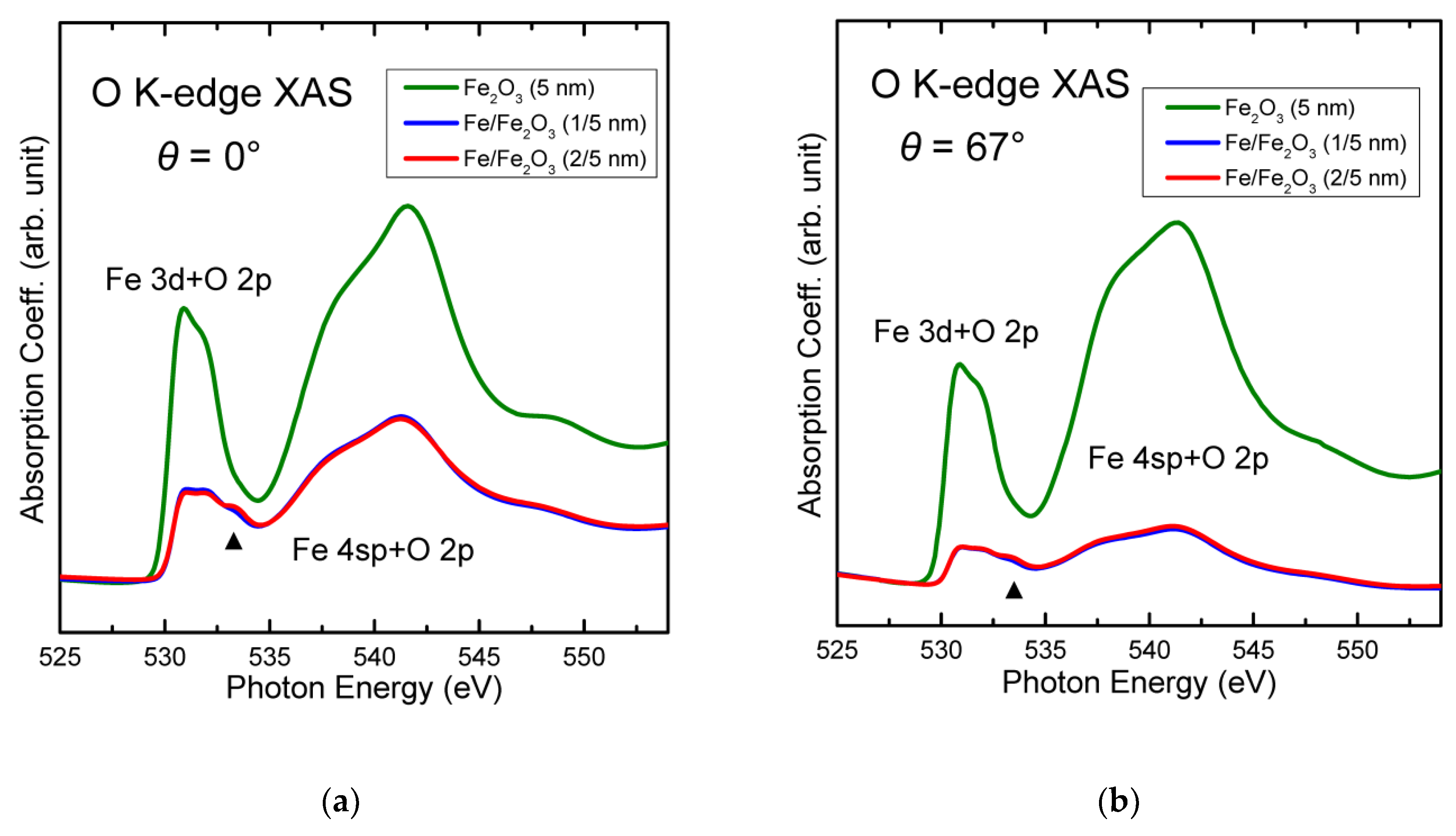
Figure 3 shows the O K-edge XAS spectra of 1/5 nm and 2/5 nm samples together with Fe2O3 taken at (a) θ = 0° and (b) θ = 67°. O K-edge XAS reflects the electron transition from O 1s core level to O 2p unoccupied states that are hybridized with the orbitals of neighboring ions (here, Fe). The lower energy part (529–535 eV) represents the Fe 3d state hybridized with O 2p while the higher energy part (535–545 eV) does Fe 4sp state hybridized with O 2p. The lower energy part in the spectrum of Fe2O3 exhibits the typical two-peak structure of Fe3+ (d5, high spin), which can be attributed to t2g-eg split in the case of octahedral sites or e-t2 split in the case of tetrahedral sites [16,32].
From comparison of Figure 3a with Figure 3b, it was noticed that the overall intensity of the Fe/Fe2O3 films relative to that of Fe2O3 decreased with increasing θ. The angle dependence can be interpreted as a finite probing depth effect, in which the information from the deep layers cannot be collected as much as those from the surface, particularly when the incident X-rays are grazing to the sample plane [33]. Thus, the significant decrease in overall intensity of the Fe/Fe2O3 samples in Figure 3b compared to Figure 3a (approximately by half) suggests that the O K-edge XAS signals are mostly from deep below the Fe layers. Since the O K-edge XAS signals manifest that there exist Fe-oxide, it can be told that certain Fe-oxide exists at the Fe/Fe2O3 interface. Any surface oxidation effect is not consistent with the angle dependence.
It should be noted that there are additional Fe 3d states observed at 534 eV (highlighted by triangles) in the spectra of the two Fe/Fe2O3 samples, indicative of the distinct chemistry of the interface oxide from Fe2O3. No such peaks were reported in stoichiometric oxides, including FeO, Fe3O4, or Fe2O3 [21,34,35,36,37,38]. Therefore, the high energy states should be attributed to an additional energy level splitting due to a lower Fe-O coordination symmetry, which can be the case of sub-stoichiometric oxide, FeOx<1 [9,32,39,40]. The formation of FeOx at the Fe/Fe2O3 interface is plausible because Fe-oxides are generally more stable than Fe metal thermodynamically, and O would be easily supplied by Fe2O3 to partially oxidize Fe near the interface region.
Figure 3 shows that the onsets of the lowest energy peaks in the spectra of Fe/Fe2O3 samples are higher (by 0.3 eV) than that of Fe2O3. The higher onset energy suggests that the interfacial FeOx is an insulator. Besides, no noticeable peak or dip features, other than those for Fe or γ-Fe2O3, were observed in the Fe L-edge XMCD data (Figure 2b) in spite of nonzero nominal Fe valence in FeOx (+2x > 0). This implies that the FeOx most plausibly has negligible FM order, i.e., should be almost an antiferromagnet [30,40,41]. Therefore, to summarize, an AFM FeOx is formed spontaneously between the FM Fe and wFM Fe2O3 layers. This finding is illustrated in the right panel of Figure 1.

4. Discussion

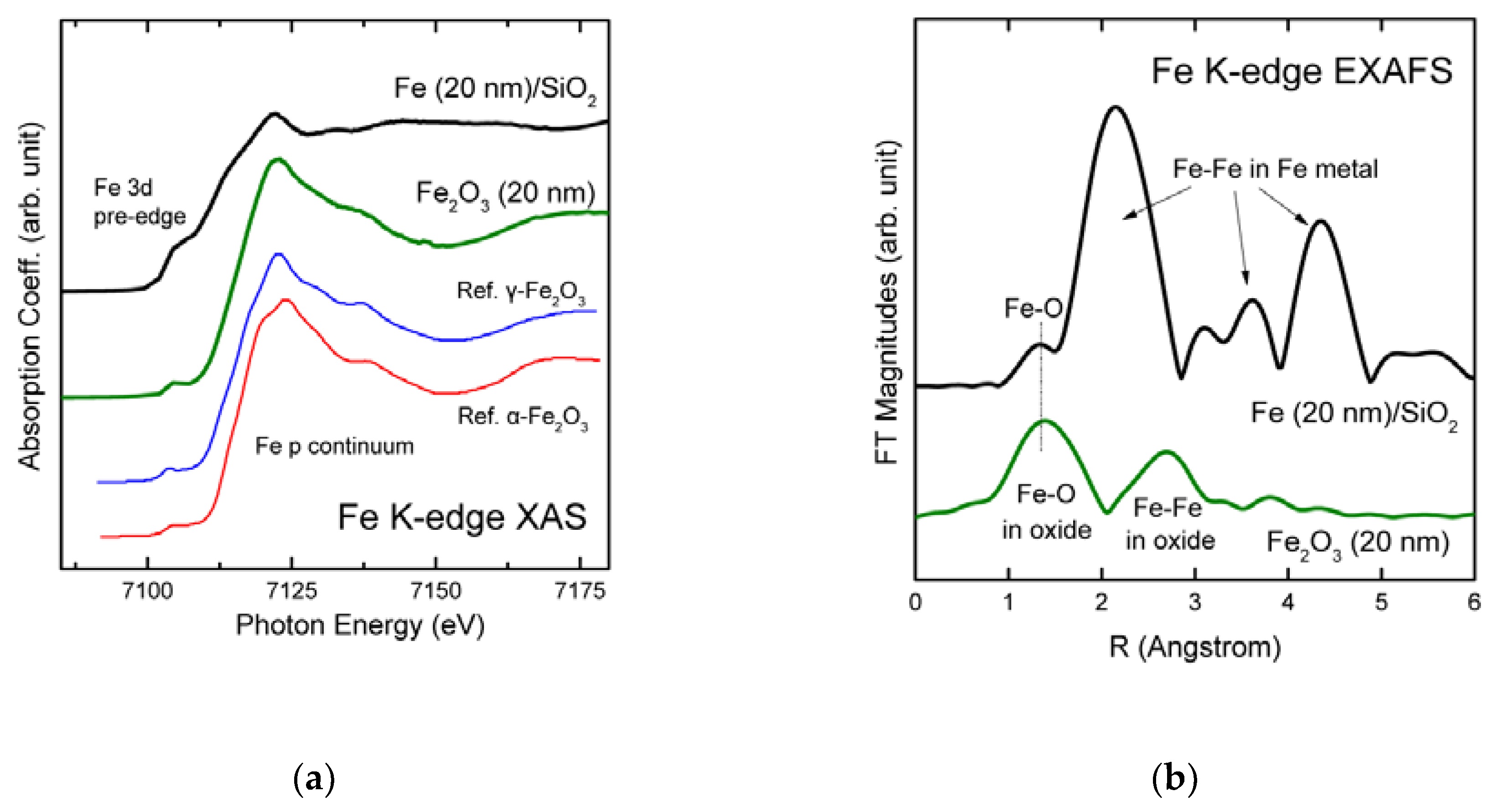
Regarding the origin of the interfacial FeOx, it needs to be clarified whether certain external factors in the growth process, e.g., remnant oxygen gas in the vacuum chamber, or uncleaned evaporation source, are responsible for the oxidation or not. Figure 4a shows the Fe K-edge XAS spectra of a thick Fe (20 nm) and Fe2O3 (20 nm) films on SiO2 prepared by the same growth condition, except for the increased thickness to enhance the signals of the hard XAS. Fe K-edge XAS reflects the transition from Fe 1s core level to Fe 3d (pre-edge region 7110 eV) or p-continuum (>7110 eV), the fine structures of which result from the virtual scatterings of final state electrons with the neighboring atoms (here, O or Fe). Thus, the local structures near Fe can be analyzed by inspecting the lineshapes. The lineshape of the Fe sample was very similar to a reference Fe foil, while that of the Fe2O3 sample was likely a combination of α- and γ-Fe2O3 [31,42,43,44]. The spectra of reference α- and γ-Fe2O3 taken from Sanson [45] are appended in the figure.
Figure 4b shows the Fourier-transformed (FT) extended X-ray absorption fine structures (EXAFS) magnitudes of the two samples. The FT was processed within a range of electron momentum k = 0–10 Å−1 on k2-weighted EXAFS oscillations by using ATHENA [46]. The coordination shells can be identified according to the phase-uncorrected interatomic distance (R) in Å. For instance, the peak at R ≈ 1.4 Å can be attributed to Fe–O bonds, while the peak at R ≈ 2.1 Å can be to the shortest Fe–Fe bonds in a bcc Fe metal. It is clearly shown in the FT spectrum that Fe sample is most likely in the form of Fe metal (short Fe–Fe bonds), while a small portion of Fe-oxide (Fe–O bonds) can exist. The latter might reflect the oxidized Fe in Fe/SiO2 film, which is already demonstrated in Figure 2. Nevertheless, Figure 4b conclusively shows that the composition of the Fe film was dominantly Fe metal. Hence, it can be concluded that the sample growth process was under control so that the Fe layers were not oxidized readily in the growth stage. Thus, the formation of FeOx should be regarded as a consequence of the interfacial interactions with Fe2O3.
Figure 5 shows the Fe L2,3-edge XMCD spectra of (a,b) Fe/Fe2O3 (1/5 nm and 2/5 nm) and (c) Fe (5 nm) taken at different θ’s, after normalization by the XAS intensity in order to represent the magnetism per Fe atom. For all the samples, the intensities of the dips for Fe metal (≈709 eV) and γ-Fe2O3 (≈711 eV) and the peaks for γ-Fe2O3 (≈710 eV) increase monotonically with increasing θ from –23° to +67°.
In principle, such an angle dependence can be ascribed either to the poling efficiency of the magnetic moments (both spin and angular moments) or to the spin sensitivity of the XMCD measurement itself [47]. The former concern is for cases where the coercivity (threshold value of B to align the spins) is large and comparable with B, so that the size of the FM spins increases in accordance with B’s along the magnetic easy-axis of the specimen. In this case, the overall XMCD intensity would increase as a function of the angle between B and the sample’s easy direction. On the other hand, the latter concern is that the circularly polarized X-rays can excite spins only parallel or antiparallel to the beam, so that the magnetic contrast observed in the XMCD appears inherently as a function of the angle between X-rays and the sample’s easy-axis. In this case, the overall XMCD intensity will increase with the absolute value of θ.
Since the XMCD intensities of the Fe/Fe2O3 samples increased with increasing θ, not the absolute value of θ, it can be concluded that the ultrathin Fe/Fe2O3 samples fall under the former case, i.e., the preferred orientations of the magnetic moments were in the sample planes for both the samples. For the case of the Fe (5 nm) sample, the two angle dependences would compete at θ < 0° to result in almost equal intensity of the θ = –23° and θ = 0° spectra.
Compared to 2/5 nm sample (Figure 5b), the θ dependence is less abrupt in the 1/5 nm sample (Figure 5a). This reflects the weaker (more robust) tendency of the in-plane magnetization in 1/5 nm (2/5 nm) sample. As the thickness of the Fe layer decreases, the magnetic anisotropy tends to change from an in-plane magnetization to a perpendicular magnetization with a crossover thickness 1 nm [29,48]. Moreover, the presence of FeOx might reduce the magnetic volume of the heterostructure effectively, so as to expedite the tendency of the perpendicular magnetization. Therefore, it is reasonable that the magnetic anisotropy in 1/5 nm sample is less significant than 2/5 nm sample.
In conclusion, the XAS/XMCD study on ultrathin Fe/Fe2O3 films provides unequivocal information on the chemistry, magnetism, and the local structures in the heterostructure system. The results of the detailed analyses indicate that the resultant composition is FM Fe/AFM FeOx/wFM (α+γ)-Fe2O3, the spin orders of which are depicted with the arrows in the right panel of Figure 1. Although it is difficult to quantify the thickness of FeOx in 1/5 nm and 2/5 nm samples (due to small volume), the EXAFS data for thick Fe/SiO2 implies that the Fe–O intensity is a few tens of percent of Fe2O3, suggesting FeOx can form up to a few-nm in thickness. The AFM FeOx layer can act as a physical barrier for spin transport from FM Fe to wFM Fe2O3, influencing the efficiency of the spin injection. Thereby, the e-beam growth of the FM/wFM (or AFM) heterostructure system can be a promising method for tailoring the spin tunneling properties for spintronics application by utilizing the parasitic FeOx formation.

Author Contributions

Conceptualization, D.-Y.C.; methodology, W.G.P.; formal analysis, A.Y.M.; writing, A.Y.M. and D.-Y.C. All authors have read and agreed to the published version of the manuscript.

Funding

This research was funded by National Research Foundation of Korea, grant number NRF-2018R1D1A1B07043427 and by Research Base Construction Fund Support Program in Jeonbuk National University in 2020.

Conflicts of Interest

The authors declare no conflict of interest. The funders had no role in the design of the study; in the collection, analyses, or interpretation of data; in the writing of the manuscript; or in the decision to publish the results.

References

  1. Khurshid, H.; Phan, M.-H.; Mukherjee, P.; Srikanth, H. Tuning exchange bias in Fe/γ-Fe2O3 core-shell nanoparticles: Impacts of interface and surface spins. Appl. Phys. Lett. 2014, 104, 072407. [Google Scholar] [CrossRef] [Green Version]
  2. Crisan, O.; von Haeften, K.; Ellis, A.; Binns, C. Structure and magnetic properties of Fe/Fe oxide clusters. J. Nanopart. Res. 2008, 10, 193–199. [Google Scholar] [CrossRef] [Green Version]
  3. Kaur, M.; McCloy, J.S.; Qiang, Y. Exchange bias in core-shell iron-iron oxide nanoclusters. J. Appl. Phys. 2013, 113, 17D715. [Google Scholar] [CrossRef]
  4. Signorini, L.; Pasquini, L.; Boscherini, F.; Bonetti, E.; Letard, I.; Brice-Profeta, S.; Sainctavit, P. Local magnetism in granular iron/iron oxide nanostructures by phase-and site-selective x-ray magnetic circular dichroism. Phys. Rev. B 2006, 74, 014426. [Google Scholar] [CrossRef]
  5. Tang, D.; Wang, P.; Speriosu, V.; Le, S.; Kung, K. Spin-valve RAM cell. IEEE Trans. Magn. 1995, 31, 3206–3208. [Google Scholar] [CrossRef]
  6. Parkin, S.S.P.; Roche, K.P.; Samant, M.G.; Rice, P.M.; Beyers, R.B.; Scheuerlein, R.E.; O’sullivan, E.J.; Brown, S.L.; Bucchigano, J.; Abraham, D.W.; et al. Exchange-biased magnetic tunnel junctions and application to nonvolatile magnetic random access memory. J. Appl. Phys. 1999, 85, 5828–5833. [Google Scholar] [CrossRef]
  7. Quindeau, A.; Fina, I.; Marti, X.; Apachitei, G.; Ferrer, P.; Nicklin, C.; Pippel, E.; Hesse, D.; Alexe, M. Four-state ferroelectric spin-valve. Sci. Rep. 2015, 5, 9749. [Google Scholar] [CrossRef] [Green Version]
  8. Wang, S.; Han, G.; Yu, G.; Jiang, Y.; Wang, C.; Kohn, A.; Ward, R. Evidence for FeO formation at the Fe/MgO interface in epitaxial TMR structure by X-ray photoelectron spectroscopy. J. Magn. Magn. Mater. 2007, 310, 1935–1936. [Google Scholar] [CrossRef]
  9. Telesca, D.; Sinkovic, B.; Yang, S.-H.; Parkin, S. X-ray studies of interface Fe-oxide in annealed MgO based magnetic tunneling junctions. J. Electron. Spectrosc. Relat. Phenom. 2012, 185, 133–139. [Google Scholar] [CrossRef]
  10. Lu, Y.; Claydon, J.S.; Ahmad, E.; Xu, Y.; Thompson, S.M.; Wilson, K.; van der Laan, G. XPS and XMCD study of Fe3O4/GaAs interface. IEEE Trans. Magn. 2005, 41, 2808–2810. [Google Scholar] [CrossRef] [Green Version]
  11. Kim, D.; Lee, H.; Kim, G.; Koo, Y.; Jung., J.; Shin, H.; Kim, J.-Y.; Kang, J.-S. Interface electronic structures of BaTiO3@ X nanoparticles (X= γ-Fe2O3, Fe3O4, α-Fe2O3, and Fe) investigated by XAS and XMCD. Phys. Rev. B 2009, 79, 033402. [Google Scholar] [CrossRef]
  12. Genuzio, F.; Menteş, T.; Freindl, K.; Spiridis, N.; Korecki, J.; Locatelli, A. Chemistry-dependent magnetic properties at the FeNi oxide–metal interface. J. Mater. Chem. C 2020, 8, 5777–5785. [Google Scholar] [CrossRef]
  13. Tong, H.; Qian, C.; Miloslavsky, L.; Funada, S.; Shi, X.; Liu, F.; Dey, S. Studies on antiferromagnetic/ferromagnetic interfaces. J. Magn. Magn. Mater. 2000, 209, 56–60. [Google Scholar] [CrossRef]
  14. Robbennolt, S.; Nicolenco, A.; Mercier Fernandez, P.; Auffret, S.; Baltz, V.; Pellicer, E.; Menéndez, E.; Sort, J. Electric field control of magnetism in iron oxide nanoporous thin films. ACS Appl. Mater. Interfaces 2019, 11, 37338–37346. [Google Scholar] [CrossRef] [PubMed]
  15. Li, P.; Xia, C.; Zhu, Z.; Wen, Y.; Zhang, Q.; Alshareef, H.N.; Zhang, X.X. Ultrathin Epitaxial Ferromagnetic γ-Fe2O3 Layer as High Efficiency Spin Filtering Materials for Spintronics Device Based on Semiconductors. Adv. Funct. Mater. 2016, 26, 5679–5689. [Google Scholar] [CrossRef]
  16. Lu, Y.; Claydon, J.; Xu, Y.; Thompson, S.; Wilson, K.; Van der Laan, G. Epitaxial growth and magnetic properties of half-metallic Fe3O4 on GaAs (100). Phys. Rev. B 2004, 70, 233304. [Google Scholar] [CrossRef]
  17. Lu, Y.; Ahmad, E.; Xu, Y.; Thompson, S.M. Annealing-induced Fe oxide nanostructures on GaAs. IEEE Trans. Magn. 2005, 41, 3328–3330. [Google Scholar] [CrossRef] [Green Version]
  18. Hassan, S.S.; Xu, Y.; Wu, J.; Thompson, S.M. Epitaxial Growth and Magnetic Properties of Half-Metallic Fe3O4 on Si (100) Using MgO Buffer Layer. IEEE Trans. Magn. 2009, 45, 4357–4359. [Google Scholar] [CrossRef]
  19. Paul, M.; Müller, A.; Ruff, A.; Schmid, B.; Berner, G.; Mertin, M.; Sing, M.; Claessen, R. Probing the interface of Fe3O4/GaAs thin films by hard x-ray photoelectron spectroscopy. Phys. Rev. B 2009, 79, 233101. [Google Scholar] [CrossRef] [Green Version]
  20. Nozaki, T.; Kubota, H.; Fukushima, A.; Yuasa, S. Interface engineering using an Fe oxide insertion layer for growing a metastable bcc-Co on MgO (001). Appl. Phys. Lett. 2015, 106, 022405. [Google Scholar] [CrossRef]
  21. Wong, P.J.; Zhang, W.; Wang, K.; van der Laan, G.; Xu, Y.; van der Wiel, W.G.; de Jong, M.P. Electronic and magnetic structure of C60/Fe3O4 (001): A hybrid interface for organic spintronics. J. Mater. Chem. C 2013, 1, 1197–1202. [Google Scholar] [CrossRef] [Green Version]
  22. Jiménez-Villacorta, F.; Prieto, C.; Huttel, Y.; Telling, N.; van der Laan, G. X-ray magnetic circular dichroism study of the blocking process in nanostructured iron-iron oxide core-shell systems. Phys. Rev. B 2011, 84, 172404. [Google Scholar] [CrossRef] [Green Version]
  23. Stöhr, J.; Siegmann, H. Magnetism: From Fundamentals to Nanoscale Dynamics; Springer: Berlin/Heidelberg, Germany, 2006. [Google Scholar]
  24. Miedema, P.S.; De Groot, F.M. The iron L edges: Fe 2p X-ray absorption and electron energy loss spectroscopy. J. Electron. Spectrosc. Relat. Phenom. 2013, 187, 32–48. [Google Scholar] [CrossRef]
  25. Pauling, L.; Hendricks, S.B. The crystal structures of hematite and corundum. J. Am. Chem. Soc. 1925, 47, 781–790. [Google Scholar] [CrossRef]
  26. Schulz, D.L.; McCarthy, G.J. X-ray powder data for an industrial maghemite (γ-Fe2O3). Powder Diffr. 1988, 3, 104–105. [Google Scholar] [CrossRef]
  27. Lee, E.; Kim, D.; Hwang, J.; Lee, K.; Yoon, S.; Suh, B.; Hyun Kim, K.; Kim, J.-Y.; Jang, Z.; Kim, B.; et al. Size-dependent structural evolution of the biomineralized iron-core nanoparticles in ferritins. Appl. Phys. Lett. 2013, 102, 133703. [Google Scholar] [CrossRef] [Green Version]
  28. Brice-Profeta, S.; Arrio, M.-A.; Tronc, E.; Menguy, N.; Letard, I.; dit Moulin, C.C.; Nogues, M.; Chanéac, C.; Jolivet, J.-P.; Sainctavit, P. Magnetic order in γ-Fe2O3 nanoparticles: A XMCD study. J. Magn. Magn. Mater. 2005, 288, 354–365. [Google Scholar] [CrossRef]
  29. Lee, J.-M.; Kim, J.-Y.; Yang, S.-U.; Park, B.-G.; Park, J.-H.; Oh, S.-J.; Kim, J.-S. Magnetism of pristine Fe films on GaAs (100). Phys. Rev. B 2007, 76, 052406. [Google Scholar] [CrossRef] [Green Version]
  30. Radaelli, G.; Petti, D.; Plekhanov, E.; Fina, I.; Torelli, P.; Salles, B.; Cantoni, M.; Rinaldi, C.; Gutiérrez, D.; Panaccione, G.; et al. Electric control of magnetism at the Fe/BaTiO3 interface. Nat. Commun. 2014, 5, 1–9. [Google Scholar] [CrossRef] [Green Version]
  31. Cao, L.; Jiang, Z.-X.; Du, Y.-H.; Yin, X.-M.; Xi, S.-B.; Wen, W.; Roberts, A.P.; Wee, A.T.; Xiong, Y.-M.; Liu, Q.-S.; et al. Origin of magnetism in hydrothermally aged 2-line ferrihydrite suspensions. Environ. Sci. Technol. 2017, 51, 2643–2651. [Google Scholar] [CrossRef]
  32. Pollak, M.; Gautier, M.; Thromat, N.; Gota, S.; Mackrodt, W.; Saunders, V. An in-situ study of the surface phase transitions of α-Fe2O3 by X-ray absorption spectroscopy at the oxygen K edge. Nucl. Instrum. Methods Phys. Res. B 1995, 97, 383–386. [Google Scholar] [CrossRef]
  33. Trainor, T.P.; Templeton, A.S.; Eng, P.J. Structure and reactivity of environmental interfaces: Application of grazing angle X-ray spectroscopy and long-period X-ray standing waves. J. Electron. Spectrosc. Relat. Phenom. 2006, 150, 66–85. [Google Scholar] [CrossRef]
  34. Bora, D.K.; Braun, A.; Erat, S.; Ariffin, A.K.; Löhnert, R.; Sivula, K.; Töpfer, J.; Grätzel, M.; Manzke, R.; Graule, T.; et al. Evolution of an oxygen near-edge X-ray absorption fine structure transition in the upper Hubbard band in α-Fe2O3 upon electrochemical oxidation. J. Phys. Chem. C 2011, 115, 5619–5625. [Google Scholar] [CrossRef] [Green Version]
  35. Yang, C.K.; Chiou, J.W.; Tsai, H.M.; Pao, C.W.; Jan, J.C.; Ray, S.C.; Yeh, C.L.; Huang, K.C.; Hsueh, H.C.; Pong, W.F.; et al. Electronic structure and magnetic properties of Al-doped Fe3O4 films studied by X-ray absorption and magnetic circular dichroism. Appl. Phys. Lett. 2005, 86, 062504. [Google Scholar] [CrossRef]
  36. Chen, C.L.; Dong, C.L.; Rao, S.M.; Chern, G.; Chen, M.C.; Wu, M.K.; Chang, C.L. Investigation of the valence states of Fe and Co in Fe1−xCoxOy (0 < x < 1) thin films by x-ray absorption spectroscopy. J. Condens. Matter Phys. 2008, 20, 255236. [Google Scholar] [CrossRef]
  37. Kumar, S.; Kumar, R.; Thakur, P.; Chae, K.H.; Sharma, S.K. Electronic structure studies of Mg0. 95Mn0. 05Fe2−2xTi2xO4 (0 ⩽ x ⩽ 0.8). J. Magn. Magn. Mater. 2008, 320, e121–e124. [Google Scholar] [CrossRef]
  38. Shen, S.; Zhou, J.; Dong, C.-L.; Hu, Y.; Tseng, E.N.; Guo, P.; Guo, L.; Mao, S.S. Surface engineered doping of hematite nanorod arrays for improved photoelectrochemical water splitting. Sci. Rep. 2014, 4, 6627. [Google Scholar] [CrossRef]
  39. Gautam, S.; Kane, S.N.; Park, B.G.; Kim, J.Y.; Varga, L.; Song, J.H.; Chae, K.H. XAS and XMCD studies of amorphous FeCo-based ribbons. J. Non Cryst. Solids 2011, 357, 2228–2231. [Google Scholar] [CrossRef]
  40. Miyokawa, K.; Saito, S.; Katayama, T.; Saito, T.; Kamino, T.; Hanashima, K.; Suzuki, Y.; Mamiya, K.; Koide, T.; Yuasa, S. X-ray absorption and X-ray magnetic circular dichroism studies of a monatomic Fe (001) layer facing a single-crystalline MgO (001) tunnel barrier. Jpn. J. Appl. Phys. 2004, 44, L9–L11. [Google Scholar] [CrossRef]
  41. Xu, M.; Li, M.; Khanal, P.; Habiboglu, A.; Insana, B.; Xiong, Y.; Peterson, T.; Myers, J.C.; Ortega, D.; Qu, H.; et al. Voltage-Controlled Antiferromagnetism in Magnetic Tunnel Junctions. Phys. Rev. Lett. 2020, 124, 187701. [Google Scholar] [CrossRef]
  42. Pellegrain, E.; Hagelstein, M.; Doyle, S.; Moser, H.; Fuchs, J.; Vollath, D.; Schuppler, S.; James, M.; Saxena, S.; Niesen, L.; et al. Characterization of Nanocrystalline γ-Fe2O3 with Synchrotron Radiation Techniques. Phys. Status Solidi B 1999, 215, 797–801. [Google Scholar] [CrossRef]
  43. Olimov, K.; Falk, M.; Buse, K.; Woike, T.; Hormes, J.; Modrow, H. X-ray absorption near edge spectroscopy investigations of valency and lattice occupation site of Fe in highly iron-doped lithium niobate crystals. J. Condens. Matter Phys. 2006, 18, 5135. [Google Scholar] [CrossRef]
  44. Bora, D.K.; Braun, A.; Erat, S.; Safonova, O.; Graule, T.; Constable, E.C. Evolution of structural properties of iron oxide nano particles during temperature treatment from 250 oC–900 oC: X-ray diffraction and Fe K-shell pre-edge X-ray absorption study. Curr. Appl. Phys. 2012, 12, 817–825. [Google Scholar] [CrossRef] [Green Version]
  45. Sanson, A. Comment on ‘On the discrimination between magnetite and maghemite by XANES measurements in fluorescence mode’. Meas. Sci. Technol. 2013, 24, 118001. [Google Scholar] [CrossRef]
  46. Ravel, B.; Newville, M. ATHENA, ARTEMIS, HEPHAESTUS: Data analysis for X-ray absorption spectroscopy using IFEFFIT. J. Synchrotron Radiat. 2005, 12, 537–541. [Google Scholar] [CrossRef] [Green Version]
  47. Stöhr, J. Exploring the microscopic origin of magnetic anisotropies with X-ray magnetic circular dichroism (XMCD) spectroscopy. J. Magn. Magn. Mater. 1999, 200, 470–497. [Google Scholar] [CrossRef]
  48. Kim, W.; Choi, J.H.; Nahm, T.U.; Song, S.H.; Oh, S.J. Magnetic properties of ultrathin Fe films on Pt (111) and Pd (111): A surface magneto-optic Kerr effect study. J. Korean Phys. Soc. 2004, 44, 722–725. [Google Scholar]
Figure 1. Schematics of the geometry of X-ray absorption spectroscopy (XAS) and X-ray magnetic circular dichroism (XMCD) measurements, and the atomic structures and magnetism of e-beam evaporated Fe/Fe2O3 films according to the results of the analyses in this work.
Magnetochemistry 06 00033 g001
Figure 2. (a) Fe L2,3-edge XAS spectra and (b) Fe L2,3-edge XMCD spectra un-normalized. The spectra of Fe (1 or 2 nm)/Fe2O3, and Fe (5 nm)/SiO2 samples exhibit the signature of Fe metal (or FeOx<1) as well as that of mixed (α+γ)-Fe2O3, suggesting a substantial degree of oxidation of the top Fe layers.
Magnetochemistry 06 00033 g002
Figure 3. Un-normalized O K-edge XAS spectra taken (a) at θ = 0° (beam normal) and (b) at θ = 66.5°. Oxidized Fe formed FeOx<1 mainly at the Fe/Fe2O3 interface, which exhibits a fundamentally different electronic structure from (α+γ)-Fe2O3.
Magnetochemistry 06 00033 g003
Figure 4. (a) Fe K-edge near-edge XAS spectra and (b) the Fourier-transformed extended X-ray absorption fine structures (FT EXAFS) magnitudes of the thick Fe and Fe2O3 films. The results show the films are indeed in their respective intended compositions, implying that the spontaneous Fe oxidations observed in the soft XAS data cannot be attributed to possible oxidation during the growth process.
Magnetochemistry 06 00033 g004
Figure 5. Fe L2,3-edge XMCD spectra normalized as to represent the signals per Fe atom at different θ’s of (a) 1/5 nm, (b) 2/5 nm and (c) Fe (5 nm) samples. Overall XMCD intensity increases according to the incidence angle of the magnetic field (not the X-rays), indicating that the easy direction of the Fe and γ-Fe2O3 spins lie in the sample planes for all the samples.
Magnetochemistry 06 00033 g005