Article

Spin Crossover in Bipyridine Derivative Bridged One-Dimensional Iron(III) Coordination Polymer

1
Department of Chemistry, Faculty of Science, Fukuoka University, 8-19-1 Nanakuma, Jonan-ku, Fukuoka 814-0180, Japan
2
Chemistry Course, Department of Science, Faculty of Science and Engineering, Kindai University, 3-4-1 Kowakae, Higashi-Osaka, Osaka 577-8502, Japan
3
RI center, Fukuoka University, 8-19-1 Nanakuma, Jonan-ku, Fukuoka 814-0180, Japan
*
Author to whom correspondence should be addressed.
Magnetochemistry 2020, 6(3), 29; https://doi.org/10.3390/magnetochemistry6030029
Submission received: 8 June 2020 / Revised: 24 June 2020 / Accepted: 28 June 2020 / Published: 1 July 2020
(This article belongs to the Special Issue Stimuli-Responsive Magnetic Molecular Materials)

Abstract

:
Herein, the syntheses, solid-state molecular structures, and characterization of two types of one-dimensional FeIII coordination polymers showing thermally induced spin crossover are reported. The reaction of [Fe(acen)Cl] (acen2− = N,N′-ethylenebis(acetylacetonylideneaminate) with 3,3′-bpy or 4,4′-bpy (bpy = bipyridine) produced zigzag and linear one-dimensional chain complexes, [Fe(acen)(3,3′-bpy)][BPh4] (1) or [NEt3H][Fe(acen)(4,4′-bpy)][BPh4]2·0.5(4,4′-bpy) (2), respectively, as confirmed by single crystal X-ray diffraction analysis. Variable-temperature single crystal X-ray diffraction measurements, continuous-wave X-band electron paramagnetic resonance (EPR) spectra, 57Fe Mössßauer spectra, and DC magnetic susceptibility data revealed that complex 1 exhibited a gradual and complete spin crossover at a transition temperature of 212 K, while complex 2 undergoes an incomplete spin crossover even at 400 K.

Graphical Abstract

1. Introduction

First-row transition metal complexes with a d4–d7 electron configuration can adopt two electron configurations, high spin (HS), and low spin (LS), which differ in terms of spin ground states. When both HS and LS states are energetically similar, switching between the two spin states reversibly occurs when external stimuli including temperature, pressure, light irradiation, and magnetic fields are applied in a process known as spin crossover (SCO) [1,2,3,4,5,6,7,8,9,10,11]. Complexes exhibiting SCO have attracted significant attention because of their wide range of potential applications, such as in thermochromic displays, actuators, (multi-modal) sensors, and data storage systems, as well as in molecular spintronics [12,13,14,15,16,17,18,19]. Moreover, a number of approaches of the nanostructural patterning based on SCO complexes have also been studied [20,21,22,23,24,25,26].
The most extensively studied SCO moieties are FeII complexes with a 3d6 electron configuration because of a distinct spin transition from a paramagnetic HS state (S = 2) to a diamagnetic LS state (S = 0), or vice versa, which generally show abrupt spin transitions and high cooperativity in the solid state [1,2,3,4,5,6,7,8,9,10,11]. In contrast, FeIII SCO complexes have not been as extensively reported as FeII SCO complexes, mostly due to the lack of cooperativity associated with FeIII SCO complexes [27,28]. To overcome this obstacle, several fascinating FeIII SCO complexes that exhibit high cooperativity have been realized by introducing relatively weak supramolecular contacts, including hydrogen bonding and π-π stacking interactions between SCO active centers in the packed crystal. These have also been shown to result in abrupt spin transition, multistep spin transition, wide thermal hysteresis loop, and light-induced excited spin state trapping effect [27,28]. However, previously reported almost FeIII SCO complexes are discrete mononuclear and dinuclear complexes. To further expand the basic understanding of magnetostructural correlations in highly cooperative FeIII SCO complexes, the development of FeIII SCO coordination polymer candidates with structurally high dimensionality is necessary. As an initial attempt to create novel FeIII SCO coordination polymers, a one-dimensional (1D) coordination polymer structure was proposed.
To date, SCO-active FeIII complexes have been explored using Schiff base ligands with a N4O2 coordination environment [27,28]. From this perspective, constructing coordination polymers can be achieved by crystal engineering via combination of an FeIII precursor with a macrocyclic-type Schiff base ligand furnished with a N2O2 coordination donor set, as a building block and a N-donor bridging ligand.
Herein, the syntheses, solid-state molecular structures, and characterization of two new one-dimensional FeIII SCO coordination polymers composed of [Fe(acen)]+ (acen2− = N,N′-ethylenebis(acetylacetonylideneaminate)) as a building block and 3,3′- and 4,4′-bpy (3,3′- and 4,4′-bipyridine) as bridging ligands are reported.

2. Results and Discussion

2.1. Syntheses and General Characterization

The complex, [Fe(acen)(3,3′-bpy)][BPh4] (1), was obtained via stepwise assembly reactions. The first reaction is the synthesis of the cationic FeIII building block, [Fe(acen)]+, which is readily prepared via the reaction of acen2– deprotonated by triethylamine (NEt3) in situ with anhydrous FeCl3 in EtOH [29,30]. Subsequent reaction of [Fe(acen)]+ and 3,3′-bpy with NaBPh4 in a 1:1:1 molar ratio in MeOH afforded dark green block-shaped crystals of 1, suitable for single crystal X-ray diffraction (SCXRD) analysis. Unfortunately, the single crystals suitable for SCXRD analysis were not obtained using the same assembly reaction as that for 1 with 4,4′-bpy instead of 3,3′-bpy. Alternatively, isolation of dark green plate-shaped crystals of [NEt3H][Fe(acen)(4,4′-bpy)][BPh4]2·0.5(4,4′-bpy) (2) via one-pot reaction between H2acen, NEt3, FeCl3, 4,4′-bpy, and NaBPh4 in a 1:2:1:1:1 molar ratio in MeOH was successful.
Both 1 and 2 showed characteristic Fourier-transform infrared (FTIR) peaks, indicating the presence of BPh4 (~700 and 730 cm–1) and a FeIII-coordinated imine moiety (~1580 cm−1), as indicated in Figure S1. The phase purity of 1 and 2 was confirmed via powder X-ray diffraction (PXRD) measurements. Experimental PXRD patterns of polycrystalline 1 and 2 were in agreement with the calculated patterns based on SCXRD data for the complexes. Therefore, no complexes exhibited crystal polymorphism and both polycrystalline samples adopted a uniform crystalline phase (Figure S2). The purities of 1 and 2 were confirmed by elemental analyses (Section 3). Thermogravimetric analyses (TGA) indicated that polycrystalline samples 1 and 2 are thermally stable up to ~420 K (Figure S3).

2.2. Structural Descriptions

The SCXRD measurements of 1 and 2 were performed at 100 and 300 K (Table S1).
At both temperatures, complex 1 crystallized in a monoclinic space group P21/c, with the asymmetric unit containing [Fe(acen)(3,3′-bpy)]+ and a BPh4 anion (upper part of Figure 1a). The FeIII center approximated a distorted octahedral geometry (octahedral distortion parameter, Σ [31]; Σ = 35.82(6) and 36.81(7)° at 100 and 300 K), with an equatorial plane occupied by a N2O2 coordination donor from an acen2− ligand, while each axial site was bridged via two N ends of a 3,3′-bpy ligand in a trans configuration with dihedral angles between pyridine rings of 36.35 and 32.68° at 100 and 300 K, forming a zigzag 1D chain (lower part of Figure 1a). At 100 K, the average Fe–Oequatorial, Fe–Nequatorial, and Fe–Naxial distances were consistent with those in the LS FeIII complexes with similar coordination environments of related FeIII(acen)-type complexes (Table S2) [32,33,34,35,36,37,38]. In contrast, the corresponding distances at 300 K were significantly longer than those observed at 100 K, suggesting a thermally induced SCO between 100 and 300 K (Table 1, Table 2 and Table S2). The changes of these coordination bond distances are comparable with those previously reported for FeIII(acen)-type SCO complexes (Table 2 and Table S2). No lattice solvent molecules were present in the crystal packing and the intrachain FeIII···FeIII separations were determined to be 9.5895(7) and 9.8189(7) Å at 100 and 300 K, respectively. Each zigzag 1D chain of 1 propagated along the crystallographic c axis in the crystal packing, separated by bulky BPh4 anions, with the closest interchain FeIII···FeIII separation of 9.3105(5) and 9.6025(8) Å at 100 and 300 K, respectively (Figure 2a).
Complex 2 crystallized in the monoclinic space group P21/n at 100 and 300 K. The asymmetric unit consisted of a [Fe(acen)(4,4′-bpy)]+ unit, a NEt3H+ cation, two BPh4 anions, and half of a non-coordinating 4,4′-bpy molecule. Similar to 1, the FeIII center was situated in a distorted octahedral geometry (Σ = 32.8(1) and 26.92(9)° at 100 and 300 K, respectively), with the same coordination environment as 1 (the upper part of Figure 1b). No significant changes in the coordination bonds were observed at 100 or 300 K, while coordination bond angles did show some changes (Table 1, Table 2 and Table S2). Unlike 1, the FeIII center linearly coordinated to neighboring FeIII centers via bridging 4,4′-bpy ligands, thereby adopting a linear 1D chain arrangement. Neighboring 1D chains were aligned parallel along the ac direction, with intrachain FeIII···FeIII separations of 11.098(1) and 11.1610(8) Å at 100 and 300 K, respectively (lower part of Figure 1b). Remarkable hydrogen bonding interactions were observed between the NEt3H+ cations and pyridyl N ends of a non-coordinating 4,4′-bpy molecule in the crystal packing (Figure S4). This dense assembly containing cationic 1D chains of [Fe(acen)(4,4′-bpy)]+, BPh4 anions, and hydrogen bonding between NEt3H+ cations and 4,4′-bpy molecule excluded lattice solvent molecules in the crystal packing (Figure 2b). The closest interchain FeIII···FeIII separations were 8.707(1) and 8.9505(8) Å at 100 and 300 K, respectively.
Although the coordination environments of 1 and 2 were similar, with the exception of chain topology and arrangement, different conformations of the C2N2 backbone were observed with respect to the acen2− ligand moiety. In particular, the acen2− geometry of 1 adopted a meso conformation, whereas that of 2 adopted an envelope conformation. Therefore, the substantial distortion of the acen2− geometry induces the LS state for complexes containing [Fe(acen)]+, consistent with previous studies (Table S2) [32,33,34,35,36,37,38].

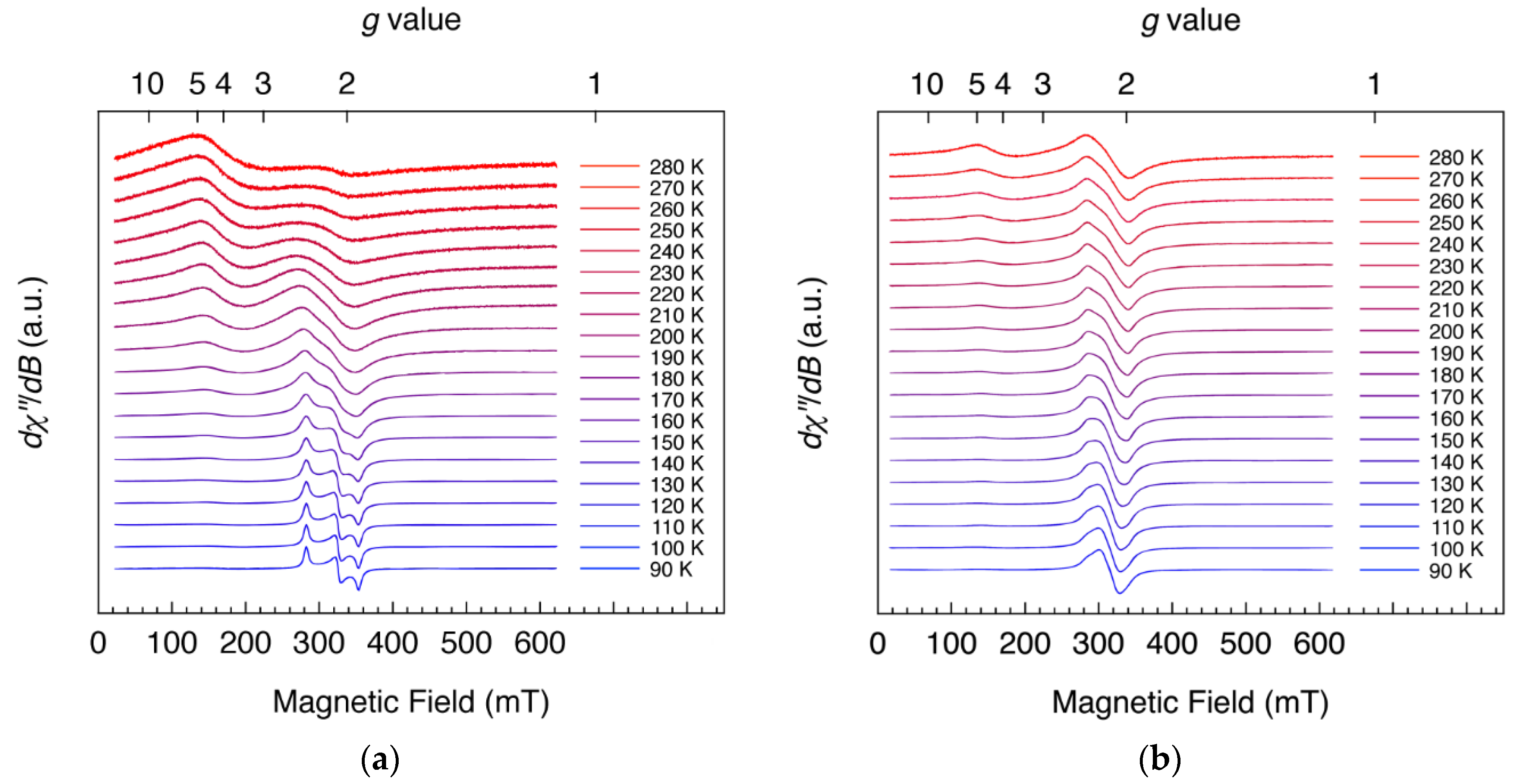
2.3. Continuous-Wave X-Band Electron Paramagnetic Spectroscopy

Continuous-wave X-band electron paramagnetic resonance (CW X-band EPR) spectroscopy is useful, because FeIII complexes with octahedral coordination geometry are Kramers systems, and are generally EPR-active at X-band frequencies (~0.3 cm−1) under magnetic fields (~1 T), regardless of the electron configuration of the FeIII center (i.e., LS (S = 1/2) or HS (S = 5/2)). However, it should be noted that for the HS FeIII center effective g values were obtained, which are only useful in a qualitative sense. The lack of quantitation arises from the dependence on the magnitude of the zero-field splitting energies for the HS state of S = 5/2 [39]. The CW X-band EPR spectra of polycrystalline 1 and 2 were collected at 90–280 K (Figure 3).
At low temperatures, the EPR spectra of 1 can be described as a complete and well-resolved LS state, S = 1/2, indicating highly rhombicity of the gx, gy, and gz values (1.919, 2.086, and 2.408, respectively), and an average g value of 2.138 (Figure 3a). The observed g values are characteristic of LS FeIII complexes with similar coordination environments [40,41,42,43,44,45,46,47]. With the increasing temperature, the EPR signals centered at the average g value of 2.138 for 1 gradually broadened from rhombic to isotropic symmetry, and became steadily indistinct. Instead, a new signal centered at approximately g = 4.3 appeared and steadily grew, corresponding to a gradual change from the LS to HS state with highly rhombicity (ED/3; where D and E are the axial and rhombic zero-field splitting parameters, respectively) [48], ultimately becoming very broad dominated by the HS signal at 280 K.
The low temperature EPR spectra of 2 showed isotropic signals centered at g = 2.137, which is considerably similar to the average g value of 2.138 observed for the LS state of 1 (Figure 3b). With increasing temperature, the EPR signals of 2 showed slight line broadening, while a strongly rhombic HS signal at g ≈ 4.3 appeared only at the high temperature region and was not overly dominant. Therefore, the FeIII center of 2 remained in a quasi-LS state throughout the temperature range measured.
Therefore, both 1 and 2 undergo thermally induced SCO from LS to HS in the solid-state. These results also suggest that 1 exhibited a gradual and complete SCO, while 2 displayed incomplete SCO over the temperature range measured herein.

2.4. Magnetic Properties

To further prove thermally induced SCO for 1 and 2, the temperature dependence of molar DC magnetic susceptibility (χM), measurements were performed using a SQUID magnetometer on the polycrystalline samples over the temperature range of 2–400 K, under an applied field of 0.5 T at a scan rate of 2 K min−1 (Figure 4).
At 2 K, the χMT (χM times temperature) value for 1 was determined to be 0.52 cm3 K mol−1, which is consistent with the expected χMT value at g > 2 for an essentially complete LS state for the FeIII center. With the increasing temperature, the χMT values increased steadily, with a more rapid increase in the χMT data between 120 and 300 K, which is indicative of thermally induced SCO. This thermal spin transition (T1/2) occurred at ~210 K without thermal hysteresis, as confirmed by differential scanning calorimetry (DSC; Figure S5). At temperatures of >120 K, the χMT values again gradually increased to 4.24 cm3 K mol−1 at 400 K, characteristic of a fully HS state for the FeIII center. The χMT value for 2 was approximately 0.60 cm3 K mol−1 at 2 K, remaining constant up to 200 K, and then gradually increases to 2.78 cm3 K mol−1 at 400 K. In contrast to 1, the FeIII center of 2 remained in a near LS state even at 400 K (inflection point at ~380 K), undergoing incomplete SCO.
To quantify the energy separation (ΔE) between the zero-point levels of the LS (2T2) and HS (6A1) states in 1, which exhibited complete SCO, the temperature dependence of χMT data was fitted to the following molecular vibrational partition function (Equation (1)) [27,49,50]:
χ M T = 1 8 3 4 g 2 + 8 ( ζ k B T ) 1 [ 1 exp ( 3 2 ζ k B T ) + 105 C exp { ζ k B T ( 1 + Δ E ζ ) } ] 1 + 2 exp ( 3 2 ζ k B T ) + 3 C exp { ζ k B T ( 1 + Δ E ζ ) }
where T is the absolute temperature, g is the Landé g factor, C is the molecular partition function ratio in the LS and HS states, and ζ is the spin-orbit coupling. The adequate parameters determined from fitting the data obtained from 1 are summarized in Table 3 (Figure S6). Compared to values previously reported for related FeIII(acen)-type SCO complexes, those of 1 are similar in magnitude, whereas those of 2 are relatively large in magnitude. These large values of 2, in contrast to 1, likely result from crystal packing differences brought about by the dense assembly. In fact, the difference in the crystal packing between 1 and 2 are to be reflected in the thermodynamic parameters (vide infra).
To determine the thermodynamic parameters associated with the thermally induced SCO for 1, the enthalpy (ΔH) and entropy (ΔS) changes were calculated using the temperature dependence of γHS (molar fraction of the HS state), which also used the following model proposed by Slichter and Drickamer (Equation (2)) [51]:
T = Δ H + Γ ( 1 2 γ HS ) R ( 1 γ HS γ HS ) + Δ S
where T is the absolute temperature, Γ is the cooperativity evaluation parameter (C = Γ/2RT1/2), and R is the gas constant (8.314 J K−1 mol−1). The obtained parameters are listed in Table 4 (Figure S7). The calculated ΔS values were larger than those expected for SCO between the HS and LS states (Rln(2SHS + 1)/(2SLS + 1) = 9.134 J K−1 mol−1), indicating a vibrational contribution to ΔS associated with bond softening (especially in the ligand-metal bonds) via SCO transition [52,53]. The value of Γ for 1 and 2 was absent, indicative of a less cooperative SCO.
While energetic studies of several FeIII(acen)-type SCO complexes have been previously reported, only one report of [Fe(acen)(pin)][BPh4]·3(MeOH) showed a relatively abrupt SCO [37]. However, to date, no structurally characterized example of an FeIII(acen)-type SCO complex has been shown to exhibit abrupt and complete SCO, as well as thermal hysteresis. The parameters associated with SCO for the previously reported FeIII(acen)-type SCO complexes with six-coordinate geometry are summarized in Table 5.

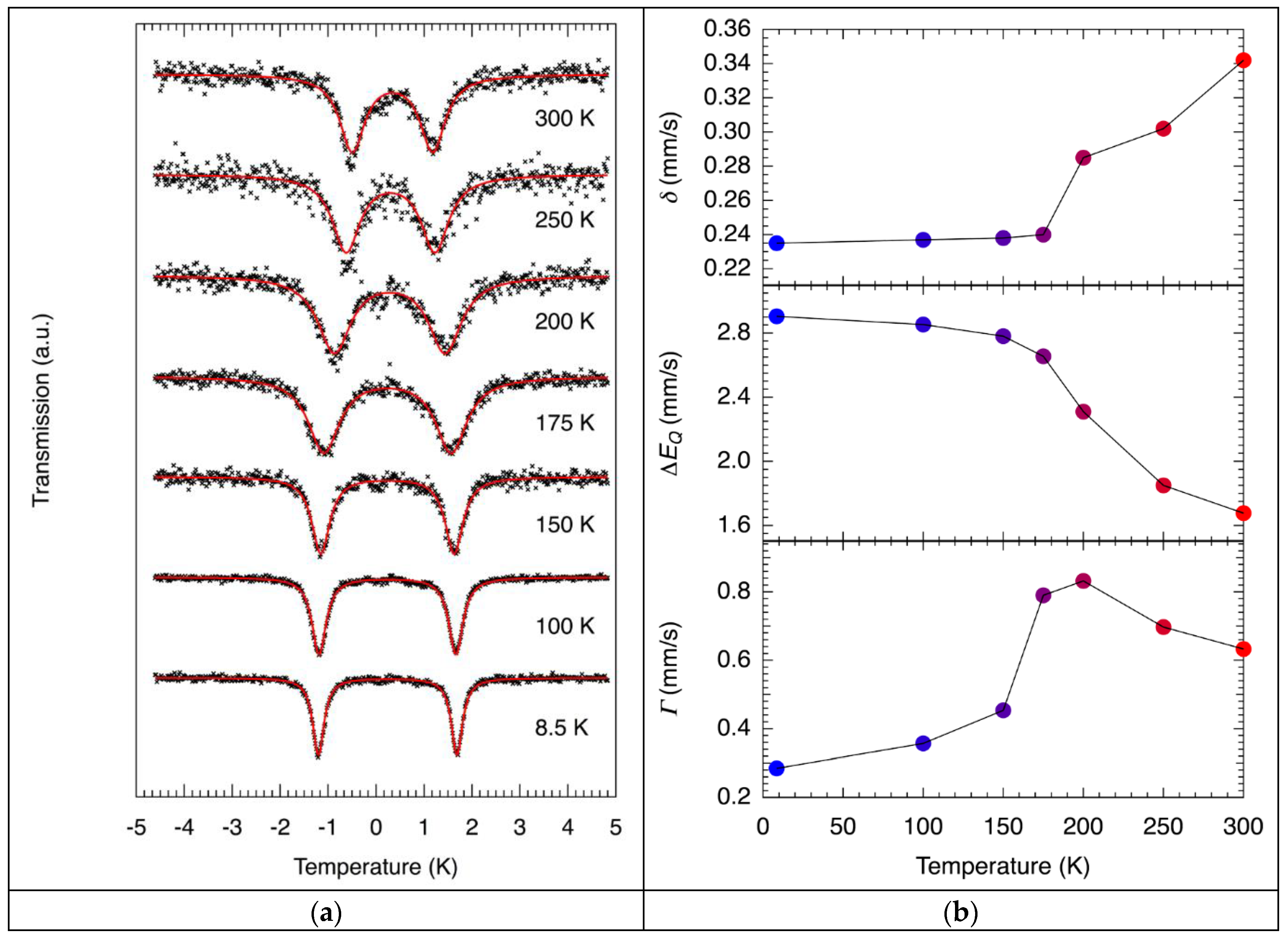
2.5. 57Fe Mössßauer Spectroscopy

For further spectroscopic verification of complete SCO in 1, variable-temperature 57Fe Mössßauer spectra were collected on the polycrystalline solids at select temperatures (Figure 5a) and Table 6). 57Fe Mössßauer spectral data of 2 were not collected as the FeIII center remained the LS state throughout the measured temperature range. Despite undergoing complete SCO, each 57Fe Mössßauer spectrum of 1 at different temperatures contained only a single symmetric doublet. The observation of the single doublet in the 57Fe Mössßauer spectra of FeIII SCO complexes arises from a rapid electronic relaxation between the LS and HS states. Thus, the electronic relaxation of 1 between the two spin states is faster than the time scale of 57Fe Mössßauer spectroscopy (~10−7 s), and the 57Fe Mössßauer spectra report the average between the LS and HS states [49,50]. At high temperatures, the respective 57Fe Mössßauer spectra exhibit an asymmetric quadrupole doublet, due to the spin-lattice relaxation effect [54]. The considerable broadening of the 57Fe Mössßauer spectra with increasing temperature was attributed to the Debye-Waller factor [54]. Indeed, absorption at 300 K is no more than ~1% and all spectra could be fitted with a single quadrupole doublet featuring a single line width. The resulting parameters (isomer shifts, δ, quadrupole splittings, ΔEQ, and line widths, Γ) are listed in Table 2 and shown in Figure 5b. The estimated δ, ΔEQ, and Γ parameters indicated a notable thermal dependence, consistent with the variable-temperature X-band EPR spectra and magnetic susceptibility data (vide supra).

3. Experimental Section

3.1. Material and Methods

Anhydrous FeCl3, MeOH, and EtOH were purchased from Wako Pure Chemical Industries, Ltd. (Osaka, Japan). 3,3′-bpy, 4,4′-bpy, and NaBPh4 were purchased from Tokyo Chemical Industry (TCI) Co., Ltd. (Tokyo, Japan). All chemicals were of reagent grade and were used as received. All reactions and manipulations were performed under aerobic conditions at 20 °C. [Fe(acen)Cl] was prepared using the published procedure [29,30].

3.2. Synthesis of [Fe(acen)(3,3′-bpy)][BPh4] (1)

A colorless solution of 3,3′-bpy (78 mg, 0.5 mmol) in 10 mL MeOH was added dropwise to a stirred purple solution of [Fe(acen)Cl] (157 g, 0.5 mmol) in 30 mL of MeOH, affording a green mixture. The mixture was stirred and heated at 50 °C for 15 min and then hot filtered. A colorless solution of NaBPh4 (171 mg, 0.5 mmol) in 30 mL MeOH was added to the hot filtrate. The resulting dark green solution was allowed to stand for several days at 20 °C, to afford dark green block-shaped crystals. The crystals were collected via filtration, washed with a small amount of ice-cold MeOH, and subsequently air-dried. Yield: 196 mg (52%). IR (ATR): νB–C = 701 and 733 cm–1, and νC=N = 1580 cm–1. Anal. Calcd. For C46H46BFeN4O2: C, 73.32%; H, 6.15%, N, 7.44%. Found: C, 73.28%; H, 6.21%, N, 7.37%. Phase purity was confirmed by PXRD measurements.

3.3. Synthesis of [NEt3H][Fe(acen)(4,4′-bpy)][BPh4]2·0.5(4,4′-bpy) (2)

A colorless solution of H2acen (112 mg, 0.5 mmol) in 40 mL MeOH was added to solid anhydrous FeCl3 (81 mg, 0.5 mmol). To the resulting mixture, NEt3 (101 mg, 1 mmol) was added dropwise, and stirred and heated at 50 °C for 15 min, during which time the color of the reaction mixture changed from red to purple. Solid 4,4′-bpy (78 mg, 0.5 mmol) was then added, and the reaction mixture was stirred for another 15 min and subsequently hot filtered. A colorless solution of NaBPh4 (171 mg, 0.5 mmol) in 30 mL MeOH was added to the hot filtrate. The resulting dark green solution was allowed to stand for several days at 20 °C, affording dark green plate-shaped crystals. The crystals were collected via filtration, washed with a small amount of ice-cold MeOH, and subsequently air-dried. Yield: 232 mg (37%). IR (ATR): νB–C = 704 and 733 cm−1, and νC=N = 1580 cm−1. Anal. Calcd. For C81H86B2FeN6O2: C, 77.64%; H, 6.92%, N, 6.71%. Found: C, 77.29%; H, 6.88%, N, 6.56%. Phase purity was confirmed by PXRD measurements.

3.4. Single Crystal X-ray Crystallography

Single crystals of 1 and 2 were coated with Nujol, quickly mounted on MicroLoops (MiTeGen LLC., Ithaca, NY, USA), and immediately cooled in a cold dinitrogen stream. The data collections were performed on a R-AXIS RAPID II IP diffractometer (Rigaku Corporation, Tokyo, Japan) with graphite-monochromated Mo-Kα radiation (λ = 0.71075 Å) and a low-temperature device. The data integration, preliminary data analysis, and absorption collections were performed on a Rigaku CrystalClear-SM 1.4.0 SP1 [55], using the CrystalStructure 4.2.2 [56] crystallographic software packages. The molecular structures were solved by the direct methods included in SIR2011 [57] and refined with the SHELXL [58] program. All non-hydrogen atoms were refined anisotropically. CCDC 2007100–2007103 for 1 and 2 contain the supplementary crystallographic data for this paper and can be obtained free of charge via www.ccdc.cam.ac.uk/data_request/cif, or by emailing [email protected], or by contacting The Cambridge Crystallographic Data Centre, 12 Union Road, Cambridge CB2 1EZ, UK; fax: +44 1223 336033. All the hydrogen atoms were included in the calculated positions. Table S1 summarizes the lattice constants and structure refinement parameters for complexes 1 and 2.

3.5. Physical Measurements

The elemental analyses were performed on a J-Science Lab Micro Corder JM10 (J-Science Lab Co., Ltd., Kyoto, Japan). IR spectra were recorded on a JASCO FT/IR-6200 spectrometer equipped with an attenuated total reflectance accessory (ATR) (JASCO Corporation, Tokyo, Japan) in the range of 650–4000 cm−1 at 293 K. PXRD data were collected at 293 K with a RIGAKU MultiFlex diffractometer (Rigaku Corporation, Tokyo, Japan) (50 kV/32 mA, 1.6 kW) with Cu Kα radiation (λ = 1.5406 Å) in the 2θ range of 5–50° and a step width of 0.02°. TGA were carried out on a Seiko Instruments SSC5200 Thermo Analyzer (Seiko Instruments Inc., Tokyo, Japan), with a heating rate of 10 K min−1 in the temperature range of 293–550 K, under a N2 atmosphere. For CW X-band EPR measurements, finely ground microcrystalline powders were sealed in quartz tubes of 4 mm diameter under a N2 atmosphere. EPR spectra were recorded on a Bruker EMXmicro spectrometer equipped with a continuous flow liquid N2 cryostat and a temperature controller (Bruker BioSpin Ltd., Yokohama, Japan). All EPR spectra were analyzed with the Bruker Xenon software package (Bruker BioSpin Ltd., Yokohama, Japan). All the EPR data were collected under the following experimental conditions: microwave frequency, 9.44 GHz; microwave power, 0.11 mW; modulation amplitude, 4 G; modulation frequency, 100 kHz. The magnetic data were collected using a Quantum Design MPMSXL7 SQUID magnetometer (Quantum Design Japan, Inc., Tokyo Japan). The measurements were performed with polycrystalline samples in a calibrated gelatin capsule. The dc magnetic susceptibility measurements were performed in the temperature range of 2–400 K in a dc field of 0.5 T. The obtained dc magnetic susceptibility data were corrected for diamagnetic contributions from the sample holder, as well as for the core diamagnetism of each sample, estimated from Pascal’s constants [59]. DSC measurements were carried out on a Seiko Instruments EXSTAR DSC 6200 Differential Scanning Calorimeter (Seiko Instruments Inc., Tokyo, Japan) with a heating rate of 10 K min−1 in the temperature range of 150–350 K under a N2 atmosphere. The Mössbauer spectra were measured between 8.5, 100, 150, 175, 200, 250, and 300 K, respectively, and performed with a Wissel MVT-1000 Mössßauer spectrometer (Wissenschaftliche Elektronik GmbH, Starnberg, Germany), with a 57Co/Ph source equipped with a closed-cycle He refrigerator cryostat in transmission mode. All spectra were calibrated at 300 K with α-Fe and were fitted using the MossA software package [60].

4. Conclusions

Two novel 3,3′- and 4,4′-bipyridine-bridged FeIII 1D chain complexes (1 and 2) were synthesized herein using a cationic mononuclear precursor, [FeIII(acen)]+, as a building unit for constructing 1D FeIII spin crossover coordination polymers. The SCXRD structures of 1 and 2 featured 1D chains, which can be considered as extended 1D zigzag and linear coordination polymer arrangements, respectively. Notably, 1 underwent thermally induced gradual and complete SCO with a transition temperature of 212 K, whereas 2 also showed thermally induced incomplete SCO even at 400 K. Current efforts are focusing on preparing coordination architectures with higher dimensionality, such as two-dimensional layers and three-dimensional MOF-like structures, which may induce cooperative SCO behavior, accompanied by abrupt and wide thermal hysteresis.

Supplementary Materials

The following are available online at https://www.mdpi.com/2312-7481/6/3/29/s1, Table S1: Single crystal X-ray crystallographic data for 1 and 2.; Table S2: Comparison of coordination bond distances for 1, 2, and related complexes.; Figure S1: FTIR spectra for 1 and 2.; Figure S2: PXRD data for 1 and 2.; Figure S3: TG profiles for 1 and 2.; Figure S4: View of N–H···N hydrogen bonding interactions between NEt3H and 4,4′-bpy for 2.; and Figure S5: DSC curves for 1.; Figure S6: Temperature dependence of χMT data with best fits for 1 and 2.; Figure S7: Temperature dependence of γHS data with best fits for 1 and 2.

Author Contributions

Conceptualization, R.I.; Data Curation, R.I.; Formal Analysis, R.I. and S.K.; Funding acquisition, R.I., T.O. and S.K.; Investigation, R.I., T.N., S.U., T.O., H.Y. and K.-i.S.; Methodology, R.I.; Software, R.I. and S.K.; Project Administration, R.I.; Resources, R.I.; Supervision, S.K.; Validation, R.I., T.O. and S.K.; Visualization, R.I.; Writing—Original Draft Preparation, R.I.; Writing—Review and Editing, R.I., T.O. and S.K. All authors have read and agreed to the published version of the manuscript.

Funding

This work was supported financially by the Ministry of Education, Culture, Sports, Science and Technology (MEXT) KAKENHI (Grant-in-Aid for Scientific Research on Innovative Areas), Grant Number 18H04529 and 18H04527 “Soft Crystals” to R.I. and T.O. This work was also supported financially by the Central Research Institute of Fukuoka University, Grant Number 171041 and 171011 to R.I. and S.K.

Conflicts of Interest

The authors declare no conflict of interest.

References

  1. Sato, O. Optically Switchable Molecular Solids: Photoinduced Spin-Crossover, Photochromism, and Photoinduced Magnetization. Acc. Chem. Res. 2003, 36, 692–700. [Google Scholar] [CrossRef] [PubMed]
  2. Gütlich, P.; Goodwin, H.A. Spin Crossover in Transition Metal Compounds I–III, Topics in Current Chemistry; Springer: Berlin, Germany, 2004. [Google Scholar]
  3. Murray, K.S. Advances in polynuclear iron(II), iron(III) and cobalt(II) spin-crossover compounds. Eur. J. Inorg. Chem. 2008, 2008, 3101–3121. [Google Scholar] [CrossRef]
  4. Gamez, P.; Costa, J.S.; Quesada, M.; Aromí, G. Iron Spin-Crossover compounds: From fundamental studies to practical applications. Dalton Trans. 2009, 38, 7845–7853. [Google Scholar] [CrossRef] [PubMed]
  5. Halcrow, M.A. Structure:function relationships in molecular spin-crossover complexes. Chem. Soc. Rev. 2011, 40, 4119–4142. [Google Scholar] [CrossRef]
  6. Gütlich, P.; Gaspar, A.B.; Garcia, Y. Spin state switching in iron coordination compounds. Beilstein J. Org. Chem. 2013, 9, 342–391. [Google Scholar] [CrossRef] [Green Version]
  7. Halcrow, M.A. Spin-Crossover Materials: Properties and Applications, 1st ed.; John Wiley & Sons, Ltd.: Chichester, UK, 2013. [Google Scholar]
  8. Halcrow, M.A. Spin-crossover Compounds with Wide Thermal Hysteresis. Chem. Lett. 2014, 43, 1178–1188. [Google Scholar] [CrossRef]
  9. Brooker, S. Spin crossover with thermal hysteresis: Practicalities and lessons learnt. Chem. Soc. Rev. 2015, 44, 2880–2892. [Google Scholar] [CrossRef] [Green Version]
  10. Sato, O. Dynamic molecular crystals with switchable physical properties. Nat. Chem. 2016, 8, 644–656. [Google Scholar] [CrossRef] [PubMed]
  11. Meng, Y.-S.; Liu, T. Manipulating Spin Transition to Achieve Switchable Multifunctions. Acc. Chem. Res. 2019, 52, 1369–1379. [Google Scholar] [CrossRef]
  12. Kahn, O.; Martinez, C.J. Spin-Transition Polymers: From Molecular Materials toward Memory Devices. Science 1998, 279, 44–48. [Google Scholar] [CrossRef]
  13. Gaspar, A.B.; Ksenofontov, V.; Seredyuk, M.; Gütlich, P. Multifunctionality in spin crossover materials. Coord. Chem. Rev. 2005, 249, 2661–2676. [Google Scholar] [CrossRef]
  14. Bousseksou, A.; Molnár, G.; Salmon, L.; Nicolazzi, W. Molecular spin crossover phenomenon: Recent achievements and prospects. Chem. Soc. Rev. 2011, 40, 3313–3335. [Google Scholar] [CrossRef] [PubMed]
  15. Shepherd, H.J.; Gural’skiy, I.A.; Quintero, C.M.; Tricard, S.; Salmon, L.; Molnár, G.; Bousseksou, A. Molecular actuators driven by cooperative spin-state switching. Nat. Commun. 2013, 4, 2607. [Google Scholar] [CrossRef] [Green Version]
  16. Gaspar, A.B.; Seredyuk, M. Spin crossover in soft matter. Coord. Chem. Rev. 2014, 268, 41–58. [Google Scholar] [CrossRef]
  17. Gentili, D.; Demitri, N.; Schäfer, B.; Liscio, F.; Bergenti, I.; Ruani, G.; Ruben, M.; Cavallini, M. Multi-modal sensing in spin crossover compounds. J. Mater. Chem. C 2015, 3, 7836–7844. [Google Scholar] [CrossRef] [Green Version]
  18. Lefter, C.; Davesne, V.; Salmon, L.; Molnár, G.; Demont, P.; Rotaru, A.; Bousseksou, A. Charge Transport and Electrical Properties of Spin Crossover Materials: Towards Nanoelectronic and Spintronic Devices. Magnetochemistry 2016, 2, 18. [Google Scholar] [CrossRef] [Green Version]
  19. Kumar, K.S.; Ruben, M. Emerging trends in spin crossover (SCO) based functional materials and devices. Coord. Chem. Rev. 2017, 346, 176–205. [Google Scholar] [CrossRef]
  20. Armand, F.; Badoux, C.; Bonville, P.; Ruaudel-Teixier, A.; Kahn, O. Langmuir-Blodgett Films of Spin Transition Iron(II) Metalloorganic Polymers. 1. Iron(II) Complexes of Octadecyl-1,2,4-triazole. Langmuir 1995, 11, 3467–3472. [Google Scholar] [CrossRef]
  21. Cobo, S.; Molnár, G.; Real, J.A.; Bousseksou, A. Multilayer Sequential Assembly of Thin Films That Display Room-Temperature Spin Crossover with Hysteresis. Angew. Chem. Int. Ed. 2006, 45, 5786–5789. [Google Scholar] [CrossRef]
  22. Molnár, G.; Cobo, S.; Real, J.A.; Carcenac, F.; Daran, E.; Vieu, C.; Bousseksou, A. A Combined Top-Down/Bottom-Up Approach for the Nanoscale Patterning of Spin-Crossover Coordination Polymers. Adv. Mater. 2007, 19, 2163–2167. [Google Scholar] [CrossRef]
  23. Agustí, G.; Cobo, S.; Gaspar, A.B.; Molnár, G.; Moussa, N.O.; Szilágyi, P.Á.; Pálfi, V.; Vieu, C.; Carmen Muñoz, M.; Real, J.A.; et al. Thermal and Light-Induced Spin Crossover Phenomena in New 3D Hofmann-Like Microporous Metalorganic Frameworks Produced as Bulk Materials and Nanopatterned Thin Films. Chem. Mater. 2008, 20, 6721–6732. [Google Scholar] [CrossRef]
  24. Cavallini, M.; Bergenti, I.; Milita, S.; Ruani, G.; Salitros, I.; Qu, Z.-R.; Chandrasekar, R.; Ruben, M. Micro- and Nanopatterning of Spin-Transition Compounds into Logical Structures. Angew. Chem. Int. Ed. 2008, 47, 8596–8600. [Google Scholar] [CrossRef]
  25. Cavallini, M. Inhomogeneous thin deposits: A strategy to exploit their functionality. J. Mater. Chem. 2009, 19, 6085–6092. [Google Scholar] [CrossRef]
  26. Cavallini, M.; Bergenti, I.; Milita, S.; Kengne, J.C.; Gentili, D.; Ruani, G.; Salitros, I.; Meded, V.; Ruben, M. Thin Deposits and Patterning of Room-Temperature-Switchable One-Dimensional Spin-Crossover Compounds. Langmuir 2011, 27, 4076–4081. [Google Scholar] [CrossRef] [PubMed]
  27. Nihei, M.; Shiga, T.; Maeda, Y.; Oshio, H. Spin crossover iron(III) complexes. Coord. Chem. Rev. 2007, 251, 2606–2621. [Google Scholar] [CrossRef]
  28. Harding, D.J.; Harding, P.; Phonsri, W. Spin crossover in iron(III) complexes. Coord. Chem. Rev. 2016, 313, 38–61. [Google Scholar] [CrossRef]
  29. Gerloch, M.; Lewis, J.; Mabbs, F.E.; Richards, A. The preparation and magnetic properties of some Schiff base–iron(III) halide complexes. J. Chem. Soc. A. 1968, 112–116. [Google Scholar] [CrossRef]
  30. Nishida, U.; Oshio, S.; Kida, S. Synthesis and Magnetic Properties of Iron(III) Complexes with Several Quadridentate Schiff Bases. Bull. Chem. Soc. Jpn. 1977, 50, 119–122. [Google Scholar] [CrossRef]
  31. McCusker, J.K.; Rheingold, A.L.; Hendrickson, D.N. Studies of Laser-Initiated 5T21A1 Intersystem Crossing in Spin-Crossover Complexes: Empirical Correlations between Activation Parameters and Ligand Structure. Inorg. Chem. 1996, 35, 2100–2112. [Google Scholar] [CrossRef]
  32. Nishida, Y.; Kino, K.; Kida, S. Crystal structures of low- and high-spin iron(III) complexes with quadridentate Schiff bases. J. Chem. Soc. Dalton Trans. 1987, 1157–1161. [Google Scholar] [CrossRef]
  33. Wang, X.; Pennington, W.T.; Ankers, D.L.; Fanning, J.C. Comparative crystal structure examination of some iron(III) quadridentate schiff base complexes. Polyhedron 1992, 11, 2253–2264. [Google Scholar] [CrossRef]
  34. Maeda, Y.; Oshio, H.; Toriumi, K.; Takashima, Y. Crystal structures, mössbauer spectra and magnetic properties of two iron(III) spin-crossover complexes. J. Chem. Soc. Dalton Trans. 1991, 1227–1235. [Google Scholar] [CrossRef]
  35. Imatomi, S.; Kitashima, R.; Hamamastu, T.; Okeda, M.; Ogawa, Y.; Matsumoto, N. One-dimensional Polynuclear Spin-crossover Iron(III) Complex Axially Bridged by 1,3-Bis(4-pyridyl)propane. Chem. Lett. 2006, 35, 502–503. [Google Scholar] [CrossRef]
  36. Imatomi, S.; Hashimoto, S.; Matsumoto, N. Inter- and intrachain spin-transition processes in one-dimensional polynuclear iron(III) complexes of N,N′-ethylenebis(acetylacetonylideneimine) bridged by 1,3-bis(4-pyridyl)propane and 1,4-bis(imidazolyl)butane. Eur. J. Inorg. Chem. 2009, 721–726. [Google Scholar] [CrossRef]
  37. Ross, T.M.; Neville, S.M.; Innes, D.S.; Turner, D.R.; Moubaraki, B.; Murray, K.S. Spin crossover in iron(III) Schiff-base 1-D chain complexes. Dalton Trans. 2010, 39, 149–159. [Google Scholar] [CrossRef]
  38. Okuhata, M.; Funasako, Y.; Takahashi, K.; Mochida, T. A spin-crossover ionic liquid from the cationic iron(iii) Schiff base complex. Chem. Commun. 2013, 49, 7662–7664. [Google Scholar] [CrossRef] [PubMed] [Green Version]
  39. Krzystek, J.; Ozarowski, A.; Telser, J. Multi-frequency, high-field EPR as a powerful tool to accurately determine zero-field splitting in high-spin transition metal coordination complexes. Coord. Chem. Rev. 2006, 250, 2308–2324. [Google Scholar] [CrossRef]
  40. Nishida, Y.; Oshio, S.; Kida, S. ESR spectra of low-spin iron(III) complexes with several quadridentate schiff bases. Inorganica Chim. Acta 1977, 23, 59–61. [Google Scholar] [CrossRef]
  41. Shongwe, M.S.; Al-Rashdi, B.A.; Adams, H.; Morris, M.J.; Mikuriya, M.; Hearne, G.R. Thermally Induced Two-Step, Two-Site Incomplete 6A12T2 Crossover in a Mononuclear Iron(III) Phenolate−Pyridyl Schiff-Base Complex: A Rare Crystallographic Observation of the Coexistence of Pure S = 5/2 and 1/2 Metal Centers in the Asymmetric Unit. Inorg. Chem. 2007, 46, 9558–9568. [Google Scholar] [CrossRef]
  42. Tang, J.; Sánchez Costa, J.; Smulders, S.; Molnár, G.; Bousseksou, A.; Teat, S.J.; Li, Y.; van Albada, G.A.; Gamez, P.; Reedijk, J. Two-Step Spin-Transition Iron(III) Compound with a Wide [High Spin-Low Spin] Plateau. Inorg. Chem. 2009, 48, 2128–2135. [Google Scholar] [CrossRef] [PubMed]
  43. Shongwe, M.S.; Al-Rahbi, S.H.; Al-Azani, M.A.; Al-Muharbi, A.A.; Al-Mjeni, F.; Matoga, D.; Gismelseed, A.; Al-Omari, I.A.; Yousif, A.; Adams, H.; et al. Coordination versatility of tridentate pyridyl aroylhydrazones towards iron: Tracking down the elusive aroylhydrazono-based ferric spin-crossover molecular materials. Dalton Trans. 2012, 41, 2500–2514. [Google Scholar] [CrossRef] [PubMed]
  44. Domracheva, N.E.; Pyataev, A.V.; Vorobeva, V.E.; Zueva, E.M. Detailed EPR Study of Spin Crossover Dendrimeric Iron(III) Complex. J. Phys. Chem. B 2013, 117, 7833–7842. [Google Scholar] [CrossRef] [PubMed]
  45. Pogány, L.; Brachňaková, B.; Moncol, J.; Pavlik, J.; Nemec, I.; Trávníček, Z.; Mazúr, M.; Bučinský, L.; Suchánek, L.; Šalitroš, I. Impact of Substituent Variation on the Presence of Thermal Spin Crossover in a Series of Mononuclear Iron(III) Schiff Base Complexes with Terminal Pseudohalido Co-ligands. Chem. Eur. J. 2018, 24, 5191–5203. [Google Scholar] [CrossRef] [PubMed] [Green Version]
  46. Dey, B.; Roy, S.; Titiš, J.; Boča, R.; Bera, S.P.; Mondal, A.; Konar, S. Above Room Temperature Spin Transition in Thermally Stable Mononuclear Fe(III) Complexes. Inorg. Chem. 2019, 58, 1134–1146. [Google Scholar] [CrossRef] [PubMed]
  47. Sundaresan, S.; Kühne, I.A.; Kelly, C.T.; Barker, A.; Salley, D.; Müller-Bunz, H.; Powell, A.K.; Morgan, G.G. Anion Influence on Spin State in Two Novel Fe(III) Compounds: [Fe(5F-sal2333)]X. Crystals 2019, 9, 19. [Google Scholar] [CrossRef] [Green Version]
  48. Aasa, R. Powder Line Shapes in the Electron Paramagnetic Resonance Spectra of High-Spin Ferric Complexes. J. Chem. Phys. 1970, 52, 3919–3930. [Google Scholar] [CrossRef]
  49. Maeda, Y.; Ohshio, H.; Takashima, Y. Rapid Electronic Relaxation Phenomenon in the Transition between 6A1 and 2T2 states. Chem. Lett. 1982, 11, 943–946. [Google Scholar] [CrossRef] [Green Version]
  50. Ohshio, H.; Maeda, Y.; Takashima, Y. Rapid Electronic Relaxation Phenomenon in a 2T ⇄ 6A Spin-Equilibrium System. Inorg. Chem. 1983, 22, 2684–2689. [Google Scholar] [CrossRef]
  51. Slichter, C.P.; Drickamer, H.G. Pressure-Induced Electronic Changes in Compounds of Iron. J. Chem. Phys. 1972, 56, 2142–2160. [Google Scholar] [CrossRef]
  52. Sorai, M.; Seki, S. Phonon coupled cooperative low-spin 1A1high-spin 5T2 transition in [Fe(phen)2(NCS)2] and [Fe(phen)2(NCSe)2] crystals. J. Phys. Chem. Solids 1974, 35, 555–570. [Google Scholar] [CrossRef]
  53. Nakamoto, T.; Tan, Z.-C.; Sorai, M. Heat Capacity of the Spin Crossover Complex [Fe(2-pic)3]Cl2·MeOH: A Spin Crossover Phenomenon with Weak Cooperativity in the Solid State. Inorg. Chem. 2001, 40, 3805–3809. [Google Scholar] [CrossRef] [PubMed]
  54. Gütlich, P.; Bill, E.; Trautwein, A.X. Mössbauer Spectroscopy and Transition Metal Chemistry, Fundamentals and Applications; Springer: Berlin, Germany, 2011. [Google Scholar]
  55. CrystalClear-SM; Version 1.4.0 SP1; Rigaku and Rigaku/MSC: The Woodlands, TX, USA, 2008.
  56. CrystalStructure; Version 4.2.2; Rigaku and Rigaku/MSC: The Woodlands, TX, USA, 2017.
  57. Burla, M.C.; Caliandro, R.; Camalli, M.; Carrozzini, B.; Cascarano, G.L.; Giacovazzo, C.; Mallamo, M.; Mazzone, A.; Polidori, G.; Spagna, R. SIR2011: A new package for crystal structure determination and refinement. J. Appl. Crystallogr. 2012, 45, 357–361. [Google Scholar] [CrossRef]
  58. Sheldrick, G.M. Crystal structure refinement with SHELXL. Acta Crystallogr. Sect. C 2015, 71, 3–8. [Google Scholar] [CrossRef]
  59. Bain, G.A.; Berry, J.F. Diamagnetic Corrections and Pascal’s Constants. J. Chem. Educ. 2008, 85, 532. [Google Scholar] [CrossRef]
  60. Prescher, C.; McCammon, C.; Dubrovinsky, L. MossA: A program for analyzing energy-domain Mössbauer spectra from conventional and synchrotron sources. J. Appl. Crystallogr. 2012, 45, 329–331. [Google Scholar] [CrossRef]
Figure 1. Solid-state molecular structures at 100 K with thermal ellipsoids drawn at 30% probability level (upper), showing numbering schemes for central metal and coordinating atoms only, where symmetry codes: (i) and (ii) x, −y + 3/2, z − 1/2 and their 1D chain arrangements (lower) for (a) 1 and (b) 2. Orange, gray, blue, and red spheres represent Fe, C, N, and O atoms. H atoms are omitted for clarity.
Magnetochemistry 06 00029 g001
Figure 2. Crystal packing diagrams of (a) 1 and (b) 2 at 100 K, showing top (left) and side (right) arrangements of the cationic 1D chains as a green colored ball and stick model and other molecules as a blue colored space-filling model. H atoms are omitted for clarity.
Magnetochemistry 06 00029 g002
Figure 3. Continuous-wave (CW) X-band electron paramagnetic resonance (EPR) spectra of polycrystalline samples for (a) 1 and (b) 2 collected in the temperature 90–280 K in 10 K intervals.
Magnetochemistry 06 00029 g003
Figure 4. Plots of the χMT versus temperature over the temperature range 2–400 K in an applied dc field of 0.5 T at scan rate of 2 K min–1 for 1 (black) and 2 (blue).
Magnetochemistry 06 00029 g004
Figure 5. (a) Variable-temperature 57Fe Mössßauer spectra of 1 between 8.5 and 300 K under zero applied dc magnetic field. Black crosses and solid red lines represent experimental data and fits, respectively. (b) Temperature dependence of δ (upper plot), ΔEQ (middle plot), and Γ (lower plot) of 1. Solid black lines serve as guides.
Magnetochemistry 06 00029 g005
Table 1. Selected bond distances (Å) and angles (°) 1 for 1 and 2 at 100 and 300 K.
1 (100 K)1 (300 K)2 (100 K)2 (300 K)
Bond distances
Fe1–N11.911(2)2.025(2)1.890(3)1.912(3)
Fe1–N21.921(2)2.039(2)1.912(3)1.922(2)
Fe1–N32.048(2)2.199(2)2.004(3)2.036(3)
Fe1–N4 i2.049(2)2.188(2)1.998(4)2.035(2)
Fe1–O11.912(1)1.929(1)1.906(2)1.904(2)
Fe1–O21.901(1)1.921(2)1.896(2)1.894(2)
Bond angles
N1–Fe1–N285.51(7)81.91(7)85.1(1)84.7(1)
N1–Fe1–N388.33(6)94.49(6)88.2(1)88.9(1)
N1–Fe1–N4 i94.10(6)88.98(6)94.1(1)93.8(1)
N2–Fe1–N393.62(6)90.58(6)93.4(1)92.5(1)
N2–Fe1–N4 i90.62(6)94.72(7)89.3(1)90.0(1)
N1–Fe1–O194.51(6)90.77(6)93.8(1)93.23(9)
N2–Fe1–O293.77(6)90.15(7)94.4(1)93.89(9)
N3–Fe1–O191.50(6)84.23(6)89.1(9)89.79(8)
N3–Fe1–O287.87(6)88.94(6)86.9(9)87.18(8)
N4 i–Fe1–O184.27(6)90.88(6)88.2(9)87.82(9)
N4 i–Fe1–O289.76(6)88.30(6)90.8(9)90.19(9)
O1–Fe1–O286.56(6)97.47(6)86.9(9)88.30(8)
N3–Fe1–N4 i175.27(6)174.03(6)176.6(1)176.50(9)
N1–Fe1–O2176.08(5)171.36(7)175.1(1)175.8(1)
N2–Fe1–O1174.88(6)170.68(7)177.3(1)176.9(1)
Σ35.82(6) 36.70(6)32.8(1)26.92(9)
1 Symmetry codes: i x, −y + 3/2, z − 1/2.
Table 2. Changes of coordination bond distances (Å) 1 between the LS and HS states in 1, 2, related complexes.
Complex 2ΔFe–OequatorialΔFe–NequatorialΔFe–NaxialReference
[Fe(acen)(3,4-Me2py)2][BPh4]0.0240.1410.150[34]
[Fe(acen)(bpyp)][BPh4]0.0200.1380.165[35,36]
[Fe(acen)(bimb)][BPh4]0.0300.1170.116[36]
10.0190.1160.146this work
20.0020.0160.034this work
1 Average values. 2 Ligand abbreviations: 3,4-Me2py = 3,4-dimethylpyridine, bpyp = 1,3-bis(4-pyridyl)propane, and bimb = bimb = 1,4-bis(imidazolyl)butane.
Table 3. Diagnostic spin crossover (SCO) parameters for 1 and related complexes.
Complex 1g2ζ (cm–1)ΔE (cm−1)CReference
[Fe(acen)(4-Mepy)][BPh4]2.1436065276[50]
[Fe(acen)(3,4-Me2py)][BPh4]2.13430528151[50]
[Fe(acen)(bpyp)][BPh4]2.1416353676[49,50]
12.14150(5)675(7)125(4)this work
22.14334(4)1431(11)311(9)this work
1 Ligand abbreviations: 4-Mepy = 4-methylpyridine, 3,4-Me2py = 3,4-dimethylpyridine, and bpyp = 1,3-bis(4-pyridyl)propane. 2 Values obtained from EPR spectra, which were fixed in the simulation.
Table 4. Thermodynamic parameters and T1/2 for 1 and related complexes.
Complex 1ΔH (kJ mol−1)ΔS (J K–1 mol−1)T1/2 (K) 2Reference
[Fe(acen)(4-Mepy)][BPh4]12.0650.21240[50]
[Fe(acen)(3,4-Me2py)][BPh4]11.2550.21224[50]
[Fe(acen)(bpyp)][BPh4]8.8846.02193[49,50]
18.97(3)42.25(14)212this work
219.37(2)50.42(5)384this work
1 Ligand abbreviations: 4-Mepy = 4-methylpyridine, 3,4-Me2py = 3,4-dimethylpyridine, and bpyp = 1,3-bis(4-pyridyl)propane. 2 Values were calculated from ΔHS.
Table 5. Summary of FeIII(acen)-type SCO complexes.
Complex 1Type of SCO 2T1/2 (K) 3Conformation 4Reference
discrete mononuclear system
[Fe(acen)(Him)2][BPh4]n.a.n.a.envelope[30,32,40]
[Fe(acen)(py)2][BPh4]n.a.n.a.n.a.[30]
[Fe(acen)(4-NH2py)2][ClO4]n.a.n.a.n.a.[30,40]
[Fe(acen)(3-Mepy)2][ClO4]n.a.n.a.n.a.[30]
[Fe(acen)(4-Mepy)2][BPh4]g, c240n.a.[30,40,50]
[Fe(acen)(3,4-Me2py)2][BPh4]g, c224meso[34,40,50]
[Fe(acen)(1-Buim)2][PF6]g, icn.aenvelope[38]
[Fe(acen)(1-Buim)2][Tf2N]g, icn.an.a.[38]
discrete dinuclear system
(tvpH)[{Fe(acen)}2(μ-tvp)(tvp)(tvpH)][BPh4]4·1.5(MeOH)g, icn.aenvelope[37]
1D chain system
[Fe(acen)(bpe)][BPh4]g, icn.an.a.[37]
[Fe(acen)(bpdh)][BPh4]·(MeOH)g, icn.an.a.[37]
[Fe(acen)(bix)][BPh4]·(MeOH)g, icn.an.a.[37]
[Fe(acen)(pin)][BPh4]·3(MeOH)a, c135n.a.[37]
[Fe(acen)(bpyp)][BPh4]g, c193meso[35,36,37,49,50]
[Fe(acen)(bimb)][BPh4]g, ic~296meso[36]
1g, c212mesothis work
2g, ic384envelopethis work
1 Abbreviations: Him = imidazole, py = pyridine, 4-NH2py = 4-aminopyridine, 3-Mepy = 3-methylpyridine, 4-methylpyridine, 3,4-Me2py = 3,4-dimethylpyridine, 1-Buim = 1-butylimidazole, Tf2N = bis[(trifluoromethyl)sulfonyl]azanide, tvp = 1,2-bis(4-pyridyl)ethylene, bpe = 1,2-bis(4-pyridyl)ethane, bpdh = 2,5-bis(4-pyridyl)-3,4-diaza-2,4-hexadiene, bix = 1,4-bis(1-methylimidazolyl)benzene, pin = N-(4-pyridyl)isonicotinamide, bpyp = 1,3-bis(4-pyridyl)propane, and bimb = 1,4-bis(imidazolyl)butane. 2–4 n.a., g, a, c, and ic denote not analyzed, gradual, abrupt, complete, and incomplete, respectively. 4 The C2N2 backbone conformation of the acacen2 ligand.
Table 6. Summary of 57Fe Mössßauer spectral parameters for 1.
T (K)δ (mm s−1)ΔEQ (mm s−1)Γ (mm s−1)
3000.342(10)1.677(20)0.633(31)
2500.302(18)1.850(35)0.697(50)
2000.285(12)2.310(24)0.832(40)
1750.240(8)2.655(16)0.790(27)
1500.238(6)2.781(12)0.454(19)
1000.237(7)2.853(4)0.357(7)
8.50.235(2)2.905(4)0.285(6)
1 The δ is referenced to α-iron at 300 K.

