Article

pH and Magnetic-Responsive Carboxymethyl Chitosan/Sodium Alginate Composites for Gallic Acid Delivery

1
Henan Key Laboratory of Tea Plant Biology, College of Tea and Food Science, Xinyang Normal University, Xinyang 464000, China
2
Dabie Mountain Laboratory, Xinyang 464000, China
3
Huaihe Campus Administrative Committee, Xinyang Normal University, Xinyang 464300, China
*
Author to whom correspondence should be addressed.
Magnetochemistry 2025, 11(10), 85; https://doi.org/10.3390/magnetochemistry11100085
Submission received: 29 August 2025 / Revised: 20 September 2025 / Accepted: 24 September 2025 / Published: 28 September 2025

Abstract

Gallic acid (GA) exhibits a broad range of biological activities; however, its clinical application is significantly limited by poor stability, rapid degradation, and low bioavailability. Consequently, developing responsive delivery platforms to enhance GA stability and targeted release has become an important research focus. Herein, GA was encapsulated within novel composite hydrogel beads (CMC-SA-Fe3O4@GA) prepared via crosslinking carboxymethyl chitosan (CMC) and sodium alginate (SA) with Fe3O4 nanoparticles (NPs) to facilitate efficient drug delivery. The formulation was characterized and evaluated in terms of drug-loading capacity, controlled-release properties, antioxidant activity, antibacterial performance, and biocompatibility. The results indicated that the GA loading efficiency reached 31.07 ± 1.23%. Application of an external magnetic field accelerated GA release, with the observed release kinetics fitting the Ritger–Peppas model. Furthermore, antioxidant capacity, evaluated by DPPH assays, demonstrated excellent antioxidant activity of the CMC-SA-Fe3O4@GA composite beads. Antibacterial tests confirmed sustained inhibitory effects against Escherichia coli and Staphylococcus aureus. In vitro, cellular assays indicated favorable biocompatibility with normal hepatic cells (HL-7702) and effective inhibition of hepatocellular carcinoma cells (HepG2). Overall, the novel pH- and magnetic field-responsive CMC-SA-Fe3O4@GA hydrogel system developed in this work offers considerable potential for controlled delivery of phenolic compounds, demonstrating promising applicability in biomedical and food-related fields.

1. Introduction

Gallic acid (GA) is a naturally occurring weakly acidic phenolic compound widely distributed in fruits, leaves, and roots, including tea leaves, blueberries, and strawberries [1]. Structurally classified as a trihydroxybenzoic acid, GA is slightly soluble in water at room temperature and readily soluble in solvents such as ether, acetone, and boiling water, while it remains insoluble in chloroform and benzene [2]. Previous studies have demonstrated that GA exhibits potent bioactive properties, including anti-allergic [3], anti-inflammatory [4], anti-mutagenic [5], antibacterial [6], and anti-cancer [7] activities, rendering it valuable in various fields such as cosmetics [8], agriculture [9], pharmaceuticals [10], food preservation [11], and organic synthesis [12]. Nevertheless, the practical utility of GA is limited due to its low bioavailability, instability, and susceptibility to rapid degradation, particularly within the sensitive and complex environment of the human digestive tract, where efficient intestinal absorption is challenging [13]. Furthermore, before reaching its intestinal absorption sites, GA is highly prone to degradation or polymerization into dimers, oligomers, and polymers in the upper gastrointestinal tract, leading to significant reductions in its bioavailability [14]. In addition, GA tends to undergo auto-oxidation in aqueous environments, further decreasing its biological efficacy since phenolic monomers typically possess superior absorption compared to their polymerized counterparts [15]. Consequently, the pharmacokinetic limitations and environmental sensitivities of GA significantly hinder its broader industrial application. Developing advanced delivery platforms capable of targeted release, improved stability, and enhanced retention represents a promising strategy to increase the bioavailability of GA and overcome these pharmacokinetic barriers.
Recently, numerous studies have been conducted on delivery systems for GA [16,17]. For instance, Chen et al. [18] prepared an injectable hydrogel (PBSIH) composed of SA-GA through hydrogen bonding and hydrophobic interactions as a wound dressing material. This hydrogel exhibited favorable cytocompatibility, antimicrobial and antioxidant properties, and significantly accelerated the healing of infected wounds by suppressing bacterial growth and reducing inflammation within 11 days of treatment. In another research, Bahtiyar et al. [19] successfully fabricated functional composite nanofibers (PVA-CS-GAs) via electrospinning of poly(vinyl alcohol) (PVA), chitosan (CS), and GA. Antioxidant assays and in vitro release experiments revealed that the crosslinked PVA-CS-GA nanofibers possessed excellent antioxidant activity and pH-responsive drug-release characteristics, indicating potential applications in wound management and drug delivery systems. However, the PBSIH hydrogels suffer from insufficient mechanical properties, restricting their practical storage and handling, whereas PVA-CS-GA nanofibers lack responsiveness to magnetic fields and are composed of non-renewable PVA, limiting their functional versatility and sustainability.
The development of magnetic-responsive delivery systems has received considerable attention. These systems enable targeted delivery of encapsulated substances to specific body sites under external magnetic fields and can generate thermal effects induced by electromagnetic fields [20,21]. Fe3O4 NPs, characterized by superparamagnetism and low toxicity, have been extensively explored in biomedical fields, including magnetic targeting and imaging [22]. Surface functionalization of Fe3O4 NPs is typically necessary; coating materials reduce particle toxicity, enhance aqueous dispersion and biocompatibility, and prevent oxidation and aggregation [23]. Additionally, functional groups on these surface coatings facilitate bio-conjugation with encapsulated substances [24]. Natural polymers have attracted significant interest for delivery applications due to their biocompatibility, biodegradability, and non-immunogenic nature [25,26]. Among these, SA, an anionic polysaccharide composed of 1,4-linked β-d-mannuronic acid (M-block) and α-l-glucuronic acid (G-block) units, has demonstrated substantial potential [27]. In the presence of divalent ions (e.g., Ca2+ or Zn2+), SA forms egg-box structures via ionic bridging between glutamine-rich G-block regions of adjacent chains [28]. However, SA hydrogels may dissolve under harsh chemical conditions [29]. Moreover, SA-based carriers generally exhibit high permeability and rapid release profiles due to their porous and hydrophilic nature [30]. Therefore, natural polysaccharides such as starch [31], chitosan [32], and cellulose [33] are often incorporated with SA to address shortcomings related to mechanical properties, durability, and diffusion rates. CMC, derived by carboxymethylation of chitosan, retains the beneficial properties of chitosan, such as biocompatibility, biodegradability, hygroscopicity, and film-forming capacity, and is soluble in aqueous solutions across a broad pH range [34]. Thus, CMC is widely utilized in biomedical applications, including tissue engineering [35], wound healing [36], ophthalmic formulations [37], and delivery systems [38]. In delivery systems, CMC hydrogels provide biocompatible and controlled-release platforms that enable targeted and sustained delivery [39]. Nevertheless, limitations of CMC hydrogels include low mechanical strength, limited drug loading efficiency, rapid degradation, and inadequate control over drug release rates [40]. To address these issues, we use SA and Fe3O4 as filling materials, minimizing the use of chemical crosslinking agents to enhance the structural stability of the hydrogel.
To our knowledge, no previous studies have reported the encapsulation of GA using magnetic-responsive CMC-SA hydrogel beads. Hence, we developed an innovative delivery platform integrating CMC, SA, and Fe3O4 NPs into hydrogel beads loaded with GA via ionic gelation. We further evaluated the release behavior of GA from the beads under varying pH conditions and external magnetic fields. Release kinetics data were analyzed using multiple kinetic models to elucidate the underlying release mechanisms. Additionally, antioxidant capacity, antibacterial efficacy, and biocompatibility of the prepared hydrogel beads were thoroughly examined. We anticipate that the magnetic-responsive hydrogel beads developed in this study will provide a robust foundation for future sustainable applications of bioactive compounds within food and biomedical oral delivery systems.

2. Experimental Section

2.1. Materials

The chemicals and materials employed in the present investigation comprised carboxymethyl chitosan (CMC; deacetylation ≥ 95%, viscosity range: 100–200 mPa·s), sodium alginate (SA; molecular weight approximately 4.8 × 10⁶ ± 1.8 × 10⁵ Da; mannuronic to guluronic acid ratio (M/G): 0.59/1), gallic acid (GA; purity ≥ 99.0%), 2-Diphenyl-1-picrylhydrazyl (DPPH; molecular weight: 394.32, purity: 97.0%), hydrochloric acid (HCl, 35%), ethanol (EtOH; purity ≥ 99.7%), and calcium chloride (CaCl2), all of which were obtained from Mackin Co., Ltd. (Chengdu, China). Additionally, Fe3O4 nanoparticles (particle size: 20–30 nm, purity ≥ 99.5%, CAS: 1317-61-9), phosphate-buffered saline (PBS) solutions at pH values of 1.8, 6.8, and 7.8, Dulbecco’s Modified Eagle Medium (DMEM, high glucose), and MTT reagent (3-(4,5-dimethylthiazol-2-yl)-2,5-diphenyltetrazolium bromide) were purchased from Yuanye Biotechnology Co. (Shanghai, China). The bacterial strains used in this research included Gram-positive Staphylococcus aureus (ATCC 6538) and Gram-negative Escherichia coli (ATCC 25922). All other organic solvents employed were supplied by Sinopharm Chemical Reagent Co. (Shanghai, China). Ultrapure water prepared using a Milli-Q purification system (Milli-Q SQ2, Merck KGaA, Darmstadt, Germany) in our laboratory was consistently utilized throughout all experimental procedures.

2.2. Construction of CMC-SA-Fe3O4@GA Hydrogel Beads

CMC-SA-Fe3O4@GA hydrogel beads were prepared according to a modified procedure based on our previous research [41]. Initially, SA (1.0 g) and CMC (0.75 g) were maintained constant, and Fe3O4 NPs (1.0 g) were dispersed into 100 mL GA solutions (0, 100, 300, and 500 µg/mL) in a beaker. The mixture underwent mechanical stirring in an 80 °C water bath for 4 h, followed by brief sonication to eliminate entrapped air bubbles. Subsequently, the uniform solution was extruded through a rubber-tipped syringe into a calcium chloride solution (3% w/v) to form hydrogel beads. Following 48 h of stabilization at room temperature, the beads were isolated through filtration and subsequently rinsed extensively with distilled water to remove any residual Ca2+ ions. The purified hydrogel beads were then lyophilized and stored under controlled conditions for further characterization. Figure 1 depicts a schematic illustration of the fabrication process for CMC-SA-Fe3O4@GA hydrogel beads. For clarity, the prepared hydrogel beads were labeled as follows, based on varying GA concentrations: CMC-SA-Fe3O4 (without GA), CMC-SA-Fe3O4@GA-1 (GA amounts, 0.1 g), CMC-SA-Fe3O4@GA-1 (GA amounts, 0.3 g), and CMC-SA-Fe3O4@GA-3 (GA amounts, 0.5 g).

2.3. Characterizations

The morphological properties and elemental composition of the prepared materials were investigated by scanning electron microscopy (SEM; Sirion 200, FEI, Hillsboro, OR, USA), operated at a voltage of 10 kV. Additionally, particle size and detailed structural information were evaluated by transmission electron microscopy (TEM; Tecnai G2 F20, FEI Co., Hillsboro, OR, USA). Fourier transform infrared spectroscopy (FT-IR) was performed using an FT-IR spectrophotometer (Nicolet iS10, Nicolet Instrument Corp., Madison, WI, USA) over the spectral range of 4000–450 cm−1, with samples prepared as KBr pellets. X-ray diffraction (XRD) analysis was conducted on a Rigaku Ultima IV diffractometer (Rigaku Corp., Tokyo, Japan) utilizing Cu-Kα radiation (λ = 0.15418 nm) under the following operational parameters: accelerating voltage of 40 kV, current intensity of 30 mA, 2θ scanning interval from 5° to 80°, scanning speed of 5°·min−1, and step size of 0.02°. Thermogravimetric (TG/DTG) analysis was conducted with a Netzsch 449 F5 simultaneous thermal analyzer (Applied Biosystems, Waltham, MA, USA). Under a nitrogen atmosphere (flow rate: 20 mL·min−1), heating 5–10 mg samples in alumina crucibles from 30 to 800 °C at 10 °C·min−1. Magnetic properties were assessed using a vibrating sample magnetometer (VSM 8604, Lake Shore, Ohio, USA) at 37 °C under an applied magnetic field from −2.0 T to 2.0 T. Fluorescence distribution within samples was visualized by confocal laser scanning microscopy (CLSM; Leica TCS SP8 DIVE, Wetzlar, Germany).

2.4. Determination of GA Loading

The amount of GA encapsulated within the hydrogel beads was quantified via a slightly modified Folin–Ciocalteu assay previously described by Li et al. [42]. Briefly, 0.5 g of the dried beads was dispersed in 200 mL of distilled water and stirred at ambient temperature until complete dissolution occurred. One milliliter of the resulting solution was combined with an equivalent volume of Folin–Ciocalteu reagent and incubated in the dark for 5 min. Subsequently, 1 mL of 10% (w/v) Na2CO3 solution was introduced, and the final volume was adjusted to 25 mL with distilled water. The mixture underwent a further incubation period of 2 h in darkness, followed by absorbance measurements at 765 nm using a UV-visible spectrophotometer (1000 Series, Talbot Scientific Ltd., Salisbury, UK). The loading capacity (LC, %) and efficiency were calculated using Equations (1) and (2).
L C = W GA W s × 100 %
DLE = W GA W tm × 100 %
where WGA denotes the mass of GA encapsulated in the CMC-SA-Fe3O4@GA hydrogel beads (g), WS represents the total mass of the CMC-SA-Fe3O4@GA hydrogel beads (g), and Wtm is the total mass of GA (g). The calibration curve relating GA concentration (C, μg/mL) to absorbance (A) was established before sample analysis, with the resulting linear relationship expressed as A = 0.01109C + 0.00177 (R2 = 0.9991).

2.5. GA Release Under pH and Magnetic Response

The release behavior of GA from CMC-SA-Fe3O4@GA hydrogel beads was investigated according to previously published procedures [43]. To evaluate the release profile, freeze-dried CMC-SA-Fe3O4@GA-3 beads (0.2 g) were enclosed in dialysis membranes (molecular weight cutoff: 3000 Da) and immersed in sterile simulated digestive fluids (30 mL) at physiological pH conditions (1.8, 6.8, and 7.8), without enzymatic supplementation. Solutions with pH values of 1.8, 6.8, and 7.8 are used to simulate gastric fluid, small intestinal fluid, and colonic fluid, respectively. Samples were incubated at 37 ± 1 °C in a thermostatic shaker operating at 150 rpm for 24 h. At specific intervals, aliquots (3.0 mL) were withdrawn, and GA concentration was spectrophotometrically analyzed at 765 nm. Following each sampling, an equal volume of fresh buffer solution with the corresponding pH was replenished to maintain a constant experimental volume. To assess the effect of external magnetic fields (EMF) on GA release behavior, cylindrical NdFeB permanent magnets (dimensions: 60 × 20 × 10 mm) were placed adjacent to the sample-containing tubes at the specified pH levels (1.8, 6.8, and 7.8). Aliquots were taken at predetermined time points (30, 60, 120, 180, 240, 360, 480, 600, 720, 960, and 1440 min), and the absorbance of each sample was measured at 765 nm. The quantity of GA released was calculated based on a previously constructed calibration curve. The cumulative release percentage of GA was determined using Equation (3):
R = V 1 × C n + V 2 × C n 1 W 0 × 100 %
where R is the cumulative release percentage, V1 is the total medium volume, V2 is the sampling volume at each interval, Cₙ and Cn−1 represent GA concentrations at the nth and ith sampling points, respectively, and W0 is the initial GA content in the hydrogel beads.

2.6. DPPH Free Radical Scavenging Activity

The DPPH radical scavenging activity of the hydrogel beads was determined according to an adapted method initially described by Feng et al. [44]. Specifically, a fresh 0.1 mM DPPH solution was prepared by dissolving DPPH powder in ethanol. A volume of 2 mL of the sample solutions (0.1 mg/mL) was then mixed with 1 mL of the DPPH solution and incubated in darkness at 25 °C for 30 min. The absorbance of each mixture was subsequently measured at a wavelength of 517 nm, using ethanol alone as the blank reference. The percentage of DPPH radical scavenging efficiency was calculated using Equation (4):
DPPH ( % ) = ( 1 A t A 0 ) × 100 %
where At represents the absorbance of the samples, and A0 denotes the absorbance of the control.

2.7. Antibacterial Activity

The antibacterial efficacy of the synthesized hydrogel beads (CMC-SA-Fe3O4@GA), as well as the individual components (CMC, Fe3O4, SA, GA, and CMC-SA-Fe3O4), was evaluated against Gram-negative Escherichia coli (E. coli) and Gram-positive Staphylococcus aureus (S. aureus) strains according to a previously established protocol [45]. Briefly, bacterial strains underwent three successive subcultures, and the resulting bacterial suspensions were uniformly plated onto sterile beef extract-peptone agar media, previously sterilized at 121 °C for 30 min. The prepared hydrogel samples were then placed onto the inoculated agar plates and incubated for 24 h at 37 °C. The diameter of the inhibition zones around the samples, representing antibacterial effectiveness, was measured using the agar well diffusion assay.

2.8. Cell Cytocompatibility and Toxicity

The cytotoxic effects of powdered CMC-SA-Fe3O4@GA hydrogel beads on hepatic HepG2 and HL-7702 cells were evaluated by the MTT assay [46]. In brief, both cell lines were cultured in DMEM supplemented with 10% fetal bovine serum (FBS) and antibiotics (1% penicillin/streptomycin) at 37 °C under a 5% CO2 atmosphere. Cells were seeded into 96-well plates at a volume of 100 µL per well and incubated for 24 h. Subsequently, cells were exposed for an additional 24 h to varying concentrations (0, 6.25, 12.5, 25, 50, and 100 μg/mL) of pure GA or the prepared hydrogel beads. After treatment, the medium was removed, and the cells were washed twice with PBS. Then, each well received 100 µL fresh DMEM and 10 µL MTT solution (5 mg/mL). Following a 4 h incubation, the resultant purple formazan crystals were dissolved in 100 µL sodium dodecyl sulfate (SDS) solution prepared in a water-DMSO mixture. Optical density (OD) values were recorded at 570 nm with a microplate reader. All assays were performed in quintuplicate, and cell viability (Cv, %) was computed according to Equation (5):
C v = O D sample O D blank O D control O D blank × 100 %
Additionally, fluorescence micrographs were acquired using confocal laser scanning microscopy (CLSM) following live/dead fluorescent staining of cells from each experimental group.

2.9. Statistical Analysis

Data are presented as mean ± standard deviation (SD) from triplicate measurements. Statistical analyses were conducted using Origin 8.5 software. Statistical significance was established at p < 0.05.

3. Results and Discussion

3.1. Construction of CMC-SA-Fe3O4@GA Hydrogel Beads Analysis

Figure 1 illustrates the schematic preparation process of CMC-SA-Fe3O4@GA hydrogel beads. A mixed solution of SA, Fe3O4, GC, and SA that has undergone blending and crosslinking forms hydrogel beads when dropped into an environment containing Ca2+. The physicochemical interactions between components in hydrogel bead networks might include: hydrogen bonding, electrostatic adsorption, and n-π interactions. When hydrogel beads are used as an oral delivery system, they exhibit varying amounts of GC release under simulated human gastrointestinal conditions, including simulated gastric fluid (pH = 1.8), small intestinal fluid, and colonic fluid. Additionally, GC release increases under the influence of an externally applied magnetic field. In the subsequent Section 3.3, we validate this conclusion through the release curve.

3.2. Characterizations Analysis

SEM was utilized to examine the surface morphologies of the synthesized samples. As illustrated in Figure 2a, CMC exhibits an amorphous morphology with irregular, folded, and uneven surfaces. In contrast, Figure 2b depicts SA having a smooth and homogeneous surface devoid of observable pores. Figure 2c presents Fe3O4 nanoparticles, which possess rough surfaces and uniform particle sizes ranging approximately between 20 and 30 nm, along with noticeable particle aggregation caused by inherent magnetic interactions. The slight image blurring observed in Figure 2c results from the aggregation phenomenon and high magnification, corroborating previous literature reports [47]. Figure 2d provides a digital image of the prepared spherical CMC-SA-Fe3O4@GA hydrogel beads. SEM images at varying magnifications (Figure 2e,f) demonstrate distinct surface topographies characterized by visible folds and wrinkles. These hydrogel beads display an average diameter of approximately 3.3 mm, presenting sufficient surface area advantageous for drug encapsulation. Higher magnification images revealed that the pronounced surface textures result from intensified cross-linking interactions between SA, CMC, and Fe3O4 nanosheets embedded within the matrix, as well as from structural deformation and wrinkling associated with water removal during lyophilization [48]. Despite these morphological transformations, the hydrogel beads effectively maintained their structural integrity, demonstrating favorable mechanical stability suitable for practical biomedical applications.
To gain deeper insights into the microstructural characteristics, transmission electron microscopy (TEM) was utilized to examine Fe3O4 NPs and powdered CMC-SA-Fe3O4@GA hydrogel beads. The obtained TEM images, high-resolution TEM (HR-TEM), and selected area electron diffraction (SAED) patterns are illustrated in Figure 3. As depicted in Figure 3a,d, the morphologies of both Fe3O4 and CMC-SA-Fe3O4@GA were predominantly spherical, with nanoparticle sizes ranging between 20 and 30 nm. Notably, particle aggregation was frequently observed. The HR-TEM micrograph of Fe3O4 NPs (Figure 3b) displayed uniform lattice fringes, indicative of substantial crystallinity. Measured interplanar spacings of approximately 0.25 nm and 0.30 nm were indexed to the (311) and (220) planes, respectively, consistent with cubic spinel magnetite structures [49]. The SAED pattern of Fe3O4 (Figure 3c) displays a spotty ring pattern indicative of a poly-nanocrystalline structure composed of randomly oriented crystallites, in agreement with previous reports [50]. By contrast, Figure 3d depicts the TEM image of CMC-SA-Fe3O4@GA as large flakes of CMC and SA matrices containing dispersed Fe3O4 NPs. The HR-TEM image in Figure 3e reveals a distinct core–shell structure, showing NPs (~30 nm) embedded within the polymeric matrix. Clear lattice fringes (Figure 3f) correspond to crystallographic planes (311), (400), (422), and (511), with interplanar distances of approximately 0.25, 0.21, 0.17, and 0.16 nm, respectively, characteristic of magnetite. The SAED pattern of CMC-SA-Fe3O4@GA exhibits fewer rings compared to Fe3O4 alone; nonetheless, the remaining indexed rings align with the cubic spinel iron oxide structure. Furthermore, these SAED findings correlate closely with the XRD results depicted in Figure 4b. Thus, the presence of CMC and SA reduced the visibility of some Fe3O4 diffraction rings but maintained the crystalline integrity of the cubic spinel structure.
FTIR spectra obtained for the synthesized samples are presented in Figure 4a, providing detailed insight into characteristic functional groups and their corresponding vibrational modes. The CMC spectrum revealed a distinct absorption peak near 3400 cm−1, attributable to O–H group stretching vibrations. The aliphatic C–H bond vibrations were identified by the absorption band around 2866 cm−1, while the COO stretching vibration gave rise to a notable absorption peak at approximately 1597 cm−1 [51]. In the spectrum obtained for SA, characteristic absorption peaks emerged between 2700 and 2900 cm−1 due to aliphatic C–H stretching vibrations. Moreover, asymmetric and symmetric vibrations of COO were responsible for peaks at approximately 1590 cm−1 and 1428 cm−1, respectively. A distinct absorption at about 617 cm−1 corresponded to the bending vibration of C–H bonds [52]. For Fe3O4, characteristic absorption bands at 578 cm−1 originated from Fe-O bond vibrations [53]. In the FTIR profile for GA, aromatic ring vibration bands (C=C) appeared prominently at 1427 cm−1 and 1525 cm−1, accompanied by a significant peak at approximately 1662 cm−1, reflecting characteristic C=O stretching vibrations [54]. The successful integration of Fe3O4 nanoparticles into the CMC-SA-Fe3O4, CMC-SA-Fe3O4@GA was confirmed by the characteristic Fe–O stretching vibration band located around 587 cm−1. The FTIR spectrum of the composite CMC-SA-Fe3O4 @GA exhibited two additional absorption bands at 1631 cm−1 and 1551 cm−1, attributed to the combined contributions of C=O stretching from GA and SA, as well as N–H bending vibrations originating from CMC [55]. These observations imply the formation of amide linkages through electrostatic interactions among GA, SA, and CMC components. Moreover, the observed enhancement in intensity and blue shift of O–H stretching bands suggested strengthened hydrogen bonding among the composite constituents. Additionally, red shifts of SA’s asymmetric and symmetric carboxyl peaks result from cross-linking interactions between Ca2+ and carboxyl groups [56]. Figure S1 (Supporting Information) is used to detail the IR information of the Fe3O4, GA, CMC-SA-Fe3O4, and CMC-SA-Fe3O4@GA.
XRD patterns (Figure 4b) reveal the crystalline properties of CMC, SA, Fe3O4, CMC-SA-Fe3O4, and CMC-SA-Fe3O4@GA samples. The CMC diffraction profile displays two broad peaks at 9.4° and 20.2°, corresponding to the (020) and (200) planes, indicating its semi-crystalline nature [57]. SA shows characteristic broad diffraction peaks at 2θ = 13.64° and 20.94°, suggesting low crystallinity and an amorphous structure [58]. Fe3O4 NPs, CMC-SA-Fe3O4, and CMC-SA-Fe3O4@GA share characteristic diffraction peaks at 30.1°, 35.7°, 43.1°, 57.3°, and 62.9°, assigned, respectively, to the (220), (311), (400), (511), and (440) crystal planes of spinel Fe3O4 [59]. In CMC-SA-Fe3O4@GA composites, characteristic SA and CMC diffraction peaks are absent, reflecting increased amorphousness. This structural change likely results from Ca2+-mediated cross-linking between SA and CMC, disrupting their original crystalline regions [60]. Importantly, incorporating GA did not significantly alter peak positions compared to CMC-SA-Fe3O4, indicating a negligible influence on crystallinity.
Thermal stability and decomposition behaviors were evaluated using thermogravimetric analysis (TGA) and corresponding derivative thermogravimetric (DTG) plots (Figure 4c,d). Fe3O4 nanoparticles demonstrated minimal weight reduction across the temperature range examined, primarily associated with desorption of physically adsorbed moisture, without significant thermal decomposition even at temperatures approaching 800 °C. In contrast, TGA curves for SA, GA, CMC-SA-Fe3O4, and CMC-SA-Fe3O4@GA samples revealed comparable three-step thermal decomposition patterns between 30 and 800 °C (Figure 4c). The initial stage (105–200 °C) is predominantly related to evaporation of adsorbed moisture [61]. Polymer degradation processes, including cleavage of glycosidic linkages, fragmentation of polymer chains, and rupture of saccharide ring structures, dominate the second decomposition stage (200–380 °C) [62]. The final decomposition phase, observed beyond 500 °C, arises from the oxidative degradation of residual organic material and the subsequent generation of carbon-based residues [63]. Final residues were 33.27%, 26.51%, 10.73%, 38.80%, and 39.12% for CMC, SA, GA, CMC-SA-Fe3O4, and CMC-SA-Fe3O4@GA, respectively, indicating the highest thermal stability in the CMC-SA-Fe3O4@GA composite. DTG curves (Figure 4d) show peak decomposition temperatures (Tmax) at 264.2, 247.5, 278.0, 285.7, and 288.6 °C, respectively, confirming improved thermal stability with CMC incorporation into SA matrices. This enhancement can be attributed to restricted polymer chain mobility and rigid intermolecular structures formed through cross-linking interactions, requiring increased thermal energy for decomposition [64].
Evaluating the magnetic strength is crucial for determining the efficiency of magnetic separation and the responsiveness of composite materials. Therefore, the magnetic properties of pure Fe3O4 and CMC-SA-Fe3O4@GA composites were investigated via vibrating sample magnetometry (VSM). Magnetization values were plotted against the applied magnetic field (M–H curves) at ambient temperature (Figure 5). Maximum saturation magnetization (Ms) values of approximately 58.64 emu/g and 16.57 emu/g were recorded for Fe3O4 and CMC-SA-Fe3O4@GA, respectively. The MS value is enough for long-term storage and precise local release of drugs from CMC-SA-Fe3O4@GA in a targeted location with an external magnetic field. Additionally, the magnetic hysteresis loops of both Fe3O4 and CMC-SA-Fe3O4@GA displayed typical superparamagnetic behavior characterized by overlapping S-shaped curves [65]. The reduced Ms of CMC-SA-Fe3O4@GA composites compared to pure Fe3O4 NPs was attributed to the inclusion of non-magnetic constituents such as GA, SA, and CMC, which effectively lowered the relative proportion of Fe3O4, resulting in weakened magnetism. This observation aligns with prior findings that non-magnetic coatings, including organic molecules, polymers, or inorganic materials, diminish the overall magnetic performance [66].

3.3. GA Loading and Release Studies

As shown in Table 1, the loading capacities of GA in CMC-SA-Fe3O4@GA-1, CMC-SA-Fe3O4@GA-2, and CMC-SA-Fe3O4@GA-3 were 13.3%, 28.6%, and 31.1%, respectively. The GA loading capacity of CMC-SA@Fe3O4@GA hydrogel beads increased with increasing GA content. The abundant –OH present in CMC, SA, and Fe3O4 materials enables hydrogen bonding interactions with GA molecules. Fe3O4 NPs, due to their nanoscale size, further enhance GA adsorption via intermolecular forces. Moreover, –OH in GA molecules form hydrogen bonds with O–H groups in Fe3O4 structures [67]. Additionally, CMC and SA materials contain abundant anionic groups, which interact electrostatically with protonated GA molecules. Therefore, GA is not only adsorbed onto Fe3O4 NPs but also loaded onto the SA/CMC surfaces. At lower GA concentrations, numerous active binding sites on CMC-SA-Fe3O4 remain unoccupied. As GA concentration increases, these sites become gradually occupied, leading to enhanced GA loading. Notably, when the GA addition reached 0.5 g, the loading capacity in CMC-SA-Fe3O4@GA-3 increased by only 2.51%, primarily because most active sites were already occupied by GA molecules, limiting further enhancement of loading capacity despite higher GA concentrations. Compared to CMC-SA-Fe3O4@GA-2, CMC-SA-Fe3O4@GA-1′s low GA loading rate was insufficient to fully leverage GA’s efficacy. CMC-SA-Fe3O4@GA-2 utilized a higher amount of GA during preparation but failed to significantly increase its loading capacity, resulting in GA wastage. Therefore, subsequent studies will proceed using CMC-SA-Fe3O4@GA-2 as the reference sample.
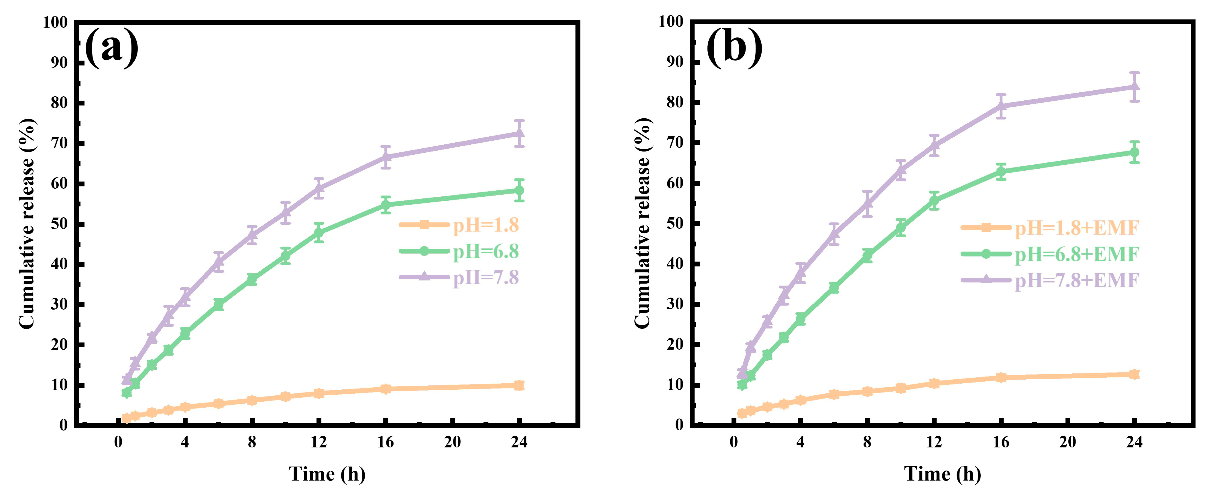
In vitro GA release from CMC-SA-Fe3O4@GA hydrogel beads was evaluated under various conditions, with and without an electromagnetic field (EMF). Figure 6a,b demonstrates that cumulative GA release strongly correlated with the pH of the medium and the application of an EMF. After a 24 h incubation period without EMF, cumulative GA release was 9.96% at pH 1.8, while it increased to approximately 58.4% and 72.5% at pH 6.8 and 7.8, respectively. The minimal GA release at acidic pH corresponds to the reduced solubility and stability of the hydrogel carrier. The significant increase in GA release at pH 7.8 compared to pH 1.8 is attributed to ionization of polymeric carboxyl groups (CMC and SA) under weakly alkaline conditions, leading to a looser hydrogel network and increased solubility, thereby facilitating GA dissolution. In addition, rapid GA release occurred within the initial 4 h, followed by sustained release from 4 h to 24 h. The initial burst release phenomenon is attributed to surface-bound GA molecules rapidly detaching from hydrogel exteriors. Previous studies indicate that drug redistribution during lyophilization can alter the spatial distribution within gel matrices, potentially accelerating the release profile [68]. Furthermore, applying an EMF-enhanced GA release. Specifically, after 24 h in an intestinal environment (pH 7.8), cumulative GA release from hydrogel beads increased by 11.4% compared to conditions without EMF, whereas only minimal enhancement was observed at pH 1.8. Previous reports suggest that an EMF aligns magnetic particles, facilitating their movement and enhancing polymer chain relaxation, which results in network expansion [69]. In conclusion, magnetic and pH-responsive CMC-SA-Fe3O4@GA hydrogel beads effectively protect GA under acidic conditions while enabling sustained release in alkaline media, highlighting their potential for improved GA encapsulation, delivery, and controlled release.

3.4. GA Release Kinetics Analysis

Mathematical kinetic models were applied to experimental release data to elucidate GA release mechanisms from CMC-SA-Fe3O4@GA beads.
Q t = M t M × 100 %
The zero-order model equation
Q t = k 0 t
The first-order model equation
Q t = 1 exp ( k 1 t )
The Higuchi model equation
Q t = k H t 1 2
The Ritger–Peppas model equation
Q t = k p t n
In these equations, Mt represents cumulative drug release at time t; M denotes maximum releasable amount; and k, Qt, n, and t indicate the kinetic constant, fractional release, diffusion exponent, and elapsed time, respectively.
Kinetic parameters derived from model fitting are summarized in Table S1, with corresponding regression plots displayed in Figures S2 and S3 (Supporting Information). Under external magnetic field (EMF) conditions at pH 7.8, the Higuich (R2 = 0.9903) and Ritger–Peppas (R2 = 0.9915) kinetic models yielded the highest regression coefficient, surpassing other evaluated models, including zero-order (R2 = 0.9059), first-order (R2 = 0.9423). Without an EMF at pH 7.8, the regression coefficient of the Higuich and Ritger–Peppas model were 0.9923 and 0.9920, higher than that of zero-order (R2 = 0.9062), first-order (R2 = 0.9422). Similar results were observed at different pH levels. Thus, the Higuich and Ritger–Peppas model accurately represents the GA release kinetics under these conditions.
According to Ritger–Peppas model interpretations, diffusion exponent (n) values reflect the nature of the release mechanism: n values below 0.43 indicate mainly Fickian diffusion; values between 0.43 and 0.85 correspond to anomalous transport involving simultaneous Fickian diffusion and swelling; and values exceeding 0.85 suggest swelling-controlled (Case-II) transport [70]. Based on the calculated kinetic parameters, GA release at pH 1.8 predominantly followed Fickian diffusion, whereas GA release at pH values of 6.8 and 7.8 was mediated by multiple mechanisms combining diffusion, swelling, and erosion processes. Consequently, GA release from CMC-SA-Fe3O4@GA hydrogel beads may involve a three-stage sequential mechanism [71]: Initially, GA-loaded beads exhibit limited water uptake due to small pore sizes, restricting GA mobility. Subsequently, water diffusion into beads increases pore size, leading to polymer relaxation and enhanced hydrogel flexibility, thereby facilitating GA release. Finally, complete bead relaxation and hydration significantly enlarge pore sizes, further promoting GA diffusion into the surrounding medium.

3.5. Antioxidant Activity Test Analysis

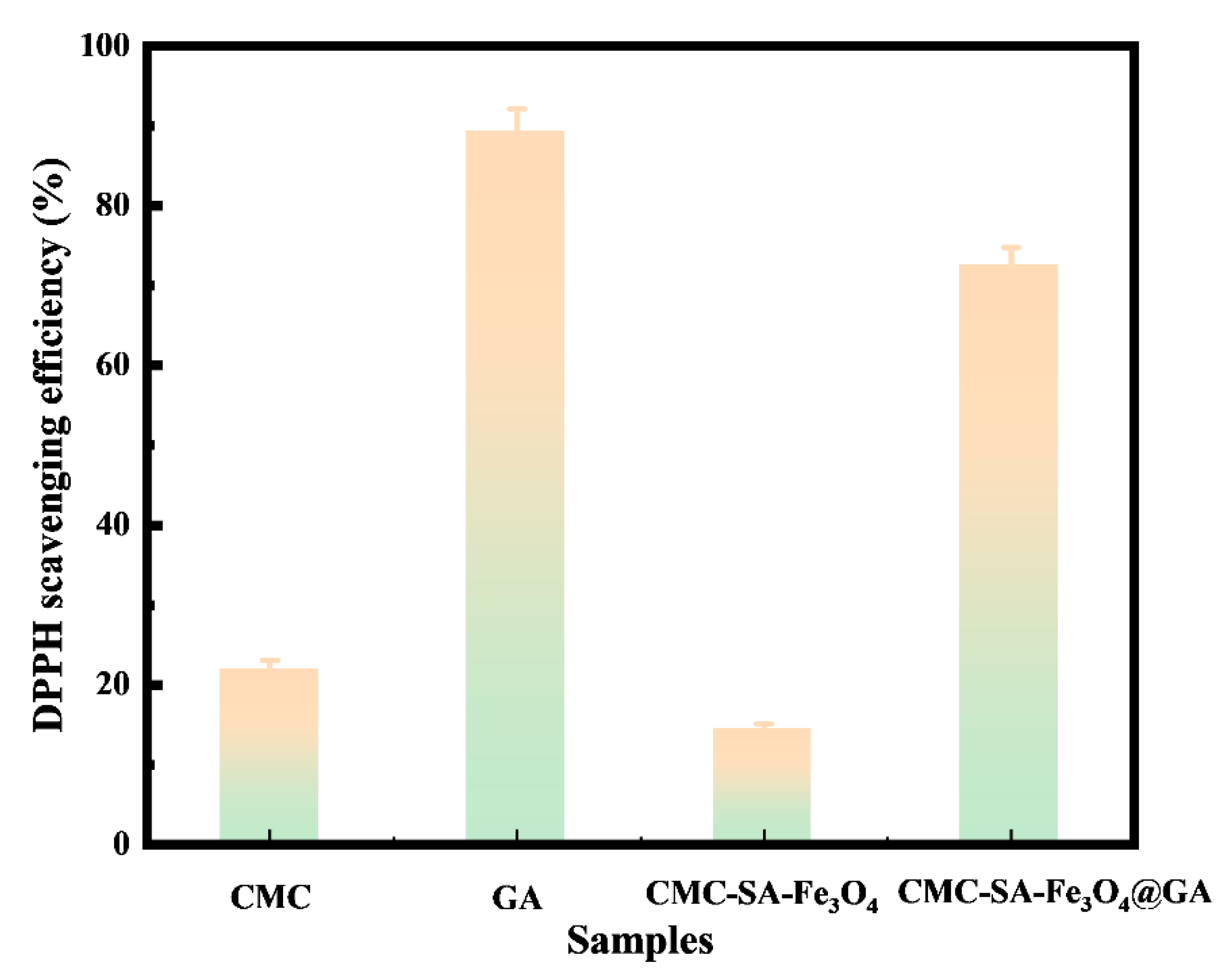
The evaluation of antioxidant potential was conducted using the DPPH radical scavenging assay, a broadly accepted method for objectively assessing antioxidant activity. The comparative results for DPPH radical scavenging are illustrated in Figure 7. CMC demonstrated intermediate antioxidant performance, exhibiting a scavenging efficiency of approximately 21.31%. This antioxidant effect can primarily be attributed to the negatively charged, hydrophilic carboxymethyl groups, facilitating interactions such as hydrogen bonding and electrostatic attraction with radicals, thereby neutralizing reactive species (e.g., -OH radicals and superoxide anions). Additionally, the antioxidant capability of CMC may correlate closely with its molecular weight [72]. Conversely, GA showed pronounced antioxidant activity (89.35%), likely resulting from hydrogen atom transfer mechanisms or proton-coupled electron transfer processes [73]. The phenolic -OH in GA molecules donate hydrogen atoms (H·) to DPPH radicals (DPPH·), converting them into stable, non-radical forms (DPPH-H). GA can also directly donate electrons (e) to DPPH radicals, neutralizing their unpaired electrons. The predominance of these mechanisms is influenced by solvent characteristics and radical types [74]. Due to the combined antioxidant properties of CMC and GA, the DPPH free radical scavenging rate of CMC-SA-Fe3O4@GA reached 72.56%.

3.6. Antibacterial Activity Analysis

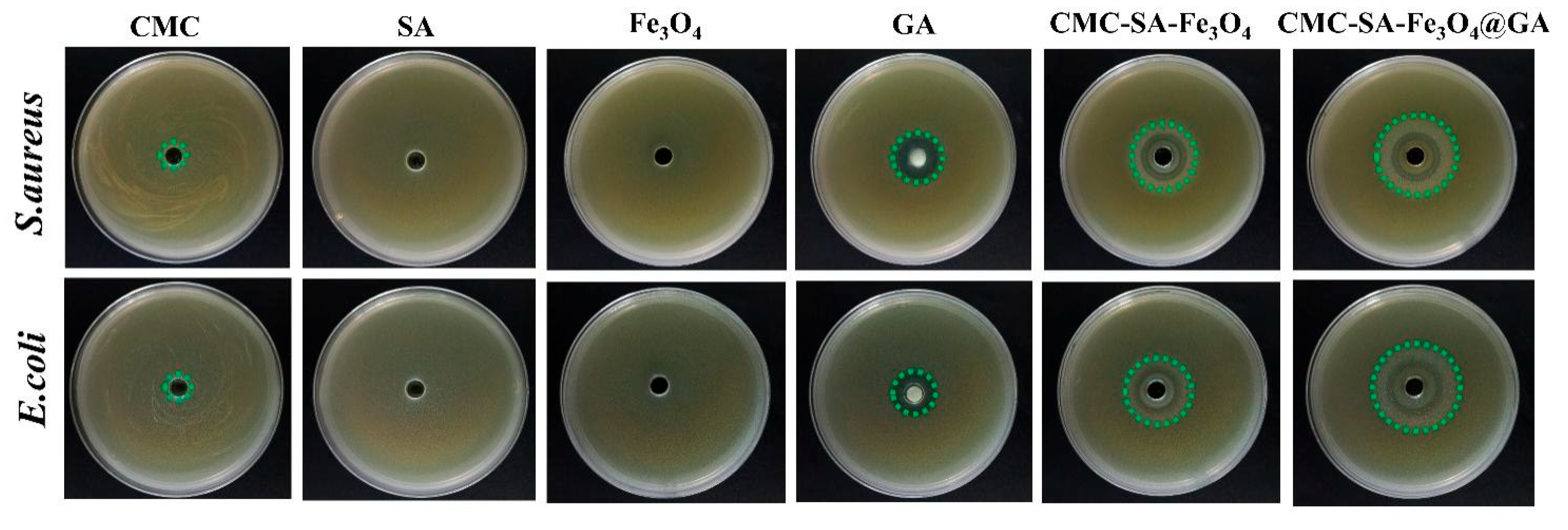
E. coli and S. aureus were selected as representative bacterial strains to evaluate the antibacterial efficacy of different samples using the disk diffusion method (Figure 8). As expected, SA and Fe3O4, serving as controls, exhibited negligible antibacterial effects, showing no inhibition zones. CMC, GA, and CMC-SA-Fe3O4@GA demonstrated inhibitory activity against E. coli and S. aureus. CMC has previously been reported to exhibit antimicrobial effects against various microorganisms, including E. coli, S. aureus, and B. subtilis [75]. For example, the antibacterial activity of N-CMC is greater than that of chitosan [76]; however, the antimicrobial effectiveness of O-CMC depends on its degree of deacetylation, concentration, molecular weight, and solution pH [77]. In this study, the observed moderate antibacterial activity of CMC might be attributed to its reduced NH3+ content and high substitution degree. Notably, GA exhibited significantly stronger inhibitory effects against S. aureus (inhibition zone diameter: 23.27 mm) than against E. coli (inhibition zone diameter: 19.81 mm). CMC-SA-Fe3O4@GA displayed a similar trend, which could be related to differences in cell wall structures between the two bacterial strains. As a Gram-positive bacterium, S. aureus possesses a cell wall primarily composed of peptidoglycan and lacks the outer membrane structure typical of Gram-negative bacteria. Phenolic -OH in GA molecules can form hydrogen bonds with polysaccharides, disrupting bacterial cell wall integrity and thereby inhibiting bacterial growth. Collectively, the results demonstrated that CMC-SA-Fe3O4@GA has substantial antimicrobial activity.

3.7. Cell Cytocompatibility and Toxicity Test Analysis

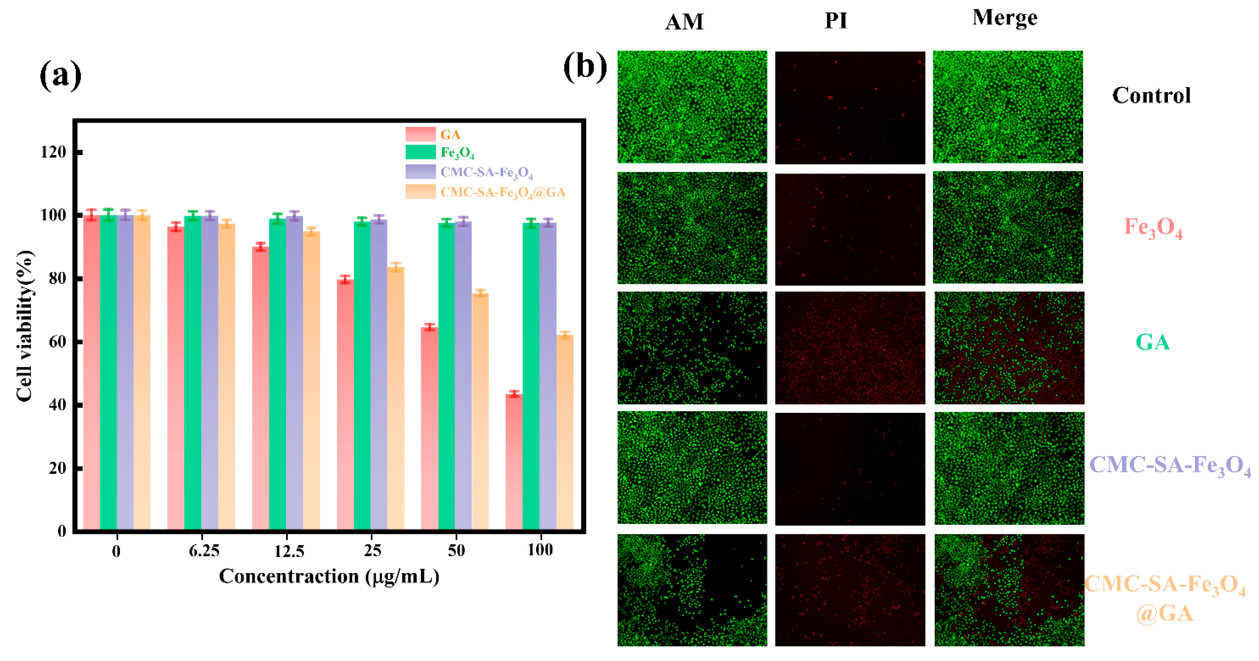
The cytotoxicity profiles of Fe3O4, GA, CMC-SA-Fe3O4, and CMC-SA-Fe3O4@GA were evaluated in HL-7702 hepatic cells using the MTT assay. Figure 9a illustrates a clear concentration-dependent decrease in HL-7702 cell viability following GA treatment, suggesting dose-dependent cytotoxic effects. In contrast, exposure to Fe3O4 and CMC-SA-Fe3O4@GA hydrogel beads at concentrations lower than 50 μg/mL maintained HL-7702 cell viability predominantly above 80%, indicating low cytotoxicity. This outcome could be attributed to enhanced biocompatibility resulting from the protective coating provided by polysaccharides (CMC, SA) combined with Fe3O4 nanoparticles. Cell viability was further verified through fluorescence-based live/dead assays visualized by confocal microscopy. Specifically, viable cells produce green fluorescence upon intracellular esterase activation of calcein-AM, while nonviable cells exhibit red fluorescence from propidium iodide (PI) staining due to compromised membrane integrity [78]. As shown in Figure 9b, nearly all cells exhibited green fluorescence across groups treated with Fe3O4, CMC-SA-Fe3O4, and CMC-SA-Fe3O4@GA, confirming their minimal apoptotic effects. However, cells exposed to high doses of GA showed partial apoptosis. Collectively, these findings indicate that the engineered CMC-SA-Fe3O4@GA hydrogel matrices exhibit minimal cytotoxicity at therapeutic doses and only marginal toxicity at higher concentrations, supporting their suitability as biocompatible drug delivery systems.
The assessment of in vitro antiproliferative activity is a critical step in evaluating novel drug delivery strategies for cancer treatment. Against this backdrop, researchers examined differences in cell survival rates after incubation with pure GA at various concentrations and encapsulated GA formulations (CMC-SA-Fe3O4@GA). Figure 10a demonstrates that both GA and the prepared hydrogel microspheres exhibit concentration-dependent cytotoxic effects on HepG2 hepatocellular carcinoma cells. Furthermore, Figure 10b reveals that cells exposed to GA and CMC-SA-Fe3O4@GA predominantly exhibit red fluorescence, except in the untreated control group. These observations confirm that both GA and its encapsulated form can induce apoptosis, highlighting the immense potential of this delivery system for tumor therapy applications.

4. Conclusions

In summary, this study successfully developed novel pH- and magnetic field-responsive CMC-SA-Fe3O4 hydrogel beads capable of efficiently loading and controlling the release of GA. GA release from the hydrogel beads demonstrated sustained behavior after an initial burst, with a higher cumulative release rate observed at pH 7.8. Among the kinetic models assessed, the Ritger–Peppas model provided the best fit, exhibiting a high regression coefficient (R2 = 0.992). Notably, applying a magnetic field enhanced the cumulative release of GA. The DPPH radical scavenging activity of CMC-SA-Fe3O4@GA reached 89.35%, indicating strong antioxidant properties. Antibacterial assays demonstrated effective inhibitory activity of CMC-SA-Fe3O4@GA against E. coli and S. aureus. In addition, the developed bio-composite system exhibited selective cytotoxicity toward cancer cells while maintaining minimal toxicity toward normal cells within 24 h, underscoring its promising biological compatibility for both in vivo and in vitro applications. Collectively, these biocompatible, antibacterial, antioxidant, and stimuli-responsive hydrogel beads provide an important strategy for future bioactive substance delivery systems.

Supplementary Materials

The following supporting information can be downloaded at: https://www.mdpi.com/article/10.3390/magnetochemistry11100085/s1 Figure S1. FT-IR spectra of Fe3O4, GA, CMC-SA-Fe3O4, and CMC-SA-Fe3O4@GA; Figure S2: Fitted curves of experimental GA release data from CMC-SA-Fe3O4@GA hydrogel beads by various drug release models at pH of 1.8, 6.8, and 7.8 in the absent of EMF; Figure S3: Fitted curves of experimental GA release data from CMC-SA-Fe3O4@GA hydrogel beads by various drug release models at pH of 1.8, 6.8, and 7.8 in the present of EMF; Table S1: Parameters of GA release models of CMC-SA-Fe3O4@GA hydrogel beads at pH of 1.8, 6.8, and 7.8 in the presence or absence of an EMF.

Author Contributions

Conceptualization, P.L. and X.H.; methodology, K.F., P.L. and X.H.; software, X.H.; validation, K.F., P.L. and H.W.; formal analysis, K.F.; investigation, K.F. and Y.L.; resources, K.F. and Y.L.; data curation, H.W.; writing—original draft preparation, K.F.; writing—review and editing, P.L.; visualization, Y.L. and H.W.; supervision, H.W.; project administration, K.F.; funding acquisition, P.L. and K.F. All authors have read and agreed to the published version of the manuscript.

Funding

This work was financially supported by the Project of Youth Research Fund of Xinyang Normal University (Grant No. 24039), the Nanhu Scholars Program for Young Scholars of XYNU, and the Open Fund of Dabie Mountain Laboratory (Grant No. DMLOF2024012).

Institutional Review Board Statement

Not applicable.

Informed Consent Statement

Not applicable.

Data Availability Statement

The original contributions presented in this study are included in the article/Supplementary Material. Further inquiries can be directed to the corresponding author.

Acknowledgments

The authors would like to thank Jie Tao from SCl-GO (www.sci-go.com) for the TEM analysis.

Conflicts of Interest

The authors declare no conflicts of interest.

References

  1. Hadidi, M.; Liñán-Atero, R.; Tarahi, M.; Christodoulou, M.C.; Aghababaei, F. The potential health benefits of gallic acid: Therapeutic and food applications. Antioxidants 2024, 13, 1001. [Google Scholar] [CrossRef]
  2. Karthammaiah, G.N.; Venkataramanan, N.; Solomon, K.A. Synthesis, characterization, and computational studies of a gallic acid-nicotinic acid salt hydrate. J. Mol. Struct. 2025, 1326, 141119. [Google Scholar] [CrossRef]
  3. Zhang, X.; Wang, Z.; Li, Y.; Zhou, Z.; Wei, B.; Dong, T.; Zhao, Y.; Ye, C.; Li, J.; Cui, J. Sea buckthorn leaves and gallic acid inhibit C48/8o-induced pseudo-allergic reaction via the PLC/IP3 signaling pathway both in vitro and in vivo. Int. Immunopharmacol. 2025, 154, 114563. [Google Scholar] [CrossRef]
  4. Alhyari, D.; Qinna, N.A.; Sheldrake, H.M.; Kantamneni, S.; Ghanem, B.Y.; Paluch, K.J. Antioxidant, Anti-inflammatory, and oral bioavailability of novel sulfonamide derivatives of gallic acid. Antioxidants 2025, 14, 374. [Google Scholar] [CrossRef]
  5. Liu, K.; Luo, B.; Zhang, L.; Hou, P.; Pan, D.; Liu, T.; Zhao, C.; Li, A. Flexible and wearable sensor for in situ monitoring of gallic acid in plant leaves. Food Chem. 2024, 460, 140740. [Google Scholar] [CrossRef] [PubMed]
  6. Liu, X.; Yuan, Q.; Yang, Y.; Li, K.; Pang, N.; Wang, J.; Yang, M.; Li, X.; Guo, J.; Liao, X. Self-assembled gallic acid-rare earth nanocomplexes against MRSA with multi-targeting antibacterial mechanisms robustly combating bacterial resistance. Chem. Eng. J. 2025, 510, 161698. [Google Scholar] [CrossRef]
  7. Holghoomi, R.; Kiani, M.H.; Rahdar, A.; Hashemi, S.M.; Ferreira, L.F.R.; Fathi-karkan, S. Nanoparticle-delivered gallic acid: A new frontier in cancer therapy. J. Drug Deliv. Sci. Tec. 2024, 101, 106129. [Google Scholar] [CrossRef]
  8. Zhang, K.q.; Lin, L.l.; Xu, H.j. Research on antioxidant performance of diglucosyl gallic acid and its application in emulsion cosmetics. Int. J. Cosmetic Sci. 2022, 44, 177–188. [Google Scholar] [CrossRef] [PubMed]
  9. Sowndarya, J.; Rubini, D.; Sinsinwar, S.; Senthilkumar, M.; Nithyanand, P.; Vadivel, V. Gallic acid an agricultural byproduct modulates the biofilm matrix exopolysaccharides of the phytopathogen Ralstonia solanacearum. Curr. Microbiol. 2020, 77, 3339–3354. [Google Scholar] [CrossRef]
  10. Jiang, Y.; Pei, J.; Zheng, Y.; Miao, Y.; Duan, B.; Huang, L. Gallic acid: A potential anti-cancer agent. Chin. J. Integr. Med. 2022, 28, 661–671. [Google Scholar] [CrossRef]
  11. Xu, Z.; Yang, B.; Fan, J.; Yuan, Q.; He, F.; Liang, H.; Chen, F.; Liu, W. Gallic acid regulates primary root elongation via modulating auxin transport and signal transduction. Front. Plant Sci. 2024, 15, 1464053. [Google Scholar] [CrossRef]
  12. Guo, J.; Ren, X.; Lu, L.; An, N.; Li, S.; Geng, M.; Li, G.; Shen, X.; Sun, X.; Wang, J. Microbial synthesis of gallic acid and its glucoside β-glucogallin. Biotechnol. Bioeng. 2024, 121, 3527–3536. [Google Scholar] [CrossRef] [PubMed]
  13. Zahrani, N.A.A.; El-Shishtawy, R.M.; Asiri, A.M. Recent developments of gallic acid derivatives and their hybrids in medicinal chemistry: A review. Eur. J. Med. Chem. 2020, 204, 112609. [Google Scholar] [CrossRef] [PubMed]
  14. Nag, S.; Majumder, S. Starch, gallic acid, their inclusion complex and their effects in diabetes and other diseases—A review. Food Sci. Nutr. 2023, 11, 1612–1621. [Google Scholar] [CrossRef] [PubMed]
  15. Tan, J.; Vincken, J.-P.; van Zadelhoff, A.; Hilgers, R.; Lin, Z.; de Bruijn, W.J. Presence of free gallic acid and gallate moieties reduces auto-oxidative browning of epicatechin (EC) and epicatechin gallate (ECg). Food Chem. 2023, 425, 136446. [Google Scholar] [CrossRef]
  16. Harwansh, R.K.; Deshmukh, R.; Shukla, V.P.; Khunt, D.; Prajapati, B.G.; Rashid, S.; Ali, N.; Elossaily, G.M.; Suryawanshi, V.K.; Kumar, A. Recent advancements in gallic acid-based drug delivery: Applications, clinical trials, and future directions. Pharmaceutics 2024, 16, 1202. [Google Scholar] [CrossRef] [PubMed]
  17. Xu, J.; Wang, Q.; Yan, S.; Qi, B. Hydrogel formed in situ through Schiff base reaction between gallic acid-grafted soybean protein isolate and oxidized dextran: Interactions, physicochemical properties, and digestive characteristics. Food Chem. 2025, 471, 142783. [Google Scholar] [CrossRef]
  18. Chen, Q.; Yang, Z.-R.; Du, S.; Chen, S.; Zhang, L.; Zhu, J. Polyphenol-sodium alginate supramolecular injectable hydrogel with antibacterial and anti-inflammatory capabilities for infected wound healing. Int. J. Biol. Macromol. 2024, 257, 128636. [Google Scholar] [CrossRef]
  19. Bahtiyar, C.; Cakir, N.T.; Kahveci, M.U.; Acik, G.; Altinkok, C. Fabrication of gallic acid containing poly(vinyl alcohol)/chitosan electrospun nanofibers with antioxidant and drug delivery properties. Int. J. Biol. Macromol. 2024, 281, 136055. [Google Scholar] [CrossRef]
  20. Graham, W.; Torbett-Dougherty, M.; Islam, A.; Soleimani, S.; Bruce-Tagoe, T.A.; Johnson, J.A. Magnetic Nanoparticles and Drug Delivery Systems for Anti-Cancer Applications: A Review. Nanomaterials 2025, 15, 285. [Google Scholar] [CrossRef]
  21. Fang, K.; Li, P.; Huang, X.; Wang, H.; Li, Y.; Zhu, D.; Luo, B. Recent advancements in magnetic starch-based composites for biomedical applications: A review. Carbohyd. Polym. 2025, 362, 123689. [Google Scholar] [CrossRef]
  22. Li, H.; Yang, S.; Hui, D.; Hong, R. Progress in magnetic Fe3O4 nanomaterials in magnetic resonance imaging. Nanotechnol. Rev. 2020, 9, 1265–1283. [Google Scholar] [CrossRef]
  23. Lesiak-Orłowska, B.; Rangam, N.V.; Sahu, N.K.; Jiricek, P.; Houdkova, J.; Atrei, A. Surface chemical and electronic properties of functionalized Fe3O4 nanoparticles influencing their cytotoxicity. Appl. Surf. Sci. 2025, 684, 161873. [Google Scholar] [CrossRef]
  24. Zhang, Y.; He, S.; Yang, T.; Feng, Y.; Yang, B.; Zhao, J.; Feng, Y. One stone two birds: Simultaneous control of eutrophication and microplastic pollution via surface functionalized nano-Fe3O4. Chem. Eng. J. 2024, 499, 156351. [Google Scholar] [CrossRef]
  25. Jiang, Z.; Song, Z.; Cao, C.; Yan, M.; Liu, Z.; Cheng, X.; Wang, H.; Wang, Q.; Liu, H.; Chen, S. Multiple natural polymers in drug and gene delivery systems. Curr. Med. Chem. 2024, 31, 1691–1715. [Google Scholar] [CrossRef]
  26. Patel, R.; Kuwar, U.; Dhote, N.; Alexander, A.; Nakhate, K.; Jain, P.; Ajazuddin. Natural polymers as a carrier for the effective delivery of antineoplastic drugs. Curr. Drug Deliv. 2024, 21, 193–210. [Google Scholar] [CrossRef]
  27. Fang, K.; Li, P.; Zhang, B.; Liu, S.; Zhao, X.; Kou, L.; Xu, W.; Guo, X.; Li, J. Insights on updates in sodium alginate/MXenes composites as the designer matrix for various applications: A review. Int. J. Biol. Macromol. 2024, 269, 132032. [Google Scholar] [CrossRef]
  28. Wang, Y.; Zhao, Y.; He, J.; Sun, C.; Lu, W.; Zhang, Y.; Fang, Y. Doubling growth of egg-box structure during Calcium-mediated molecular assembly of alginate. J. Colloid. Interf. Sci. 2023, 634, 747–756. [Google Scholar] [CrossRef]
  29. Zhou, W.; Zhang, H.; Yu, Y.; Zou, X.; Shi, J.; Zhao, Y.; Ye, Y. Dissolution mechanism of sodium alginate and properties of its regenerated fiber under low temperature. Int. J. Biol. Macromol. 2020, 162, 810–819. [Google Scholar] [CrossRef] [PubMed]
  30. Liew, C.V.; Chan, L.W.; Ching, A.L.; Heng, P.W.S. Evaluation of sodium alginate as drug release modifier in matrix tablets. Int. J. Pharmaceut. 2006, 309, 25–37. [Google Scholar] [CrossRef]
  31. Zhang, M.; Chen, H. Development and characterization of starch-sodium alginate-montmorillonite biodegradable antibacterial films. Int. J. Biol. Macromol. 2023, 233, 123462. [Google Scholar] [CrossRef] [PubMed]
  32. Liu, X.; Liu, L.; Huang, F.; Meng, Y.; Chen, Y.; Wang, J.; Wang, S.; Luo, Y.; Li, J.; Liang, Y. pH-sensitive chitosan/sodium alginate/calcium chloride hydrogel beads for potential oral delivery of rice bran bioactive peptides. Food Chem. 2025, 470, 142618. [Google Scholar] [CrossRef]
  33. Bai, W.; Chen, H.; Li, J.; Cai, W.; Kong, Y.; Zuo, X. Calcium carbonate hollow microspheres encapsulated cellulose nanofiber/sodium alginate hydrogels as a sequential delivery system. Int. J. Biol. Macromol. 2025, 309, 142839. [Google Scholar] [CrossRef]
  34. Javanbakht, S.; Shaabani, A. Carboxymethyl cellulose-based oral delivery systems. Int. J. Biol. Macromol. 2019, 133, 21–29. [Google Scholar] [CrossRef]
  35. Zennifer, A.; Senthilvelan, P.; Sethuraman, S.; Sundaramurthi, D. Key advances of carboxymethyl cellulose in tissue engineering & 3D bioprinting applications. Carbohyd. Polym. 2021, 256, 117561. [Google Scholar]
  36. Kanikireddy, V.; Varaprasad, K.; Jayaramudu, T.; Karthikeyan, C.; Sadiku, R. Carboxymethyl cellulose-based materials for infection control and wound healing: A review. Int. J. Biol. Macromol. 2020, 164, 963–975. [Google Scholar] [CrossRef] [PubMed]
  37. Yang, Y.; Nan, W.; Zhang, R.; Shen, S.; Wu, M.; Zhong, S.; Zhang, Y.; Cui, X. Fabrication of carboxymethyl cellulose-based thermo-sensitive hydrogels and inhibition of corneal neovascularization. Int. J. Biol. Macromol. 2024, 261, 129933. [Google Scholar] [CrossRef]
  38. Zhao, M.; Zhou, H.; Hao, L.; Chen, H.; Zhou, X. Natural rosin modified carboxymethyl cellulose delivery system with lowered toxicity for long-term pest control. Carbohyd. Polym. 2021, 259, 117749. [Google Scholar] [CrossRef]
  39. Hu, Y.; Hu, S.; Zhang, S.; Dong, S.; Hu, J.; Kang, L.; Yang, X. A double-layer hydrogel based on alginate-carboxymethyl cellulose and synthetic polymer as sustained drug delivery system. Sci. Rep. 2021, 11, 9142. [Google Scholar] [CrossRef]
  40. Gong, J.; Hou, L.; Ching, Y.C.; Ching, K.Y.; Dai Hai, N.; Chuah, C.H. A review of recent advances of cellulose-based intelligent-responsive hydrogels as vehicles for controllable drug delivery system. Int. J. Biol. Macromol. 2024, 264, 130525. [Google Scholar] [CrossRef] [PubMed]
  41. Fang, K.; Zhang, Y.; Yin, J.; Yang, T.; Li, K.; Wei, L.; Li, J.; He, W. Hydrogel beads based on carboxymethyl cassava starch/alginate enriched with MgFe2O4 nanoparticles for controlling drug release. Int. J. Biol. Macromol. 2022, 220, 573–588. [Google Scholar] [CrossRef]
  42. Li, J.; Kim, S.Y.; Chen, X.; Park, H.J. Calcium-alginate beads loaded with gallic acid: Preparation and characterization. LWT Food Sci. Technol. 2016, 68, 667–673. [Google Scholar] [CrossRef]
  43. Çakır, D.; Gümüşderelioğlu, M. Nopal mucilage-encapsulated poly (butylene adipate-co-terephthalate) nanoparticles for efficient gallic acid delivery in glioblastoma cells. J. Drug Deliv. Sci. Technol. 2025, 110, 107025. [Google Scholar] [CrossRef]
  44. Sripunya, A.; Chittasupho, C.; Mangmool, S.; Angerhofer, A.; Imaram, W. Gallic acid-encapsulated PAMAM dendrimers as an antioxidant delivery system for controlled release and reduced cytotoxicity against ARPE-19 cells. Bioconjugate Chem. 2024, 35, 1959–1969. [Google Scholar] [CrossRef]
  45. Abbasi, A.R.; Sohail, M.; Minhas, M.U.; Khaliq, T.; Kousar, M.; Khan, S.; Hussain, Z.; Munir, A. Bioinspired sodium alginate based thermosensitive hydrogel membranes for accelerated wound healing. Int. J. Biol. Macromol. 2020, 155, 751–765. [Google Scholar] [CrossRef]
  46. Khainskaya, K.; Hileuskaya, K.; Nikalaichuk, V.; Ladutska, A.; Akhmedov, O.; Abrekova, N.; You, L.; Shao, P.; Odonchimeg, M. Chitosan-gallic acid conjugate with enhanced functional properties and synergistic wound healing effect. Carbohyd. Res. 2025, 553, 109496. [Google Scholar] [CrossRef]
  47. Li, T.; Jiang, Y.; Zeng, L.; Zhang, H.; Yuan, Z.; Liu, J.; Liu, C.; Li, H.; Zhang, L.; Yang, C. Surface sizing introducing Fe3O4 nanoparticles for magnetic properties strengthening of basalt fibers. Ceram. Int. 2025, 51, 2216–2225. [Google Scholar] [CrossRef]
  48. Grenier, J.; Duval, H.; Barou, F.; Lv, P.; David, B.; Letourneur, D. Mechanisms of pore formation in hydrogel scaffolds textured by freeze-drying. Acta Biomater. 2019, 94, 195–203. [Google Scholar] [CrossRef] [PubMed]
  49. Dawn, R.; Kiran, C.; Tjiu, W.W.; Aabdin, Z.; Zzaman, M.; Faizal, F.; Panatarani, C.; Joni, I.M.; Chandra, G.; Amemiya, K.; et al. Effect of spin and orbital selectivity of DNA induced SiO2 coated Fe3O4 nanoparticles: X-ray magnetic circular dichroism study. Colloid. Surface. A. 2025, 709, 136111. [Google Scholar] [CrossRef]
  50. Jeevakumari, J.F.; Suresh, G. Comprehensive evaluation of solvothermally synthesized Fe3O4 and PEG capped Fe3O4 nanoparticles for magnetic and biomedical applications. Inorg. Chem. Commun. 2025, 174, 113904. [Google Scholar] [CrossRef]
  51. Xing, X.; Liu, C.; Zheng, L. Preparation of photo-crosslinked microalgae-carboxymethyl chitosan composite hydrogels for enhanced wound healing. Carbohyd. Polym. 2025, 348, 122803. [Google Scholar] [CrossRef]
  52. Fei, Y.; Li, Y.; Han, S.; Ma, J. Adsorptive removal of ciprofloxacin by sodium alginate/graphene oxide composite beads from aqueous solution. J. Colloid. Interf. Sci. 2016, 484, 196–204. [Google Scholar] [CrossRef] [PubMed]
  53. Zhang, Y.; Cheng, Y.; Xu, M.; Fang, C.; Li, M.; Han, H.; Du, Y. Construct of flexible Fe3O4@VO2/Ti3C2Tx composite films with absorption-dominated and tunable terahertz electromagnetic shielding. Ceram. Int. 2025, 51, 18987–18999. [Google Scholar] [CrossRef]
  54. Neisi, E.; Dadkhah Tehrani, A.; Shamloei, H.R. Development of cellulose nanowhisker–gallic acid antioxidant bioconjugate via covalent conjugation and supramolecular interactions: A comparative study. Int. J. Biol. Macromol. 2024, 271, 132561. [Google Scholar] [CrossRef]
  55. Lü, T.; Ma, R.; Ke, K.; Zhang, D.; Qi, D.; Zhao, H. Synthesis of gallic acid functionalized magnetic hydrogel beads for enhanced synergistic reduction and adsorption of aqueous chromium. Chem. Eng. J. 2021, 408, 127327. [Google Scholar] [CrossRef]
  56. Hao, X.; Yang, S.; Li, Y. High efficiency and selective removal of Cu (II) via regulating the pore size of graphene oxide/montmorillonite composite aerogel. J. Hazard. Mater. 2022, 424, 127680. [Google Scholar] [CrossRef] [PubMed]
  57. Li, X.; Sánchez del Río Sáez, J.; Liu, Y.; Du, S.; Cruz, C.; Wei, G.; Wang, D.-Y. Carboxymethyl chitosan-based composite film for fire warning under high humidity conditions with wireless signal transmission. Int. J. Biol. Macromol. 2025, 316, 144540. [Google Scholar] [CrossRef]
  58. Liu, H.; Chen, X.; Chen, D.; Zhang, Z.; Xia, Y.; Zhang, K.; Ming, J.; Lei, X. Decoding the molecular interaction mechanisms of tannic acid in modulating Fe3+-sodium alginate microgel delivery systems. Food Chem. 2025, 489, 144935. [Google Scholar] [CrossRef]
  59. Asl, A.M.; Abdouss, M.; Kalaee, M.R.; Homami, S.S.; Pourmadadi, M. Targeted delivery of quercetin using gelatin/starch/Fe3O4 nanocarrier to suppress the growth of liver cancer HepG2 cells. Int. J. Biol. Macromol. 2024, 281, 136535. [Google Scholar] [CrossRef]
  60. Feng, Q.; Fan, B.; He, Y.-C. Antibacterial, antioxidant, Cr(VI) adsorption and dye adsorption effects of biochar-based silver nanoparticles-sodium alginate-tannic acid composite gel beads. Int. J. Biol. Macromol. 2024, 271, 132453. [Google Scholar] [CrossRef]
  61. Sun, L.; Zhao, J.; Chen, W.; Wang, G. Electrostatically sprayed alginate-carboxymethyl chitosan microgel for colon-targeted melatonin delivery ameliorates ulcerative colitis. Food Biosci. 2025, 68, 106616. [Google Scholar] [CrossRef]
  62. Wu, P.; He, R.-H.; Fang, Y.; Chen, K.; Wu, M.; Zhang, W.; Lv, J.; Zhao, Y. The study of double-network carboxymethyl chitosan/sodium alginate based cryogels for rapid hemostasis in noncompressible hemorrhage. Int. J. Biol. Macromol. 2024, 266, 131399. [Google Scholar] [CrossRef] [PubMed]
  63. El-Basiouny, N.M.; Soliman, S.M.A.; Khalil, N.M.; Abd El-Ghany, M.N. Chitosan and alginate/Aspergillus flavus-mediated nanocomposite films for preservation of postharvest tomatoes. Int. J. Biol. Macromol. 2025, 297, 139559. [Google Scholar] [CrossRef]
  64. Sellappan, L.K.; Mishra, S.; Sundaresan, S.; Rathinavelan, T.; Majumdar, S. Multifunctional NiO-curcumin nanocomposite-loaded chitosan-alginate scaffolds for enhanced bone tissue regeneration and antibacterial activity. J. Drug Deliv. Sci. Tec. 2025, 110, 107114. [Google Scholar] [CrossRef]
  65. Sahayaraj, R.; Enoch, K.; Pati, S.S.; Somasundaram, A.A. Cobalt doped Fe3O4 nanoparticles with improved magnetic anisotropy and enhanced hyperthermic efficiency. Ceram. Int. 2025, 51, 20786–20797. [Google Scholar] [CrossRef]
  66. Abdolrahimi, M.; Vasilakaki, M.; Slimani, S.; Ntallis, N.; Varvaro, G.; Laureti, S.; Meneghini, C.; Trohidou, K.N.; Fiorani, D.; Peddis, D. Magnetism of nanoparticles: Effect of the organic coating. Nanomaterials 2021, 11, 1787. [Google Scholar] [CrossRef]
  67. Levy, L.; Gurov, A.; Radian, A. The effect of gallic acid interactions with iron-coated clay on surface redox reactivity. Water Res. 2020, 184, 116190. [Google Scholar] [CrossRef]
  68. Zhao, Y.; Li, B.; Zhang, W.; Zhang, L.; Zhao, H.; Wang, S.; Huang, C. Recent advances in sustainable antimicrobial food packaging: Insights into release mechanisms, design strategies, and applications in the food industry. J. Agric. Food Chem. 2023, 71, 11806–11833. [Google Scholar] [CrossRef] [PubMed]
  69. Gong, B.; Sanford, A.R.; Ferguson, J.S.; Filipcsei, G.; Csetneki, I.; Szilágyi, A.; Zrínyi, M. Magnetic field-responsive smart polymer composites. In Oligomers-Polymer Composites-Molecular Imprinting; Springer: Berlin/Heidelberg, Germany, 2007; pp. 137–189. [Google Scholar]
  70. Sulttan, S.; Rohani, S. Controlled drug release of smart magnetic self-assembled micelle, kinetics and transport mechanisms. J. Pharm. Sci. 2022, 111, 2378–2388. [Google Scholar] [CrossRef]
  71. Kamaly, N.; Yameen, B.; Wu, J.; Farokhzad, O.C. Degradable controlled-release polymers and polymeric nanoparticles: Mechanisms of controlling drug release. Chem. Rev. 2016, 116, 2602–2663. [Google Scholar] [CrossRef]
  72. Sun, T.; Yao, Q.; Zhou, D.; Mao, F. Antioxidant activity of N-carboxymethyl chitosan oligosaccharides. Bioorganic Med. Chem. Lett. 2008, 18, 5774–5776. [Google Scholar] [CrossRef]
  73. Milenković, D.; Đorović, J.; Petrović, V.; Avdović, E.; Marković, Z. Hydrogen atom transfer versus proton coupled electron transfer mechanism of gallic acid with different peroxy radicals. React. Kinet. Mech. Cat. 2018, 123, 215–230. [Google Scholar] [CrossRef]
  74. Mittal, A.; Vashistha, V.K.; Das, D.K. Free radical scavenging activity of gallic acid toward various reactive oxygen, nitrogen, and sulfur species: A DFT approach. Free Radical Res. 2023, 57, 81–90. [Google Scholar] [CrossRef]
  75. Shariatinia, Z. Carboxymethyl chitosan: Properties and biomedical applications. Int. J. Biol. Macromol. 2018, 120, 1406–1419. [Google Scholar] [CrossRef] [PubMed]
  76. Xu, T.; Xin, M.; Li, M.; Huang, H.; Zhou, S.; Liu, J. Synthesis, characterization, and antibacterial activity of N, O-quaternary ammonium chitosan. Carbohyd. Res. 2011, 346, 2445–2450. [Google Scholar] [CrossRef] [PubMed]
  77. Xing, K.; Chen, X.G.; Li, Y.Y.; Liu, C.S.; Liu, C.G.; Cha, D.S.; Park, H.J. Antibacterial activity of oleoyl-chitosan nanoparticles: A novel antibacterial dispersion system. Carbohyd. Polym. 2008, 74, 114–120. [Google Scholar] [CrossRef]
  78. Chan, L.L.; Wilkinson, A.R.; Paradis, B.D.; Lai, N.J. Rapid image-based cytometry for comparison of fluorescent viability staining methods. J. Fluoresc. 2012, 22, 1301–1311. [Google Scholar] [CrossRef]
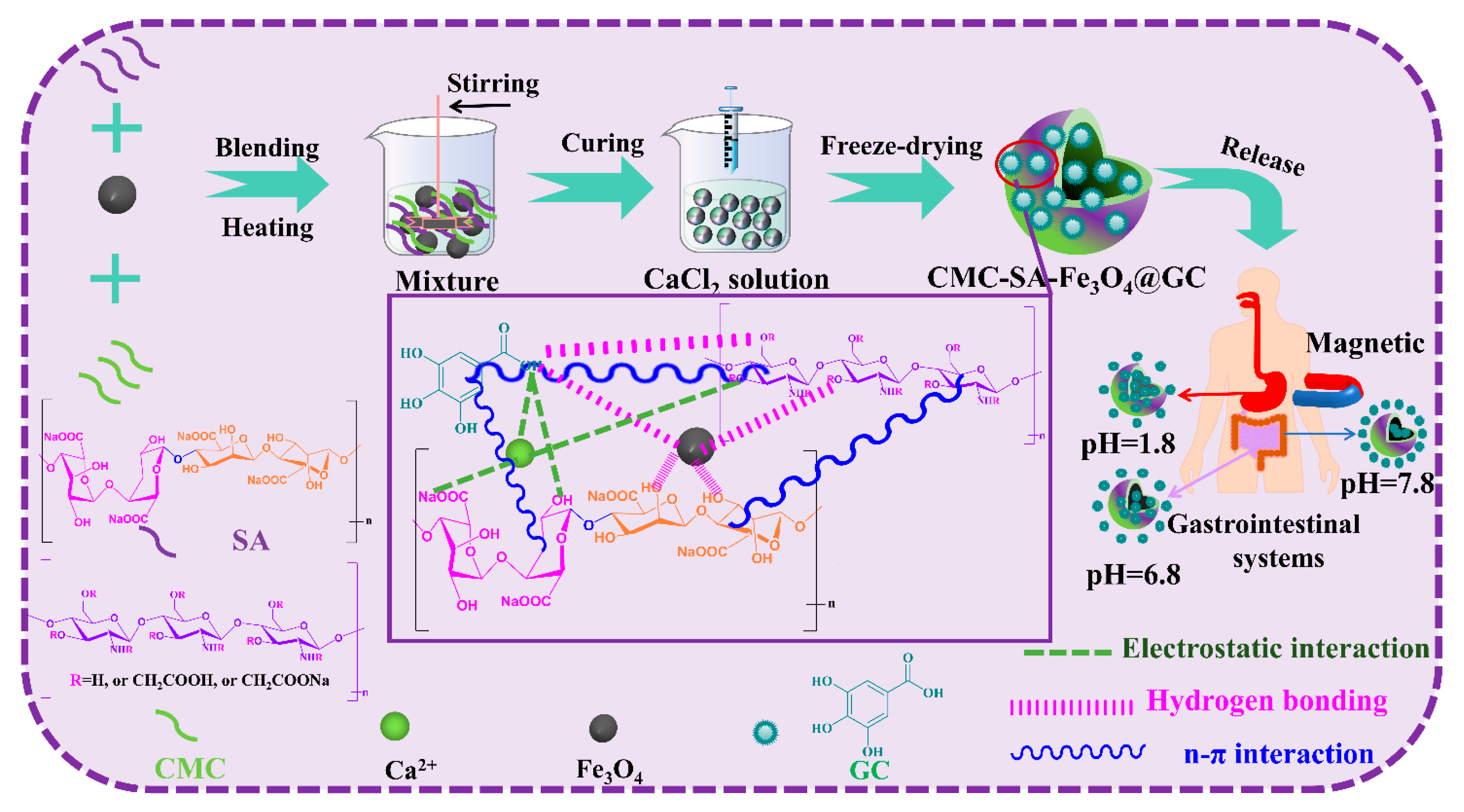
Figure 1. Schematic diagram of the preparation and release process of CMC-SA-Fe3O4@GA hydrogel beads.
Magnetochemistry 11 00085 g001
Figure 2. SEM photographs: CMC, SA, and Fe3O4; ((a)–(c), respectively); digital and SEM images of CMC-SA- Fe3O4@GA hydrogel beads ((d)–(f), respectively).
Magnetochemistry 11 00085 g002
Figure 3. TEM, HR-TEM, and SAED photographs of Fe3O4 (ac) and CMC-SA-Fe3O4@GA hydrogel beads after crushing (df).
Magnetochemistry 11 00085 g003
Figure 4. (a) FT-IR spectra of CMC, SA, Fe3O4, GA, CMC-SA-Fe3O4, and CMC-SA-Fe3O4@GA; (b) XRD patterns of CMC, SA, Fe3O4, CMC-SA-Fe3O4, and CMC-SA-Fe3O4@GA; (c,d) TG and DTG curves of Fe3O4, CMC, SA, Fe3O4, GA, CMC-SA-Fe3O4, and CMC-SA-Fe3O4@GA.
Magnetochemistry 11 00085 g004
Figure 5. Magnetization curves of Fe3O4 and CMC-SA-Fe3O4@GA.
Magnetochemistry 11 00085 g005
Figure 6. (a) Cumulative drug release patterns from CMC-SA-Fe3O4@GA in different solutions with an EMF; (b) Cumulative drug release patterns from CMC-SA-Fe3O4@GA in different solutions without an EMF.
Magnetochemistry 11 00085 g006
Figure 7. The DPPH free radical scavenging capacity of various samples.
Magnetochemistry 11 00085 g007
Figure 8. Bacteriostatic capacity of different samples for E. coli and S. aureus. (Initial zone: 6 mm).
Magnetochemistry 11 00085 g008
Figure 9. (a) In vitro cytocompatibility against HL-7702 cell lines; (b) Fluorescent images of HL-7702 cells after treated with different samples and followed with Cal-cein-AM and PI staining.
Magnetochemistry 11 00085 g009
Figure 10. (a) In vitro cytocompatibility against HePG2 cell lines; (b) Fluorescent images of HepG2 cells after treated with different samples and followed with Calcein-AM and PI staining.
Magnetochemistry 11 00085 g010
Table 1. Loading capacity and efficiency of CMC-SA-Fe3O4@GA hydrogel beads.
SamplesAdditions of GA (g)Loading Capacity (LC%)Loading Efficiency (LE%)
CMC-SA-Fe3O4@GA-10.113.3± 0.4 a66.5± 2.0 a
CMC-SA-Fe3O4@GA-20.328.6 ± 0.9 b47.7± 1.5 b
CMC-SA-Fe3O4@GA-30.531.1 ± 1.2 c31.1 ± 1.2 c
Values are means ± SD of triplicate. Different superscript letters mean significant differences in the same column (p < 0.05).
Disclaimer/Publisher’s Note: The statements, opinions and data contained in all publications are solely those of the individual author(s) and contributor(s) and not of MDPI and/or the editor(s). MDPI and/or the editor(s) disclaim responsibility for any injury to people or property resulting from any ideas, methods, instructions or products referred to in the content.