Article

Syntheses, Structures, and Properties of Mono- and Dinuclear Acetylacetonato Ruthenium(III) Complexes with Chlorido or Thiocyanato Ligands

1
Department of Chemistry, Graduate School of Natural Science and Technology, Shimane University, 1060 Nishikawatsu, Matsue 690-8504, Japan
2
Research Institute of Frontier Science and Technology, Okayama University of Science, 1-1 Ridaicho, Kita-Ku, Okayama 700-0005, Japan
3
School of Biological and Environmental Sciences, Kwansei Gakuin University, 1 Gakuen Uegahara, Sanda 669-1330, Japan
*
Author to whom correspondence should be addressed.
Magnetochemistry 2024, 10(3), 16; https://doi.org/10.3390/magnetochemistry10030016
Submission received: 26 January 2024 / Revised: 21 February 2024 / Accepted: 22 February 2024 / Published: 27 February 2024
(This article belongs to the Section Molecular Magnetism)

Abstract

:
The mononuclear and dinuclear ruthenium(III) complexes trans-Ph4P[RuIII(acac)2Cl2] (1), Ph4P[{RuIII(acac)Cl}2(μ-Cl)3] (2) and trans-Ph4P[RuIII(acac)2(NCS)2]·0.5C6H14 (3·0.5C6H14) were synthesized. Single crystals of 1, 2·H2O and 3·CH3CN suitable for X-ray crystal structure analyses were obtained through recrystallization from DMF for 1 and 2·H2O and from acetonitrile for 3·CH3CN. An octahedral Ru with bis-chelate-acac ligands and axial chlorido or κ-N-thiocyanido ligands (for 1 and 3·CH3CN) and triply µ-chlorido-bridged dinuclear Ru2 for 2·H2O were confirmed through the structure analyses. The Ru–Ru distance of 2.6661(2) of 2·H2O is indicative of the existence of the direct metal–metal interaction. The room temperature magnetic moments (μeff) are 2.00 and 1.93 μB for 1 and 3·0.5C6H14, respectively, and 0.66 μB for 2. The temperature-dependent (2–300 K) magnetic susceptibility showed that the strong antiferromagnetic interaction (J ≤ −800 cm−1) is operative between the ruthenium(III) ions within the dinuclear core. In the 1H NMR spectra measured in CDCl3 at 298 K, the dinuclear complex 2 showed signals for the acac ligand protons at 2.50 and 2.39 ppm (for CH3) and 5.93 ppm (for CH), respectively, while 1 and 3·0.5C6H14 showed signals with large paramagnetic shifts; −17.59 ppm (for CH3) and −57.01 ppm (for CH) for 1 and −16.89 and −17.36 ppm (for CH3) and −53.67 and −55.53 ppm (for CH) for 3·0.5C6H14. Cyclic voltammograms in CH2Cl2 with an electrolyte of nBu4N(ClO4) showed the RuIII → RuIV redox wave at 0.23 V (vs. Fc/Fc+) for 1 and the RuIII → RuII waves at −1.39 V for 1 and −1.25 V for 3·0.5C6H14 and the RuIII–RuIII → RuIII–RuIV and RuIII–RuIII → RuIII–RuIV waves at 0.91 V and −0.79 V for 2.

Graphical Abstract

1. Introduction

Acetylacetone (Hacac) is well known as one of useful bidentate ligands, which can form chelate complexes, such as [MII(acac)2], [MIII(acac)3] and [MIII(acac)2X2], through reactions with various transition metal ions [1,2,3,4,5,6,7,8,9,10,11,12,13,14,15,16,17,18,19,20,21,22,23,24,25,26,27,28]. The complexes have been widely investigated, for example, as building blocks for magnetic materials [29,30,31], NMR shift reagents and paramagnetic relaxation reagents [32,33,34,35] and catalysts [36,37]. The trans-[RuIII(acac)2(CN)2] anion has an unpaired electron and has been reported to work as a paramagnetic linker in combination with Mn2+ and [FeIIIsalen]+ to produce ferri- or ferromagnetic compounds {MnII[RuIII(acac)2(CN)2]2}n and [{FeIII(salen)}{RuIII(acac)2(CN)2}] with two- and one-dimensional structures, respectively [38,39]. The cyanido linker anion trans-[RuIII(acac)2(CN)2] is prepared from trans-[RuIII(acac)2Cl2] through the axial ligand substitution of Cl with CN, with the substitution reaction shown in Scheme 1 [38]. Through a research project, we studied using the anionic complex trans-[RuIII(acac)2(CN)2] with more various metal ions or complexes to develop the chemistry of this type of assembled magnetic compounds.
During the course of our study, we found the formation of a dinuclear anion [{RuIII(acac)Cl}2(μ-Cl)3], of which the structure is shown in Scheme 2, when the reaction of RuCl3·nH2O and Hacac was performed in the presence of KCl, followed by the addition of Ph4PCl to obtain trans-PPh4[RuIII(acac)2Cl2] (1), and we could elucidate the dinuclear structure of Ph4P[{RuIII(acac)Cl}2(μ-Cl)3] (2) through an X-ray crystal structure analysis. Although Hasegawa et al. reported the formation of a dinuclear complex Ph4As[{RuIII(acac)Cl}2(μ-Cl)3] as a by-product when isolating trans-Ph4As[RuIII(acac)2Cl2], the dinuclear complex was characterized based on an elemental analysis and the 1H NMR spectrum, their X-ray crystal structural data were incomplete to establish the dinuclear structure [40] and structurally elucidated examples of this kind of triply bridged diruthenium(III) complexes are still limited. Therefore, we decided to investigate the dinuclear complex Ph4P[{RuIII(acac)Cl}2(μ-Cl)3] (2) in more detail. For such a study, the mononuclear complexes trans-Ph4P[RuIII(acac)2Cl2] (1) and trans-Ph4P[RuIII(acac)2(NCS)2] (3) were also prepared and investigated. Here, we report on structures and magnetic, spectral, and electrochemical properties of trans-Ph4P[RuIII(acac)2Cl2] (1), Ph4P[{RuIII(acac)Cl}2(μ-Cl)3] (2) and trans-Ph4P[RuIII(acac)(NCS)2]·0.5C6H14 (3·0.5C6H14).

2. Results and Discussion

2.1. Synthesis and Characterizations

Refluxing a mixture of RuCl3·nH2O, acetylacetone (Hacac) and KCl in water and the addition of excess Ph4PCl gave the mononuclear complex trans-Ph4P[RuIII(acac)2Cl2] (1) in the yield of 11% (based on RuCl3). The chromatographical purifications with Al2O3 columns (eluent: chloroform/MeOH and acetonitrile/chloroform) confirmed the formation of the dinuclear complex Ph4P[{RuIII(acac)Cl}2(μ-Cl)3] (2) in very low amounts. However, the yield of complex 2 increased to 1.6% (based on RuCl3) when passing oxygen gas through the reacting solution. The monomeric complex trans-Ph4P[RuIII(acac)2(NCS)2]·0.5C6H14 (3·0.5C6H14) was synthesized by refluxing a methanolic solution of 1 and an excess amount of KSCN in the yield of 64% (based on 1). The chemical formulae of the obtained complexes of 1, 2 and 3·0.5C6H14 were confirmed through elemental analyses. ESI-TOF-MS spectra also confirmed the formations of anionic units of 369.9307 m/z (calcd for [M] 369.9318) for 1, 577.7369 m/z (calcd for [M] 577.6223) for 2 and 415.9426 m/z (calcd for [M] 415.4562) for 3·0.5C6H14. IR spectra of the complexes are given in Figures S1–S3. Powder X-ray diffraction (PXRD) analyses were also performed for complexes 1, 2 and 3·0.5C6H14. The obtained results are displayed in Figures S4–S6, respectively. The PXRD pattern of 1 agreed with its simulated pattern derived from the crystal structure of 1 (Figure S4). However, the patterns of 2 and 3·0.5C6H14 did not agree with those simulated from the crystal structures of 2·H2O and 3·CH3CN, respectively. The disagreement was more remarkable in 3·0.5C6H14. The reason was explained to be the facts that crystals of 2·H2O and 3·CH3CN have the crystal solvents H2O and CH3CN, respectively, though the crystalline powder of 2 has no crystal solvent and that of 3·0.5C6H14 has solvent molecules of C6H14. Detailed discussions of the crystal structures of 1, 2·H2O and 3·CH3CN are given in the next section.

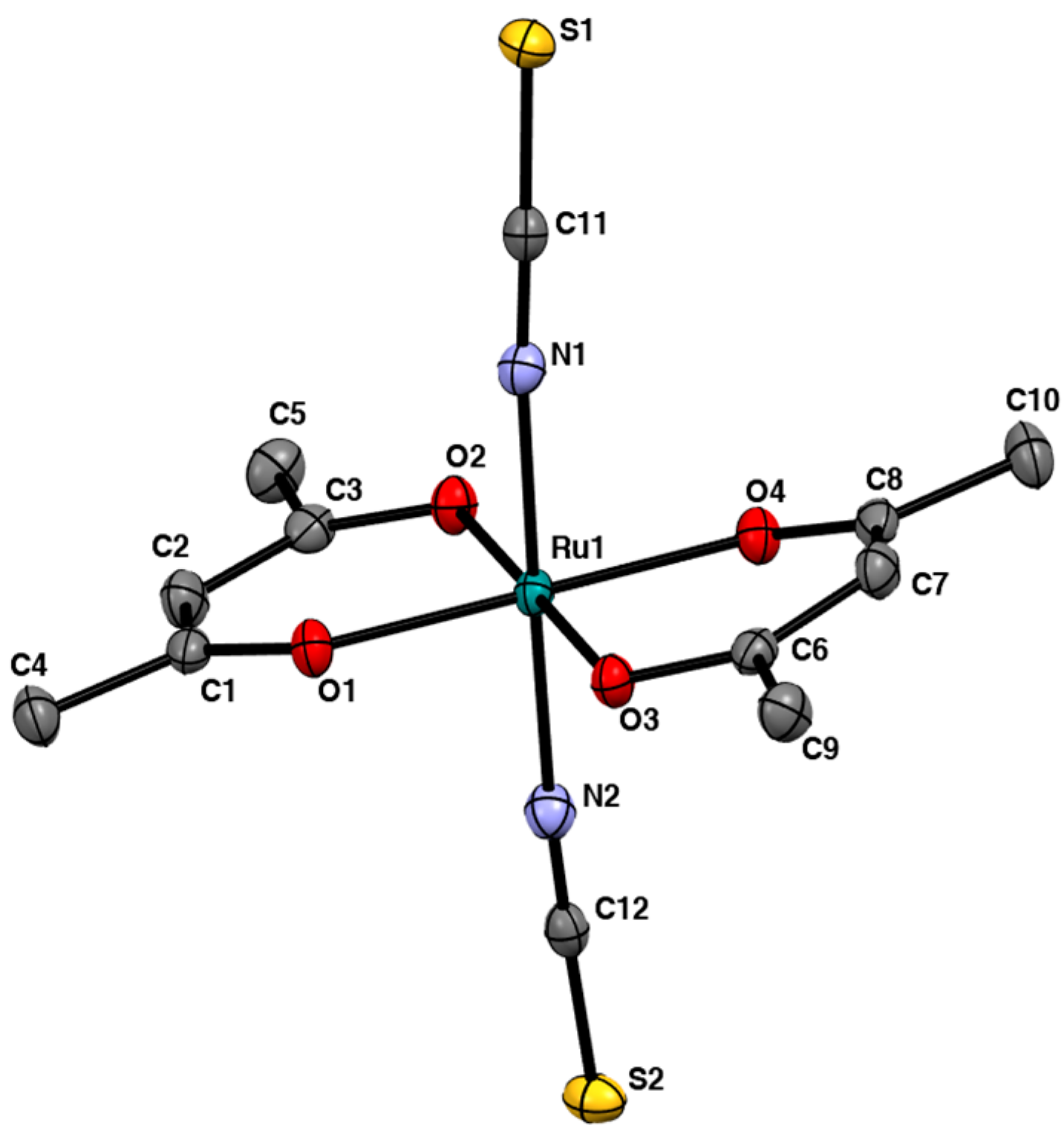
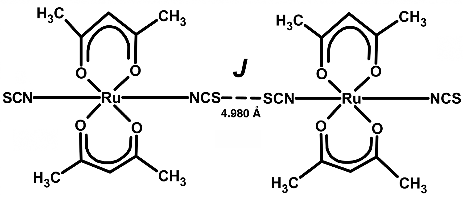
2.2. Crystal Structures

Single crystals of 1 and 2·H2O suitable for the X-ray crystal structure analysis were obtained through slow diffusion of diethyl ether to solutions of 1 and 2·H2O in DMF, respectively, while, for 3, the crystals were grown through recrystallization from acetonitrile and isolated as 3·CH3CN. Crystallographic data are listed in Table 1. Selected bond distances and angles are given in Tables S1–S6, respectively. The mononuclear complex trans-Ph4P[RuIII(acac)2Cl2] (1) crystallized in the P 1 ¯ space group. The crystal packing diagram of 1 is shown Figure S7. In this crystal, there are crystallographically different mononuclear trans-[RuIII(acac)2Cl2] anions designated as Ru1 and Ru2 for the central ruthenium atoms, while the tetraphenylphosphate (Ph4P+) cation exists among the [RuIII(acac)2Cl2] units. The packing feature of the present complex is essentially the same as that for trans-Ph4As[Ru(acac)2Cl2] [40], though different counter cations of Ph4As+ existed in the crystal. A perspective drawing of the structure of one of the [RuIII(acac)2Cl2] units of 1 is shown in Figure 1. The inversion center is located at the ruthenium atom. The equatorial positions of each Ru atom are occupied with four oxygen atoms of the two acac ligands with Ru–Oeq distances of 2.015(1) and 2.012(1) Å for Ru1 and 2.018(1) and 2.011(1) Å for Ru2, respectively, which are comparable to those of trans-Ph4As[RuIII(acac)2Cl2] (Ru–Oeq = 2.010(3)–2.016(3) Å). The chlorido ligands are coordinated to Ru1 and Ru2 with distances of Ru–Clax = 2.360(1) and 2.363(1) Å, respectively, which are also comparable to those of trans-Ph4As[RuIII(acac)2Cl2] (Ru–Clax = 2.355(2) and 2.632(1) Å). The Clax–Ru–Oeq bond angles are 88.46(4) and 91.54(4)° for Ru1 and 86.63(5) and 93.37(5)° for Ru2, respectively, of which values are comparable to those of trans-Ph4As[Ru(acac)2Cl2] (∠Clax–Ru–Oeq = 87.1(1)–91.5(1)°).
The dinuclear complex Ph4P[{RuIII(acac)Cl}2(μ-Cl)3]·H2O (2·H2O) crystallized in P 1 ¯ . The crystal packing diagram and the dinuclear anionic unit of 2·H2O are shown in Figure S8 and Figure 2, respectively. The crystal consists of Ph4P+ cations, [{RuIII(acac)Cl}2(μ-Cl)3] anions and crystallization water molecules. There is no specific interaction between them. As shown in Figure S8, the dinuclear structure is composed of two RuIII(acac)Cl units with triple chlorido-bridges. Octahedral geometries around both the Ru(III) atoms are accomplished with the bidentate chelate of two acac ligands with Ru–O distances of 1.995(1) (for Ru1–O1), 1.991(1) (for Ru1–O2), 2.001(2) (for Ru2–O3) and 2.001(1) Å (for Ru2–O4), respectively. The chlorido bridges link the two ruthenium(III) atoms with distances of Ru1–Cl3 = 2.367(1), Ru1–Cl4 = 2.359(1), Ru1–Cl5 = 2.378(1), Ru2–Cl3 = 2.364(1), Ru2–Cl4 = 2.359(1) and Ru2–Cl5 = 2.378(1) Å. The bond distances are relatively long compared with those of the terminal Ru–Cl bonds in each RuIII(acac)Cl unit (Ru1–Cl1 = 2.340(1) and Ru2–Cl2 = 2.331(1) Å), reflecting the bridging property. The O–Ru–O chelate bite angles in the RuIII(acac)Cl units are close to 90° (∠O1–Ru1–O2 = 93.60(6) and ∠O3–Ru2–O4 = 93.80(7)°). The face-sharing octahedral structure shown by 2·H2O has been reported for the dinuclear complex Cs3[(RuIIICl3)2(μ-Cl)3] [41]. The structure and bond distances and angles of the anionic unit, which has the D3h symmetry, are illustrated in Scheme 3.
The dimensions of the dinuclear cores of Cs3[(RuIIICl3)2(μ-Cl)3] and 2·H2O are very similar; Ru-Cl (terminal) = 2.332(4) Å (for Cs3[(RuIIICl3)2(μ-Cl)3]) and 2.340(1) and 2.331(1) Å (for 2·H2O), Ru–Cl (bridging) = 2.391(4) Å (for Cs3[(RuIIICl3)2(μ-Cl)3]) and 2.359(1)–2.378(1) Å (for 2·H2O), and ∠Ru–Cl(bridging)–Ru = 69.5(2)° (for Cs3[(RuIIICl3)2(μ-Cl)3]) and 68.36(1)–68.83(1)° (for 2·H2O). The Ru–Ru distances are 2.725(3) Å for Cs3[(RuIIICl3)2(μ-Cl)3] and 2.6661(2) Å for 2·H2O, respectively. The short Ru–Ru distances are indicative of the direct M–M interaction between the ruthenium(III) ions. The mixed valent diruthenium(II,III) complexes with triply chlorido-bridges [{Ru2.5(NH3)3}2(μ-Cl)3](BPh4)2 and [{Ru2.5(tacn)}2(μ-Cl)3](PF6)2·4H2O (tacn = 1,4,7-triazacyclononane) had similar, however, relatively longer, Ru–Ru distances, 2.753(4) Å for the former complex and 2.830(1) Å for the latter complex [42,43]. The mixed valence dinuclear complexes were interpreted to have an unpaired electron with an MO diagram for the eleven 4d electrons shown in Scheme 4a [43,44], where an unpaired electron resides in an a2” (σ*) orbital and the bond order becomes 0.5. When this interpretation is applied to the Ru2III,III complexes 2 and Cs3[(RuIIICl3)2(μ-Cl)3], the bond order becomes one, because an electron is removed from the a2” (σ*) orbital, leading to relatively short Ru–Ru bonds. As discussed later, the diamagnetism of the Ru2III,III complexes can be interpreted based on the MO diagram (Scheme 4b), where ten 4d electrons are arranged pairwise in the orbitals.
The complex 3·CH3CN crystallized in the monoclinic lattice (P21/n). The crystal consists of Ph4P+ cations, trans-[RuIII(acac)2(NCS)2] anions and crystallization solvent molecules of CH3CN, as can be seen in the packing diagram (Figure S9). The anionic unit of trans-[RuIII(acac)2(NCS)2] is shown in Figure 3. The Ru–Oeq (acac) distances are 2.002(1)–2.012(1)Å, comparable to those of 1 and 2·H2O. The Ru(III) center is further coordinated by nitrogen atoms of the NCS ligands with Ru–N bond distances of 2.016(2) (for Ru1–N1) and 2.007(2) Å (for Ru1–N2), respectively. The bond angles of O1–Ru1–O2, O1–Ru1–O3, O2–Ru–O4 and O3–Ru1–O4 are 93.71(5), 85.99(5), 87.03(5) and 93.28(5)°, respectively. The sum of the four ∠Oeq–Ru–Oeq’ values is 360.0°, which indicates that the ruthenium atom is located without deviation on the plane composed of oxygen atoms O1, O2, O3 and O4. Although three atoms of each NCS- ligands are arranged almost linearly (∠N1–C11–S1 = 178.9(2) and ∠N2–C12–S2 = 177.9(2)°), the bond angle values of ∠Ru1–N1–C11 = 170.9(1) and ∠Ru1–N2–C12 = 173.0(1) means that the axial coordination of the NCS- ligands are slightly tilted from the perpendicular vector to the Ru(Oeq)4 plane, while the N1–Ru1–N2 bond angle (177.5(1)°) is close to 180°.
As can been seen from the bond distances and angles for the cations of Ph4P+ listed in Tables S1–S6, the structural features of the cations are basically the same among the complexes 1, 2·H2O and 3·CH3CN.

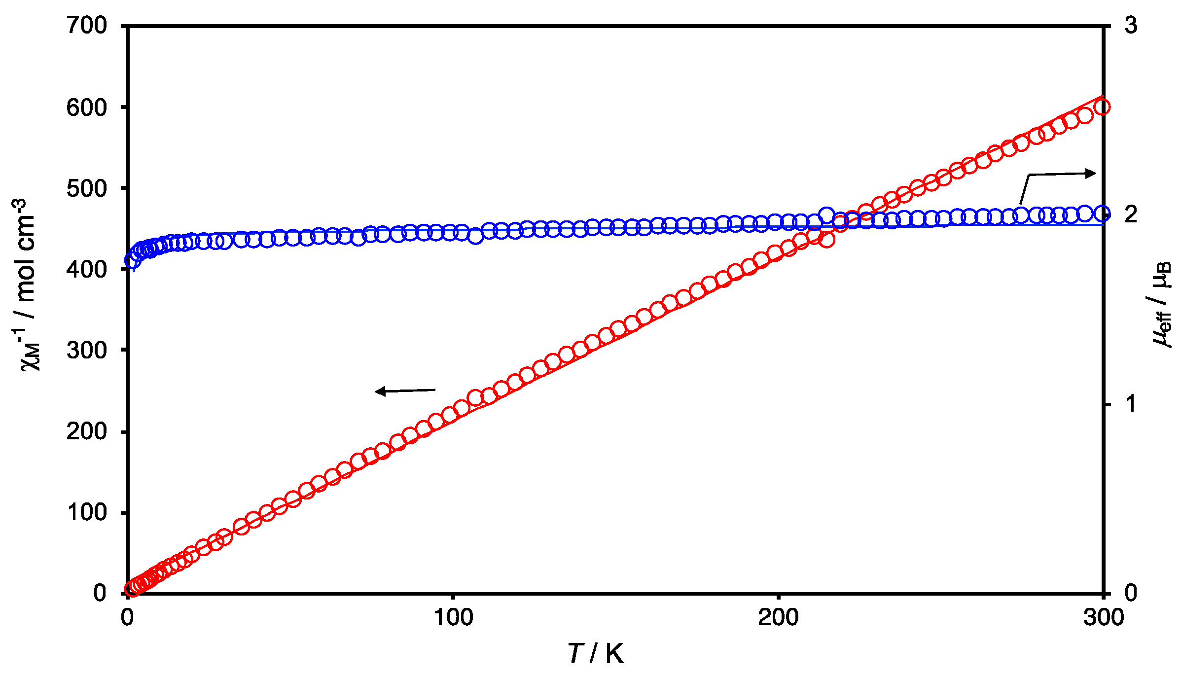
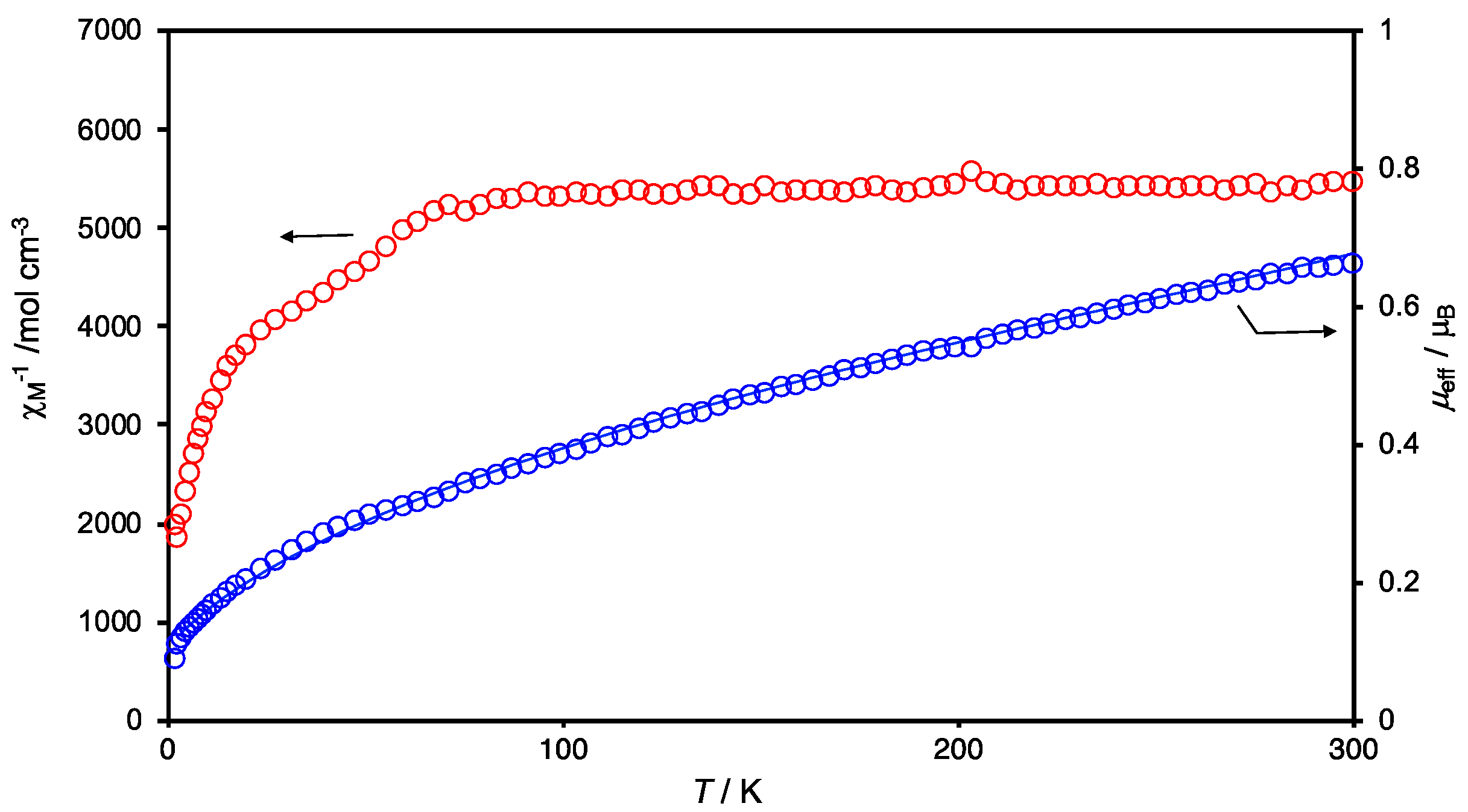
2.3. Magnetic Properties

The temperature dependencies of the effective magnetic moment (μeff) (per Ru(III) for 1 and 3·0.5C6H14 and per Ru(III)2 for 2) and reciprocal magnetic susceptibility (1/χM) values of 1, 2 and 3·0.5C6H14 are given in Figure 4, Figure 5 and Figure 6, respectively. The temperature-dependent behaviors are essentially different between the mononuclear complexes 1 and 3·0.5C6H14 and the dinuclear complex 2.
The magnetic moments at 300 K are 2.00 and 1.93 μB for 1 and 3·0.5C6H14, respectively, indicating the existences of an unpaired electron for both the complexes when considering that the spin-only value is 1.73 μB for an S = 1/2 system. Although both of the complexes have rather large moment values, the temperature dependences of χM−1 obey the Curie–Weiss law, χM = C/(Tθ) with C = 0.498 cm3 mol−1 K and θ = −6.3 K for 1 and C = 0.464 cm3 mol−1 K and θ = −4.8 K for 3·0.5C6H14, meaning that the interaction between the mononuclear Ru(III) units is limited and weakly antiferromagnetic overall. A difference in the temperature-dependent profile between 1 and 3·0.5C6H14 was observed when the temperature fell below 5 K; the moment value (μeff) decreased for 1 and increased for 3·0.5C6H14, which may have occurred due to the difference in the weak interaction between 1 and 3·0.5C6H14. We looked into the X-ray crystal structure data of 1 and 3·CH3CN to search the origin of such interactions. The closest distance between chlorine atoms (designated with Cl1 and Cl2) of neighboring [Ru(acac)2Cl2] units is 4.921 Å for 1 (see Figure S7), leading to a chain structure, as shown in Scheme 5, and that between sulfur atoms (designated with S2) of [Ru(acac)2(NCS)2] units is 4.980 Å (the second and third closest distances are considerably long; 7.363 and 7.539 Å for S1···S1 separation) (see Figure S9), leading to a dimer structure, as shown in Scheme 6. Taking these contacts (4.921 Å for 1 and 4.980 Å for 3·CH3CN) into consideration, we analyzed their temperature-dependent magnetic moments with the equation introduced by Bonner–Fisher for the chain S = 1/2 local spins (Equation (1)) in the case of 1 (see Scheme 5) [45] and the Bleaney–Bowers equation for the two S = 1/2 local spins (Equation (2)) in the case of 3 (see Scheme 6) [46].
χM = (Ng2β2/kT)(0.25 + 0.14995x + 0.30094x2)/(1.0 + 1.9862x + 0.68854x2 + 6.062x3) + Nα, with x = |J|/kT
χM = 2(Ng2β2/kT)[3 + exp(−2J//kT)]−1 + 2Nα
where N is the Avogadro number, g is the g factor, β is the Bohr magneton, k is the Boltzmann constant, J is the exchange integral between the ruthenium (III) ions and is the temperature-independent paramagnetism (TIP). The temperature-dependent profiles could be reproduced with the parameter values of g = 2.21, J = −0.29 cm−1 and Nα = 60 × 10−6 emu mol−1 for 1 and g = 2.14, J = 0.22 cm−1 and Nα = 60 × 10−6 emu mol−1 for 3·0.5C6H14, which are included as blue solid lines in Figure 4 and Figure 6, respectively. The results support that the Cl···Cl (=4.921 Å in 1) and S···S (=4.980 Å in 3·CH3CN) contacts found in the crystal structures gave rise to the weak antiferromagnetic and ferromagnetic interactions observed below 5 K in 1 and 3·0.5C6H14, respectively.
The magnetic moment (μeff) of 2 is 0.66 μB at 300 K, which is much lower than the spin-only value (1.73 μB), indicating the existence of a strong antiferromagnetic interaction in the anionic unit of [{RuIII(acac)Cl}2(μ-Cl)3]. In fact, due to the strong antiferromagnetic interaction, the temperature dependence of χM−1 no longer obeys the Curie–Weiss law (Figure 5). The magnetic moment decreases steadily with lowering the temperature. Due to each Ru(III) center having an unpaired electron (S = 1/2), the magnetic behavior was simulated using the modified Bleaney–Bowers equation (Equation (3)), including a correction term (ρ) for paramagnetic impurities:
χM = 2{(1 − ρ)(Ng2β2/kT)[3 + exp(−2J//kT)]−1 + ρ(Ng2β2/4kT) + Nα}
The simulation results gave the following parameter values: g = 2.2, J ≤ −800 cm−1, Nα = 90 × 10−6 emu mol−1, ρ = 0.0015. The fitting quality was nearly the same, as long as J ≤ −800 cm−1, when other parameter values were fixed at g = 2.2, Nα = 90 × 10−6 emu mol−1, and ρ = 0.0015. This large negative J value (J ≤ −800 cm−1) obviously means that there is a very strong antiferromagnetic interaction between the Ru(III) centers, leading to the fact that complex 2 is practically diamagnetic (μeff = 0.66 μB). The dinuclear complex Cs3[(RuIIICl3)2(μ-Cl)3] was reported to have a moment value of μeff = 0.51 μB at 300 K [41], indicating that the strong antiferromagnetic interaction is also operative between the Ru(III) centers, like in the case of 2. As to this type of face-sharing bioctahedral complex anion [(RuIIICl3}2(μ-Cl)3]3−, calculations using the broken-symmetry density functional theory have been performed [47]. The calculation results indicated that the Ru(III) ions (low-spin state) were strongly coupled to result in the formation of a metal–metal σ bond and the minimum energy was at Ru–Ru = 2.74 Å, which was in good agreement with the observed bond length value of Ru–Ru = 2.725(3) Å for Cs3[(RuIIICl3}2(μ-Cl)3]. Due to 2·H2O having nearly the same Ru–Ru distance (2.6661(2) Å) as that of Cs3[(RuIIICl3}2(μ-Cl)3], the strong antiferromagnetic interaction (J ≤ −800 cm−1) is considered to be based on the direct interaction between the Ru(III) centers and pairwise arrangement of ten 4d electrons in the molecular orbitals (Scheme 4b).
The Ru–Ru distance is an important piece of evidence used to determine the presence of the direct metal–metal interactions. But this is not enough, like in the case of the face-sharing octahedral complex [{RuIIICl2(nBu3P)}{RuIIICl(nBu3P)}(μ-Cl)3], which had a rather long Ru–Ru distance of 3.176(1) Å, and it was difficult to determine whether or not the direct interaction existed. The magnetic susceptibility data could have given the answer to the question, although this complex was obtained only once, in a very small amount, as single crystals and no magnetic measurement has been carried out [48].
Field-dependent magnetizations were measured at 2K for 1, 2 and 3·0.5C6H14, the results being given in Figures S10, S11 and S12, respectively. On the increase in the external magnetic field, magnetizations of 1 and 3·0.5C6H14 increased to 1.10 and 0.94 , respectively, at 70,000 Oe. Brillouin function curves with g = 2.3 for 1 and g = 2.1 for 3·0.5C6H14 were drawn with red solid lines in Figures S10 and S12, respectively. The deviation from the theoretical curve (Brillouin function) could have occurred due to the magnetic anisotropy and/or magnetic interactions. The field dependence for the magnetization of 2 is typical of the one for diamagnetism due to the strong antiferromagnetic interaction between the two Ru(III) ions (Figure S11).

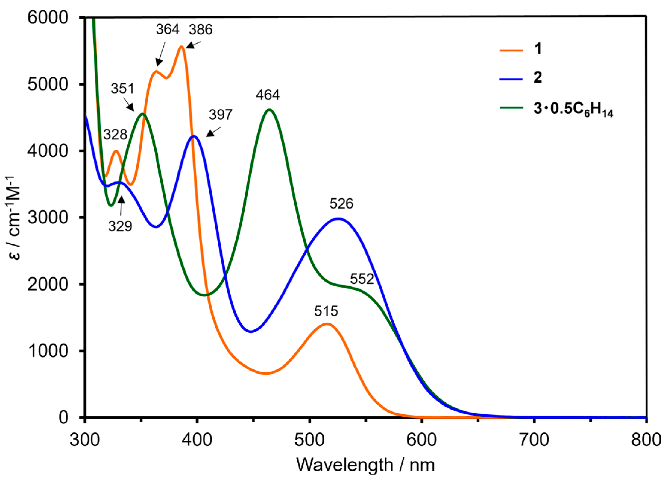
2.4. Reflectance and Absorption Spectra

Diffuse reflectance and absorption spectra for 1, 2 and 3·0.5C6H14 were measured in solid and solution (CH2Cl2) and given in Figure S13 and Figure 7, respectively. As to trans-Ph4As[RuIII(acac)2Cl2], which have been measured in MeOH, Hasegawa et al. assigned the lowest energy band at 521 nm as πd←π (acac) and the second lowest energy band at 376 nm as πd←Cl, referring to the assignment reported for [RuIII(acac)3] [40,49]. The assignment is also applicable for complexes 1, 2 and 3·0.5C6H14. The similarity in the band positions between the reflectance and absorption spectra indicates that the mononuclear and dinuclear core structures (trans-[RuIII(acac)2Cl2] (for 1), [{RuIII(acac)Cl}2(μ-Cl)3] (for 2) and trans-[RuIII(acac)2(NCS)2] (for 3·0.5C6H14) are maintained in the solution.

2.5. 1H NMR Spectra

The 1H NMR spectra were measured for 1, 2 and 3·0.5C6H14 in chloroform-d1 at 298 K, and are given in Figures S14, S15 and S16, respectively. In the case of 2, other than the signals at 7.8–8.1 ppm for the phenyl protons of Ph4P+, signals at 2.39 and 2.50 ppm and at 5.93 ppm were observed; the set of the former two signals and the latter signal was assigned as methyl (CH3) and methine (CH) protons, respectively, for the acac ligand. It should be noted that the signals appeared in this region due to the strong antiferromagnetic interaction within the dinuclear anion of 2 to be diamagnetic. Although the reason for the splitting of the signals for the methyl protons (CH3) is unclear, a similar assignment had been determined for the signals for the protons of the dinuclear complex trans-Ph4As[RuIII(acac)2Cl2] measured in acetone-d6 (2.40 (6H,s, CH3), 2.46 (6H, s, CH3), 5.99 (~2H, s, CH), 7.90 (16H, m, o, m-H) and 7.96 ppm (4H, t, p-H)) [40]. In the cases of the mononuclear complexes 1 and 3·0.5C6H14, large paramagnetic shifts were observed for the signals of the acac ligand protons (−17.59 ppm (for CH3) and −57.01 ppm (for CH) for 1 and −16.89 and −17.36 ppm (for CH3) and −53.67 and −55.53 ppm (for CH) for 3·0.5C6H14 other than the phenyl protons of Ph4P+; the signals for the CH3 and CH protons of 3·0.5C6H14 are split, respectively.

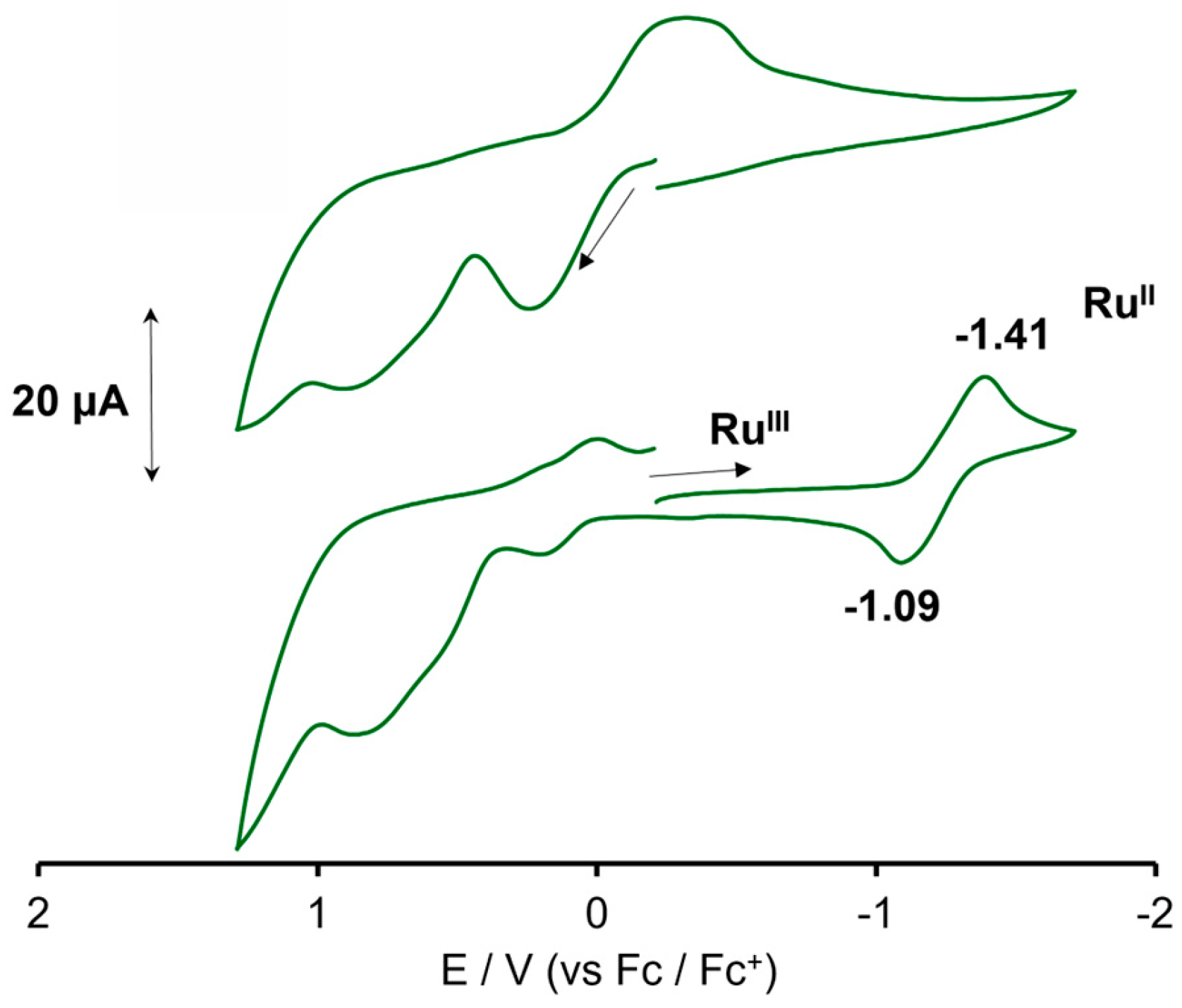
2.6. Cyclic Voltammograms

Cyclic voltammograms (CVs) were obtained for 1, 2 and 3·0.5C6H14 in dichloromethane solutions containing 0.1 M of nBu4N(ClO4). The voltammograms of 1 and 2 are given in Figure 8. Complex 1 showed a redox wave at E1/2 ((Epa + Epc)/2) = 0.23 V (vs. Fc/Fc+), which was attributed to the RuIII → RuIV process, and an irreversible wave at E1/2 (Epc/2) = −1.39 V, which was attributed to the RuIII → RuII process. Hasegawa et al. reported that the corresponding waves were observed at 0.93 and −0.45 V (vs. NHE) for trans-Ph4As[RuIII(acac)2Cl2] in the acetone solution, the potential values being calculated to be 0.29 and −1.09 V (vs. Fc/Fc+), respectively [40], when quoting E1/2 = 0.64 V (vs. NHE) for the Fc/Fc+ redox couple in the literature [50]. The dinuclear complex 2 showed a redox wave at E1/2 = 0.91 V and another wave at E1/2 = −0.79 V. The former wave was attributed to the RuIII–RuIII → RuIII–RuIV process, and the latter wave was attributed to the RuIII–RuIII → RuII–RuIII process.
Complex 3·0.5C6H14 showed a rather complicated redox behavior. As shown in Figure 9, in the oxidation process, irreversible waves were observed. They should have been related to the oxidation of the NCS ligands and decomposition of the complex [51] because a quasi-reversible redox wave at E1/2 = −1.25 V disappeared when the CV measurement started toward the oxidation side. The redox wave at −1.25 V was attributed to the RuIII → RuII process and positively shifted compared with that for 1 (−1.39 V), probably due to the stronger donating nature of NCS (in 3·0.5C6H14) than Cl (in 1).

3. Materials and Methods

3.1. General Aspects

The elemental analyses for C, H and N were carried out using YANACO CHN CORDER MT-6 (Yanako, Tokyo, Japan). Infrared spectra were recorded as KBr disk using a JASCO FT/IR-4600 (JASCO, Tokyo, Japan). Powder X-ray diffraction analysis was performed on a Rigaku SmartLab X-ray diffractometer (Rigaku, Tokyo, Japan) with Cu Kα. Magnetic susceptibilities were measured by using Quantum Design MPMS-XL7 (Quantum Design, San Diego, CA, USA) (installed at the Institute of Molecular Science (IMS), Okazaki, Japan) for 1 and 3·0.5C6H14 and MPMS3 (Quantum Design, San Diego, CA, USA) (installed at Shimane University) for 2 over the temperature range of 2–300 K with a magnetic field of 5000 Oe. The measured data were corrected for diamagnetic contribution [52]. Field-dependent magnetization measurements were performed from 0 to 70,000 Oe at 2 K for 1, 2 and 3·0.5C6H14 with MPMS 3. Absorption and diffuse reflectance spectra were measured with Shimadzu UV-2450 (Shimadzu, Kyoto, Japan). Cyclic voltammograms were obtained in dichloromethane containing 0.1 M of tetra-n-butylammonium perchlorate (nBu4N(ClO4)) on a BAS 100BW Electrochemical Workstation (Bioanalytical Systems, West Lafayette, IN, USA). A glassy carbon disk (1.5 mm radius), a platinum wire and a Ag/Ag+ (TBAP/CH3CN) electrode were used as the working, counter and reference electrodes, respectively. Ferrocene (Fc) was used as an internal standard, and the potentials were quite relative to the Fc/Fc+ couple. ESI-TOF-MS spectra were recorded on a Bruker micrOTOF II (Bruker, Billerica, MA, USA) with an acetonitrile solution. 1H spectra were obtained with a JEOL JNM-AL 400 spectrometer (JEOL, Tokyo, Japan) in chloroform-d1. Chemical shifts (δ/ppm) were determined using the residual solvent signal: 7.26 ppm for the proton of CHCl3 in CDCl3 for 1H NMR spectra [53,54].

3.2. Synthesis of Complexes

3.2.1. Synthesis of trans-Ph4P[RuIII(acac)2Cl2] (1)

This complex was synthesized using a modified method described in the literature [40]. A mixture of RuCl3·nH2O (0.40g, 1.93 mmol (based on RuCl3)), acetylacetone (2 mL) and 1 M KCl in water (2 mL) was refluxed for 30 min. Then, the solution was evaporated to dryness and the residue was dissolved in c.a. 20 mL of water. The addition of Ph4PCl (0.36 g, 0.961 mmol) in 2 mL of water to the solution gave an orange precipitate, which was collected and dried over P2O5 under vacuum overnight. The obtained orange powder was dissolved in chloroform and purified chromatographically using an Al2O3 column (eluent: chloroform/methanol (99:1 v/v.). The second fraction separated from the small amount of the first fraction was evaporated to dryness and employed again for chromatographic purification using an Al2O3 column (eluent: methanol/acetonitrile (1:1 v/v.). The eluent was evaporated to dryness, dissolved in a small amount of chloroform, followed by the addition of n-hexane, giving an orange precipitation, which was collected through filtration and dried under vacuum at 100 °C for 3 h. The yield was 0.15g (11%, based on RuCl3). Anal. found: C; 56.94, H; 4.73. calcd for C34H34Cl2O4PRu: C; 57.55, H; 4.83. IR data (KBr disk, cm−1) 3059 w, 1585 w, 1546 s, 1520 vs, 1482 w, 1435 m, 1387 s, 1267 m, 1199 w, 1164 w, 1109 s, 1022 w, 997 w, 935 w, 788 w, 755 w, 726 s, 693 s, 660 m, 526 s, 456 m. UV–Vis (in CH2Cl2, λmax) 515, 386, 364, 328, 276, 232 nm. HR-MS (ESI-TOF) 369.9307 m/z (calcd for [M] 369.9318). 1H NMR (chlroform-d1, 298 K) δ 9.00–8.00 (m, o, m, p-H), −17.59 (s, CH3), −57.01 (br.s., CH).

3.2.2. Synthesis of Ph4P[{RuIII(acac)Cl}2(μ-Cl)3] (2)

A mixture of RuCl3·nH2O (0.31 g, 1.49 mmol (based on RuCl3)), acetylacetone (1.5 mL) and 1.0 M aqueous solution of KCl (1.5 mL) was refluxed for 30 min. During the reaction, oxygen gas was passed through the reacting solution. Then, the solution was evaporated to dryness and the residue was dissolved in c.a. 30 mL of water. Ph4PCl (0.30 g, 0.81 mmol) was added to the aqueous solution, giving a purple precipitate, which was collected through suction filtration. The obtained powder was dissolved in chloroform and purified chromatographically using an Al2O3 column (eluent: chloroform/methanol (10:3 v/v.)). The first fraction was evaporated to dryness and employed again for chromatographic purification using an Al2O3 column (eluent: chloroform/acetonitrile (1:1 v/v.)). The eluted solution was evaporated to dryness and dissolved in a small amount of chloroform, followed by the addition of n-hexane to give a purple precipitate, which was collected through filtration and dried under vacuum at 110 °C for 3 h. The yield was 0.021 g (1.6% based on RuCl3). Anal found: C, 44.27, H, 3.64. calcd for C34H34Cl5O4PRu2: C, 44.53, H, 3.74. IR data (KBr disk, cm−1) 3056 w, 1627 w, 2656 vs, 1520 vs, 1483 s, 1438 vs, 1368 vs, 1274 s, 1191 m, 1166 w, 1108 s, 1025 m, 996 m, 937 m, 789 w, 757 m, 723 s, 690 s, 645 m, 526 vs, 463 s, 432 w. UV–Vis (in CH2Cl2, λmax) 527, 396, 330, 295, 235 nm. HR-MS (ESI-TOF) found 577.7369 m/z (calcd for [M] 577.6223). 1H NMR (chlroform-d1, 298 K) δ 8.08 (m, 4H, p-H), 7.97 (m, 8H), 7.88 (m, 8H), 5.93 (s, 2H, CH), 2.50 (s, 6H, CH3) and 2.39 (s, 6H, CH3).

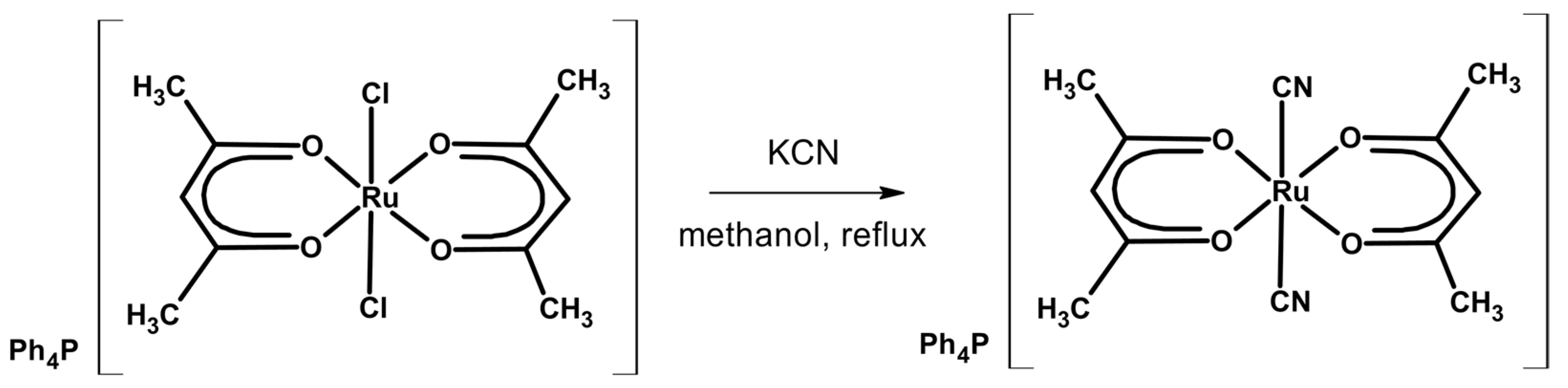
3.2.3. Synthesis of trans-Ph4P[RuIII(acac)2(NCS)2] (3·0.5C6H14)

A methanolic solution of 1 (0.60 g, 0.84 mmol) and KSCN (0.85 g, 8.76 mmol) was refluxed for 24 h. Then, the solution was evaporated to dryness and the residue was dissolved in dichloromethane and filtered. The filtrate was evaporated to ca. 3 mL, followed by the addition of n-hexane to give a reddish-purple precipitate, which was collected through filtration and dried under vacuum at 100 °C for 3 h. The yield was 0.43 g (64% based on trans-Ph4P[Ru(acac)2Cl2] (1)). Anal found: C, 58.94; H, 4.56; N, 3.75. calcd for C39H41N2O4S2PRu: C, 58.70; H, 5.18; N, 3.51. IR data (KBr disk, cm−1) 3057 w, 2087 vs, 2054 s, 1522 vs, 1483 m, 1436 s, 1378 s, 1270 m, 1188 w, 1108 s, 1024 m, 996 m, 935 m, 791 w, 754 m, 723 s, 689 m, 658 w, 526 vs, 456 m. UV–Vis (in CH2Cl2, λmax) 552, 464, 351, 276, 270, 237. HR-MS (ESI-TOF) found 415.9426 m/z (calcd for [M] 415.4562). 1H NMR (chlroform-d1, 298 K) δ 8.50–7.80 (m, o,m,p-H), −16.89 and −17.36 (s, CH3), −53.67 and −55.53 (s, CH3).

3.3. Crystal Structure Determination

X-ray crystallographic data for 1, 2·H2O and 3·CH3CN (Table 1) were collected for each single crystal at 293 K on a RIGAKU Saturn 724 CCD system equipped with a Mo rotating-anode X-ray generator with monochromate Mo Kα radiation (λ = 0.71075 Å) (installed at the Okayama University of Science). The structures were solved using direct methods (SHELXT and SIR-2011, respectively) and refined using the full-matrix least-squares technique (F2) with SHELXL-2014 as part of the SAINT Crystal Structure 4.2.5 (RIGAKU) software, respectively. Non-hydrogen atoms were refined with anisotropic displacement parameters, and all hydrogen atoms were refined with a riding model [55,56]. Selected bond distances and angles for 1, 2·H2O and 3·CH3CN are given in Tables S1–S6. CCDC-2325719, 2327661 and 2325718 contained the supplementary crystallographic data for trans-Ph4P[RuIII(acac)2Cl2] (1), Ph4P[{RuIII(acac)Cl}2(μ-Cl)3]·H2O (2·H2O) and trans-Ph4P[RuIII(acac)2(NCS)2] (3·CH3CN), respectively. These data can be obtained free of charge from the Cambridge Crystallographic Data Centre via www.ccdc.cam.ac.uk/data_request/cif (accessed on 31 January 2024).

4. Conclusions

The mononuclear and dinuclear ruthenium(III) complexes trans-Ph4P[RuIII(acac)2Cl2] (1) and Ph4P[{RuIII(acac)Cl}2(μ-Cl)3] (2) were synthesized through the reactions of RuCl3·nH2O with acetylacetone. The dinuclear complex 2 was isolated by passing oxygen gas during the reaction and repeated chromatographic purifications using Al2O3 columns (eluents: chloroform/MeOH and acetonitrile/chloroform). The mononuclear complex trans-Ph4P[RuIII(acac)2(NCS)2]·0.5C6H14 (3·0.5C6H14) was synthesized through the substitution reaction of the axial Cl of 1 with NCS. The mononuclear structures of 1 and 3·CH3CN and a dinuclear structure of 2·H2O were confirmed through X-ray crystal structure analyses. The Ru–Ru distance of 2.6661(2) in the dinuclear core of 2·H2O was indicative of the existence of the direct metal–metal interaction. The room temperature magnetic moments (μeff) were 2.00 and 1.93 μB for 1 and 3·0.5C6H14, respectively, and 0.66 μB for 2. The strong antiferromagnetic interaction (J ≤ −800 cm−1) between the ruthenium(III) ions within the dinuclear core was confirmed with a temperature-dependent magnetic susceptibility measurement at the 2–300 K range. The field dependence for magnetization measured from 0 to 70,000 Oe at 2 K showed that 2 was typical of the one for diamagnetism due to the strong antiferromagnetic interaction. The strong antiferromagnetic interaction between the unpaired electrons of the ruthenium(III) centers was considered to come from the direct metal–metal interaction. The mononuclear and dinuclear cores of 1, 2, and 3·C6H14 were maintained in the solution of CH2Cl2, which was verified by similarity in the absorption band positions in the visible region between the spectra measured in solid (diffuse reflectance spectrum) and solution (absorption spectrum) for each complex. In the 1H NMR spectra measured in chlroform-d1 at 298 K, the dinuclear complex 2 showed signals for the acac ligand protons at 5.93 ppm (for CH) and 2.50 and 2.39 ppm (for CH3), respectively, while 1 and 3·0.5C6H14 showed signals with large paramagnetic shifts: −17.59 ppm (for CH3) and −57.01 ppm (for CH) for 1 and −16.89 and −17.36 ppm (for CH3) and −53.67 and −55.53 ppm (for CH) for 3·0.5C6H14. In the CVs (the potential was quoted relative to the Fc/Fc+ couple), which were measured in CH2Cl2 with an electrolyte of nBu4N(ClO4), the RuIII → RuIV redox wave was shown at 0.23 V for 1, but not for 3·0.5C6H14 due to the decomposition, and RuIII → RuII waves were shown at −1.39 V for 1 and −1.25 V for 3·0.5C6H14, while, for 2, the RuIII–RuIII → RuIII–RuIV and RuIII–RuIII → RuIII–RuIV waves were shown at 0.91 V and −0.79 V, respectively.

Supplementary Materials

The following supporting information can be downloaded at: https://www.mdpi.com/article/10.3390/magnetochemistry10030016/s1, Table S1: Bond lengths of 1; Table S2: Bond angles of 1; Table S3: Bond lengths of 2·H2O; Table S4: Bond angles of 2·H2O; Table S5: Bond lengths of 3·CH3CN; Table S6: Bond angles of 3·CH3CN; Figure S1: IR spectrum of 1; Figure S2: IR spectrum of 2; Figure S3: IR spectrum of 3·0.5C6H14; Figure S4: Observed (top) and simulated (bottom) XRD patterns of 1; Figure S5: Observed (top for 2) and simulated (bottom for 2·H2O) XRD patterns; Figure S6: Observed (top for 3·0.5C6H14) and simulated (bottom for 3·CH3CN) XRD patterns; Figure S7: Crystal packing diagram of 1 without hydrogen for clarity; Figure S8: Packing diagram of 2·H2O without hydrogen atoms for clarity; Figure S9: Packing diagram of 3·CH3CN without hydrogen atoms for clarity; Figure S10: Field dependence of magnetization for 1 at 2 K. The red solid line represents the Brillouin function with g = 2.3; Figure S11: Field dependence of magnetization for 2 at 2 K; Figure S12: Field dependence of magnetization for 3·0.5C6H14 at 2 K. The red solid line represents the Brillouin function with g = 2.1; Figure S13: Diffuse reflectance spectra of 1 (orange solid line), 2 (blue solid line) and 3·0.5C6H14 (green solid line); Figure S14: 1H NMR Spectrum of 1 in chlroform-d1 at 298 K; Figure S15: 1H NMR spectrum of 2 in chlrooform-d1 at 298 K; Figure S16: 1H NMR spectrum of 3·0.5C6H14 in chloroform-d1 at 298 K.

Author Contributions

Conceptualization, methodology and funding acquisition, M.H.; investigation, K.N., C.H., S.N., H.A. and M.M.; data curation, M.H.; writing—original draft preparation, K.N. and M.H.; writing—review and editing, M.H. All authors have read and agreed to the published version of the manuscript.

Funding

A part of this work (SQUID measurement) was conducted at the Institute for Molecular Science, supported by Advanced Research Infrastructure for Materials and Nanotechnology in Japan (JPMXP1223MS1053) of the Ministry of Education, Culture, Sport, Science and Technology (MEXT), Japan.

Institutional Review Board Statement

Not applicable.

Informed Consent Statement

Not applicable.

Data Availability Statement

Crystallographic data of 1, 2·H2O and 3·CH3CN can be obtained free of charge from the Cambridge Crystallographic Data Centre (CCDC); deposition numbers of 1, 2 and 3·CH3CN are CCDC-2325719, 2327661 and 2325718, respectively.

Acknowledgments

The authors are grateful to Michiko Egawa (Shimane University) for her measurements of the elemental analyses, Masataka Maeyama (Rigaku Corporation) for his useful suggestion in the crystal structure analysis of complex 2·H2O and Yusuke Kataoka (Shimane University) for his help to measure the field-dependent magnetization and PXRD.

Conflicts of Interest

The authors declare no conflicts of interest.

References

  1. Chen, G.J.-J.; McDonald, J.W.; Newton, W.E. Preparation of OMoCl(acac)2 by a novel oxygen-chlorine atom exchange and its use as a reagent for the synthesis of monomeric molybdenum(V) complexes. Inorg. Chim. Acta 1979, 35, 93–97. [Google Scholar] [CrossRef]
  2. Doyle, G. The Reaction of Some Molybdenum and Tungsten Halides with β-Diketones. Inorg. Chem. 1971, 10, 2348–2350. [Google Scholar] [CrossRef]
  3. Dallmann, K.; Preetz, W. Crystal Structures, Vibrational Spectra and Normal Coordinate Analyses of cis- and trans-[OsX2(acac)2], X = Cl, Br, I. Z. Für Naturforschung B 1997, 52, 965–974. [Google Scholar] [CrossRef]
  4. Young, K.J.H.; Mironov, O.A.; Nielsen, R.J.; Chen, M.J.; Stewart, T.; Goddard, W.A., III; Periana, R.A. Synthesis and Characterization of the κ2-acac-O,O Complex OsIV(acac)2PhCl and Study of CH Activation with Benzene. Organometallics 2011, 30, 5088–5094. [Google Scholar] [CrossRef]
  5. Ferguson, G.; Glidewell, C. Enantiomeric disorder in racemic cis dichlorobis(pentane-2,4-dionato) titanium(IV). Acta Cryst. 2001, C57, 264–265. [Google Scholar]
  6. Wilkie, C.A.; Lin, G.Y.; Haworth, D.T. A comparative study of the cis-disubstituted bis(β-diketonato)titanium(IV) complexes. J. Inorg. Nucl. Chem. 1978, 40, 1009–1012. [Google Scholar] [CrossRef]
  7. Lindmark, A.F.; Fay, R.C. Dipseudohalobis (β-diketonato)titanium(IV) complexes. Synthesis, stereochemistry, configurational rearrangements, and vibrational spectra. Inorg. Chem. 1975, 14, 282–287. [Google Scholar] [CrossRef]
  8. Bradley, D.C.; Holloway, C.E. Nuclear magnetic resonance and infrared spectral studies on labile cis-dialkoxy-bis(acetylacetonato)titanium(IV) compounds. J. Chem. Soc. A 1969, 282–285. [Google Scholar] [CrossRef]
  9. Brown, S.N.; Chu, E.T.; Hull, M.W.; Noll, B.C. Electronic Dissymmetry in Chiral Recognition. J. Am. Chem. Soc. 2005, 127, 16010–16011. [Google Scholar] [CrossRef] [PubMed]
  10. Fay, R.C.; Lowry, R.N. Structure and lability of dihalobis(acetylacetonato)titanium(IV) complexes. Inorg. Nucl. Chem. Lett. 1967, 3, 117–120. [Google Scholar] [CrossRef]
  11. Cox, M.; Clark, R.J.H.; Milledge, H.J. Structural Studies on Group IV Metal Acetylacetonates. Nature 1966, 212, 1357. [Google Scholar] [CrossRef]
  12. Sarkhel, P.; Paul, B.C.; Poddar, R.K. Rhodium(III) complexes containing acetylacetonate and mono- or bidentate ligands. Indian J. Chem. 1999, 38A, 150–155. [Google Scholar]
  13. Sodhi, R.K.; Paul, S. An Overview of Metal Acetylacetonates: Developing Areas/Routes to New Materials and Applications in Organic Syntheses. Catal. Surv. Asia 2018, 22, 31–62. [Google Scholar] [CrossRef]
  14. Behzadi, K.; Thompson, A. Preparation, Characterization and Reactions of Vanadium(IV) β-Diketonate Complexes. J. Less Common Met. 1987, 128, 281–296. [Google Scholar] [CrossRef]
  15. Paul, B.C.; Poddar, R.K. Synthesis, characterization and reactivity studies of dichloroacetylacetonato acetylacetone ruthenium(III). Transit. Met. Chem. 1993, 18, 96–100. [Google Scholar] [CrossRef]
  16. Janiak, C.; Scharmann, T.G.; Lange, C.H.K. Zirconium β-diketonate/methylaluminoxane systems as singlesite catalysts for the preparation of high-molecular-weight polyethylene. Macromol. Rapid Commun. 1994, 15, 655–658. [Google Scholar] [CrossRef]
  17. Pinnavia, T.J.; Fay, R.C. Preparation and properties of some six- and seven-coordinate halo(acetylacetonato) complexes of zirconium(IV) and hafnium(IV). Inorg. Chem. 1968, 7, 502–508. [Google Scholar] [CrossRef]
  18. Bagnall, K.W.; Edwards, J.; Rickard, C.E.F.; Tempest, A.C. Cyclopentadienyluranium(IV) acetylacetonates. J. Inorg. Nucl. Chem. 1979, 41, 1321–1323. [Google Scholar] [CrossRef]
  19. Shibata, S.; Ohta, M. Molecular structure of bis(acetylacetonato)zinc(II) in the gas phase as determined from electron diffraction data. J. Mol. Struct. 1981, 77, 265–270. [Google Scholar] [CrossRef]
  20. Korochentsev, V.V.; Vovna, V.I.; L’vov, I.B.; Shapkin, N.P. Electronic and geometric structure of the protonated forms of nickel β-diketonates. Russ. J. Coord. Chem. 2011, 37, 371–376. [Google Scholar] [CrossRef]
  21. Funaioli, T.; Marchetti, F.; Pampaloni, G.; Zacchini, S. Ligand-interchange reactions between M(iv) (M = Ti, V) oxide bis-acetylacetonates and halides of high-valent group 4 and 5 metals. A synthetic and electrochemical study. Dalton Trans. 2013, 42, 14168–14177. [Google Scholar] [CrossRef]
  22. Stabinikov, P.A.; Pervukhina, N.V.; Baidina, I.A.; Sheludyakova, L.A.; Borisov, S.V. On the Symmetry of iron(III) tris-acetylacetonate crystals. J. Struct. Chem. 2007, 48, 186–192. [Google Scholar] [CrossRef]
  23. Shalhoub, G.M. Co(acac)3 Synthesis, reactions, and spectra: An experiment for general chemistry. J. Chem. Educ. 1980, 57, 525–526. [Google Scholar] [CrossRef]
  24. Ledneva, A.Y.; Artemkina, S.B.; Piryazev, D.A.; Fedorov, V.E. Structure and Thermal Properties of the molybdenum Complex Mo(acac)3. J. Struct. Chem. 2015, 56, 1021–1023. [Google Scholar] [CrossRef]
  25. Dedeian, K.; Djurovich, P.I.; Garces, F.O.; Carlson, G.; Watts, R.J. A new synthetic route to the preparation of a series of strong photoreducing agents: Fac-tris-ortho-metalated complexes of iridium(III) with substituted 2-phenylpyridines. Inorg. Chem. 1991, 30, 1685–1687. [Google Scholar] [CrossRef]
  26. Dallmann, K.; Preetz, W. Darstellung, Kristallstruktur, Schwingungsspektren und Normalkoordinatenanalyse von [Os(acac)3]/ Synthesis, Crystal Structure, Vibrational Spectra, and Normal Coordinate Analysis of [Os(acac)3]. Z. Für Naturforschung B 1998, 53, 232–238. [Google Scholar] [CrossRef]
  27. Jackson, A.B.; White, P.S.; Templeton, J.L. Bis(acetylacetonato)tricarbonyl Tungsten(II):  A Convenient Precursor to Chiral Bis(acac) Tungsten(II) Complexes. Inorg. Chem. 2006, 45, 6205–6213. [Google Scholar] [CrossRef] [PubMed]
  28. Chang, Q.; Yan, C.; Li, X.-N.; Cui, H.; Ye, Q.; Jiang, J.; Yu, J.; Chen, J.; Liu, W. The influence of sodium and barium ions on the structures of iridium (III) acetylacetonate complexes. Res. Chem. Intermed. 2015, 41, 7631–7640. [Google Scholar] [CrossRef]
  29. Nakanishi, R.; Yatoo, M.A.; Katoh, K.; Breedlove, B.K.; Yamashita, M. Dysprosium Acetylacetonato Single-Molecule Magnet Encapsulated in Carbon Nanotubes. Materials 2017, 10, 7. [Google Scholar] [CrossRef]
  30. Zhang, D.; Zhang, L.-F.; Yuting, C.; Hailong, W.; Ni, Z.-H.; Wolfgang, W.; Jianzhuang, J. Heterobimetallic porphyrin-based single-chain magnet constructed from manganese(iii)-porphyrin and trans-dicyanobis(acetylacetonato) ruthenate(iii) containing co-crystallized bulk anions and cations. Chem. Commun. 2010, 46, 3550–3552. [Google Scholar] [CrossRef]
  31. Lan, W.; Hao, X.; Dou, Y.; Zhou, Z.; Yang, L.; Liu, H.; Li, D.; Dong, Y.; Kong, L.; Zhang, D. Various Structural Types of Cyanide-Bridged FeIII–MnIII Bimetallic Coordination Polymers (CPs) and Polynuclear Clusters Based-on A New mer-Tricyanoiron(III)Building Block: Synthesis, Crystal Structures, and Magnetic Properties. Polymers 2019, 11, 1585. [Google Scholar] [CrossRef]
  32. Hirayama, M.; Kitani, K. Observation of 27Al n.m.r. paramagnetic shifts (large contact shift contributions) induced by lanthanoid shift-reagents in tris(acetylacetonato)aluminium(III). J. Chem. Soc. Chem. Commun. 1980, 21, 1030–1031. [Google Scholar] [CrossRef]
  33. Goff, H.M.; Hines, J.; Griesel, J.; Mossman, C. Synthesis, Characterization, and Use of a Cobalt(II) Complex as an NMR Shift Reagent. J. Chem. Educ. 1982, 59, 422–423. [Google Scholar] [CrossRef]
  34. Levy, G.C.; Komoroski, R.A. Paramagnetic Relaxation Reagents. Alternatives or Complements to Lanthanide Shift Reagents in Nuclear Magnetic Resonance Spectral Analysis. J. Am. Chem. Soc. 1974, 96, 678–681. [Google Scholar] [CrossRef]
  35. Gillies, E.; Szarek, W.A.; Baird, M.C. Application of Paramagnetic Shift Reagents in Nuclear Magnetic Resonance Studies of Complex Alcohols and Amines. Can. J. Chem. 1971, 49, 211–216. [Google Scholar] [CrossRef]
  36. Sirkecioglu, O.; Karliga, B.; Talinli, N. Benzylation of alcohols by using bis[acetylacetonato]copper as catalyst. Tetrahedron Lett. 2003, 44, 8483–8485. [Google Scholar] [CrossRef]
  37. Pichugina, D.; Kuz’menko, N.; Shestakov, A. Gold complexes with Oxygen-containing Ligands as a Catalyst for Methane Oxidation. Gold Bull. 2007, 40, 115–120. [Google Scholar] [CrossRef]
  38. Yeung, W.-F.; Man, W.-L.; Wong, W.-T.; Lau, T.-C.; Gao, S. Ferromagnetic Ordering in a Diamond-Like Cyano-Bridged MnIIRuIII Bimetallic Coordination Polymer. Angew. Chem. Int. Ed. 2001, 40, 3031–3033. [Google Scholar] [CrossRef]
  39. Guo, J.-F.; Wang, X.-T.; Wang, B.-W.; Xu, G.-C.; Gao, S.; Szeto, L.; Wong, W.-T.; Wong, W.-Y.; Lau, T.-C. One-Dimensional Ferromagnetically Coupled Bimetallic Chains Constructed with trans-[Ru(acac)2(CN)2]: Syntheses, Structures, Magnetic Properties, and Density Functional Theoretical Study. Chem. Eur. J. 2010, 16, 3524–3535. [Google Scholar] [CrossRef]
  40. Hasegawa, T.; Lau, T.C.; Taube, H.; Schaefer, W.P. Electronic Effects of Bis(acetylacetone) in Ruthenium(II) and Ruthenium(III) Complexes. Inorg. Chem. 1991, 30, 2921–2928. [Google Scholar] [CrossRef]
  41. Darriet, J. Crystal structure and magnetic properties of the (Ru2Cl9)3− ion in Cs3Ru2Cl9. Rev. Chim. Mine. 1981, 18, 27–32. [Google Scholar]
  42. Hughes, M.N.; O’Reardon, D.; Poole, R.K.; Hursthouse, M.B.; Thornton-Pett, M. The Structure of the mixed-valence chloro-bridged dimeric complex cation [Ru2(NH3)6Cl3]2+. Polyhedron 1987, 6, 1711–1713. [Google Scholar] [CrossRef]
  43. Clucas, W.A.; Armstrong, R.S.; Buys, I.E.; Hambley, T.W.; Nugent, K.W. X-ray Crystallographic Study of the Ruthenium Blue Complexes [Ru2Cl3(tacn)2](PF6)2·H2O, [Ru2Br3(tacn)2](PF6)2·2H2O, and [Ru2I3(tacn)2](PF6)2: Steric Interactions and the Ru-Ru “Bond Length”. Inorg. Chem. 1996, 35, 6789–6794. [Google Scholar] [CrossRef] [PubMed]
  44. Neubold, P.; Vedova, B.S.P.C.D.; Wieghardt, K.; Nuber, B.; Weiss, J. Novel Cofacial Bioctahedral Complexes of Ruthenium: Syntheses and Properties of the Mixed-Valence Species [LRu2.5(μ-X)3Ru2.5L]2+ (X = Cl, Br, I, OH). Crystal Structures of [LRu2.5(μ-OH)3Ru2.5L](PF6)2·H2O and [LRuIV(μ-O)3RuIVL](PF6)2·H2O (L = 1,4,7-Trimethyl-1,4,7-triazacyclononane). Inorg. Chem. 1990, 29, 3355–3363. [Google Scholar]
  45. Bonner, J.C.; Fisher, M.E. Linear Magnetic Chains with Anisotropic Coupling. Phys. Rev. 1964, 135, 640–658. [Google Scholar] [CrossRef]
  46. Bleaney, B.; Bowers, K.D. Anomalous paramagnetism of copper acetate. Proc. R. Soc. Lond. 1952, 214, 451–465. [Google Scholar] [CrossRef]
  47. Lovel, T.; Stranger, R.; McGrady, J.E. Mutal Interdependence of Spin Crossover and Metal-Metal Bond Formation in M2Cl93− (M = Fe, Ru, Os). Inorg. Chem. 2001, 40, 39–43. [Google Scholar] [CrossRef]
  48. Cotton, F.A.; Matusz, M.; Torralba, R.C. New Di- and Trinuclear Complexes of Ruthenium with Octahedra Joined on Faces or Edges: Ru2Cl6(PBu3)3, Ru2Cl6(PBu3)4 and Ru3Cl8(PBu3)4 (Bu = CH3CH2CH2CH2). Inorg. Chem. 1989, 28, 1516–1520. [Google Scholar] [CrossRef]
  49. Grobelny, R.; Jezowska-Trzebiatowska, B.; Wojciechowski, W. The Absorption Spectra and Magnetic Properties of the chelated compounds of Ru(III) with β-diketones. J. Inorg. Nucl. Chem. 1966, 28, 2715–2718. [Google Scholar] [CrossRef]
  50. Cardona, C.M.; Li, W. Electrochemical Considerations for Determining Absolute Frontier Orbital Energy Levels of Conjugated Polymers for Solar Cell Applications. Adv. Mater. 2011, 23, 2367–2371. [Google Scholar] [CrossRef]
  51. Hashem, E.; Platts, J.A.; Hartl, F.; Lorusso, G.; Evangelisti, M.; Schulzke, C.; Baker, R.J. Thiocyanate Complexes of Uranium in Multiple Oxidation States: A Combined Structural, Magnetic, Spectroscopic, Spectroelectrochemical, and Theoretical Study. Inorg. Chem. 2014, 53, 8624–8637. [Google Scholar] [CrossRef]
  52. Bain, G.A.; Berry, J.F. Diamagnetic Corrections and Pascal’s Constants. J. Chem. Educ. 2008, 85, 532–536. [Google Scholar] [CrossRef]
  53. Gottlieb, H.E.; Kotlyar, V.; Nudelman, A. NMR Chemical Shifts of Common Laboratory Solvents as Trace Impurities. J. Org. Chem. 1997, 62, 7512–7515. [Google Scholar] [CrossRef]
  54. Fulmer, G.R.; Miller, A.J.M.; Sherden, N.H.; Hugo, E.G.; Nudelman, A.; Brian, M.S.; John, E.B.; Karen, I.G. NMR Chemical Shifts of Trace Impurities: Common Laboratory Solvents, Organics, and Gases in Deuterated Solvents Relevant to the Organometallic Chemist. Organometallics 2010, 29, 2176–2179. [Google Scholar] [CrossRef]
  55. Sheldrick, G.M. SHELXT—Integrated space-group and crystal-structure determination. Acta Cryst. 2015, A71, 3–8. [Google Scholar] [CrossRef] [PubMed]
  56. Sheldrick, G.M. Crystal structure refinement with SHELXL. Acta Cryst. 2015, C71, 3–8. [Google Scholar]
Scheme 1. Axial ligand substitution reaction from trans-[RuIII(acac)2Cl2] to trans-[RuIII(acac)2(CN)2].
Magnetochemistry 10 00016 sch001
Scheme 2. Chemical structure of [{Ru(acac)Cl}2(μ-Cl)3].
Magnetochemistry 10 00016 sch002
Figure 1. ORTEP view of one of the anionic units of 1, showing thermal ellipsoids at the 50% probability level. Hydrogen atoms were omitted for clarity.
Magnetochemistry 10 00016 g001
Figure 2. ORTEP view of the anionic dinuclear unit for 2·H2O, showing thermal ellipsoids at the 50% probability level. Hydrogen atoms were omitted for clarity.
Magnetochemistry 10 00016 g002
Scheme 3. Chemical structure of [(RuIIICl3)2(μ-Cl)3]3−, the bond distances and angles being shown together [41].
Magnetochemistry 10 00016 sch003
Scheme 4. MO diagram for 4d electrons of the mixed-valent diruthenium(II,III) (a) and diruthenium(III,III) (b) with the face-sharing octahedral structure [43,44].
Magnetochemistry 10 00016 sch004
Figure 3. ORTEP view of the anionic unit for 3·CH3CN, showing thermal ellipsoids at the 50% probability level. Hydrogen atoms were omitted for clarity.
Magnetochemistry 10 00016 g003
Figure 4. Temperature dependences of reciprocal magnetic susceptibility1/χM (red circles) and magnetic moment μeff (blue circles) of trans-Ph4P[RuIII(acac)2Cl2] (1). The red solid line was drawn with C = 0.498 cm3 mol−1 K and θ = −6.3 K and the blue solid line was calculated and drawn with g = 2.21, J = −0.29 cm−1 and Nα = 60 × 10−6 emu mol−1 (see text).
Magnetochemistry 10 00016 g004
Figure 5. Temperature dependencies of reciprocal magnetic susceptibility χM (red circles) and magnetic moment μeff (blue circles) for Ph4P[{RuIII(acac)Cl}2(μ-Cl)3] (2). The blue solid line was calculated and drawn with the parameter values of g = 2.2, J = −800 cm−1, Nα = 90 × 10−6 emu mol−1 and ρ = 0.0015 (see text).
Magnetochemistry 10 00016 g005
Figure 6. Temperature dependences of reciprocal magnetic susceptibility 1/χM (red circles) and magnetic moment μeff of trans-Ph4P[RuIII(acac)2(NCS)2] (3·0.5C6H14). The red solid line was drawn with C = 0.464 cm3 mol−1 K and θ = −4.8 K and the blue solid line was calculated and drawn with g = 2.14, J = 0.22 cm−1 and Nα = 60 × 10−6 emu mol−1 (see text).
Magnetochemistry 10 00016 g006
Scheme 5. Chain structure comprised of [Ru(acac)2Cl2] units in 1. J is the parameter for the magnetic interaction between [Ru(acac)2Cl2] units in the chain.
Magnetochemistry 10 00016 sch005
Scheme 6. Dimer structure comprised of [Ru(acac)2(NCS)2] units in 3. J is the parameter for the magnetic interaction between [Ru(acac)2(NCS)2] units in the dimer.
Magnetochemistry 10 00016 sch006
Figure 7. Absorption spectra (measured in CH2Cl2) of 1 (orange line), 2 (blue line) and 3·0.5C6H14 (green line).
Magnetochemistry 10 00016 g007
Figure 8. Cyclic voltammograms of 1 (blue line) and 2 (orange line) at 1.0 × 10⁻³ M in CH2Cl2 containing 0.1 M nBu4N(ClO4) (glassy carbon working electrode; scan rate = 50 mV/s; room temperature; under Ar).
Magnetochemistry 10 00016 g008
Figure 9. Cyclic voltammograms of 3·0.5C6H14 at 1.0 × 10⁻³ M in CH2Cl2 containing 0.1 M nBu4N(ClO4) (glassy carbon working electrode; scan rate = 50 mV/s; room temperature; under Ar).
Magnetochemistry 10 00016 g009
Table 1. Crystallographic data and structure refinement of 1, 2·H2O and 3·CH3CN.
Complexes12·H2O3·CH3CN
Chemical formulaC34H34Cl2O4PRuC34H36Cl5O5PRu2C38H37N3O4PRuS2
FW709.55935.03795.87
Temperature, T (K)939393
Crystal systemTriclinicTriclinicMonoclinic
Space group P 1 ¯ P 1 ¯ P21/n
a (Å)9.8418(2)10.91020(10)20.0627(3)
b (Å)13.3562(3)12.05510(10)7.36340(10)
c (Å)14.1498(2)16.1720(2)24.6645(5)
α (°)103.441(2)69.5690(10)90
β (°)106.783(2)74.3220(10)98.403(2)
γ(°)107.998(2)70.0890(10)90
V3)1584.42(6)1846.19(4)3604.56(10)
Z224
Dcalcd (g cm−3)1.4871.6781.478
Crystal size (mm)0.2 × 0.1 × 0.050.15 × 0.1 × 0.050.2 × 0.1 × 0.05
μ (mm−1)0.7511.2630.643
θ range for data collection (°)1.607–31.4641.980–31.5461.669–31.629
Reflections collected30,58034,22967,229
[R1 (I < 2σ(I)); wR2 (all data)] (a)R1 = 0.0301
ωR2 = 0.0826
R1 = 0.0283
ωR2 = 0.0743
R1 = 0.0310
ωR2 = 0.0947
GOF1.1061.0721.099
(a) R 1 = F O F c F O ; ω R 2 = ω F O 2 F C 2 2 F O 2 2 1 2 .
Disclaimer/Publisher’s Note: The statements, opinions and data contained in all publications are solely those of the individual author(s) and contributor(s) and not of MDPI and/or the editor(s). MDPI and/or the editor(s) disclaim responsibility for any injury to people or property resulting from any ideas, methods, instructions or products referred to in the content.

