Article

Plant Regeneration via Somatic Embryogenesis and Indirect Organogenesis in Blue Honeysuckle (Lonicera caerulea L.)

1
College of Horticulture & Landscape Architecture, Northeast Agricultural University, Harbin 150030, China
2
College of Life Science, Northeast Agricultural University, Harbin 150030, China
3
Key Laboratory of Biology and Genetic Improvement of Horticultural Crops (Northeast Region), Ministry of Agriculture and Rural Affairs, Harbin 150030, China
4
National-Local Joint Engineering Research Center for Development and Utilization of Small Fruits in Cold Regions, National Development and Reform Commission, Harbin 150030, China
*
Authors to whom correspondence should be addressed.
Horticulturae 2023, 9(9), 996; https://doi.org/10.3390/horticulturae9090996
Submission received: 6 August 2023 / Revised: 28 August 2023 / Accepted: 2 September 2023 / Published: 4 September 2023
(This article belongs to the Special Issue Plant Tissue and Organ Cultures for Crop Improvement in Omics Era)

Abstract

:
Blue honeysuckle (Lonicera caerulea L.), which belongs to the Caprifoliaceae family, is an emerging fruit crop worldwide. For the development of a transgenic system and multipurpose tissue culture, this study for the first time established an in vitro regeneration system via somatic embryogenesis, as well as improving the previously established indirect organogenesis-based regeneration system. For embryogenesis, Murashige and Skoog (MS) medium supplemented with 1.0 mg/L 2,4-dichlorophenoxyacetic acid (2,4-D) showed the highest induction rate of the embryogenic callus (97.6%), and MS supplemented with 0.1 mg/L 6-benzyladenine (6-BA), 0.1 mg/L α-naphthaleneacetic acid (NAA), and 0.5 g/L activated carbon (AC) achieved the highest somatic embryo rate (28.3%). For indirect organogenesis, MS medium supplemented with 1.0 mg/L 6-BA and 0.1 mg/L NAA resulted in the highest non-embryogenic callus induction rate (98.9%) and adventitious shoot induction rate (51.6%). For adventitious root induction, MS supplemented with 1.0 mg/L indole-3-butyric acid (IBA) achieved the highest root induction rate (96.0%) and average root length (4.6 cm), whereas MS supplemented with 0.5 mg/L indole-3-acetic acid (IAA) resulted in the highest average regenerated root number (8.8). The total time for the regeneration from explants to soil-planted seedlings (10 euphylla) was 105 and 150 days with an efficiency of 44.1% and 23.9% through organogenesis and somatic embryogenesis, respectively. This study provides a powerful tool for rapid propagation, proliferation, and transformation, as well as laying a technological foundation for gene function research and genetic improvement of blue honeysuckle.

1. Introduction

Blue honeysuckle (Lonicera caerulea Linn.), also known as “Haskap” or “Honeyberry”, is a deciduous shrub belonging to the Caprifoliaceae family [1]. The fruit of blue honeysuckle is small berries with attractive azure to dark-blue color and can be eaten directly without the need for peeling or denucleation, with a sour–sweet taste resembling that of blueberry, bilberry, and blackcurrant [2]. Due to its abundant anthocyanins, it is widely processed into juice, jam, wine, and candy, or added to processed foods as a natural pigment or nutritional additive [3]. Besides anthocyanins, the fruit of blue honeysuckle have also been reported to be rich in vitamin C, phenolics, and flavonoids, which confer them with strong antibacterial, anti-inflammatory, antioxidant, and antidiabetic activities [1,4,5]. Blue honeysuckle fruit also contain high levels of iridoids, and therefore can be used for adjunctive treatment of depression [6]. In addition, blue honeysuckle has some highly desirable agronomic traits, including tolerance to extreme freezing (−50 °C tolerance during dormancy), early fruiting, and stable annual yield (about 2 to 2.5 kg for a 6-year-old tree), which make it a well-accepted emerging fruit crop [7,8]. The commercial cultivation of blue honeysuckle has been rapidly expanded in cold-climate regions worldwide during the past two decades, such as Russia, northeast China, Canada, and Hokkaido of Japan [9,10]. However, despite its promising potential, the basic biological research on blue honeysuckle largely lags behind that on major fruits, emerging fruit crops [11], or minor horticultural plants [12]. Particularly, the in vitro propagation of blue honeysuckle is a key procedure for its industrialized seedling production and development of biotechnology tools [10].
Establishment of an efficient in vitro plant regeneration system is the initial step to setting up a genetic transformation system and the subsequent application of CRISPR/Cas9 gene editing technology [13]. Organogenesis and somatic embryogenesis are two major pathways for plant in vitro regeneration. Organogenesis refers to the development of roots, shoots, and flowers directly from an explant or indirectly from a callus culture, resulting in the final regeneration of a whole plant [14]. Somatic embryogenesis is the development of somatic embryos directly from the explant or indirectly from callus-induced explant to finally generate new plants with somatic cells going through a series of morphological and biochemical changes [15,16]. In these two pathways, both direct and indirect organogenesis (DO and IDO) are widely applied in germplasm conservation [17], rapid propagation [18], virus elimination [19], somatic hybrid regeneration [20], homozygous plant regeneration [21], and gene function research [22]. Compared with the rare direct somatic embryogenesis (DSE), indirect somatic embryogenesis (ISE) plays a versatile role in plant biotechnology, including the construction of somatic hybrids, regeneration of homozygous lines, virus elimination, and genetic transformation. Notably, although both organogenesis and somatic embryogenesis (particularly IDO and ISE) are applied in plant genetic transformation, somatic embryogenesis generally has an advantage over organogenesis because it can avoid the risk of chimera development [23]. However, few studies have been conducted on the in vitro propagation of blue honeysuckle. After the first study that focused on shoot proliferation of blue honeysuckle (L. caerulea and L. caerulea var. edulis) [24], subsequent research studies have demonstrated the micropropagation efficiency of blue honeysuckle is influenced by genotypes [25], base medium compositions (MS, 3/4 MS, 1/4 MS, and MS supplemented with wheat starch) [24,26,27,28], plant growth regulators (6-BA and thidiazuron + IAA) [25,29], chelated irons (FeNaEDDHA, Fe(III)-EDTA, Fe(III)-DTPA, Fe(III)-EDDHA, Fe(III)-HEDP, and Fe(II)-HEDP) [27,28], and culture temperatures [24]. To the best of our knowledge, there have been no reports about plant regeneration through the ISE pathway on blue honeysuckle. Furthermore, it is crucial to comprehensively evaluate the efficiency of organogenesis regeneration to facilitate downstream development of a transgenic regeneration system—a fundamental technique for functional genomic study and further genetic improvement of selected cultivars.
This study for the first time established an in vitro plant regeneration system through indirect somatic embryogenesis in blue honeysuckle (L. caerulea L.), and the previously established regeneration system based on indirect organogenesis was comprehensively improved by assessing the efficiency of different explant types, genotypes, and concentrations and types of plant growth regulators (PGRs). The findings are expected to facilitate the further development of biotechnologies for blue honeysuckle.

2. Materials and Methods

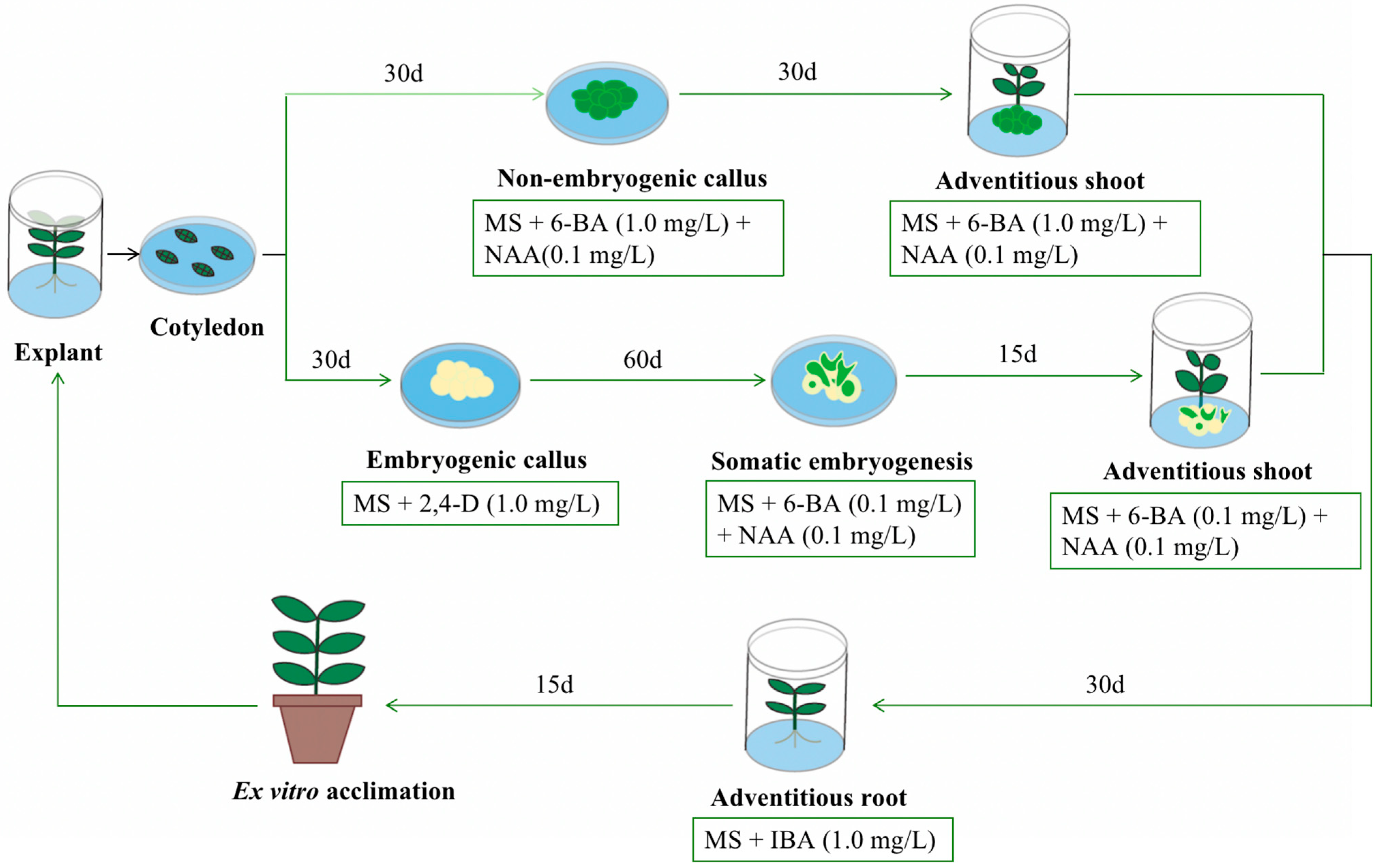
2.1. Plant Materials and Preparation of Tissue Culture

Blue honeysuckle cultivar ‘Berel’ (L. caerulea L.) was used in all procedures in this study. This cultivar was officially imported from Russia at 2001 [30] (Heilongjiang province regional registration of crop varieties: 2011037). Three genotypes, ‘Lanjingling’ (L. caerulea L., China National Plant Variety Protection: CNPVP-20200389) [31], ‘Berel’, and a wild accession “Altai-4” (L. caerulea subsp. altaica), were used in adventitious shoot induction. Mature blue honeysuckle fruits were collected from the Horticultural Station of Northeast Agricultural University (126.73° E, 45.74° N, BD-09 coordinate system, https://api.map.baidu.com/lbsapi/getpoint/index.html, (accessed on 1 September 2023)), Harbin, China, from June to July of 2022. The fruit samples were kept in a freezer (–20 °C) for at least 20 h to terminate the possible seed dormancy. The seeds were collected from the fruit and washed thoroughly with running tap water for 30 min and then soaked in 0.05 mol/L NaOH solution for 10 min for pectin removal. The cleaned seeds were sterilized with 75% ethanol for 30 s and then with 10% NaClO2 solution for 5 min under a laminar airflow chamber (DL-CJ-1NDII, HDL, Beijing, China). The seeds were further in vitro washed with autoclaved double distilled water three times to eliminate the residual chemicals. The prepared seeds were sown on 1/2 MS (half-strength MS medium, pH = 5.8) solid medium containing 20 g/L sucrose and 7.5 g/L agar powder (PB8190, Promisebio, Japan) in autoclaved culture jars (10 cm in height and 6 cm in diameter) under long-day conditions (16 h/8 h light/dark, light intensity of 2000 lx, and temperature of 25 ± 2 °C) for 30 d.

2.2. Callus Induction

In vitro, cotyledons and euphyllas developed from the seeds were cut into approximately 1 cm2 pieces, and the tender stems and roots developed from the seeds were cut into about 0.2 cm segments. The prepared explants were inoculated on MS basal medium (pH = 5.8) [32] in the jars with different combinations of 6-BA + NAA and 2,4-D (Table 1 and Table S1). The jars were incubated in a dark room with a temperature of 25 ± 2 °C for at least 7 d, and then the induced callus was transferred to the long-day environment mentioned above. All the experiments consisted of three replicates, with each replicate including 30 explants. Sub-culture was performed every two weeks on fresh medium to maintain high callus competence. The frequency of callus induction was investigated after in vitro culturing for 30 d with the equation of callus induction rate (%) = number of explant-produced calli/total number of inoculated explants.

2.3. Somatic Embryogenesis

To induce somatic embryogenesis, the embryogenic callus induced from MS supplemented with 1.0 mg/L 2,4-D (yellowish to transparent color and friable texture) was transferred to MS medium with different concentrations of 6-BA + NAA and 6-BA + IBA (Table 2 and Table S1) and cultured under long-day conditions. All the experiments consisted of 3 replicates, with each replicate including 30 callus clusters. Sub-culture was performed every two weeks on fresh medium to maintain high embryogenic competence. The morphogenesis and frequency of somatic embryogenesis were investigated after culturing for 75 d. For microscopic observation, samples were gently rinsed with sterile distilled water to remove the medium on the surface, blotted dry on a piece of sterile tissue paper, and photographed using a light photomicroscope (SZN71, SOPTOP, China). For histological analysis, the samples were fixed in FAA (5 mL formaldehyde: 5 mL glacial acetic acid: 90 mL ethanol) overnight, then dehydrated through an ethanol series (70, 80, 95, and 100% at 1 h each concentration), and finally embedded in paraffin wax at 58 °C. Eight-micrometer thick sections were cut using a microtome (DM IL LED, Leica, Germany), fixed on glass slides, and stained with an aqueous 1% hematoxylin–eosin stain solution. Histological observation was conducted using a microscope (DM IL LED, Leica, Germany). The embryogenesis frequency was calculated with the equation of embryos induction rate (%) = number of callus-produced somatic embryos/total number of inoculated embryonic calli.

2.4. Adventitious Shoot Induction

After 30 d of in vitro culturing, the non-embryogenic callus induced from MS supplemented with 1.0 mg/L 6-BA + 0.1 mg/L NAA (green and dense callus) was isolated and transferred to the MS medium containing various concentrations of 6-BA + NAA and 6-BA + IBA for shoot organogenesis (Table 3, Table 4 and Table S1). All the experiments consisted of three replicates, with each replicate including 30 calli. Sub-culture was performed every two weeks on fresh medium to maintain high adventitious shoot regeneration competence. The frequency of adventitious shoot induction was recorded after 30 d with the equation of adventitious shoot induction rate (%) = number of callus-generated adventitious shoot (s)/total number of inoculated organogenic calli.

2.5. Adventitious Root Induction

After somatic embryogenesis or shoot induction through organogenesis, the regenerated shoots with four to six euphyllas were carefully separated and transferred to MS medium with IBA (0.5, 1.0 mg/L), IAA (0.5, 1.0 mg/L), and PGR-free for adventitious root induction (Table 5 and Table S1). Each root induction treatment was repeated three times, and each replicate consisted of 15 shoots. Sub-culture was carried out every two weeks on fresh medium to maintain high adventitious root induction competence. After 30 d of rooting culture, data on rooting rate, number of roots, and length of roots were recorded.

2.6. Plant Regeneration and Transplantation

The plantlets with 3–5 cm of adventitious roots were taken from the culture jars and the medium adhered to the roots was gently washed away under a running tap. For acclimatization, the plantlets were hydroponically cultured in 50 mL plastic pipes using sterile water for 3 d under shade. Then, the plantlets were planted in gallon pots containing horticultural substrate (universal type: 2265734, Scotts Miracle-Gro, Wuhan, China) in a heliogreenhouse (15–25 °C temperature and ~80% relative humidity).

2.7. Statistical Analysis

The raw data were manually recorded in Excel 2019 for data organization, and then imported to SPSS software (version 25.0, IBM Inc., Armonk, NY, USA) for statistical analysis. Data were presented by mean ± standard error. Analysis of variance (ANOVA) was used to test the statistical significance, and multiple comparisons of means were performed by Duncan’s test at the 0.05 significance level.

3. Results and Discussion

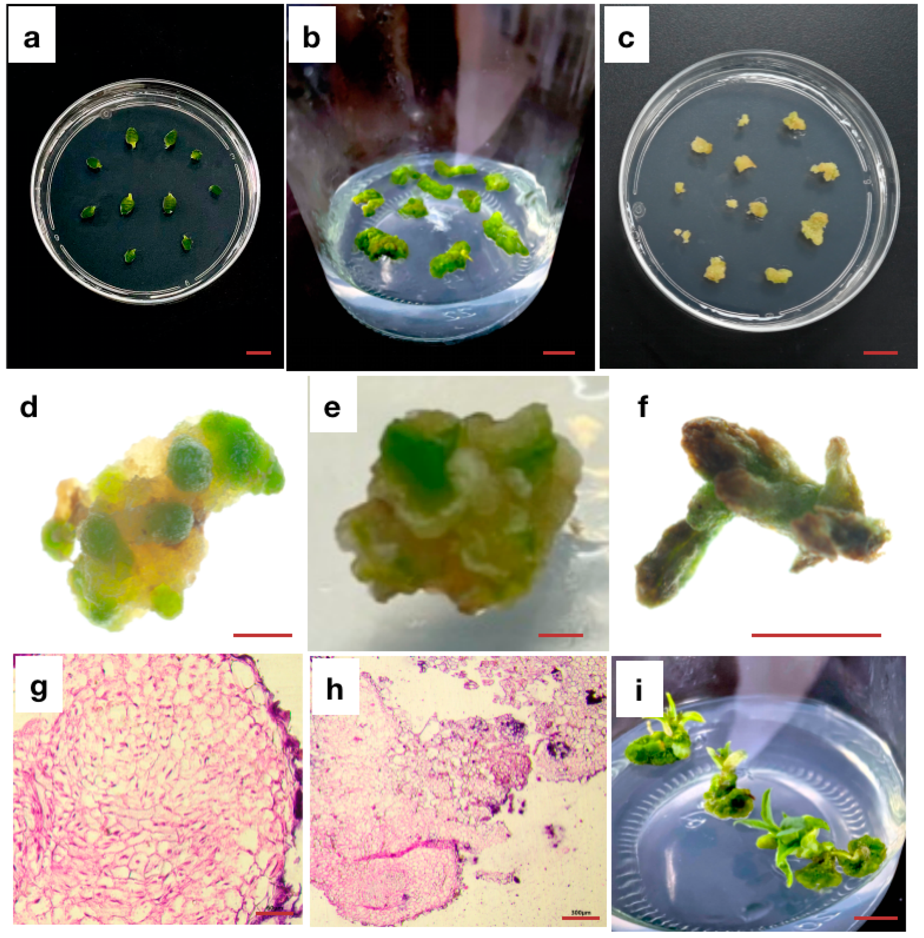
3.1. Effects of PGRs and Explant Types on Callus Induction

To comprehensively investigate the effects of PGRs and explant type on the callus induction efficiency of blue honeysuckle, we designed treatments with four explant types (cotyledon, euphylla, tender stem, and tender root) interacting with the combination of 6-BA and NAA at two concentrations or four 2,4-D concentrations (Table 1 and Supplementary Figures S1 and S2). Generally, the callus was initiated from the cutting surface after about 5 d of in vitro culturing from cotyledon or euphylla and about 7 d from tender stem or tender root (Figure 1a). For the combination of 6-BA and NAA, the highest callus induction efficiency was observed for 1.0 mg/L 6-BA and 0.1 mg/L NAA, which was 98.9%, 65.7%, 73.0%, and 83.7% on cotyledon, euphylla, tender stem, and tender root, respectively (Table 1). However, for 2,4-D, 1.0 mg/L was the most effective concentration for the callus induction of cotyledon (97.6%) and euphyllas (60.0%), and 2.0 mg/L was the optimum concentration for the callus induction of tender stem (85.3%) and tender root (86.7%). After 30 d of subculturing, the callus on 6-BA and NAA medium gradually formed a green and compact morphology (Figure 1b and Figure S2a,b), demonstrating an organogenesis tendency, whereas that induced on 2,4-D medium gradually became light-yellow and friable, and lump tissues resembling apical meristem had developed on the callus surface (Figure 1c and Supplementary Figures S1a and S2c,d), demonstrating an embryogenesis tendency. Previous studies of blue honeysuckle regeneration were mainly aimed at rapid micropropagation through organogenesis and thus primarily selected axillary buds or shoots as the explant due to their convenient accessibility [24,25,29]. However, the explant tissues collected from adult trees often have incomplete disinfection, and the vigor of the explant is easily affected by the tree vigor and external environments, such as weather, diseases, and pests, which inevitably leads to uncontrollability in the initial step of laboratory operations [33]. In this study, the explants derived from in vitro tissue culture are more stable for the following biotechnological operations such as genetic transformation. In addition, a typical embryonic callus was induced on blue honeysuckle for the first time. The induction efficiency and yield of both the embryonic and non-embryonic callus on different explant types were compared, providing a multi-purpose initialized procedure for research and propagation of blue honeysuckle.

3.2. Effect of PGRs on Somatic Embryogenesis

To induce embryogenesis and accelerate shoot development from somatic embryogenic callus, the yellowish and friable callus (after 30~35 d of culturing on 2,4-D medium from explant) was sub-cultured on MS medium containing various combinations of 6-BA with IBA/NAA at different concentrations, and the embryogenesis rate was recorded (Table 2). After about 30 d of culturing, both a proliferation of embryonic callus (yellow and friable) and induction of non-embryonic callus were observed, and some globular embryos rapidly developed from the embryonic callus (Figure 1d,g). After 40~50 d of culturing, the globular embryos gradually developed into heart-shaped embryos (Figure 1e,h). After 50~60 d, torpedo to cotyledon-shaped embryos were formed (Figure 1f). Then, after about 75 d, four to six euphyllas were developed. Generally, the combination of 6-BA and NAA showed a better effect on somatic embryogenesis than that of 6-BA and IBA. Among the combinations of 6-BA and NAA, the combination of 0.1 mg/L 6-BA + 0.1 mg/L NAA achieved the highest embryogenesis rate (28.3%), and the embryogenesis rate showed a decreasing trend with increasing NAA concentration. However, among the combinations of 6-BA and IBA, the combination of 0.2 mg/L 6-BA and 2.0 mg/L IBA resulted in the highest embryogenesis rate (21.1%). The present study observed the somatic embryogenesis of blue honeysuckle for the first time, which showed similar developmental processes and was a little longer and more time-consuming compared with direct embryogenesis from endosperm for triploid induction [34]. However, considering the difficulty in acquiring endosperm of blue honeysuckle due to its mini seed size (0.9–1.2 g/1000 seeds) [35], the somatic embryogenesis protocol proposed in this study is more practical for further laboratory operations.

3.3. Effects of PGRs and Genotypes on Adventitious Shoot Induction through Organogenesis

To develop a rapid propagation protocol for blue honeysuckle, the induction efficiency of adventitious shoots from green and compact calli was investigated on media with various combinations of 6-BA with IBA/NAA at different concentrations (Table 3). Generally, all the media successively induced adventitious shoots on the surface of the callus within 30 d (Figure 1i). For combinations of 6-BA and NAA, 1.0 mg/L 6-BA + 0.1 mg/L NAA resulted in the highest adventitious shoot induction rate (51.6%), followed by 1.0 mg/L 6-BA + 0.2 mg/L NAA (35.6%). For combinations of 6-BA and IBA, the highest adventitious shoot induction rate was observed on the medium with 1.0 mg/L 6-BA + 0.2 mg/L IBA (42.8%), followed by 2.0 mg/L 6-BA + 0.1 mg/L IBA (36.1%). However, the media supplied with combinations of 6-BA and IBA were more likely to induce hyperhydric adventitious shoots, particularly when the 6-BA concentration was 2.0 mg/L. Thus, we further investigated the adventitious shoot induction rate among three genotypes using 6-BA and NAA combinations (Table 4). For all three genotypes, namely ‘Berel’, ‘Lanjingling’, and “Altai-4”, the highest adventitious shoot induction rate was observed on the medium containing 1.0 mg/L 6-BA + 0.1 mg/L NAA (Supplementary Figure S3). Among the three genotypes, ‘Lanjingling’ showed the highest adventitious shoot induction rate (14.4% to 53.3%), followed by ‘Berel’ (12.8% to 43.9%), and then “Altai-4” (15.6% to 38.9%), which was generally higher than the previous regeneration study (8%) based on MS medium supplied with 1.0 mg/L TDZ + 0.2 mg/L IAA [29].

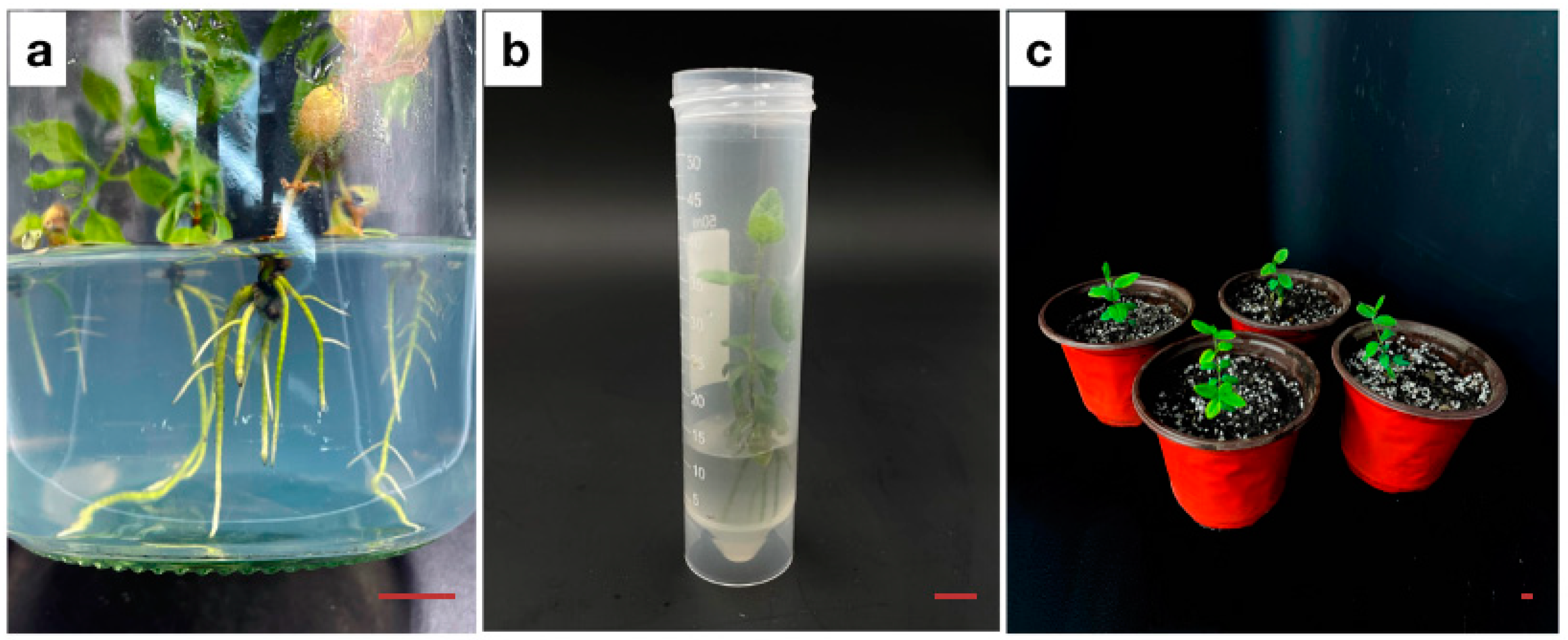
3.4. Adventitious Root Induction

To investigate the effect of PGRs on adventitious root induction, the regenerated shoots through organogenesis or embryogenesis were transferred to MS medium with or without IBA and IAA combined at various concentrations (Table 5). After about 15 d of culturing, root primordia were generated on the basal of the shoots, which gradually developed into normal adventitious roots (Figure 2a). However, 0.5 mg/L IBA and 1.0 mg/L IAA medium induced no roots until 30 d. Among the five root induction media, 1.0 mg/L IBA resulted in a higher root induction rate and longer root length (96% and 4.6 cm) than 0.5 mg/L IAA (74% and 1.8 cm) and PGR-free medium (67% and 2.4 cm), which is consistent with the previous report [25]. Intriguingly, 0.5 mg/L IAA medium resulted in a higher induced root number per plantlet (8.8) than PGR-free (7.0) and 1.0 mg/L IBA medium (4.2), suggesting that, even though auxin PGRs are well accepted to be used for root induction in micropropagation, the auxin types might affect the rooting characteristics.

3.5. Plant Regeneration and Conversion

After about 3 days of acclimatization (Figure 2b) and 15 days of growth in a pot, new leaves emerged from the plantlets (Figure 2c). The regenerated plantlets showed an average length of 26.6 cm, average leaf number of 11.3, average root number of 15.1, and average survival rate of 90.0% from tissue culture to soil culture (Table S2). The overall efficiency from explant to regenerated plant was about 44.1% and required about 105 d through the organogenetic pathway, and 23.9% and about 150 d through the somatic embryogenesis pathway (Figure S4).

4. Conclusions

To lay a foundation for genetic transformation of blue honeysuckle (L. caerulea L.), this study established an in vitro regeneration system through somatic embryogenesis for the first time, and improved the previously established regeneration protocol based on organogenesis (Figure 3). The system and protocol could also serve as a versatile tool for the application and advancement of other biotechnologies in blue honeysuckle, including micropropagation, virus elimination, and somatic hybrid/homozygous regeneration.

Supplementary Materials

The following supporting information can be downloaded at: https://www.mdpi.com/article/10.3390/horticulturae9090996/s1, Figure S1: Calli induced from different explants of L. caerulea L. after 30 days of in vitro culture; Figure S2: Morphology of L. caerulea L. callus induced on different basal culture medium; Figure S3: Adventitious shoots induced from different genotypes of L. caerulea L. after 40 days of in vitro culture; Figure S4: Time chart of blue honeysuckle (L. caerulea L.) regeneration through somatic embryogenesis and organogenesis pathways; Table S1: The referenced culture mediums in the present study; Table S2: Length, leaf number, root number, survival percentage of the plantlets regenerated from tissue culture [25,27,34,36,37,38,39,40,41,42].

Author Contributions

J.H. and C.Z. funded this project. C.Z. conceptualized, designed, and supervised the project. Y.L. contributed to the study’s conception and design. Material preparation by Y.L., Y.Z. and Q.F. Data collection by Y.L., S.L. and X.S. The analysis was performed by Y.L., Y.W., M.Y. and D.Q. The first draft of the manuscript was written by Y.L. and all authors commented on previous versions of the manuscript. All authors have read and agreed to the published version of the manuscript.

Funding

This research was funded by Natural Science Foundation of Heilongjiang Province China (Grant No. LH2021C031), “National Natural Science Foundation of China (Grant No. 32102332)”, “China Agriculture Research System (CARS-29), China Postdoctoral Science Foundation (Grant No. 2021MD703804)”, “Central government special supporting fund for reform and development of local universities (2022- Heilongjiang province -High-level talent—class B, 59009810)”.

Data Availability Statement

The datasets generated and/or analyzed during the current study are available from the corresponding author on reasonable request.

Acknowledgments

The authors wish to thank Jungang Huo, Zaimin Liu and Yonghe Zhang (Northeast Agricultural University), for their advice on this project.

Conflicts of Interest

The authors declare no conflict of interest.

References

  1. Bieniek, A.A.; Grygorieva, O.; Bielska, N. Biological Properties of Honeysuckle (Lonicera caerulea L.): A Review: The nutrition, health properties of honeysuckle. Agrobiodiversity Improv. Nutr. Health Life Qual. 2021, 5, 287–295. [Google Scholar] [CrossRef]
  2. Bao, T.; Karim, N.; Xie, L.; Xie, J.; Chen, W. Simulated gastrointestinal digestion and colonic fermentation of blue honeysuckle: Phenolic profile and protectivity on ethyl carbamate-induced oxidative damage. Process Biochem. 2022, 120, 74–84. [Google Scholar] [CrossRef]
  3. Chaovanalikit, A.; Thompson, M.M.; Wrolstad, R.E. Characterization and Quantification of Anthocyanins and Polyphenolics in Blue Honeysuckle (Lonicera caerulea L.). J. Agric. Food Chem. 2004, 52, 848–852. [Google Scholar] [CrossRef] [PubMed]
  4. Kusznierewicz, B.; Piekarska, A.; Mrugalska, B.; Konieczka, P.; Namieśnik, J.; Bartoszek, A. Phenolic composition and antioxidant properties of Polish blue-berried honeysuckle genotypes by HPLC-DAD-MS, HPLC postcolumn derivatization with ABTS or FC, and TLC with DPPH visualization. J. Agric. Food Chem. 2012, 60, 1755–1763. [Google Scholar] [CrossRef]
  5. Gowd, V.; Karim, N.; Shishir, M.R.I.; Xie, L.; Chen, W. Dietary polyphenols to combat the metabolic diseases via altering gut microbiota. Trends Food Sci. Technol. 2019, 93, 81–93. [Google Scholar] [CrossRef]
  6. Kou, Y.; Li, Z.; Yang, T.; Shen, X.; Wang, X.; Li, H.; Zhou, K.; Li, L.; Xia, Z.; Zheng, X.; et al. Therapeutic potential of plant iridoids in depression: A review. Pharm. Biol. 2022, 60, 2167–2181. [Google Scholar] [CrossRef]
  7. Holubec, V.; Smekalova, T.; Leisova-Svobodova, L. Morphological and molecular evaluation of the Far East fruit genetic resources of Lonicera caerulea L.—Vegetation, ethnobotany, use and conservation. Genet. Resour. Crop Evol. 2019, 66, 121–141. [Google Scholar] [CrossRef]
  8. Kithma, A.B.; Silva, H.; Rupasinghe, H.P.V. Polyphenols composition and anti-diabetic properties in vitro of haskap (Lonicera caerulea L.) berries in relation to cultivar and harvesting date. J. Food Compos. Anal. 2019, 88, 103402. [Google Scholar] [CrossRef]
  9. Kalisz, S.; Kieliszek, M. Influence of storage conditions on selected quality characteristics of blue honeysuckle berry juice. Agrochimica 2021, 66, 25–37. [Google Scholar] [CrossRef]
  10. Huo, J.; Sui, W.; Yang, G.; Yu, Z. Review of study on germplasm resources of blue honeysuckle (Lonicera caerulea L.). Acta Hortic. Sin. 2005, 32, 159–164. [Google Scholar]
  11. Zhu, C.; Sun, X.; Fu, Q.; Zhan, Y.; Li, S.; Liu, Y.; Yu, M.; Qin, D.; Zhang, L.; Huo, J. Complete chloroplast genome and phylogenetic analysis of Lonicera caerulea var. edulis (Caprifoliaceae). Mitochondrial DNA Part B 2023, 8, 314–318. [Google Scholar] [CrossRef] [PubMed]
  12. Casella, F.; Vurro, M.; Valerio, F.; Perrino, E.V.; Mezzapesa, G.N.; Boari, A. Phytotoxic Effects of Essential Oils from Six Lamiaceae Species. Agronomy 2023, 13, 257. [Google Scholar] [CrossRef]
  13. Zhang, X.-M.; Wu, Y.-F.; Li, Z.; Song, C.-B.; Wang, X.-P. Advancements in plant regeneration and genetic transformation of grapevine (Vitis spp.). J. Integr. Agric. 2021, 20, 1407–1434. [Google Scholar] [CrossRef]
  14. Sugiyama, M. Organogenesis in vitro. Curr. Opin. Plant Biol. 1999, 2, 61–64. [Google Scholar] [CrossRef]
  15. Yang, X.; Zhang, X. Regulation of somatic embryogenesis in higher plants. Crit. Rev. Plant Sci. 2010, 29, 36–57. [Google Scholar] [CrossRef]
  16. Gaj, M.D. Direct somatic embryogenesis as a rapid and efficient system for in vitro regeneration of Arabidopsis thaliana. Plant Cell Tissue Organ Cult. 2001, 64, 39–46. [Google Scholar] [CrossRef]
  17. Barra-Jiménez, A.; Aronen, T.S.; Alegre, J.; Toribio, M. Cryopreservation of embryogenic tissues from mature holm oak trees. Cryobiology 2015, 70, 217–225. [Google Scholar] [CrossRef]
  18. Kolbanova, E. Simultaneous direct ex vitro rooting and adaptation of the blue honeysuckle micro-sprouts (Lonicera Caerulea L. var. Kamtschatica). Proc. Natl. Acad. Sci. Belarus. Agrar. Ser. 2020, 58, 298–310. [Google Scholar] [CrossRef]
  19. Malabadi, R.B.; Chalannavar, R.K.; Meti, N.T.; Mulgund, G.S.; Odhav, N.B. Detection of Glutathione S-Transferase gene (GST2, GST3) during induction of somatic embryogenesis in grape (Vitis vinifera L.). Res. Biotechnol. 2013, 4, 1–11. [Google Scholar]
  20. Grosser, J.W.; Ollitrault, P.; Olivares-Fuster, O. Somatic hybridization in citrus: An effective tool to facilitate variety improvement. Vitr. Cell. Dev. Biol.—Plant 2000, 36, 434–449. [Google Scholar] [CrossRef]
  21. Wang, S.-M.; Lan, H.; Cao, H.-B.; Xu, Q.; Chen, C.-L.; Deng, X.-X.; Guo, W.-W. Recovery and characterization of homozygous lines from two sweet orange cultivars via anther culture. Plant Cell Tissue Organ Cult. 2015, 123, 633–644. [Google Scholar] [CrossRef]
  22. Zhu, C.; Zheng, X.; Huang, Y.; Ye, J.; Chen, P.; Zhang, C.; Zhao, F.; Xie, Z.; Zhang, S.; Wang, N.; et al. Genome sequencing and CRISPR/Cas9 gene editing of an early flowering Mini-Citrus (Fortunella hindsii). Plant Biotechnol. J. 2019, 17, 2199–2210. [Google Scholar] [CrossRef] [PubMed]
  23. Raza, G.; Singh, M.B.; Bhalla, P.L. Somatic embryogenesis and plant regeneration from commercial soybean cultivars. Plants 2020, 9, 38. [Google Scholar] [CrossRef] [PubMed]
  24. Karhu, S.T. Axillary shoot proliferation of blue honeysuckle. Plant Cell Tissue Organ Cult. 1997, 48, 195–201. [Google Scholar] [CrossRef]
  25. Sedlák, J.; Paprštein, F. In vitro propagation of blue honeysuckle. Hortic. Sci. 2007, 34, 129. [Google Scholar] [CrossRef]
  26. Dziedzic, E. Propagation of blue honeysuckle (Lonicera caerulea var. kamtschatica Pojark.) in in vitro culture. J. Fruit Ornam. Plant Res. 2008, 16, 93–100. [Google Scholar]
  27. Al, F.; Clapa, D.; Cristea, V.; Plopa, C. In vitro propagation of Lonicera kamtschatica. Agric. -Sci. Pract. 2014, 1–2, 89–90. [Google Scholar] [CrossRef]
  28. Glinushkin, A.; Akimova, S.; Nikulina, E.; Tsirulnikova, N.; Kirkach, V.; Kalinitchenko, V.; Radzhabov, A.; Radkevich, E.; Marchenko, L.; Solovyov, A.; et al. Preliminary Study: Micropropagation Using Five Types of Chelated Iron and the Subsequent Acclimation of Blue Honeysuckle (Lonicera caerulea var. kamtschatica Sevast.). Forests 2023, 14, 821. [Google Scholar] [CrossRef]
  29. Hunková, J.; Szabóová, M.; Gajdošová, A. Protocols for adventitious regeneration of Amelanchier alnifolia var. cusickii and Lonicera kamtschatica ‘Jugana’. Plants 2021, 10, 1155. [Google Scholar] [CrossRef]
  30. Smolik, M.; Ochmian, I.; Bobrowska-Chwat, A.; Chwat, G.; Arus, L.; Banaszczak, P.; Bocianowski, J.; Milczarski, P.; Ostrowska, K. Fingerprinting, structure, and genetic relationships among selected accessions of blue honeysuckle (Lonicera caerulea L.) from European collections. Biotechnol. Rep. 2022, 34, e00721. [Google Scholar] [CrossRef]
  31. Zhu, C.; Zhang, L.; Gao, Y.; Qin, D.; Huo, J. Two novel blue honeysuckle (Lonicera caerulea L.) cultivars: Lanjingling and Wulan. HortScience 2022, 57, 1145–1147. [Google Scholar] [CrossRef]
  32. Murashige, T.; Skoog, F. A revised medium for rapid growth and bioassays with tobacco tissue cultures. Physiol. Plant 1962, 15, 473–497. [Google Scholar] [CrossRef]
  33. Yu, T. Study on Rapid Propagation Techniques of Tissue Culture of Blue Honeysuckle (Lonicera caerulea L.); Northeast Agricultural Universily: Harbin, China, 2014. [Google Scholar]
  34. Miyashita, T.; Ohashi, T.; Shibata, F.; Araki, H.; Hoshino, Y. Plant regeneration with maintenance of the endosperm ploidy level by endosperm culture in Lonicera caerulea var. emphyllocalyx. Plant Cell Tissue Organ Cult. 2009, 98, 291–301. [Google Scholar] [CrossRef]
  35. Plekhanova, M.N. Blue honeysuckle (Lonicera caerulea L.)—A new commercial berry crop for temperate climate: Genetic resources and breeding. Acta Hortic. 2000, 538, 159–164. [Google Scholar] [CrossRef]
  36. Hui, J.X.; Wen, S.C.; Hua, Z.Y.; Ming, L.X. Comparative study on different methods for Lonicera japonica Thunb. micropropagation and acclimatization. J. Med. Plants Res. 2012, 6, 4389–4393. [Google Scholar]
  37. Kim, S.W.; Cheol, O.S.; In, D.S.; Liu, J.R. High frequency somatic embryogenesis and plant regeneration in zygotic embryo cultures of Japanese honeysuckle. Plant Cell Tissue Organ Cult. 2003, 72, 277–280. [Google Scholar] [CrossRef]
  38. Marcelina, K.-M.; Ochmian, I. Propagation of Blue Honeysuckles (Lonicera caerulea L.) in In Vitro Culture. J. Basic Appl. Sci. 2014, 10, 164–169. [Google Scholar] [CrossRef]
  39. Mihaljević, I.; Tomaš, V.; Vuković, D.; Dugalić, K. Propagation of Three Blue Honeysuckle (Lonicera caerulea L.) Cultivars In Vitro Culture; Agricultural Institute Osijek: Osijek, Croatia; Croatian Agency for Agriculture and Food: Osije, Croatia, 2019; Volume 23, pp. 41–48. [Google Scholar]
  40. Ochatt, S. Requirements for plant regeneration from protoplasts of the shrubby ornamental honeysuckle, Lonicera nitida cv. Maigrun. Plant Cell Tissue Organ Cult. 1991, 25, 161–167. [Google Scholar] [CrossRef]
  41. Shi, F.; Tan, M.; Mo, Q.; Chen, X.; Zhao, Y.; Guo, X.; Jiang, N. Application of the phosphomannose-isomerase/mannose selection system in the Agrobacterium-mediated transformation of Lonicera hypoglauca Miq. J. Plant Biochem. Biotechnol. 2020, 29, 528–538. [Google Scholar] [CrossRef]
  42. Zhang, Q.; Liang, Q.; Xia, X.; Yin, W. In vitro culture and plant regeneration from sprouting buds of Lonicera edulis Turcz. J. Beijing For. Univ. 2010, 32, 126–130. [Google Scholar]
Figure 1. Shoot organogenesis and somatic embryogenesis in blue honeysuckle (L. caerulea L.) under in vitro conditions. (a) Primary explant: cotyledons (bar = 1 cm); (b) Non-embryonic callus formation from cotyledon explants after 30 d of culture on MS medium with 6-BA (1.0 mg/L) + NAA (0.1 mg/L) (bar = 1 cm); (c) Embryogenic callus from cotyledon explants after 30 d of culture on MS medium with 2,4-D 1.0 mg/L (bar = 1 cm); (df) Somatic embryos were induced on MS medium with 0.1 mg/L 6-BA and 0.1 mg/L NAA after culturing for 40, 50, and 60 d, respectively (bar = 0.5 cm); (g,h) Longitudinal sections of embryos at globular embryo (bar = 200 µm) and heart-shaped embryo stages (bar = 300 µm); (i) Adventitious shoot induced directly on the surface of the callus within 30 d of culturing on the medium supplemented with 1.0 mg/L 6-BA and 0.1 mg/L NAA (bar = 1 cm).
Horticulturae 09 00996 g001
Figure 2. Adventitious root induction to planting processes of blue honeysuckle (L. caerulea L.). (a) Adventitious root after 30 d on MS supplemented with 1.0 mg/L IBA (bar = 1 cm); (b) Acclimatization for plantlet (bar = 1 cm); (c) Regenerated plantlets planted in the soil after 15 d (bar = 1 cm).
Horticulturae 09 00996 g002
Figure 3. Plant regeneration protocols of blue honeysuckle (L. caerulea L.) through somatic embryogenesis and indirect organogenesis pathways.
Horticulturae 09 00996 g003
Table 1. Effects of PGRs and explant type on callus induction of blue honeysuckle (L. caerulea L.).
MS + PGR (mg/L)Callus Induction Rate (%)Morphological Feature
CotyledonEuphyllaTender StemTender Root
6-BA 0.5 + NAA 0.173.0 ± 5.2 c0 c56.7 ± 20.8 a60.3 ± 19.0 aGreen and compact
6-BA 0.5 + NAA 0.288.4 ± 4.1 b37.3 ± 2.5 b73.3 ± 5.8 a63.0 ± 14.7 aGreen and compact
6-BA 1.0 + NAA 0.198.9 ± 1.7 a65.7 ± 9.8 a73.0 ± 6.1 a83.7 ± 14.8 aGreen and compact
6-BA 1.0 + NAA 0.286.4 ± 5.6 b46.7 ± 10.4 b68.3 ± 2.1 a69.7 ± 10.0 aGreen and compact
2,4-D 0.595.6 ± 7.7 a5.7 ± 5.1 b85.0 ± 8.5 a71.3 ± 7.5 aYellow and friable
2,4-D 1.097.6 ± 3.6 a60.0 ± 13.0 a85.3 ± 6.7 a75.7 ± 10.3 aYellow and friable
2,4-D 2.086.7 ± 13.3 a17.7 ± 13.6 b85.3 ± 13.1 a86.7 ± 17.3 aYellow and friable
2,4-D 3.093.8 ± 5.9 a4.7 ± 4.0 b71.6 ± 5.0 a69.0 ± 3.5 aYellow and friable
Note: Mean values and standard deviations are shown. Means with different letters (a–c) indicate significant differences by Duncan’s multiple range test (p ≤ 0.05) in the same column.
Table 2. Effects of different PGRs on embryogenesis of blue honeysuckle (L. caerulea L.).
MS + PGR (mg/L) +AC (0.5 g/L)Embryogenesis Induction Rate (%)
6-BA 0.1 + NAA 0.128.3 ± 1.7 a
6-BA 0.1 + NAA 0.213.3 ± 1.7 b
6-BA 0.1 + NAA 0.311.1 ± 3.5 b
6-BA 0.1 + NAA 0.49.4 ± 2.5 bc
6-BA 0.1 + NAA 0.56.1 ± 1.0 c
6-BA 0.2 + IBA 1.010.6 ± 5.9 bc
6-BA 0.2 + IBA 2.021.1 ± 5.4 a
6-BA 0.2 + IBA 3.011.1 ± 1.0 bc
6-BA 0.5 + IBA 1.03.9 ± 2.5 c
6-BA 0.5 + IBA 2.012.8 ± 3.5 b
6-BA 0.5 + IBA 3.011.8 ± 6.3 bc
Note: Mean values and standard deviations are shown. Means with different letters (a–c) indicate significant differences by Duncan’s multiple range test (p ≤ 0.05) in the same column.
Table 3. Effects of PGRs on induction of adventitious shoots of blue honeysuckle (L. caerulea L.).
MS + PGR (mg/L)Adventitious Shoot Induction Rate (%)
6-BA 0.5 + NAA 0.110.6 ± 2.5 c
6-BA 0.5 + NAA 0.213.3 ± 3.1 c
6-BA 1.0 + NAA 0.151.6 ± 6.5 a
6-BA 1.0 + NAA 0.235.6 ± 3.5 b
6-BA 1.0 + IBA 0.116.7 ± 4.2 c
6-BA 1.0 + IBA 0.242.8 ± 3.5 a
6-BA 2.0 + IBA 0.131.7 ± 6.0 b
6-BA 2.0 + IBA 0.236.1 ± 1.0 ab
Note: Mean values and standard deviations are shown. Means with different letters (a–c) indicate significant differences by Duncan’s multiple range test (p ≤ 0.05) in the same column.
Table 4. Effects of different genotypes on regeneration of adventitious shoots of blue honeysuckle (L. caerulea L.) cultivars: ‘Berel’, ‘Lanjingling’, and “Altai-4”.
GenotypeMS + PGRs (mg/L)Adventitious Shoot Induction Rate (%)
Berel6-BA 0.5 + NAA 0.112.8 ± 1.9 c
6-BA 0.5 + NAA 0.213.9 ± 1.0 c
6-BA 1.0 + NAA 0.143.9 ± 2.5 a
6-BA 1.0 + NAA 0.232.8 ± 5.8 b
Lanjingling6-BA 0.5 + NAA 0.114.4 ± 3.5 c
6-BA 0.5 + NAA 0.216.7 ± 4.4 c
6-BA 1.0 + NAA 0.153.3 ± 2.9 a
6-BA 1.0 + NAA 0.227.2 ± 1.9 b
Altai-46-BA 0.5 + NAA 0.118.3 ± 1.7 c
6-BA 0.5 + NAA 0.228.3 ± 1.4 b
6-BA 1.0 + NAA 0.138.9 ± 2.5 a
6-BA 1.0 + NAA 0.215.6 ± 3.5 c
Note: Mean values and standard deviations are shown. Means with different letters (a–c) indicate significant differences by Duncan’s multiple range test (p ≤ 0.05) in the same column.
Table 5. Effects of different PGRs on adventitious root induction of blue honeysuckle (L. caerulea L.).
MS + PGR (mg/L)Adventitious Root Induction Rate
(%)
Root Length
(cm)
Root Number per Plantlet
PGR-free67.0 ± 4.3 b2.4 ± 0.5 b7.0 ± 5.2 a
IBA 0.50 c0 c0 c
IBA 1.096.0 ± 3.3 a4.6 ± 1.8 a4.2 ± 2.4 b
IAA 0.574.0 ± 12.2 b1.8 ± 0.2 b8.8 ± 3.0 a
IAA 1.00 c0 c0 c
Note: Mean values and standard deviations are shown. Means with different letters (a–c) indicate significant differences by Duncan’s multiple range test (p ≤ 0.05) in the same column.
Disclaimer/Publisher’s Note: The statements, opinions and data contained in all publications are solely those of the individual author(s) and contributor(s) and not of MDPI and/or the editor(s). MDPI and/or the editor(s) disclaim responsibility for any injury to people or property resulting from any ideas, methods, instructions or products referred to in the content.

