Article

Trichoderma viride Isolate Tvd44 Enhances Potato Growth and Stimulates the Defense System against Potato Virus Y

1
Plant Protection and Biomolecular Diagnosis Department, Arid Lands Cultivation Research Institute, City of Scientific Research and Technological Applications (SRTA-City), New Borg El Arab City 21934, Egypt
2
Department of Botany and Microbiology, College of Science, King Saud University, P.O. Box 2455, Riyadh 11451, Saudi Arabia
3
Botany Department, Faculty of Science, Suez Canal University, Ismailia 41522, Egypt
4
Istituto Agronomico Mediterraneo di Bari, Via Ceglie 9, 70010 Valenzano, Italy
*
Authors to whom correspondence should be addressed.
Horticulturae 2023, 9(6), 716; https://doi.org/10.3390/horticulturae9060716
Submission received: 4 May 2023 / Revised: 11 June 2023 / Accepted: 15 June 2023 / Published: 17 June 2023
(This article belongs to the Section Plant Pathology and Disease Management (PPDM))

Abstract

:
Biological treatments may be employed to combat viral plant infections. In this study, Trichoderma viride was applied as a biocontrol agent to enhance the systemic resistance of potato plants against potato virus Y (PVY). T. viride isolate Tvd44 (OQ991378) was isolated and molecularly characterized before being used as an agent against PVY. The foliar application of Tvd44 on PVY-inoculated potatoes significantly promoted plant growth, height, roots, and number of leaves. Results also showed that the levels of peroxidase (POX), polyphenol oxidase (PPO), total proteins, and chlorophyll increased in potato leaves 21 days post-inoculation compared to untreated plants. Results of qPCR assays conducted on Tvd44-treated plants exhibited a reduction in PVY-CP accumulation levels up to 18.76-fold compared to untreated plants (101.82-fold). qPCR results also showed that defense-related genes (PR-1, POD, PAL, CHS, and HQT) were highly expressed in all Tvd44-treated plants. Three compounds: thiocarbamic acid, N,N-dimethyl, S-1,3-diphenyl-2-butenyl ester; 1,1-dicyano-2-methyl-4-(p-cyanophenyl) propane; and trans-[(2,3-diphenylcyclopropyl)methyl] phenyl sulfide were the most abundant compounds detected in the ethyl acetate extract of Tvd44-culture filtrate using GC–MS analysis. Our finding supports the efficacy of T. viride isolate Tvd44 as a potential agent that can successfully control PVY infections in potatoes and increase the productivity of the crop.

1. Introduction

Potato virus Y (PVY, genus Potyvirus, family Potyviridae) is characterized by a single-stranded positive-sense RNA [1]. PVY is known to have a significant impact on potato crops, the third most widely consumed crop after rice and wheat [2]. PVY has a detrimental effect on both the quantity and quality of potato tubers, leading to losses ranging from 10 to 90% [3]. The extent of these losses depends on various factors, such as the year, cultivar, and region [4]. Plants belonging to the Solanaceae family, such as peppers, tomatoes, and tobacco, exhibit susceptibility to PVY infection [5]. There are more than 40 distinct species of aphids that are responsible for the transmission of PVY. The efficacy of chemical means in managing intracellular pathogens such as viruses is limited. Therefore, in instances of PVY epidemics, plants that have been infected with the virus are rogued or insecticides are employed to reduce the population of vectors that transmit the pathogen [6]. The imperative to investigate biological control alternatives has intensified in response to the hazards associated with the utilization of chemical pesticides to manage plant diseases. The potential utilization of Trichoderma as a pathogen management strategy that is both safe for the environment and effective at preventing the spread of viral plant diseases has captured the attention of many researchers [7].
Trichoderma spp., a group of plant growth-promoting fungi (PGPF), are considered promising microorganisms with potential applications in sustainable agriculture [8]. Trichoderma mechanisms include competition, antibiosis, and induction of the synthesis of glucanase, chitinase enzymes, and mycoparasitism [9,10]. Moreover, its defensive responses are linked to germination-stimulant metabolism and the development of systemic acquired resistance [11,12,13]. Trichoderma spp. colonize plant roots and alter gene expression to induce plant metabolism. Various secondary metabolites enhance PR protein production during the Trichoderma–plant interaction, activating plant defense systems against the pathogen [14]. The introduction of various Trichoderma species into the rhizosphere has been observed to confer protection to plants against a multitude of plant pathogens. Pathogens observed in plants, comprising viral, fungal, and bacterial infections, prompt the activation of resistance mechanisms akin to the hypersensitive response, induced systemic resistance, and systemic acquired resistance [15]. There is currently little data on the role of Trichoderma spp. in the induction of plant defenses to viruses [16,17].
Trichoderma viride is mostly employed as a biofertilizer and as a biocontrol agent for fungi that cause plant disease in crops [18]. It has been observed that T. viride culture filtrates can inhibit the growth and aflatoxin production of Aspergillus flavus [19]. While there exists some evidence indicating the involvement of Trichoderma spp. in the resistance of plants to viral diseases, no prior research has investigated the impact of T. viride on plant physiology and antiviral properties in the context of plant viral infections. Thus, the present study aimed to evaluate the antiviral activity of T. viride against PVY infection in potato plants under greenhouse conditions for the first time. Furthermore, the effects of T. viride Tvd44 on plant growth parameters were evaluated. Antioxidant enzymes such as peroxidase (POX) and polyphenol oxidase (PPO), which participate in the metabolism of reactive oxygen species (ROS), were measured. The protein content and photosynthetic pigment chlorophyll were determined. The accumulation level of the PVY-CP gene was quantified. Expression levels of some defense-related genes (PR-1, POD, PAL, CHS, and HQT) were also evaluated. Additionally, potential bioactive components of the secondary metabolites of the T. viride Tvd44 isolate were identified using GC–MS.

2. Materials and Methods

2.1. Fungal Isolation, Molecular Identification, and Culture Preparation of Trichoderma viride

The Tvd44 strain of Trichoderma viride was isolated from the roots of asymptomatic tomato plants located in Damanhour, El-Behira governorate, Egypt. T. viride was characterized through morphological traits as well as molecular identification via the ITS region. The methodology of serial dilution was utilized to isolate the Trichoderma sp. The culture that was acquired underwent purification through hyphal tip isolation. Subsequently, it was sustained on PDA slants to facilitate identification. For DNA isolation, T. viride cultures were grown at room temperature in potato extract broth for 3 to 4 days. Hyphae were collected on cheesecloth in a Buchner funnel and then washed with 25 mM EDTA followed by distilled water. The samples were frozen in liquid nitrogen until used in the DNA extraction method according to Castle et al. [20]. Identification of the specimens was carried out through a combination of morphological characteristics and molecular typing utilizing the ITS1 and ITS4 primers, as described in references [21,22]. Table 1 displays the primer sequences. The PCR reactions were composed of 1 µL of both forward and reverse primer, 10 µL of 2× Taq Ready Mix, and 1 µL of DNA template. A volume of 25 µL was achieved by the addition of dsH2O. Using a Techne Prime thermal cycler, an initial denaturation at 95 °C for 3 min was followed by 35 cycles of 94 °C for 45 s, 55 °C for 45 s, and 72 °C for 1 min, and a final extension step at 72 °C for 5 min. The amplified PCR product was subjected to purification using a PCR cleanup column kit manufactured by QIAGEN, Hilden, Germany. The DNA nucleotide sequence that was acquired underwent analysis by NCBI-BLAST (http://blast.ncbi.nlm.nih.gov/Blast.cgi, accessed on 21 March 2023). MEGA 11 software was utilized to analyze the phylogenetic tree, which was generated through the application of the maximum likelihood statistic method [23]. This approach was employed to determine the evolutionary relationships among the taxa under investigation.

2.2. Viral Source and Molecular Identification

Samples of potato (Solanum tuberosum L.) exhibiting severe leaf mosaic distortion and chlorosis along with characteristic PVY-like symptoms were obtained from Borg El-Arab, Alexandria governorate, Egypt. The viral RNA was extracted using the RNeasy Mini Kit, following the guidelines provided by the manufacturer. The initial cDNA strand was synthesized according to the methodology outlined by Aseel et al. [24]. Subsequently, PCR amplification was performed on the cDNA using primers specific to the PVY coat protein (CP) gene, as listed in Table 1. The PCR program was performed as mentioned above with the annealing step at 58 °C for 45 s. The duration of the final elongation phase was 5 min, with a temperature of 72 °C. The PCR products were checked on 2% agarose gel electrophoresis, purified, and subjected to sequencing, and the phylogenetic tree was analyzed utilizing MEGA 11 software, as previously detailed.

2.3. Antiviral Activity Assay in the Greenhouse

The experimental setup involved the utilization of plastic pots with a diameter of 25 cm, which were filled with sterilized soil consisting of a 1:1 ratio of clay and sand (w/w). The experiment involved cultivating virus-free potato tubers of the Spunta cultivar for 21 days in an insect-proof greenhouse, maintaining constant conditions of 26 ± 2 °C and a 14/10 h day/night cycle. To prepare the Tvd44 culture filtrate (spraying solution), 1 mL containing 1 × 10⁹ conidia was inoculated in 100 mL of potato dextrose broth and incubated at 28 °C for 6 days on a rotary shaker at 150 rpm. Subsequently, culture filtrate was obtained by filtration through Whatman filter paper No. 1. The filtrate was passed through a 0.2 μm pore biological membrane filter before application on the plant leaves. In addition, 1 mL of PVY solution (20 µg/mL) was used as a viral inoculum. The experimental treatments utilized in this study were labeled as follows: a healthy control group denoted as “C”, potato plants that were inoculated with PVY labeled as “V”, potato plants that were subjected to leaf spraying with Tvd44 culture filtrate labeled as “T”, and potato plants that were treated with Tvd44 48 h before PVY inoculation labeled as “T + V”. The entire plant was sprayed with culture filtrate until the leaves appeared to be doused. The PVY inoculation was performed on the two upper true leaves of each potato plant via mechanical inoculation by dusting them with carborundum, as per the previously described method [25]. The experimental design consisted of five biological replicates, each of which was represented by five pots. Each container was populated with a triad of potato plants. To conduct sample analysis, each biological replication was comprised of nine potato leaves that were collected from each of the three potato plants, with three leaves being obtained from each plant in every pot. Each evaluation involved the execution of three technical replicates for every biological replicate.

2.4. Disease Assessment

According to Mansour and Al-Musa’s description, disease symptoms were visible in all infected pots 21 days after PVY inoculation [26]. As per the findings of Imran et al. [27], the assessment of disease severity in affected plants was conducted using a six-point scale that took into account the observable disease symptoms and the extent of leaf damage. The scale was as follows: 0 denoting the absence of any symptoms, 1 indicating a range of 1–20%, 2 representing 21–40%, 3 denoting 41–60%, 4 denoting 61–80%, and 5 indicating 81–100%. Subsequently, the disease severity values were converted into the percent disease index (PDI) using the subsequent formula:
PDI = a b AK × 100
where a is the number of infected plants with the same severity grade, b is the severity grade, A is the total number of plants, and K is the maximum infection grade.
By dividing the number of infected plants by the total number of plants and multiplying the result by 100, it is possible to express disease incidence as a percentage. The categorization of incidence levels was as follows: low incidence was defined as ranging from 1% to 20%; moderate incidence was defined as ranging from 21% to 49%, and high incidence was defined as ranging from 50% to 100%.

2.5. Growth Parameter Evaluation

Five plants were selected at random from each treatment, uprooted, and subsequently washed under running water. The plants were then evaluated for their height (cm), shoot and root fresh weight (g), and shoot and root lengths (cm), as well as the number of leaves.

2.6. Estimation of Antioxidant Enzyme Activity

2.6.1. Leaf Sample Preparation

A quantity of 1 g of powdered leaf tissue was homogenized using 4 mL of a 0.1 M phosphate buffer solution with a pH of 7. The extracts were filtered using a nylon cloth. Subsequently, the extracts underwent centrifugation at 10,000× g for 20 min at a temperature of 4 °C, as previously described [28]. The supernatants were preserved at −80 °C and subsequently utilized for the assessment of peroxidase and polyphenol oxidase activities, as well as for the quantification of protein content.

2.6.2. Peroxidase (POX) Activity

The procedure for measuring the activity of the peroxidase (POX) enzyme has been described by Angelini et al. [29]. This involved the addition of 80 µL of the crude extract to a solution containing 500 µL of a 0.1 M phosphate buffer with a pH of 7, 500 µL of 5 mM guaiacol, and 60 µL of 2 mM H2O2. The complete solution was incubated at 30 °C for 10 min, leading to the formation of tetraguaiacol. After this, absorbance was measured at a wavelength of 480 nm, wherein the molar extinction coefficient (ɛ) was determined to be 26.600 M−1 cm−1.

2.6.3. Polyphenol Oxidase (PPO) Activity

PPO activity was evaluated through the measurement of purpurogallin at a wavelength of 420 nm, utilizing an extinction coefficient of 26.40 M−1 cm−1 [30]. The reaction mixture, consisting of 2 mM pyrogallol in 0.1 M K-phosphate buffer pH 6, was subjected to enzymatic treatment by the addition of enzyme extract. The reaction was allowed to proceed for 5 min at 25 °C, following which it was terminated by the addition of 2.5 N H2SO4. The absorbance of the reaction mixture was measured and enzyme activity was quantified in μM g−1 FM min.

2.7. Protein Content

Total protein was extracted from potato leaves (200 mg fresh weight), which were ground with liquid nitrogen with a mortar and pestle. After that, the powdered leaves were homogenized in protein extraction buffer (0.050 g polyvinyl polypyrrolidone, 1 mL 0.05 M Tris buffer) and transferred to a new Eppendorf tube. The mixture was vigorously vortexed for 15 s before being placed on ice for 10 min. After centrifuging the lysate at 4 °C at 12.000 rpm for 20 min, 100 µL of supernatant was taken to a new Eppendorf tube for protein concentration determination with bovine serum albumin [31].

2.8. Chlorophyll Photosynthetic Pigment

For total chlorophyll content (TCC) determination, shoot tissue was immersed in 5 mL of 80% acetone and left overnight at 4 °C in the dark [32]. At A663 and A645 nm, Photometric readings were used to calculate TCC (a + b) using the following equation: (8.02 × A663 + 20.2 × A645) × V/1000 × W; where V = volume and W = fresh weight [33].

2.9. Transcriptional Level of Defense-Related Genes

2.9.1. RNA Extraction and cDNA Synthesis

The RNeasy Plant Mini Kit was utilized to isolate total RNA from 100 mg (fresh weight) potato leaves that were collected at 21 days post-inoculation (dpi), following the manufacturer’s instructions. Following the assessment of RNA purity and concentration, 2 µg of DNase-treated RNA underwent cDNA synthesis utilizing M-MuLV reverse transcriptase, according to previous studies [34,35]. The transcription reaction was conducted in a thermal cycler (Eppendorf, Hamburg, Germany) at a temperature of 42 °C for 1 h, and subsequently deactivated at a temperature of 80 °C for 10 min. The mixture of reactants was preserved at −20 °C until its utilization.

2.9.2. Quantitative PCR (qPCR) Assay

The impact of Tvd44 on the accumulation level of defense-related transcripts against PVY was assessed by qPCR. This study utilized a set of primers (as outlined in Table 1) that were specific to various genes, including PR-1, POD, PAL, CHS, HQT, and PVY-CP. The expression levels were normalized to β-actin as a housekeeping gene. qPCR reactions were performed in triplicate for each sample using the Rotor-Gene 6000 system according to the protocol described by Rashad et al. [36]. The quantification and calculation of the relative expression level of the target gene were performed using the amplification program, as previously described in reference [37].

2.10. GC–MS Analysis

As per the findings of Abdelkhalek et al. [38], the fungal culture filtrate was collected after 48 h of incubation in broth media. The culture filtrate was then combined with ethyl acetate in a 1:1 (v/v) ratio. Following 20 min of intense agitation, the amalgam was partitioned using a funnel. Subsequently, a rotary evaporator concentrated the ethyl acetate phase through evaporation at 40 °C. GC–MS analysis was conducted on the residue. Helium gas was transported through a carrier at a 1 mL/min flow rate. The temperature of the injector was 250 °C. Mass spectra were recorded for 53 min at an energy level of 70 eV. The components were identified by comparing them with data from the GC–MS libraries.

2.11. Statistical Analysis

The data acquired underwent statistical analysis via one-way ANOVA utilizing CoStat software. Concurrently, Tukey’s honest significant differences method (HSD) was employed to determine the statistical differences in the mean at a significance level of p ≤ 0.05. The standard deviation (±SD) was depicted as a column bar. There was no statistically significant difference observed among columns that share the same letter.

3. Results

3.1. Fungal Isolation and Molecular Identification

Trichoderma viride Tvd44 hyphae were identified by colony shape and color on PDA. Under a light microscope, Tvd44 hyphae were seen to be septate, have multiple nuclei, be clamped together, and have conidia. The molecular identity of Tvd44 was determined using PCR-amplified products of about 550 bp of the ITS region. The verified sequence was deposited in GenBank with the accession number OQ991378. The phylogenetic tree (Figure 1) observed that T. viride isolate Tvd44 was closely related to other T. viride isolates available in GenBank, especially those from Thailand (Acc #OM084773). Not surprisingly, the ITS locus alone was not able to clearly resolve GenBank isolates within the T. viride/atroviride/koningii species complex.

3.2. Viral Source and Identification

For the viral isolation, the typical PVY symptoms of naturally infected potato samples, including chlorosis, mosaic, and necrotic lesions, were confirmed by RT–PCR, using a specific primer of the PVY-CP gene, which amplified about 820 bp. The PCR purification and sequencing, the NCBI-BLAST alignment, and the analysis of the phylogenetic tree revealed that PVY strain DA55 was related to other PVY isolates, mainly from Mexico (Acc #AY700020) (Figure 2).

3.3. Disease Assessment

Compared to untreated plants, those that had been sprayed with a Tvd44 culture suspension on their leaves (48 h before virus inoculation) showed considerably reduced disease symptoms and increased plant development (Figure 3). The symptoms of PVY, including mosaic, chlorosis, yellowing, leaf deformation, size decrease of some plant leaves, and necrotic lesions, were shown on potato plants inoculated with PVY at 14 dpi (Figure 3). No observable symptoms were detected in either the control group or the group of plants treated with Tvd44 (Figure 3). The response of disease severity to the applied treatments (T. viride 48 h before PVY-inoculated plants) significantly limited and reduced both disease severity and incidence compared with infected potato plants treated with PVY only. No symptoms were shown on the control plants or Tvd44-treated plants (Table 2).

3.4. Growth Parameter Evaluation

Tvd44 generated a 37.7 cm plant height, followed by Tvd44 before PVY inoculation (34.3 cm). Moreover, the potato plants inoculated with PVY had a lower plant height (15.7 cm) compared to the control plants (Table 3). In addition, the root length increased with T and T + V (19.7 and 18.0 cm, respectively) more than did the shoot length for the same treatments. When compared to potato plants infected with PVY and potato plants used as a control, treatment with Tvd44 resulted in substantial increases in the fresh weights of shoot systems to 5.27 g and root systems to 1.39 g. There was also a significant increase in the treatment with Tvd44 before PVY inoculation in the fresh weight of shoots and root systems (4.53 g and 2.15 g, respectively). Potato plants treated with PVY alone had significantly lower shoot and root weights (1.5 g and 0.5 g, respectively) (Table 3).

3.5. Estimation of Antioxidant Enzyme Activity

3.5.1. Peroxidase (POX) Activity

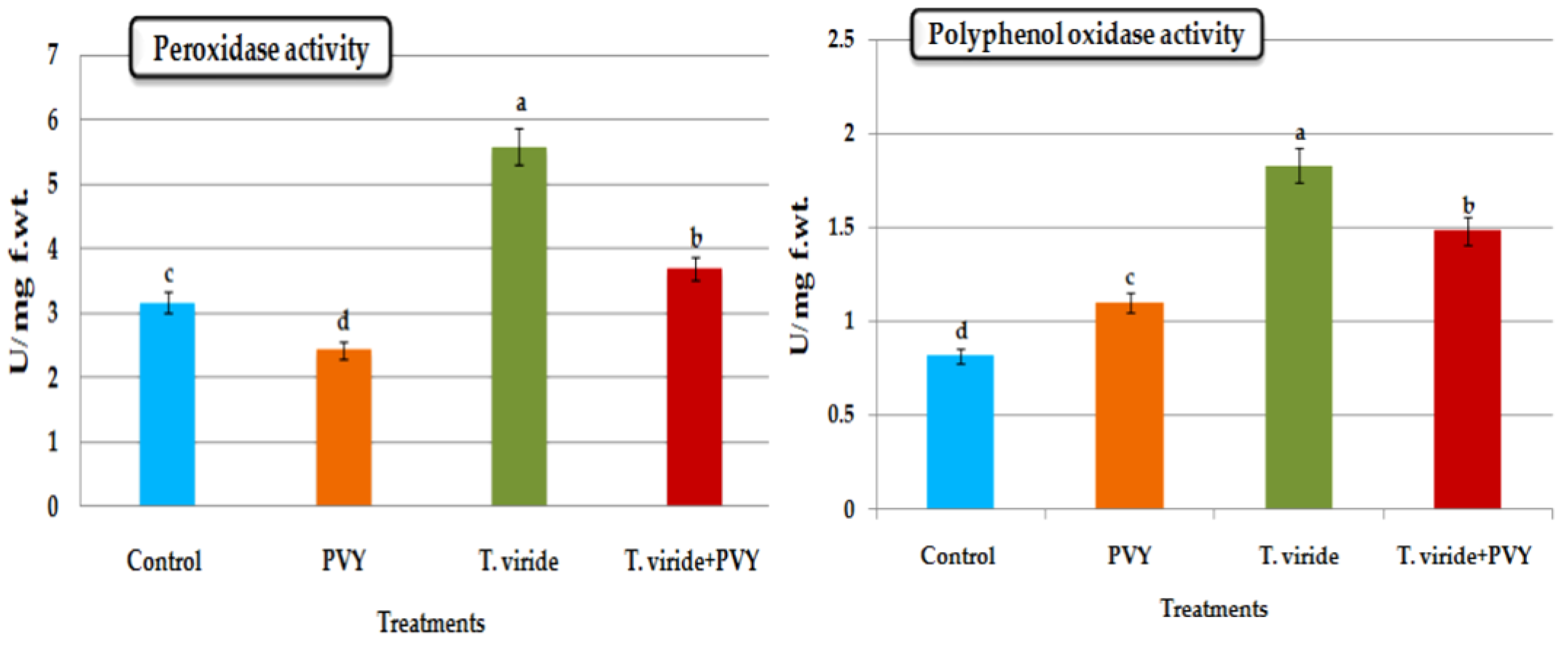
The POX enzyme activity was significantly increased in potato plants treated with Tvd44, followed by the treatment with Tvd44 and PVY inoculation. The POX activities reached 5.59 and 3.7 U L−1 min−1, respectively. When compared with untreated potato plants, those that had been infected with PVY showed significantly lower levels of POX activity, which peaked at a value of 2.42 U L−1 min−1 (Figure 4).

3.5.2. Polyphenol Oxidase (PPO) Activity

PPO activity in the Tvd44 treatment displayed the highest level (1.82 U L−1 min−1) compared to the control treatment (0.81 U L−1 min−1) and the PVV treatment (1.10 U L−1 min−1). Likewise, the Tvd44 + PVY treatment induced significantly increased PPO activity (1.48 U L−1 min−1) compared to the control and PVY treatments (Figure 4).

3.6. Protein Content

Protein content significantly increased with treatment with T. viride isolate Tvd44, reaching a maximum value of 754.1 mg mL−1 compared to the other treatments. Potato plants that were treated with T. viride 48 h before being PVY inoculated had a protein content of 589.6 mg mL−1, with no significant differences between this treatment and those inoculated with PVY alone, which had a protein content of 588.5 mg mL−1 (Figure 5).

3.7. Chlorophyll

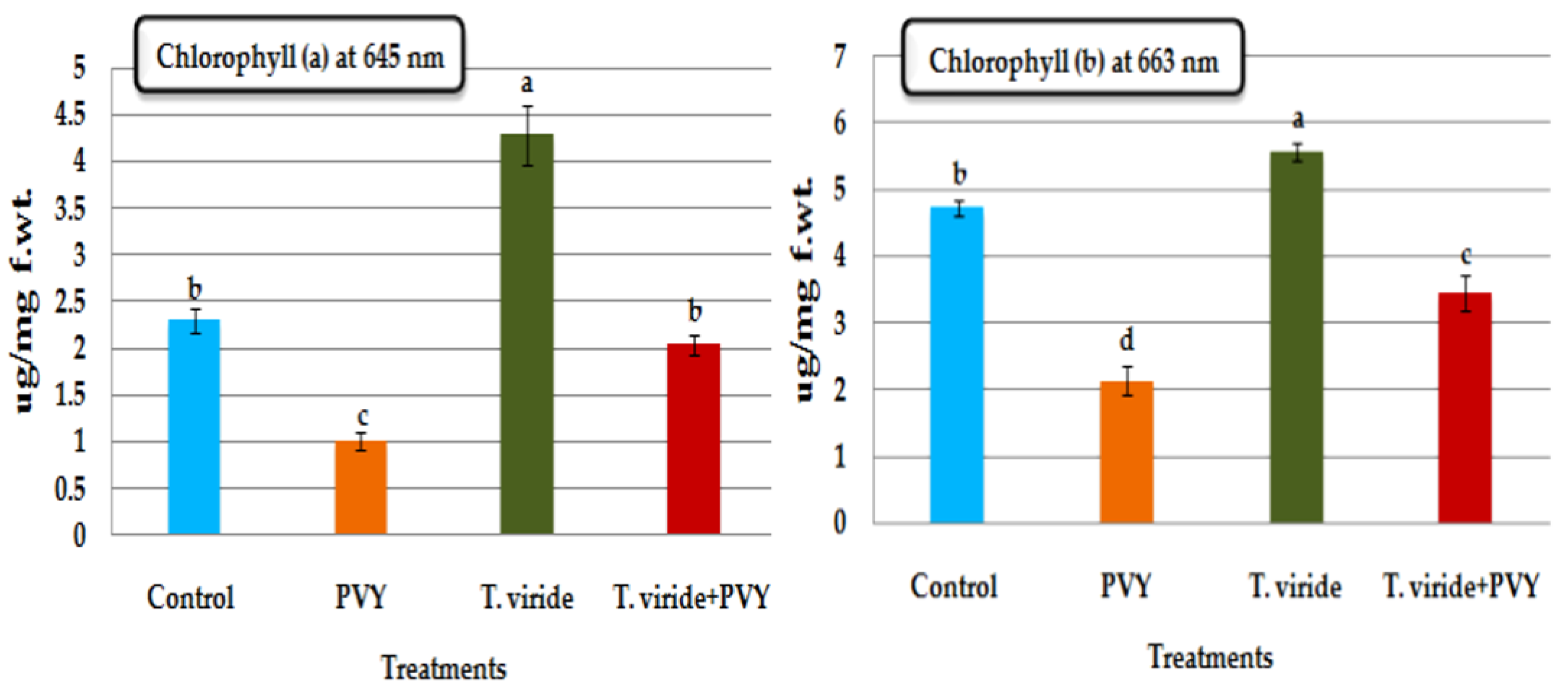
The chlorophyll a and chlorophyll b content increased in potato plants treated with T. viride isolate Tvd44 (4.28 a and 5.57 b ug/mg f.wt.), followed by the potato plants treated with T. viride 48 h before PVY inoculation (2.04 a and 3.45 b ug/mg f.wt.); these were found to be greater than those in the potato plants inoculated with PVY alone, where the chlorophyll a and b contents decreased (1.02 a and 2.14 b ug/mg f.wt.), as presented in Figure 6.

3.8. Effect of T. viride on PVY Accumulation Level

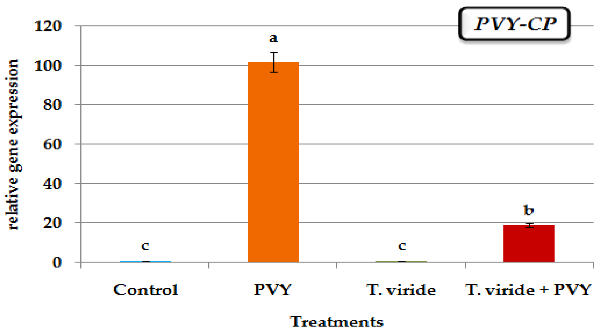
The results indicate that virus-infected plants exhibited a significantly higher relative gene expression of PVY-CP (101.82-fold) compared to the control group (Figure 7). In comparison, the plants treated with T. viride 48 h before PVY inoculation exhibited a lower level of PVY-CP accumulation, as evidenced by a relative gene expression level of 18.76-fold, as depicted in Figure 7.

3.9. Defense-Related Transcriptional Levels

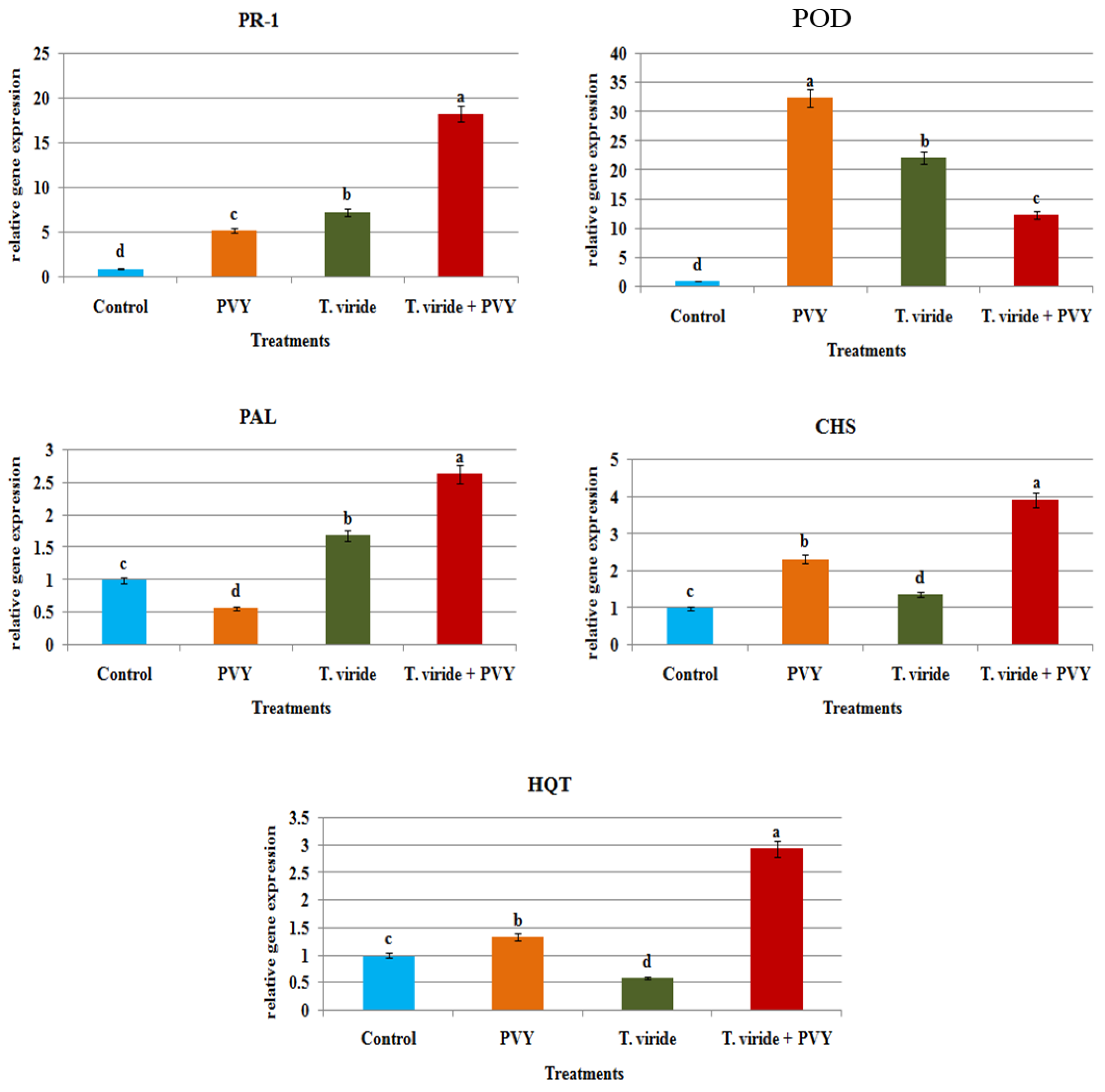
The qPCR results revealed the upregulation of PR-1 in all treatments compared with the control (Figure 8). The protective T. viride had a higher relative gene expression level (18.34-fold). The treatment with T. viride and virus-inoculated potato plants showed that the relative gene expression levels were 7.31-fold and 5.62-fold higher, respectively, compared with healthy potato plants (Figure 8). POD showed upregulation of expression in all treatments, especially potatoes with PVY only (32.44-fold), followed by treatment with T. viride (22.16-fold) and T. viride 48 h before PVY inoculation (12.41-fold) compared to the control expression level (Figure 8). All treatments showed transcriptional upregulation expression of PAL, whereas PVY-infected potato plants showed transcriptional downregulation expression that was 0.61-fold lower than the control (Figure 8). The highest relative gene expression level (2.63-fold higher compared with control) was found in protective T. viride 48 h before PVY inoculation. After that, the T. viride treatment had a relative gene expression level of 1.69-fold compared to PVY alone (Figure 8). Regarding CHS-relative gene expression, upregulation was noted in the protective treatment with T. viride 48 h before PVY inoculation (3.89-fold higher). Subsequently, potatoes with PVY showed a transcriptional upregulation of gene expression that was 2.33-fold. T. viride alone resulted in a 0.68-fold lower transcriptional downregulation expression level in potato plants than in untreated potato plants (Figure 8). Similar to CHS, the gene expression level of HQT showed upregulation in T. viride 48 h before PVY (2.93-fold higher) and in the PVY treatment (1.31-fold), while the downregulation of expression levels for the T. viride treatment was 0.48-fold lower compared to the control (Figure 8).

3.10. Identification of Bioactive Metabolites of Tvd44

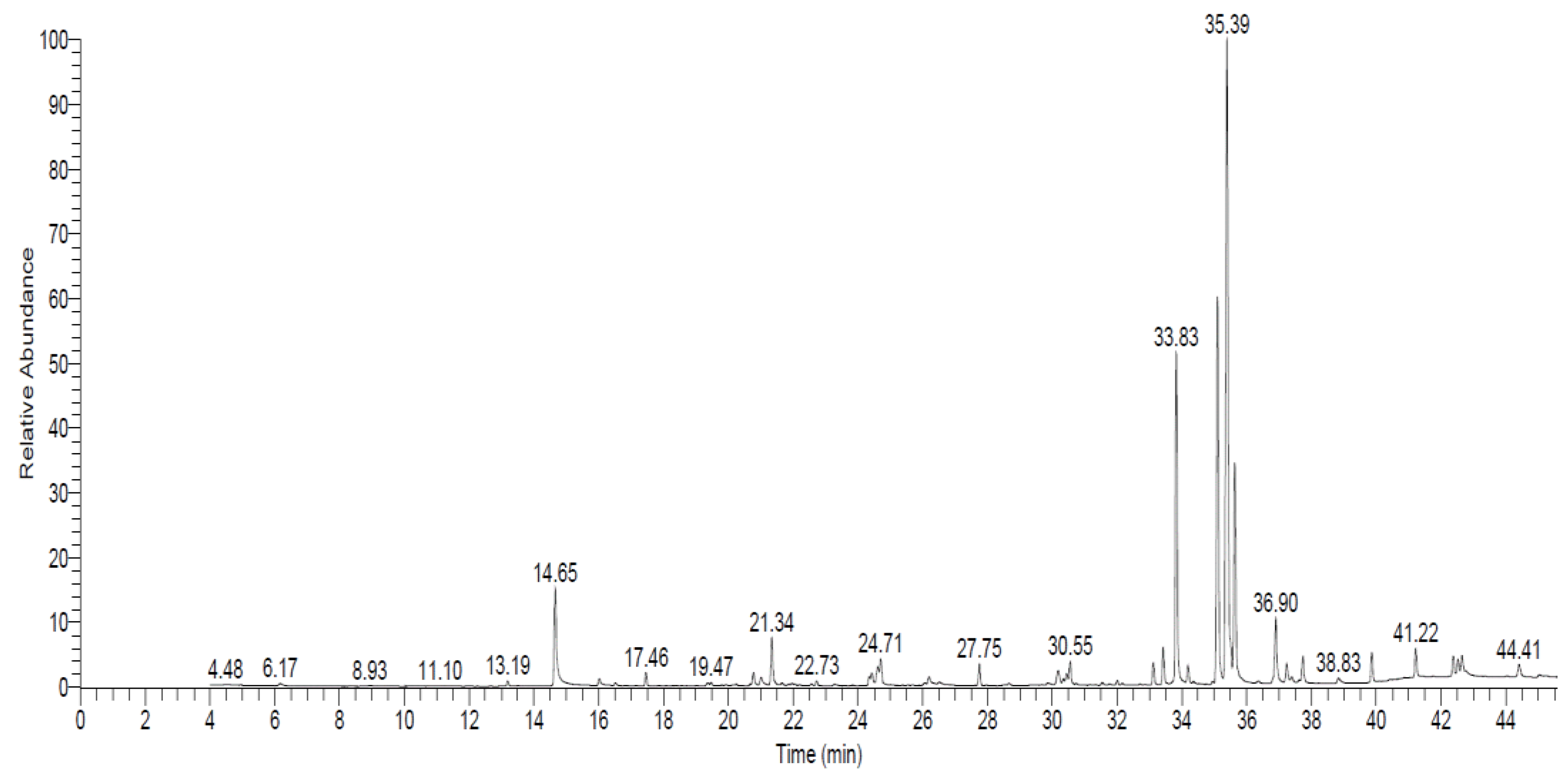
In this study, GC–MS was used to find the bioactive components of an ethyl acetate extract of Tvd44 culture filtrate. Figure 9 shows the GC–MS histogram of the compounds that were found. The GC–MS study showed that Tvd44-CF had more than 16 compounds. Table 4 presents the compounds with the highest relative abundance, along with their corresponding retention time (RT), relative abundance (RA%), molecular formula, and biological activity. The first compound, thiocarbamic acid, N,N-dimethyl, S-1,3-diphenyl-2-butenyl ester, showed the highest concentration at an RT of 35.39, while the second compound was 1,1-dicyano-2-methyl-4-(p-cyanophenyl) propene, which appeared at an RT of 35.11. The third detected compound was trans-[(2,3-diphenylcyclopropyl)methyl] phenyl sulfide, having an RT of 33.83. The other three compounds, 1-propene, 3-(2-cyclopentenyl)-2-methyl-1,1-diphenyl; 6-amyl-α-pyrone; and S-(1,3-diphenylbutyl) dimethyl thiocarbamate, were detected at RTs of 35.63, 14.65, and 36.90, respectively (Figure 9 and Table 4).

4. Discussion

To our knowledge, this is the first study to demonstrate the inhibition of PVY by T. viride isolate Tvd44. Trichoderma species such as T. viride, T. atroviride, and T. harzianum are effective and among the most adaptable biological control agents [17,46,47]. In the current study, the enhancement in potato plant growth upon the foliar application of T. viride Tvd44 agrees with previous studies that found that increased plant growth, including in stressed plants, is one of the impacts of T. harzianum-T22 treatment [48,49]. In addition, the results are similarly consistent with those obtained by Yedidia et al. [50] with T. harzianum treatment on cucumber plants, which increased by 80% in dry weight, 95% in shoot length, and 75% in root length when compared to control plants. The application of T. viride triggered systemic resistance against disease in potato plants [51]. Additionally, Jamil [52] reported that T. viride-treated tomato plants infected with Fusarium oxysporum showed lower disease severity, better growth and yield, the highest physiological activity, the least amount of disease, and the highest biochemical and antioxidant activities. In a similar case, greenhouse experiments showed that T. viride and T. harzianum were either directly protective against Fusarium solani or indirectly connected to the plant’s defense system when treated singly or in combination [53]. Additionally, according to Aggarwal et al. [54], T. viride isolate TV5-2 detoxified the Bipolaris sorokiniana toxin and decreased the disease severity of spot blotch in wheat.
The evaluation of symptoms and disease severity indicated that the administration of Tvd44 resulted in a reduction of PVY in all treated plants. The data obtained from the study indicate that Tvd44 has the potential to activate the innate immune system of the host and/or initiate systemic acquired resistance (SAR), leading to the suppression of PVY and/or inhibition of its replication. The results are in agreement with the protection shown for other plants [17,55]. It has been documented that treatment of tomato plants with two endophytic bacterial strains, B. subtilis 26D and B. subtilis Ttl2, induces systemic resistance and reduces the accumulation level of PVY at 7 and 14 dpi by approximately 1.8 to 4.7 times [56]. Furthermore, the application of Bacillus amyloliquefaciens strain MBI600 through drenching exhibited a delay in the systemic accumulation of PVY [57].
Trichoderma intermediates impact systemic resistance by affecting the levels of defense-related enzymes and metabolites such as PAL, POX, β-1–3-glucanase, PPO, phenols, and chitins. These pathogenesis-related enzymes are critical for plant resistance to viral diseases [17,58]. In the present study, POX activity was triggered and increased significantly in potato plants treated with Tvd44, followed by those treated with Tvd44 and PVY. Furthermore, the PPO activity was significantly increased in the Tvd44 and Tvd44 + PVY treatments compared to the control and PVY treatments. In another study after treatment with T. asperellum and the virus CMV, the activities of stress-resistance enzymes such as POX, SOD, PAL, LOX, and CAT in the leaves were investigated. In comparison to control plants, infection with CMV, Trichoderma, and CMV + Trichoderma resulted in an overall increase in antioxidant enzyme activity. Trichoderma asperellum treatment was found to have a different impact on the activity of these enzymes [59]. POX is a component of the plant defense system that is responsible for reducing the detrimental effects of stress by scavenging ROS [60,61]. Many studies have found an increase in POX, CAT, and PPO activity in virus-infected plants [62].
The protein content significantly increased in potato plants treated with Tvd44 compared with control plants, followed by the treatment with Tvd44 and PVY, and potato plants infected with PVY were observed to have no significant differences between each other. Similarly, Abdel-Shafiet al. [63] discovered that the total protein content of squash plants infected with ZYMV and plants treated with Trichoderma sp. filtrate with ZYMV increased significantly. This may be due to the formation of new antiviral proteins that play a role in inducing systemic resistance. Similarly, T. harzianum-T22 increased photosynthesis, as evidenced by the chlorophyll content being greater in all plants treated with T22 and inoculated with CMV [16]. Furthermore, our findings support a growing body of evidence that Trichoderma species can improve photosynthetic ratio and effectiveness in plants [49]. In contrast, there is typically a reduction in chlorophyll in plants infected with plant viruses [64,65]. To obtain a potential understanding of the mechanism underlying T. viride-induced resistance against PVY, the transcriptional activity of select plant defense-related genes in PVY-challenged plants was assessed using qPCR. By stimulating transcriptional expression levels of these genes, T. viride Tvd44 triggered defense mechanisms against PVY. The decrease in virus concentration and disease intensity showed that various defense pathways are involved in Trichoderma-induced resistance against viruses. The results agree with Tamandegani et al. [59], who reported that increased transcription levels of resistance-related genes increase the effectiveness of T. asperellum against CMV.
Generally, PR-1 is a principal regulator of SAR and a signal of the early defense response [66]. Indeed, the accumulation of SA and increasing resistance of numerous plants were linked to the induction of PR-1 [67,68]. In this study, the PR-1 gene was observed to have upregulated expression in all treatments and also in potato plants inoculated only with PVY. Our results agree with Abdelkhalek et al. [17], who found that T. hamatum Th23 can cause tomato plants to upregulate PR-1 and PR-7 upon infection with TMV. Similarly, when Arabidopsis was infected with the Beet severe curly top virus, the PR-1 gene, as well as several genes involved in the SA pathways, showed increased expression levels [69]. In other experiments, results demonstrated increased relative expression of SA-inducible genes such as PR-1 and PAL-1 in the leaves of cucumber plants treated with T. asperellum, as well as JA/ET-inducible genes such as LOX-1 and ETR-1 in the cucumber plants’ leaves [59].
POD gene activity has been related to improved plant defense against pathogens and alternative producers as a potential source of ROS [70]. In addition to the activation of antioxidant and PR genes in response to pathogen infection, ROS also increases programmed cell death at the cell level and sites of infection [71,72]. It has been reported that PVY infection increased peroxidase activity and the PR genes PR-1b and PR-1a [73]. We suggest that PVY-infected potato plants and Tvd44 treatment both stimulate the expression of the POD gene separately. Then, in the treatment of Tvd44 and PVY together, they decrease POD gene expression. PAL is a key regulator enzyme in the phenylpropanoid pathway as well as SA biosynthesis [74,75]. The downregulation of PAL activity was associated with PVY infection in the current study, whereas PAL upregulation was observed in Tvd44 only and T. viride + PVY inoculation. In contrast, POD and PAL activities were increased after trichokonin treatment, which was extracted from T. pseudokoningii SMF2 against TMV infection. After 4 days of treatment, POD and PAL reached their peaks of activity with a 5.2-fold and 8.4-fold increase, respectively, compared with control plants [76].
CHS is the first enzyme in the flavonoid pathway, and it produces primary metabolites that are essential for flavonoid synthesis in many plant tissues [77,78]. In the present study, Tvd44 induced potato plants to become resistant to PVY infection. Thus, we suggest the high expression of both CHS and HQT genes in treatment by Tvd44 and PVY accumulates both flavonoids and polyphenols in potato leaves and thus protects them from viral infection. According to a previous study, overexpression of CHS has been linked to a substantial buildup of flavonoid and isoflavonoid molecules with a broad antimicrobial action against a variety of phytopathogens [79,80]. Over-expression of HQT and PAL increased chlorogenic acid content [81,82,83], which was a plant response to viral infection, and vice versa [24]. Chlorogenic acid is a polyphenolic component that helps plants fight diseases and prevent pathogens such as viruses [84,85]. Thus, the increased transcriptional expression of CHS and HQT genes reveals their antiviral functions, showing that polyphenolic chemicals can be used by the potato plant as one of its defenses against viral infection and spread.
The GC–MS spectral analysis demonstrated that the culture filtrate derived from Tvd44 comprises more than 16 compounds. Thiocarbamic acid, N,N-dimethyl, S-1,3-diphenyl-2-butenyl ester was the major compound in ethyl acetate extract, which is one of the best-known herbicides and has insecticidal activity [44,86]. Moreover 1,1-Dicyano-2-methyl-4-(p-cyanophenyl)propene and trans-[(2,3-Diphenylcyclopropyl)methyl] phenyl sulfide exhibited different antimicrobial activities [42,43]. 6-Amyl-α-pyrone, a primary-secondary metabolite, has been found to have a notable impact on the biological regulation of pests [39]. 6-Amyl-α-pyrone is classified as an unsaturated lactone and has been recognized as a significant bioactive constituent of various Trichoderma species [39,87]. Therefore, it is plausible that Tvd44 may serve as a potential biocontrol agent for mitigating infections caused by PVY. However, additional investigations are required to validate the feasibility of implementing the findings in potential field applications.

5. Conclusions

The findings of the present investigation indicate that T. viride Tvd44 can stimulate the growth of potato plants, decrease the level of PVY accumulation, elicit systemic resistance, and enhance the production of certain defense enzymes. The potential utilization of Tvd44 as a biological control agent for mitigating PVY infection is being considered. The application of T. viride Tvd44 resulted in the upregulation of peroxidase, polyphenol oxidase, protein content, and chlorophyll content in potato plants. Treatment with Tvd44 reduced the PVY accumulation level compared to treatment with the virus alone. In addition, Tvd44 increased the plant height, number of leaves, and fresh weight of the shoots and root systems of potato plants. Treatment with Tvd44 and inoculation with PVY showed increased transcript upregulation of the relative expression levels of the PAL, PR-1, CHS, and HQT genes, except for the POD gene, whose gene expression was reduced compared with other treatments. In general, we can conclude that the utilization of T. viride for treatment or protection purposes has potential for the control and management of plant viral diseases.

Author Contributions

Conceptualization, D.G.A. and A.A.; methodology, D.G.A., S.A.S. and A.A.; software, D.G.A. and A.A.; validation, D.G.A.; formal analysis, D.G.A.; investigation, D.G.A. and A.A.; resources, D.G.A.; data curation, D.G.A. and A.A.; writing—original draft preparation, D.G.A., A.A. and S.A.S.; writing—review and editing, D.G.A., A.A., A.E. and T.E.; visualization, D.G.A. and A.A.; project administration, A.A.A.-A.; funding acquisition, A.A.A.-A. All authors have read and agreed to the published version of the manuscript.

Funding

This research was financially supported by the Supporting Researchers Project (number RSP2023R505): King Saud University, Riyadh, Saudi Arabia.

Institutional Review Board Statement

Not applicable.

Informed Consent Statement

Not applicable.

Data Availability Statement

Experimental data supporting the findings of this study are available from the corresponding authors upon request.

Acknowledgments

The authors express their sincere thanks to the City of Scientific Research and Technological Applications (SRTA-City), Alexandria, Egypt, for providing the necessary research facilities. The authors would like to extend their appreciation to the Supporting Researchers Project (number RSP2023R505), King Saud University, Riyadh, Saudi Arabia.

Conflicts of Interest

The authors declare no conflict of interest.

References

  1. Hall, J.S.; Adams, B.; Parsons, T.J.; French, R.; Lane, L.C.; Jensen, S.G. Molecular cloning, sequencing, and phylogenetic relationships of a new potyvirus: Sugarcane streak mosaic virus, and a reevaluation of the classification of the Potyviridae. Mol. Phylogenet. Evol. 1998, 10, 323–332. [Google Scholar] [CrossRef]
  2. Bhoi, T.K.; Samal, I.; Majhi, P.K.; Komal, J.; Mahanta, D.K.; Pradhan, A.K.; Saini, V.; Raj, M.N.; Ahmad, M.A.; Behera, P.P. Insight into aphid mediated Potato Virus Y transmission: A molecular to bioinformatics prospective. Front. Microbiol. 2022, 13, 1001454. [Google Scholar] [CrossRef]
  3. Valkonen, J.P.T.; Gebhardt, C.; Zimnoch-Guzowska, E.; Watanabe, K.N. Resistance to Potato virus Y in potato. In Potato Virus Y: Biodiversity, Pathogenicity, Epidemiology and Management; Springer: Berlin/Heidelberg, Germany, 2017; pp. 207–241. [Google Scholar]
  4. Elsharkawy, M.M.; Alotibi, F.O.; Al-Askar, A.A.; Adnan, M.; Kamran, M.; Abdelkhalek, A.; Behiry, S.I.; Saleem, M.H.; Ahmad, A.A.; Khedr, A.A. Systemic Resistance Induction of Potato and Tobacco Plants against Potato Virus Y by Klebsiella oxytoca. Life 2022, 12, 1521. [Google Scholar] [CrossRef]
  5. Aramburu, J.; Galipienso, L.; Matas, M. Characterization of potato virus Y isolates from tomato crops in northeast Spain. Eur. J. Plant Pathol. 2006, 115, 247–258. [Google Scholar] [CrossRef]
  6. Kopp, A.; Kondrák, M.; Bánfalvi, Z. Molecular mechanisms of resistance to potato virus X and Y in potato. Acta Phytopathol. Entomol. Hung. 2015, 50, 151–160. [Google Scholar] [CrossRef] [Green Version]
  7. Abdelkhalek, A.; Hafez, E. Plant Viral Diseases in Egypt and Their Control. In Cottage Industry of Biocontrol Agents and Their Applications; Springer: Berlin/Heidelberg, Germany, 2020; pp. 403–421. [Google Scholar]
  8. Heflish, A.A.; Abdelkhalek, A.; Al-Askar, A.A.; Behiry, S.I. Protective and Curative Effects of Trichoderma asperelloides Ta41 on Tomato Root Rot Caused by Rhizoctonia solani Rs33. Agronomy 2021, 11, 1162. [Google Scholar] [CrossRef]
  9. Metcalf, D.A.; Wilson, C.R. The process of antagonism of Sclerotium cepivorum in white rot affected onion roots by Trichoderma koningii. Plant Pathol. 2001, 50, 249–257. [Google Scholar] [CrossRef]
  10. Behiry, S.; Soliman, S.A.; Massoud, M.A.; Abdelbary, M.; Kordy, A.M.; Abdelkhalek, A.; Heflish, A. Trichoderma pubescens Elicit Induced Systemic Resistance in Tomato Challenged by Rhizoctonia solani. J. Fungi 2023, 9, 167. [Google Scholar] [CrossRef]
  11. Abeysinghe, S. Systemic resistance induced by Trichoderma harzianum RU01 against Uromyces appendiculatus on Phaseolus vulgaris. J. Natl. Sci. Found. Sri Lanka 2009, 37, 203–207. [Google Scholar] [CrossRef]
  12. Howell, C.R. Cotton seedling preemergence damping-off incited by Rhizopus oryzae and Pythium spp. and its biological control with Trichoderma spp. Phytopathology 2002, 92, 177–180. [Google Scholar] [CrossRef] [Green Version]
  13. Maheshwary, N.; Gangadhara Naik, B.; Amoghavarsha Chittaragi, M.; Naik, S.K.; Nandish, M. Compatibility of Trichoderma asperellum with fungicides. Pharma Innov. J 2020, 9, 136–140. [Google Scholar]
  14. Malmierca, M.G.; Barua, J.; McCormick, S.P.; Izquierdo-Bueno, I.; Cardoza, R.E.; Alexander, N.J.; Hermosa, R.; Collado, I.G.; Monte, E.; Gutiérrez, S. Novel aspinolide production by Trichoderma arundinaceum with a potential role in Botrytis cinerea antagonistic activity and plant defence priming. Environ. Microbiol. 2015, 17, 1103–1118. [Google Scholar] [CrossRef] [PubMed]
  15. Woo, S.L.; Scala, F.; Ruocco, M.; Lorito, M. The molecular biology of the interactions between Trichoderma spp., phytopathogenic fungi, and plants. Phytopathology 2006, 96, 181–185. [Google Scholar] [CrossRef] [Green Version]
  16. Vitti, A.; Pellegrini, E.; Nali, C.; Lovelli, S.; Sofo, A.; Valerio, M.; Scopa, A.; Nuzzaci, M. Trichoderma harzianum T-22 induces systemic resistance in tomato infected by Cucumber mosaic virus. Front. Plant Sci. 2016, 7, 1520. [Google Scholar] [CrossRef] [PubMed] [Green Version]
  17. Abdelkhalek, A.; Al-Askar, A.A.; Arishi, A.A.; Behiry, S.I. Trichoderma hamatum Strain Th23 Promotes Tomato Growth and Induces Systemic Resistance against Tobacco mosaic Virus. J. Fungi 2022, 8, 228. [Google Scholar] [CrossRef]
  18. Deepa, N.; Sreenivasa, M.Y. Biocontrol strategies for effective management of phytopathogenic fungi associated with cereals. In New and Future Developments in Microbial Biotechnology and Bioengineering; Elsevier: Amsterdam, The Netherlands, 2019; pp. 177–189. [Google Scholar]
  19. Calistru, C.; McLean, M.; Berjak, P. In vitro studies on the potential for biological control of Aspergillus flavus and Fusarium moniliforme by Trichoderma species. Mycopathologia 1997, 137, 115–124. [Google Scholar] [CrossRef]
  20. Castle, A.; Speranzini, D.; Rghei, N.; Alm, G.; Rinker, D.; Bissett, J. Morphological and molecular identification of Trichoderma isolates on North American mushroom farms. Appl. Environ. Microbiol. 1998, 64, 133–137. [Google Scholar] [CrossRef] [Green Version]
  21. Carbone, I.; Kohn, L.M. A method for designing primer sets for speciation studies in filamentous ascomycetes. Mycologia 1999, 91, 553–556. [Google Scholar] [CrossRef]
  22. Samuels, G.J.; Dodd, S.L.; Gams, W.; Castlebury, L.A.; Petrini, O. Trichoderma species associated with the green mold epidemic of commercially grown Agaricus bisporus. Mycologia 2002, 94, 146–170. [Google Scholar] [CrossRef]
  23. Tamura, K.; Stecher, G.; Kumar, S. MEGA11: Molecular Evolutionary Genetics Analysis Version 11. Mol. Biol. Evol. 2021, 38, 3022–3027. [Google Scholar] [CrossRef]
  24. Aseel, D.G.; Rashad, Y.M.; Hammad, S.M. Arbuscular mycorrhizal fungi trigger transcriptional expression of flavonoid and chlorogenic acid biosynthetic pathways genes in tomato against Tomato mosaic Virus. Sci. Rep. 2019, 9, 9692. [Google Scholar] [CrossRef] [Green Version]
  25. Aseel, D.G.; Abdelkhalek, A.; Alotibi, F.O.; Samy, M.A.; Al-Askar, A.A.; Arishi, A.A.; Hafez, E.E. Foliar Application of Nanoclay Promotes Potato (Solanum tuberosum L.) Growth and Induces Systemic Resistance against Potato Virus, Y. Viruses 2022, 14, 2151. [Google Scholar] [CrossRef]
  26. Mansour, A.; Al-Musa, A. Tomato yellow leaf curl virus: Host range and virus-vector relationships. Plant Pathol. 1992, 41, 122–125. [Google Scholar] [CrossRef]
  27. Imran, M.; Khan, M.A.; Azeem, M.; Ahmed, N.; Binyamin, R.; Riaz, A. Screening of tomato germplasm for the source of resistance and its management against ToMV. Pak. J. Phytopathol. 2012, 24, 53–57. [Google Scholar]
  28. de Souza, M.B.; Stamford, N.P.; Silva, E.V.; Berger, L.R.R.; e Silva, S.C.E.R.; Costa, A.F.; Ferraz, A.P.F. Defense response by inter-active bio-protector and chitosan to Sclerotium rolfsii Wilt disease on cowpea, Brazilian Oxisol. Afr. J. Agric. Res. 2018, 13, 1053–1062. [Google Scholar]
  29. Angelini, R.; Manes, F.; Federico, R. Spatial and functional correlation between diamine-oxidase and peroxidase activities and their dependence upon de-etiolation and wounding in chick-pea stems. Planta 1990, 182, 89–96. [Google Scholar] [CrossRef]
  30. Kumar, K.B.; Khan, P.A. Peroxidase & polyphenol oxidase in excised ragi (Eleusine corocana cv PR 202) leaves during senescence. Indian J. Exp. Biol. 1982, 20, 412–416. [Google Scholar]
  31. Bradford, M.M. A rapid and sensitive method for the quantitation of microgram quantities of protein utilizing the principle of protein-dye binding. Anal. Biochem. 1976, 72, 248–254. [Google Scholar] [CrossRef] [PubMed]
  32. Hung, R.; Lee, S.; Bennett, J.W. Arabidopsis thaliana as a model system for testing the effect of Trichoderma volatile organic compounds. Fungal Ecol. 2013, 6, 19–26. [Google Scholar] [CrossRef]
  33. Palta, J.P. Leaf chlorophyll content. Remote Sens. Rev. 1990, 5, 207–213. [Google Scholar] [CrossRef]
  34. Rashad, Y.M.; Aseel, D.G.; Hafez, E.E. Antifungal potential and defense gene induction in maize against Rhizoctonia root rot by seed extract of Ammi visnaga (L.) Lam. Phytopathol. Mediterr. 2018, 57, 73–88. [Google Scholar]
  35. Hafez, E.E.; Abdelkhalek, A.A.; Abd El-Wahab, A.S.E.-D.; Galal, F.H. Altered gene expression: Induction/suppression in leek elicited by Iris Yellow Spot Virus infection (IYSV) Egyptian isolate. Biotechnol. Biotechnol. Equip. 2013, 27, 4061–4068. [Google Scholar] [CrossRef] [Green Version]
  36. Rashad, Y.; Aseel, D.; Hammad, S.; Elkelish, A. Rhizophagus irregularis and Rhizoctonia solani differentially elicit systemic transcriptional expression of polyphenol biosynthetic pathways genes in sunflower. Biomolecules 2020, 10, 379. [Google Scholar] [CrossRef] [Green Version]
  37. Livak, K.J.; Schmittgen, T.D. Analysis of relative gene expression data using real-time quantitative PCR and the 2−ΔΔCT method. Methods 2001, 25, 402–408. [Google Scholar] [CrossRef]
  38. Abdelkhalek, A.; Behiry, S.I.; Al-Askar, A.A. Bacillus velezensis PEA1 Inhibits Fusarium oxysporum Growth and Induces Systemic Resistance to Cucumber mosaic Virus. Agronomy 2020, 10, 1312. [Google Scholar] [CrossRef]
  39. Hamrouni, R.; Molinet, J.; Dupuy, N.; Taieb, N.; Carboue, Q.; Masmoudi, A.; Roussos, S. The effect of aeration for 6-pentyl-alpha-pyrone, conidia and lytic enzymes production by Trichoderma asperellum strains grown in solid-state fermentation. Waste Biomass Valorization 2020, 11, 5711–5720. [Google Scholar] [CrossRef] [Green Version]
  40. Salwan, R.; Rialch, N.; Sharma, V. Bioactive volatile metabolites of Trichoderma: An overview. In Secondary Metabolites of Plant Growth Promoting Rhizomicroorganisms: Discovery and Applications; Springer: Berlin/Heidelberg, Germany, 2019; pp. 87–111. [Google Scholar]
  41. Stracquadanio, C.; Quiles, J.M.; Meca, G.; Cacciola, S.O. Antifungal activity of bioactive metabolites produced by Trichoderma asperellum and Trichoderma atroviride in liquid medium. J. Fungi 2020, 6, 263. [Google Scholar] [CrossRef]
  42. Gothai, S.; Vijayarathna, S.; Chen, Y.; Kanwar, J.R.; Wahab, H.A.; Sasidharan, S. In vitro-scientific evaluation on anti-Candida albicans activity, antioxidant properties, and phytochemical constituents with the identification of antifungal active fraction from traditional medicinal plant Couroupita guianensis Aubl. Flower. J. Complement. Med. Res. 2018, 8, 85. [Google Scholar] [CrossRef]
  43. Mariastutt, H.D.; Listiyowati, S.R.I.; Wahyudi, A.T.R.I. Antifungal activity of soybean rhizosphere actinomycetes producing bioactive compounds against Fusarium oxysporum. Biodiversitas J. Biol. Divers. 2018, 19, 2127–2133. [Google Scholar] [CrossRef]
  44. Abdullah, R.R. Insecticidal activity of secondary metabolites of locally isolated fungal strains against some cotton insect pests. J. Plant Prot. Pathol. 2019, 10, 647–653. [Google Scholar] [CrossRef] [Green Version]
  45. ChunYan, H.; Hong, P.; ZhenYu, Z.; Jing, S. Evaluation of antioxidant and antitumour activities of lemon essential oil. J. Med. Plant Res. 2010, 4, 1910–1915. [Google Scholar]
  46. Govindasamy, V.; Balasubramanian, R. Biological control of groundnut rust, Puccinia arachidis, by Trichoderma harzianum/Biologische Bekämpfung des Erdnuß-Rostes, Puccinia arachidis mit Trichoderma harzianum. Z. Pflanzenkrankh. Pflanzenschutz/J. Plant Dis. Prot. 1989, 96, 337–345. [Google Scholar]
  47. Mukherjee, P.K.; Horwitz, B.A.; Herrera-Estrella, A.; Schmoll, M.; Kenerley, C.M. Trichoderma research in the genome era. Annu. Rev. Phytopathol. 2013, 51, 105–129. [Google Scholar] [CrossRef] [PubMed]
  48. Harman, G.E. Myths and dogmas of biocontrol changes in perceptions derived from research on Trichoderma harzinum T-22. Plant Dis. 2000, 84, 377–393. [Google Scholar] [CrossRef] [PubMed] [Green Version]
  49. Shoresh, M.; Harman, G.E.; Mastouri, F. Induced systemic resistance and plant responses to fungal biocontrol agents. Annu. Rev. Phytopathol. 2010, 48, 21–43. [Google Scholar] [CrossRef] [PubMed] [Green Version]
  50. Yedidia, I.; Srivastva, A.K.; Kapulnik, Y.; Chet, I. Effect of Trichoderma harzianum on microelement concentrations and increased growth of cucumber plants. Plant Soil 2001, 235, 235–242. [Google Scholar] [CrossRef]
  51. Susiana, P.; Achmadi, P.; Retno, P.S.; Rina, S.K.; Kadarwati, B. The resistance of potatoes by application of Trichoderma viride antagonists fungus. E3S Web Conf. 2018, 73, 6014. [Google Scholar] [CrossRef] [Green Version]
  52. Jamil, A. Antifungal and plant growth promoting activity of Trichoderma spp. against Fusarium oxysporum f. sp. lycopersici colonizing tomato. J. Plant Prot. Res. 2021, 61, 243–253. [Google Scholar]
  53. Awad-Allah, E.F.A.; Shams, A.H.M.; Helaly, A.A.; Ragheb, E.I.M. Effective Applications of Trichoderma spp. as Biofertilizers and Biocontrol Agents Mitigate Tomato Fusarium Wilt Disease. Agriculture 2022, 12, 1950. [Google Scholar] [CrossRef]
  54. Aggarwal, R.; Gupta, S.; Singh, V.B.; Sharma, S. Microbial detoxification of pathotoxin produced by spot blotch pathogen Bipolaris sorokiniana infecting wheat. J. Plant Biochem. Biotechnol. 2011, 20, 66–73. [Google Scholar] [CrossRef]
  55. Harman, G.E.; Petzoldt, R.; Comis, A.; Chen, J. Interactions between Trichoderma harzianum strain T22 and maize inbred line Mo17 and effects of these interactions on diseases caused by Pythium ultimum and Colletotrichum graminicola. Phytopathology 2004, 94, 147–153. [Google Scholar] [CrossRef] [Green Version]
  56. Veselova, S.V.; Sorokan, A.V.; Burkhanova, G.F.; Rumyantsev, S.D.; Cherepanova, E.A.; Alekseev, V.Y.; Sarvarova, E.R.; Kasimova, A.R.; Maksimov, I. V By modulating the hormonal balance and ribonuclease activity of tomato plants Bacillus subtilis induces defense response against potato virus X and potato virus Y. Biomolecules 2022, 12, 288. [Google Scholar] [CrossRef] [PubMed]
  57. Beris, D.; Theologidis, I.; Skandalis, N.; Vassilakos, N. Bacillus amyloliquefaciens strain MBI600 induces salicylic acid dependent resistance in tomato plants against Tomato spotted wilt virus and Potato virus Y. Sci. Rep. 2018, 8, 10320. [Google Scholar] [CrossRef] [PubMed] [Green Version]
  58. El-Gendi, H.; Al-Askar, A.A.; Király, L.; Samy, M.A.; Moawad, H.; Abdelkhalek, A. Foliar Applications of Bacillus subtilis HA1 Culture Filtrate Enhance Tomato Growth and Induce Systemic Resistance against Tobacco mosaic virus Infection. Horticulturae 2022, 8, 301. [Google Scholar] [CrossRef]
  59. Tamandegani, P.R.; Sharifnabi, B.; Massah, A.; Zahravi, M. Induced reprogramming of oxidative stress responses in cucumber by Trichoderma asperellum (Iran 3062C) enhances defense against Cucumber mosaic virus. Biol. Control 2021, 164, 104779. [Google Scholar] [CrossRef]
  60. Das, K.; Roychoudhury, A. Reactive oxygen species (ROS) and response of antioxidants as ROS-scavengers during environmental stress in plants. Front. Environ. Sci. 2014, 2, 53. [Google Scholar] [CrossRef] [Green Version]
  61. Abdelkhalek, A.; Aseel, D.G.; Király, L.; Künstler, A.; Moawad, H.; Al-Askar, A.A. Induction of Systemic Resistance to Tobacco mosaic virus in Tomato through Foliar Application of Bacillus amyloliquefaciens Strain TBorg1 Culture Filtrate. Viruses 2022, 14, 1830. [Google Scholar] [CrossRef]
  62. Saravanan, T.; Bhaskaran, R.; Muthusamy, M. Pseudomonas fluorescens induced enzymological changes in banana roots (Cv. rasthali) against Fusarium wilt disease. Plant Pathol. J. 2004, 3, 72–80. [Google Scholar] [CrossRef] [Green Version]
  63. Abdel-Shafi, S.; Abdel-Gawd, S.; Sleem, E. Induction of Systemic resistance and enhanced enzyme activity by Trichoderma sp. Shmosa Tri (FJ 937359) in Squash against Zucchini Yellow Mosaic Virus (ZYMV). In Proceedings of the Egypt. J. Bot. 3rd International Conference, Helwan, Egypt, 17–18 April 2013; pp. 539–558. [Google Scholar]
  64. Abdelkhalek, A.; Yassin, Y.; Abdel-Megeed, A.; Abd-Elsalam, K.A.; Moawad, H.; Behiry, S.I. Rhizobium leguminosarum bv. viciae-Mediated Silver Nanoparticles for Controlling Bean Yellow Mosaic Virus (BYMV) Infection in Faba Bean Plants. Plants 2023, 12, 45. [Google Scholar] [CrossRef]
  65. Petrova, D.; Chaneva, G.; Stoimenova, E.; Kapchina-Toteva, V. Effect of Cucumber mosaic virus on the contents of chlorophyll, proline, the degree of lipid peroxidation and phenotypic expression of pepper lines with different susceptibility to virus. Oxid. Commun. 2012, 35, 182–189. [Google Scholar]
  66. Hoegen, E.; Strömberg, A.; Pihlgren, U.; Kombrink, E. Primary structure and tissue-specific expression of the pathogenesis-related protein PR-1b in potato. Mol. Plant Pathol. 2002, 3, 329–345. [Google Scholar] [CrossRef] [PubMed]
  67. Pellegrini, L.; Rohfritsch, O.; Fritig, B.; Legrand, M. Phenylalanine ammonia-lyase in tobacco (molecular cloning and gene expression during the hypersensitive reaction to Tobacco mosaic virus and the response to a fungal elicitor). Plant Physiol. 1994, 106, 877–886. [Google Scholar] [CrossRef] [PubMed] [Green Version]
  68. Nawrath, C.; Métraux, J.-P. Salicylic acid induction–deficient mutants of Arabidopsis express PR-2 and PR-5 and accumulate high levels of camalexin after pathogen inoculation. Plant Cell 1999, 11, 1393–1404. [Google Scholar]
  69. Chen, H.; Zhang, Z.; Teng, K.; Lai, J.; Zhang, Y.; Huang, Y.; Li, Y.; Liang, L.; Wang, Y.; Chu, C. Up-regulation of LSB1/GDU3 affects Geminivirus infection by activating the salicylic acid pathway. Plant J. 2010, 62, 12–23. [Google Scholar] [CrossRef]
  70. Bindschedler, L.V.; Dewdney, J.; Blee, K.A.; Stone, J.M.; Asai, T.; Plotnikov, J.; Denoux, C.; Hayes, T.; Gerrish, C.; Davies, D.R. Peroxidase-dependent apoplastic oxidative burst in Arabidopsis required for pathogen resistance. Plant J. 2006, 47, 851–863. [Google Scholar] [CrossRef] [Green Version]
  71. Chamnongpol, S.; Willekens, H.; Moeder, W.; Langebartels, C.; Sandermann, H.; Van Montagu, M.; Inzé, D.; Van Camp, W. Defense activation and enhanced pathogen tolerance induced by H2O2 in transgenic tobacco. Proc. Natl. Acad. Sci. USA 1998, 95, 5818–5823. [Google Scholar] [CrossRef] [PubMed] [Green Version]
  72. Han, Y.; Luo, Y.; Qin, S.; Xi, L.; Wan, B.; Du, L. Induction of systemic resistance against Tobacco mosaic virus by Ningnanmycin in tobacco. Pestic. Biochem. Physiol. 2014, 111, 14–18. [Google Scholar] [CrossRef]
  73. Xu, H.; Nie, J. Identification, characterization, and molecular detection of Alfalfa mosaic virus in potato. Phytopathology 2006, 96, 1237–1242. [Google Scholar] [CrossRef] [Green Version]
  74. Huang, J.; Gu, M.; Lai, Z.; Fan, B.; Shi, K.; Zhou, Y.-H.; Yu, J.-Q.; Chen, Z. Functional analysis of the Arabidopsis PAL gene family in plant growth, development, and response to environmental stress. Plant Physiol. 2010, 153, 1526–1538. [Google Scholar] [CrossRef] [Green Version]
  75. Su, H.; Song, S.; Yan, X.; Fang, L.; Zeng, B.; Zhu, Y. Endogenous salicylic acid shows different correlation with baicalin and baicalein in the medicinal plant Scutellaria baicalensis Georgi subjected to stress and exogenous salicylic acid. PLoS ONE 2018, 13, e0192114. [Google Scholar] [CrossRef] [Green Version]
  76. Luo, Y.; Zhang, D.-D.; Dong, X.-W.; Zhao, P.-B.; Chen, L.-L.; Song, X.-Y.; Wang, X.-J.; Chen, X.-L.; Shi, M.; Zhang, Y.-Z. Antimicrobial peptaibols induce defense responses and systemic resistance in tobacco against Tobacco mosaic virus. FEMS Microbiol. Lett. 2010, 313, 120–126. [Google Scholar] [CrossRef] [PubMed] [Green Version]
  77. André, C.M.; Schafleitner, R.; Legay, S.; Lefèvre, I.; Aliaga, C.A.A.; Nomberto, G.; Hoffmann, L.; Hausman, J.-F.; Larondelle, Y.; Evers, D. Gene expression changes related to the production of phenolic compounds in potato tubers grown under drought stress. Phytochemistry 2009, 70, 1107–1116. [Google Scholar] [CrossRef] [PubMed]
  78. Marais, J.P.J.; Deavours, B.; Dixon, R.A.; Ferreira, D. The stereochemistry of flavonoids. In The Science of Flavonoids; Springer: Berlin/Heidelberg, Germany, 2006; pp. 1–46. [Google Scholar]
  79. Dao, T.T.H.; Linthorst, H.J.M.; Verpoorte, R. Chalcone synthase and its functions in plant resistance. Phytochem. Rev. 2011, 10, 397–412. [Google Scholar] [CrossRef] [PubMed] [Green Version]
  80. Martínez, G.; Regente, M.; Jacobi, S.; Del Rio, M.; Pinedo, M.; de la Canal, L. Chlorogenic acid is a fungicide active against phytopathogenic fungi. Pestic. Biochem. Physiol. 2017, 140, 30–35. [Google Scholar] [CrossRef]
  81. Bate, N.J.; Orr, J.; Ni, W.; Meromi, A.; Nadler-Hassar, T.; Doerner, P.W.; Dixon, R.A.; Lamb, C.J.; Elkind, Y. Quantitative relationship between phenylalanine ammonia-lyase levels and phenylpropanoid accumulation in transgenic tobacco identifies a rate-determining step in natural product synthesis. Proc. Natl. Acad. Sci. USA 1994, 91, 7608–7612. [Google Scholar] [CrossRef] [Green Version]
  82. Howles, P.A.; Sewalt, V.J.H.; Paiva, N.L.; Elkind, Y.; Bate, N.J.; Lamb, C.; Dixon, R.A. Overexpression of L-phenylalanine ammonia-lyase in transgenic tobacco plants reveals control points for flux into phenylpropanoid biosynthesis. Plant Physiol. 1996, 112, 1617–1624. [Google Scholar] [CrossRef] [Green Version]
  83. Niggeweg, R.; Michael, A.J.; Martin, C. Engineering plants with increased levels of the antioxidant chlorogenic acid. Nat. Biotechnol. 2004, 22, 746. [Google Scholar] [CrossRef]
  84. Leiss, K.A.; Maltese, F.; Choi, Y.H.; Verpoorte, R.; Klinkhamer, P.G.L. Identification of chlorogenic acid as a resistance factor for thrips in chrysanthemum. Plant Physiol. 2009, 150, 1567–1575. [Google Scholar] [CrossRef] [Green Version]
  85. Tsao, R.; Marvin, C.H.; Broadbent, A.B.; Friesen, M.; Allen, W.R.; Mcgarvey, B.D. Evidence for an isobutylamide associated with host-plant resistance to western flower thrips, Frankliniella occidentalis, in chrysanthemum. J. Chem. Ecol. 2005, 31, 103–110. [Google Scholar] [CrossRef]
  86. Gupta, R.C. Carbamate pesticides. In Encyclopedia of Toxicology; Elsevier: Amsterdam, The Netherlands, 2014; pp. 661–664. [Google Scholar]
  87. Degani, O.; Khatib, S.; Becher, P.; Gordani, A.; Harris, R. Trichoderma asperellum secreted 6-Pentyl-α-Pyrone to control Magnaporthiopsis maydis, the maize late wilt disease agent. Biology 2021, 10, 897. [Google Scholar] [CrossRef]
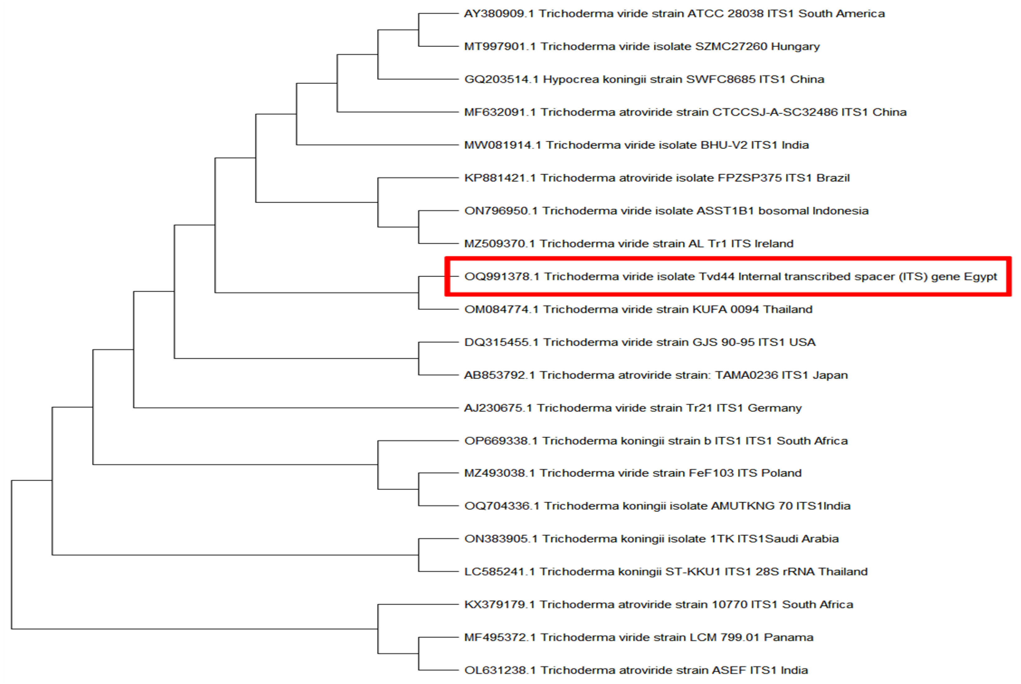
Figure 1. A maximum likelihood-based unrooted phylogenetic tree shows the connection between T. viride Tvd44 (red box) and other T. viride, Trichoderma atroviride, and Trichoderma koningii isolates based on the ITS marker sequences available in GenBank.
Horticulturae 09 00716 g001
Figure 2. A phylogenetic tree analysis using the maximum likelihood method shows the relationship between PVY strain DA55 (red box) and other PVY isolates based on PVY-CP sequences available in GenBank.
Horticulturae 09 00716 g002
Figure 3. Effect of T. viride Tvd44 on PVY infection of potato plants under greenhouse conditions.
Horticulturae 09 00716 g003
Figure 4. Effect of T. viride Tvd44 on the peroxidase activity and polyphenol oxidase activity in potato leaves at 21 dpi. Values are means and standard deviations of five biological replicates. Bars followed by the same letters are not significantly different according to Tukey’s HSD test (p ≤ 0.05).
Horticulturae 09 00716 g004
Figure 5. Effect of T. viride Tvd44 on the protein content in potato leaves at 21 dpi. Values are means and standard deviations of five biological replicates. Bars followed by the same letters are not significantly different according to Tukey’s HSD test (p ≤ 0.05).
Horticulturae 09 00716 g005
Figure 6. Chlorophyll content of potato plants under greenhouse conditions as affected by PVY inoculation and T. viride treatment. Values are means and standard deviations of five biological replicates. Bars followed by the same letters are not significantly different according to Tukey’s HSD test (p ≤ 0.05).
Horticulturae 09 00716 g006
Figure 7. The relative gene expression of PVY-CP in PVY-infected potato plants after 21 dpi. Values are means and standard deviations of five biological replicates. Bars followed by the same letters are not significantly different according to Tukey’s HSD test (p ≤ 0.05).
Horticulturae 09 00716 g007
Figure 8. The relative gene expressions of PR-1, POD, PAL, CHS, and HQT at 21 dpi of the T. viride Tvd44 treatment compared with untreated potato leaves. Values are means and standard deviations of five biological replicates. Bars followed by the same letters are not significantly different according to Tukey’s HSD test (p ≤ 0.05).
Horticulturae 09 00716 g008
Figure 9. GC–MS fractionation of ethyl acetate extract of T. viride Tvd44 culture filtrate.
Horticulturae 09 00716 g009
Table 1. Nucleotide sequence primers used in this study.
GenePrimer Sequence (5′–3′)Functional Annotation
PR-1Forward: CCAAGACTATCTTGCGGTTC
Reverse: GAACCTAAGCCACGATACCA
Pathogenesis related protein-1
PODForward: TGGAGGTCCAACATGGCAAGTTCT
Reverse: TGCCACATCTTGCCCTTCCAAATG
Peroxidase
PAL1Forward: ACGGGTTGCCATCTAATCTGACA
Reverse: CGAGCAATAAGAAGCCATCGCAAT
Phenylalanine ammonia-lyase
CHSForward: CACCGTGGAGGAGTATCGTAAGGC
Reverse: TGATCAACACAGTTGGAAGGCG
Chalcone synthase
HQTForward: CCCAATGGCTGGAAGATTAGCTA
Reverse: CATGAATCACTTTCAGCCTCAACAA
Hydroxycinnamoyl Co A quinate hydroxycinnamoyl transferase
Beta-actinForward: ATGCCATTCTCCGTCTTGACTTG
Reverse: GAGTTGTATGTAGTCTCGTGGATT
Housekeeping gene
ITSForward: TCCGTAGGTGAACCTGCGG
Reverse: TCCTCCGCTTATTGATATGC
Internal transcribed spacer
PVY-CPForward: CAACTCCAGATGGAACAATTG
Reverse: CCATTCATCACAGTTGGC
Potato virus Y coat protein
Table 2. Disease assessment of potato plants infected with PVY (21 days after inoculation) pre-inoculated or not with T. viride. C = untreated control, T = T. viride Tvd44, V = infected with PVY, T + V = T. viride 48 h before inoculation with PVY, and PDI = percent disease index.
TreatmentDisease Incidence (%) *Disease Incidence GradePDI (%)
C00.0 c-00.0 c
T00.0 c-00.0 c
V89.4 ahigh83.6 a
T + V21.4 bmoderate11.2 b
* The values presented in each column that share the same letter are not significantly different as per Tukey’s HSD test (p ≤ 0.05). Each value is representative of five biological replicates.
Table 3. Effect of T. viride on the growth parameters of potato plants infected with PVY (21 days after inoculation). C = untreated control, T = T. viride Tvd44, V = infected with PVY, and T + V = T. viride 48 h before inoculation with PVY.
Treatment *Plant Height (cm)Shoot Length (cm)Root Length (cm)Shoot Fresh Weight (g)Root Fresh Weight (g)No. of Leaves
C25.0 ± 2.65 b15.7 ± 2.08 a08.0 ± 2.00 b03.6 ± 0.61 b1.02 ± 0.24 bc29.7 ± 1.53 b
T37.7 ± 2.52 a18.0 ± 1.00 a19.7 ± 3.51 a5.27 ± 0.72 a1.39 ± 0.51 b57.7 ± 2.08 a
V15.7 ± 2.10 c10.0 ± 1.00 b05.7 ± 1.53 b1.50 ± 0.50 c0.53 ± 0.21 c11.7 ± 5.85 c
T + V34.3 ± 4.04 a16.3 ± 3.51 a18.0 ± 01.0 a4.53 ± 0.31 ab2.15 ± 0.15 a31.3 ± 2.08 b
* The values presented in each column that share the same letter are not significantly different as per Tukey’s HSD test (p ≤ 0.05). Each value is representative of five biological replicates.
Table 4. Chemical properties of the highest six compounds detected in the ethyl acetate extract of T. viride Tvd44 culture filtrate using GC–MS analysis.
* RTRA%CompoundMolecular FormulaBiological ActivityReferences
14.654.296-Amyl-α-pyroneC10H14O2Antifungal[39,40,41]
33.8314.06trans-[(2,3-Diphenylcyclopropyl)methyl] phenyl sulfideC22H20OSAnticandidal and antioxidant[42]
35.1116.901,1-Dicyano-2-methyl-4-(p-cyanophenyl)propeneC13H9N3Antifungal and insecticidal[43]
35.3931.78Thiocarbamic acid, N,N-dimethyl, S-1,3-diphenyl-2-butenyl esterC19H21NOSAntimicrobial[44]
35.639.021-Propene, 3-(2-cyclopentenyl)-2-methyl-1,1-diphenylC21H22Antifungal[42]
36.902.95S-(1,3-diphenylbutyl) dimethyl thiocarbamateC19H21NOSAntioxidant and anticancer activity[45]
* RT: retention time, RA: relative abundance.
Disclaimer/Publisher’s Note: The statements, opinions and data contained in all publications are solely those of the individual author(s) and contributor(s) and not of MDPI and/or the editor(s). MDPI and/or the editor(s) disclaim responsibility for any injury to people or property resulting from any ideas, methods, instructions or products referred to in the content.