Review

In Vitro Propagation of Caper (Capparis spinosa L.): A Review

1
Natural Resources and Local Products Research Unit, Regional Center of Agricultural Research of Agadir, National Institute of Agricultural Research, Avenue Ennasr, BP 415 Rabat Principale, Rabat 10090, Morocco
2
In Vitro Culture Laboratory, Department of Basic Sciences, National School of Agriculture, BP S/40, Meknes 50001, Morocco
3
Agro-Biotechnology Research Unit, Regional Center of Agricultural Research of Marrakech, National Institute of Agricultural Research, Avenue Ennasr, BP 415 Rabat Principale, Rabat 10090, Morocco
*
Authors to whom correspondence should be addressed.
Horticulturae 2022, 8(8), 737; https://doi.org/10.3390/horticulturae8080737
Submission received: 17 May 2022 / Revised: 25 June 2022 / Accepted: 27 June 2022 / Published: 17 August 2022
(This article belongs to the Special Issue In Vitro Technology and Micropropagated Plants)

Abstract

:
Caper (Capparis spinosa L.) is a shrubby plant species recalcitrant to vegetative propagation and generally difficult to propagate by seeds. This is due to the difficulties associated with seed germination, root induction from stem cuttings, and plant hardening. Propagation by tissue culture would be a good alternative and promising approach to overcome the limitations of conventional propagation. Tissue culture methods can be used for the clonal propagation of caper plants. Indeed, in many plant species, micropropagation has played a decisive role in the rapid and large-scale production of uniform and genetically stable plants. Tissue culture methods can also be used in genetic improvement and conservation programs. In this review, we first provided an overview on caper and its conventional means of propagation, then we described the different methods of caper micropropagation, i.e., in vitro seed germination and seedling development, propagation by nodal segmentation of elongated shoots (i.e., microcuttings), and adventitious organogenesis. These micropropagation methods can make it possible to overcome all the obstacles preventing large-scale propagation and genetic improvement of caper. Thus, the most updated information on the progress made in the field of caper micropropagation is reported and future perspectives are outlined.

1. Introduction

Caper (Capparis spinosa L.) is a shrubby plant native to the Mediterranean region. It belongs to the family Capparidaceae and genus Capparis, which includes more than 250 species generally used for ornamental, culinary, cosmetic, pharmaceutical, and medicinal purposes [1]. Capparis spinosa is characterized by high morphological and ecological diversity, which led some authors to differentiate several intraspecific variants and taxa [2,3,4,5]. It plays important socio-economic roles in the arid regions of many countries, and is well adapted to high temperatures, intense sunlight, and fluctuating climates [6,7,8].
Caper is cultivated for both unopened flowers and young fruits that are used in many traditional dishes [9]. The fruits of caper are harvested from both wild and cultivated plants. The main producers are Morocco, Spain, and Turkey [10,11].
Caper is a shrub with high medicinal values. This species is rich in bioactive compounds such as flavonoids, glucosinolates, phenolic acids, and alkaloids that can be used for medicinal, culinary, and ornamental purposes. Along this line, many health-promoting properties of caper extracts were scientifically demonstrated, in particular anti-cancer and antioxidant activities [12]. Caper is also used for ecological purposes since it helps in preventing soil erosion and preserving biodiversity and soil water [13,14].
Caper is commonly propagated by seeds. This method allows the maintenance of high genetic diversity within its populations. However, propagation by seeds cannot be used for the production of true-to-type plants [12,15]. Due to the high socio-economic and ecological importance of caper, it becomes preparatory to genetically characterize the existing material in order to identify the best and most promising genotypes, with superior agronomic characteristics. Such genotypes would be subjected to rapid and large-scale propagation, and then used for different ecological and industrial purposes. Propagation by stem cuttings may be a good alternative to sexual propagation. Nevertheless, this vegetative propagation method is associated with serious rooting problems [12]. Therefore, developing efficient propagation methods through tissue culture is needed today for caper.
In vitro propagation is a very promising approach for rapid production of caper plants. Tissue culture of caper was first reported in 1984 [16]. Since, several studies have been published describing micropropagation systems for caper, but with extremely variable rooting and acclimatization rates [9,12,17,18,19,20,21,22,23]. The high variability of findings reported in the literature may be related to several factors such as the genotype, age, and physiological state of the donor plant, explant, plant growth regulators (PGRs), and culture conditions [16]. Today, it is extremely important to develop a reproducible and efficient micropropagation system for the best-performing genotypes.
The present review provides the most updated data on the progress made in the field of caper micropropagation. This includes propagation by in vitro seed germination and seedling development, propagation by microcuttings, and through adventitious organogenesis. This review also describes the micropropagation steps of caper and discusses the difficulties encountered in each in vitro culture method.

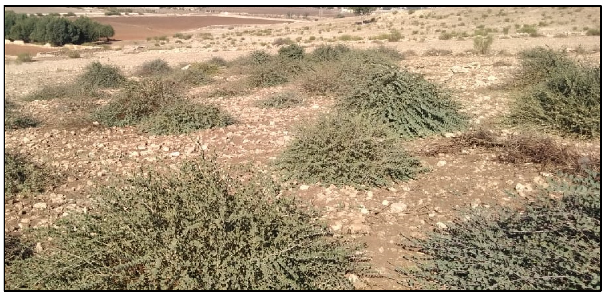
1.1. Geographic Distribution, Botanical Classification, and Ecological Characteristics of Caper

Caper is a xerophytic and heliophilic species endowed with a great adaptation capacity to difficult environmental conditions [24]. Caper is mainly located in the arid and semi-arid areas of the Mediterranean region. It is found in north and east Africa, Madagascar, southwest and central Asia (China and Iran), the Middle East, southern Europe (Greece, Italy), Australia, and Oceania [3,25]. It grows naturally from the Atlantic coasts of the Canary Islands and Morocco to the Black Sea, and to the shore of the Caspian Sea [3,26]. The wild and cultivated caper plants are used throughout its entire area of growth, from west Africa to the Norfolk Islands in the Pacific [27].
Caper belongs to the genus Capparis and family Capparidaceae [3,28,29,30]. It is a perennial species, generally thorny and hairy, with a height of about 50–80 cm. Caper is characterized by a very high level of heterogeneity within its populations [3,29,31]. The taxonomic position of C. spinosa is still difficult to assess due to the several taxa of different ranks that have been described [32]. Therefore, the classification of this species remains ambiguous and controversial. A morphological study of some caper ecotypes from north-central Morocco revealed the existence of three genotypic clusters that correspond to three species: Capparis spinosa, C. ovata, and C. cartilaginea [31]. C. spinosa is by far the most economically important one and is mainly found in the Mediterranean region [1,3,29]. It is also one of the most common medicinal and aromatic plants that grow along roads, on slopes, and in rocky and stony areas [33].
Flower buds (i.e., capers) are the most sought-after parts of caper. The firmness of capers is a key indicator of their commercial quality (i.e., an indicator of good storability). The firmness index of capers varies depending on the genotype and area of growth [34]. Caper fruit is a dehiscent berry of 2–4 cm long (Figure 1). The number of seeds per fruit is on average 130, with a minimum of 15 seeds in the smallest fruits and a maximum of 400 seeds in large fruits [13]. The roots of caper are very deep but not highly branched [13]. Indeed, the caper has a powerful root system that can mobilize large volumes of soil. These characteristics make caper highly tolerant to drought and able to grow even on the poorest soils and on steep slopes. Hence, caper has a great ecological interest as it can prevent erosion in arid and semi-arid regions [35].
As a xerophytic plant, caper has morphological and physiological characteristics that make it tolerant to the most severe climatic conditions of arid and semi-arid regions [11]. Caper is found in sites with temperatures as low as −4 °C, but generally the annual average temperature of its natural regions of growth exceeds 14 °C. It withstands temperatures of up to 40 °C and is also drought tolerant. It can survive with no rain for several months. In its area of growth, the rainfall varies from 200 to 550 mm per year [13].
Caper is a rupicolous species that can be cultivated on different types of soils. It grows on loamy clay, sandy, rocky, and gravelly soils. It also grows on clayey and poorly draining soils and on light sandy loam soils with alkaline pH (Figure 2). However, it seems that caper prefers light, well-draining soils with a neutral to alkaline pH [13,14].

1.2. Economic Importance and Medicinal Uses

The economic importance of caper lies in the value of its young shoots, tender young fruits, and flower buds, and they have a high demand in the international market [24]. Flower buds are generally harvested before flowering and used for aromatic purposes in the cosmetic industry. The wild buds are harvested by seasonal pickers then stored in salt before packaging [36].
Since ancient times, different parts of the caper have been used as traditional herbal remedies due to their beneficial effects on human health [37]. In recent years, the biological activities of caper bud extracts have been evaluated and numerous health promoting properties were reported. Caper extracts possess a high inhibitory activity against acetylcholinesterase (AChE) and butyrylcholinesterase (BChE), as well as antioxidant and metal chelating properties [38]. Caper leaves, stems, flowers, roots, and fruits were also reported to have antidiabetic, anti-obesity, antimicrobial, anti-inflammatory, and antihypertensive activities [39,40,41,42,43]. This is due to their content of flavonoids, phenols, alkaloids, tannins, and glycosides [44].
In Greek folk medicine, herbal teas made from the young shoots of caper are used against rheumatism. The bark and leaves of caper have anticarcinogenic activities. Indeed, they contain rutin, selenium, and quercetin, which contribute to the prevention of certain forms of cancer [14].

2. Conventional Propagation of Caper

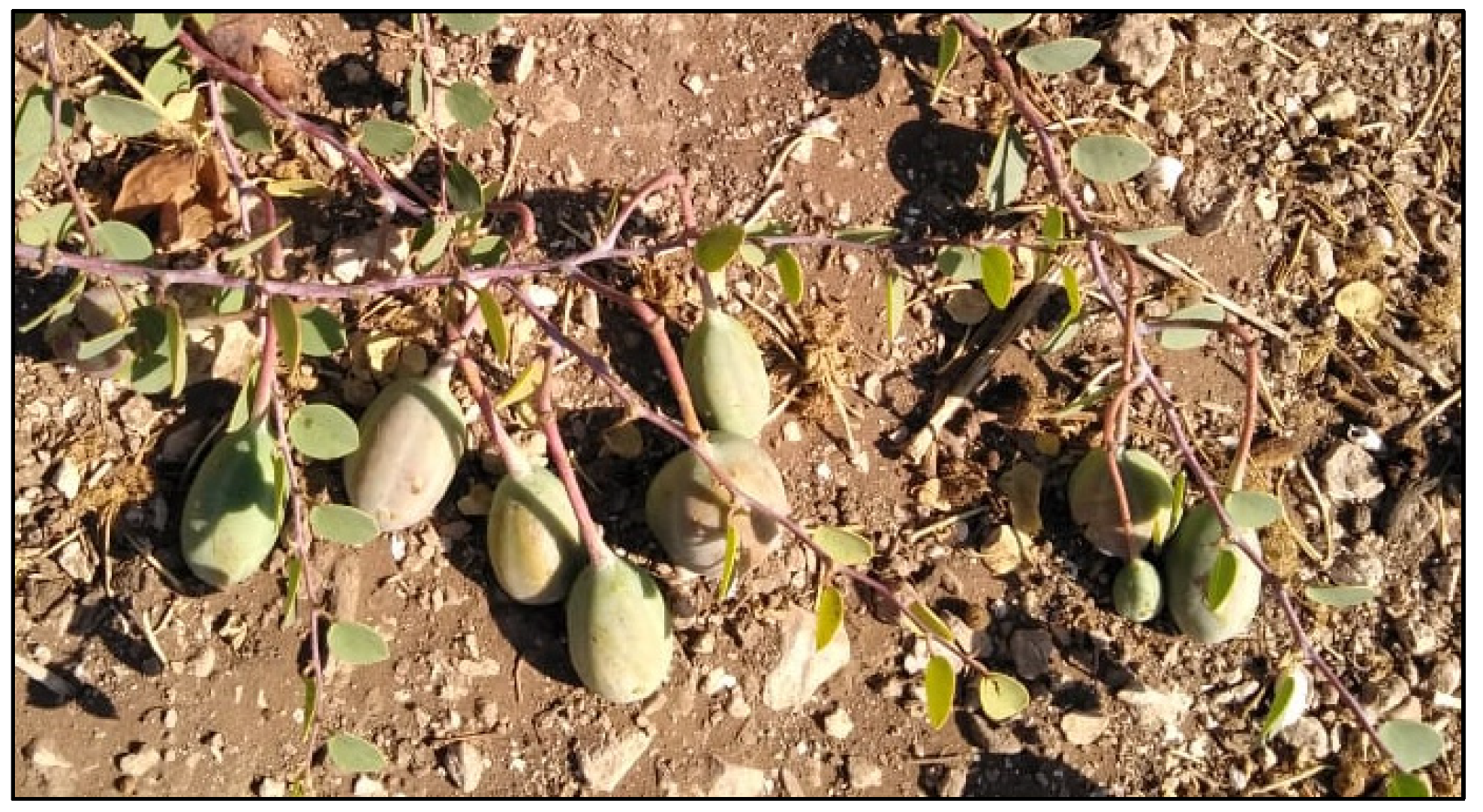
2.1. Propagation by Seeds

Propagation by seeds is the most widely used method for caper propagation. However, many factors may hamper caper seed germination, among which the most important are the hardness of seed coat, seed dormancy, the hormonal balance within seeds, and embryo immaturity. The seed coat and mucilage surrounding the seeds are ecological adaptations to prevent water loss and maintain seed viability during dry seasons [14,45,46]. Generally, viable embryos germinate within 3 to 4 days after partial removal of the lignified seed coat [47].
Several studies have been conducted to overcome plant seed dormancy. Treatments such as plant hormones, sulfuric acid (H2SO4), methanol, potassium nitrates, boiling water, and stratification were suggested to break seed dormancy [48]. In the case of caper, H2SO4, cold stratification, scarification, and treatment with gibberellic acid (GA3) or nitric acid (KNO3) have been suggested [16,49,50,51,52,53]. However, the germination rate of caper seeds was not high enough for rapid and large-scale propagation even after the application of these treatments.

2.2. Propagation by Stem Cuttings

Vegetative propagation by stem cuttings is a practicable and efficient method for the clonal multiplication of plants. However, in many species, this method is hampered by rooting difficulties [48]. Caper is a plant species difficult to root. Root induction from stem cuttings depends on many factors such as the type and age of cutting, time of cuttings’ collection, the substrate used for planting, and other seasonal and environmental parameters. Moghaddasi et al. [14] observed a root induction rate of up to 55% in one-year-old twigs of caper. Some authors used PGRs to improve the rooting ability of caper cuttings [16,46,54,55]. Bahrani et al. [46] evaluated the effects of indole-3-butyric acid (IBA) on root induction from growing season (softwood) and one year old (semi-hardwood) stems of caper. IBA did not promote root induction from softwood cuttings, while it showed a very low rooting percentage (16.1%) in semi-hardwood cuttings [46]. According to Saifi et al. [54], treating caper cuttings with 3000 ppm IBA for one minute resulted in a rhizogenesis rate of 50%. On the other hand, the use of leafy cuttings showed a slightly higher rhizogenesis rate (67.1%) than leafless cuttings (61.4%) [15]. The ability of caper cuttings to induce roots is also influenced by other endogenous and exogenous factors such as the genotype, cutting thickness, and planting season [45,55,56].
Caper propagation by stem cuttings allows for the production of true-to-type plants. However, as mentioned above, this method is hindered by rooting problems. Additionally, caper plants produced by stem cuttings are highly susceptible to drought during the first years of planting.
Based on the above, it seems that the use of in vitro culture methods would considerably help in the rapid and large-scale propagation of this medicinal plant. Up to date, few studies have been undertaken on caper micropropagation. Three different methods were described: (i) in vitro seed germination and seedling development; (ii) nodal segmentation of elongated shoots (i.e., microcuttings); and (iii) organogenesis. These micropropagation methods are described and discussed in the following sections.

3. Caper Propagation by Tissue Culture

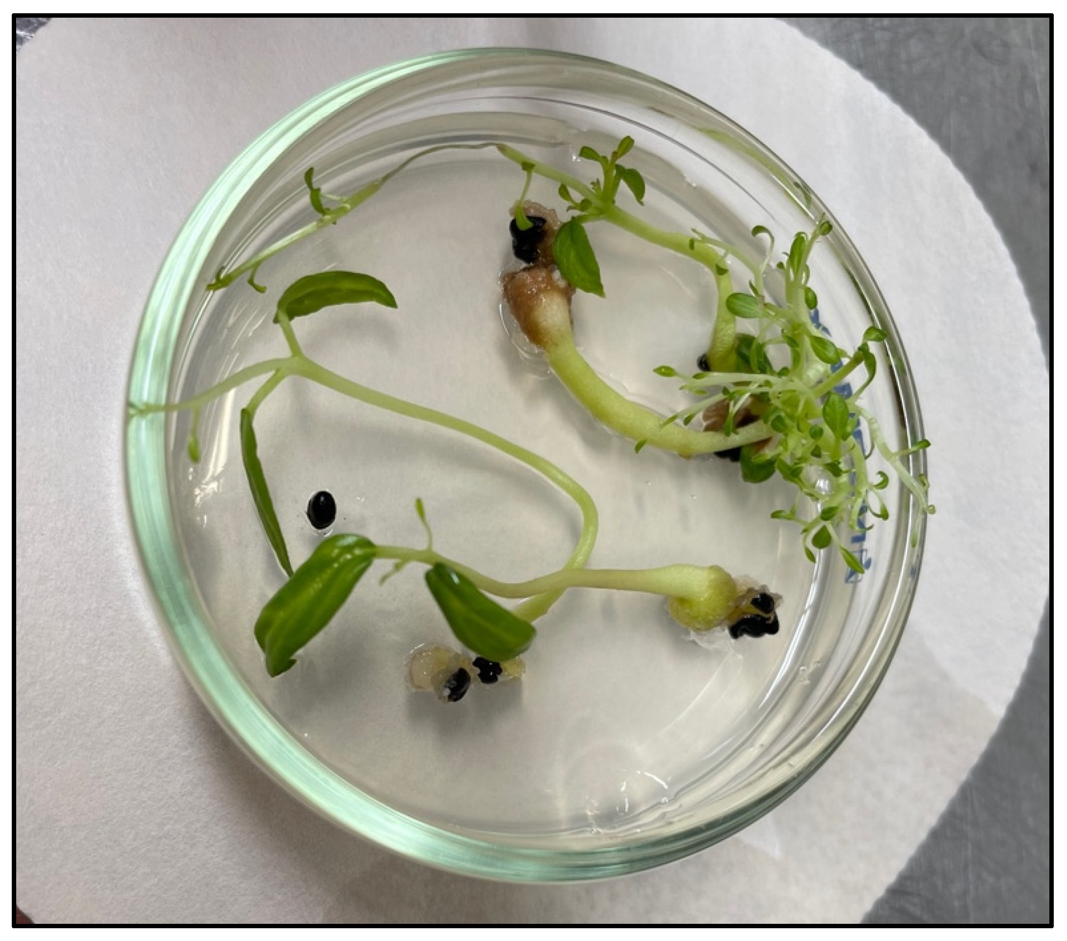
3.1. In Vitro Seed Germination and Seedling Development

Propagation by in vitro seed germination allows the conservation of caper diversity and ensures the sustainable use of this species. Under natural conditions, the propagation of caper by seeds is hampered by many factors as previously mentioned. In vitro seed germination may considerably help to overcome these constraints. Indeed, seeds are cultured on appropriate culture media and under controlled conditions that provide embryos with the optimal nutrients and conditions to promote germination and subsequent growth [48]. Accordingly, in vitro seed germination (Figure 3) can be considered the best approach to produce a large number of caper plants while preserving the genetic diversity within this species. In vitro seed germination is also a powerful tool for genetic improvement.

3.1.1. Surface Disinfection and Culture Medium

Various surface disinfection protocols were described for caper seeds. For example, Rhimi et al. [8] started the disinfection procedure by rinsing the seeds five times with sterile distilled water. The seeds were then treated with H2SO4 for 30 min, followed by immersion in 70% alcohol for 5 min and then in 6% sodium hypochlorite for 20 min, with continuous stirring. Finally, the seeds were rinsed five times (5 min each) with sterile distilled water. Germanà and Chiancone [57] soaked caper seeds in 70% ethyl alcohol for 3 min, followed by 20 min in a solution of 25% commercial bleach (approximately 1.5% active chlorine), and finally by three rinses with sterile distilled water.

3.1.2. Seed Germination

Several factors can affect seed germination in vitro, for example, plant genotype, culture medium components, PGRs, seed coat, pretreatments, and culture conditions [8,48,58,59,60,61]. In caper, a large variation was observed in germination rates among the different populations (Table 1). It appears that Murashige and Skoog medium (MS; [62]) is the most suitable for in vitro seed germination of caper. After scarification, Chalak et al. [17] observed a seed germination rate of 71% on PGR-free MS medium, while the use of sterile distilled water gelled with agar showed a germination rate of 64%. According to Rhimi et al. [8], a 75% germination rate was observed in caper seeds from the Nahli site (Tunisia) following 30-min H2SO4 and 48-h GA3 (2000 mg L−1) pretreatments. In a different work, the caper seeds harvested from a wild tree located in the Madonie Mountains and that belongs to the subspecies rupestris showed a germination rate of 80.4%. The seeds were subjected to a heat pretreatment at 40 °C for one hour, then cultured on modified MS medium [57]. Treating seeds with gamma rays may improve their germination ability in vitro and promote seedling growth and development [63,64]. Seed irradiation with gamma rays resulted in an in vitro germination rate of 50% [65].
The effect of PGRs on seed germination has also been the subject of investigation [57,66,67,68]. During in vitro seed germination, shoot and root growth depends strongly on exogenous PGRs, their concentrations, as well as the metabolism of endogenous plant hormones, their concentration, and interaction with PGRs [69,70]. Based on our experiments (data not published), the use of MS medium supplemented with GA3 and 6-benzylaminopurine (BAP) or thidiazuron (TDZ) promotes caper seed germination. This is in good agreement with results reported for other plant species [71,72]. GA3 is a growth regulator widely used for in vitro seed germination. The exogenous GA3 added to culture medium acts by increasing the concentration of endogenous GA3 and lowering that of abscisic acid (ABA), which promotes seed germination [73,74]. BAP is a potent cytokinin commonly used in tissue culture due to its efficacy and affordability [75]. Regarding TDZ, it is a phenylurea derivative that has a potent cytokinin-like activity and is widely used for the micropropagation of Mediterranean species [76].

3.1.3. Seedling Growth and Plantlet Acclimatization

Research on in vitro development of caper seedlings and their acclimatization is still very limited. According to Heydariyan et al. [77], salicylic acid and GA3 significantly improved the growth and development of caper seedlings. Germanà and Chiancone [57] noted that the combination of 1-naphthaleneacetic acid (NAA), BAP, and GA3 inhibited apical bud growth. However, new buds emerged around the cotyledons, in the region between the epicotyl and hypocotyl. According to these authors, 40% of seedlings produced new shoots [57].
Successful acclimatization of plantlets grown in vitro is a crucial step for the mass propagation of any species. Rhimi et al. [8] reported a survival rate of 95% after the acclimatization of 15-day-old seedlings in pots containing a mixture of peat and sand (2:1, w/w).

3.2. Caper Propagation by Microcuttings

Despite the large number of caper genotypes and their reputed medicinal virtues, an efficient and reproducible propagation system through microcuttings has yet to be developed. Such system should aim not only to produce a large number of true-to-type plants, but also to harness the economic potential of capers.
Propagation by microcuttings, also known as propagation by nodal segmentation of elongated shoots, is a vegetative propagation method based on the multiplication of axillary shoots derived from microcuttings (i.e., tender stem segments bearing 1–3 buds) in vitro. Elongated shoots are cut into small segments (i.e., microshoots) containing one or two nodes then placed on a rooting medium to promote the induction of adventitious roots. The plantlets are then transferred to the glasshouse for acclimatization. This method allows for the production of true-to-type plants [48].
The findings of several studies carried out on capper micropropagation by microcuttings underlined the enormous potential of this method [9,12,17]. However, successful regeneration through microcuttings depends on many factors associated with the genotype and culture conditions.

3.2.1. Plant Material and Culture Medium

In order to establish aseptic cultures and obtain leafy shoots, nodal segments were used [9,12,17,78]. In addition, the effects of different basal media on the multiplication and rooting of caper microshoots were evaluated, for example, MS medium, Linsmaier and Skoog medium [79], Nitsch and Nitsch medium [80], Woody Plant Medium (WPM; [81]), and Rugini medium [82]. However, MS medium was found to be the most suitable for culture establishment and shoot proliferation [9,20].

3.2.2. Surface Disinfection

The development of an efficient disinfection protocol is a necessary step for the setup of a micropropagation system. However, the establishment of axenic cultures from microcuttings may be very challenging [9,18]. Various protocols have been described in the literature, most of them used sodium hypochlorite as a disinfectant agent [17,20,22,23]. The use of sodium hypochlorite coupled with ethanol was also suggested and showed convincing results. Indeed, some authors suggested to first soak microcuttings in a solution of 70–80% ethanol for 2 to 5 min before surface disinfection with sodium hypochlorite [9,12,23,78].

3.2.3. Shoot Multiplication and Elongation

Shoot multiplication and elongation are strongly affected by PGRs. High levels of endogenous cytokinins may be needed to induce cell division. Addition of exogeneous PGRs promote cell elongation and multiplication. Many authors suggested the use of a BAP-auxin combination for caper shoot multiplication and elongation (Table 2). For example, El-Mekawy et al. [20] reported that the combination of 0.5 mg L−1 BAP and 0.05 mg L−1 NAA gave an average number of 3.89 shoots per explant, with an average length of 3.24 cm.
The undeniable effect of BAP on caper shoot multiplication and elongation was also reported by Rodriguez et al. [78], who indicated that 4 µM (≃0.9 mg L−1) BAP (in combination with 0.3 µM (≃0.05 mg L−1) indole-3-acetic acid (IAA) and 0.3 µM (≃0.1 mg L−1) GA3) promoted shoot proliferation and growth. This was confirmed by Sottile et al. [83], who reported that the combination of 6 µM (≃1.35 mg L−1) BAP and 0.12 µM (≃0.02 mg L−1) IBA allowed for maximum proliferation of caper leafy shoots (91%). This combination also improved the number of shoots per explant (8.7) and shoot length (2.7 cm). Multiple shoot formation from nodal buds was observed by Chalak et al. [17] on media containing 1.5 mg L−1 BAP, 0.05 mg L−1 IBA, and 0.1 mg L−1 GA3, with an average number of shoots ranging from 4.25 to 5.43.
Some other factors were reported to affect shoot multiplication and elongation of caper, such as the type and concentration of carbohydrates added to culture medium [12]. Sucrose is by far the most commonly used carbon source for the multiplication of caper shoots. Glucose promoted caper shoot growth as well and, in some cases resulted in better growth than sucrose [12]. Successive subcultures have been reported to affect the proliferation capacity of caper shoots. Shoot multiplication was enhanced by successive subcultures on fresh culture media, with an average rate exceeding 20 new shoots per explant at the end of the sixth subculture. However, the proliferation capacity decreased during the following subcultures [17].

3.2.4. In Vitro Rhizogenesis of Caper Microshoots

Adventitious root induction from microshoots is the most decisive step of the microcuttings’ technique. In many plant species, it was very difficult to induce roots from in vitro cultured shoots [84]. Root formation from microshoots depends on many factors such as the genotype, culture medium and conditions, and PGRs, particularly auxins and their concentrations. Early cell division is a prerequisite for root formation. Generally, the root induction requires high levels of auxins. Addition of cytokinins to culture medium may be necessary since they are involved in the regulation of cell division and organ differentiation [12,18,69].
In many plant species, the use of the auxins IAA, IBA and NAA resulted in high in vitro rhizogenesis rates [85,86,87]. These auxins, used either alone or in combination, have also shown convincing results in Capparis species [9,18,20]. Generally, the roots appear after 15 to 20 days of culture [9]. Attia et al. [22] observed an in vitro rooting percentage of 56.7% on MS medium containing 1.5 mg L−1 NAA. According to Carra et al. [9] and Sottile et al. [83], the use of IBA at 5 μM (≃1 mg L−1) resulted in high rhizogenesis rates (93.4–93.7%). However, increasing IBA concentration to 10 μM (≃2 mg L−1) decreased the rhizogenesis rate to 68.7% [9]. On the other hand, 1 μM (≃0.18 mg L−1) NAA showed a rhizogenesis rate of 68.7% [9]. The average number of roots per shoot ranged from 2.4 on the medium supplemented with 1 µM (≃0.18 mg L−1) NAA to 3.5 on that containing 5 μM (≃0.87 mg L−1) IAA under light conditions, whereas the highest average length of roots was 28 mm when 5 μM (≃1 mg L−1) IBA was added to culture medium [9]. The combination of NAA and IBA at 0.75 mg L−1 and 0.25 mg L−1, respectively, resulted in a rooting percentage of 67% [16]. Rodriguez et al. [78] observed 70% rooting after a 20-day incubation period in darkness on MS medium containing IAA. Chalak et al. [17] and Chalak and Elbitar [18] suggested a pulse treatment for 4 h in a solution of 100 mg L−1 IAA before transferring the explants to MS or half-strength Murashige and Skoog medium (½MS). This treatment resulted in a rhizogenesis range of 87–92%. Soaking caper microshoots in a solution of 24.6 µM (≃5 mg L−1) IBA for 10 min before culture on MS medium under a 16 h photoperiod enhanced the rooting percentage to 80.5% [47].

3.2.5. Plantlet Acclimatization

To date, only a few studies have been conducted on the acclimatization of caper plants produced by the microcuttings technique, and divergent results were published. Some authors reported low survival rates. For example, Chalak et al. [17] observed a survival rate of 40%. On the other hand, some other studies reported high survival rates. For example, Chalak and Elbitar [18] observed a survival rate of 92%. Musallam et al. [88] reported a successful transfer of caper plants regenerated in vitro to field conditions as well as normal growth and development.
The composition and texture of culture medium used before acclimatization (e.g., elongation and/or rooting medium) may significantly influence the survival rate of the plants during acclimatization [89,90]. In C. orientalis, the survival rate varied from 56% to 66% depending on the hormonal composition of rooting medium [12]. The impact of rooting medium on subsequent plantlet acclimatization was observed in other plant species [91].
Based on the above, the effects of many factors on caper plantlet survival during acclimatization are still to be evaluated, such as the genotype, substrate mixtures and ratios, nutrient solutions and irrigation, pre-acclimatization, and greenhouse/glasshouse conditions. Therefore, further research should focus on these factors in order to improve the survival rate of caper plants produced by the microcuttings technique.

3.3. Caper Propagation by Organogenesis

Organogenesis is a powerful in vitro regeneration system for the mass production of genetically uniform plants [92,93]. It is based on the totipotency of plant cells and involves the development of shoot buds and roots either directly on the explant or after callus formation under aseptic and appropriate culture conditions to subsequently form complete plants [61,92,94]. Successful regeneration through organogenesis depends on many factors including the genotype and explant type as well as culture medium and conditions. Despite the enormous potential of this method, there are only few papers dealing with it in caper. Organogenesis can be considered as the best approach available today to propagate caper and can be exploited for genetic improvement and conservation purposes.
Different PGRs were evaluated to develop an in vitro regeneration system through organogenesis for caper. Movafeghi et al. [95] suggested to culture hypocotyl explants on MS medium supplemented with 0.1 mg L−1 NAA and 0.5 mg L−1 BAP. This PGR combination promoted adventitious shoot induction and proliferation. Increasing NAA concentration decreased the formation rate of adventitious shoots. Root induction was performed on MS medium containing 0.5 mg L−1 NAA and the regenerated plantlets were successfully acclimatized to ex vitro conditions. Elmaghrabi et al. [96] cultured leaf explants on MS medium containing 1.2 mg L−1 2,4-dichlorophenoxyacetic acid (2,4-D) to induce callogenesis. For shoot and root formation, they suggested to transfer calli on MS medium supplemented with 2 mg L−1 BAP. The plantlets were successfully acclimatized to greenhouse conditions. Fahmideh et al. [97] suggested to use 2 mg L−1 kinetin and 2 mg L−1 NAA for shoot induction from callus culture, and 1 mg L−1 NAA for shoot rooting.
Al-Safadi and Elias [65] also highlighted the significant impact of PGRs on caper organogenesis. The use of 2 mg L−1 GA3 gave an average of 2.2 shoots per explant while the combination of 0.1 mg L−1 GA3, 1 mg L−1 NAA, and 2 mg L−1 zeatin riboside yielded an average of 5.5 shoots per explant. A survival rate of 86% was observed during acclimatization. Caglar et al. [47] pointed out the effectiveness of TDZ for the production and multiplication of caper shoots in vitro. Indeed, 4.54 μM (≃1 mg L−1) TDZ gave an average number of 45.7 shoots per explant.
The effects of other factors on caper organogenesis were scarcely investigated. Al-Safadi and Elias [65] found that gamma irradiation greatly improved the growth of caper shoots induced in vitro. In fact, a 10 Gy dose of gamma irradiation promoted shoot growth by up to 200% and improved rhizogenesis to reach 100%.

4. Conclusions and Future Perspectives

Caper is a shrub that has long been considered difficult to propagate conventionally and, despite the high socioeconomic and medicinal importance of this species, research on its micropropagation is still very limited. The present review reported and discussed the main findings of the literature in the field of caper micropropagation. Based on the studies reported here, it can be seen that caper micropropagation was achieved through seed germination, microcuttings, and organogenesis. However, each method presents many difficulties and divergent results. Moreover, the organogenesis technique is not well exploited and more research is still needed in this field. Based on the information gathered in this report, it is highly important to improve the existing micropropagation methods, and to develop new regeneration systems that could be used for rapid and large-scale propagation and genetic improvement of caper, such as somatic embryogenesis. In fact, the development of the various caper industries is closely linked to the development of efficient propagation methods through tissue culture.
Somatic embryogenesis is a powerful micropropagation technique that is used not only for the production of a large number of plants in a short period of time, but also for genetic transformation, artificial seed production, and germplasm conservation. Somatic embryogenesis also offers the possibility to identify and select, under in vitro conditions, the genotypes that are resistant to biotic and abiotic stresses. It can be used for secondary metabolite production, which may be very interesting in caper. Thus, developing an in vitro regeneration system through somatic embryogenesis for caper is currently needed for the sustainable utilization and improvement of this species.

Author Contributions

Conceptualization, M.K., I.B. and M.A.M.; investigation, M.K. and M.A.M.; writing—original draft preparation, M.K.; writing—review and editing, M.A.M., M.K. and I.B.; supervision, I.B. All authors have read and agreed to the published version of the manuscript.

Funding

This research received no external funding.

Institutional Review Board Statement

Not applicable.

Informed Consent Statement

Not applicable.

Data Availability Statement

Not applicable.

Conflicts of Interest

The authors declare no conflict of interest.

References

  1. Fici, S. Intraspecific variation and evolutionary trends in Capparis spinosa L. (Capparaceae). Plant Syst. Evol. 2001, 228, 123–141. [Google Scholar] [CrossRef]
  2. Zohary, M. The species of Capparis in the Mediterranean and the Near Eastern countries. Bull. Res. Counc. Isr. 1960, 8D, 49–64. [Google Scholar]
  3. Inocencio, C.; Rivera, D.; Concepción Obón, M.; Alcaraz, F.; Barreña, J.-A. A systematic revision of Capparis section Capparis (Capparaceae) 1, 2. Ann. Mo. Bot. Gard. 2006, 93, 122–149. [Google Scholar] [CrossRef]
  4. Danin, A. Capparis in the East Mediterranean countries. Fl. Medit. 2010, 20, 179–185. [Google Scholar]
  5. Gristina, A.S.; Fici, S.; Siragusa, M.; Fontana, I.; Garfì, G.; Carimi, F. Hybridization in Capparis spinosa L.: Molecular and morphological evidence from a Mediterranean island complex. Flora Morphol. Distrib. Funct. Ecol. Plants 2014, 209, 733–741. [Google Scholar] [CrossRef]
  6. Jiang, H.E.; Li, X.; Ferguson, K.D.; Wang, Y.F.; Liu, C.J.; Li, C.S. The discovery of Capparis spinosa L. (Capparidaceae) in the Yanghai Tombs (2800 years b.p.), NW China, and its medicinal implications. J. Ethnopharmacol. 2007, 113, 409–420. [Google Scholar] [CrossRef]
  7. Faran, M. Capparis spinosa—The plant on the wall. In Medicinal and Aromatic Plants of the Middle-East (Medicinal and Aromatic Plants of the World); Yaniv, Z., Dudai, N., Eds.; Springer: Dordrecht, The Netherlands, 2014; pp. 59–65. [Google Scholar]
  8. Rhimi, A.; Hannachi, H.; Hjaoujia, S.; Yousfi, H.; Boussaid, M. In vitro germination and seedling development of Tunisian caper (Capparis spinosa L.). Int. J. Agron. Agri. Res. 2017, 10, 1–8. [Google Scholar]
  9. Carra, A.; Del Signore, M.B.; Sottile, F.; Ricci, A.; Carimi, F. Potential use of new diphenylurea derivatives in micropropagation of Capparis spinosa L. Plant Growth Regul. 2012, 66, 229–237. [Google Scholar] [CrossRef]
  10. Inocencio, C.; Cowan, R.S.; Alcaraz, F.; Rivera, D.; Fay, F.M. AFLP fingerprinting in Capparis subgenus Capparis related to the commercial sources of capers. Genet. Resour. Crop. Evol. 2005, 52, 137–144. [Google Scholar] [CrossRef]
  11. Kdimy, A.; El Yadini, M.; Guaâdaoui, A.; Bourais, I.; El Hajjaji, S.; Le, H.V. Phytochemistry, biological activities, therapeutic potential, and socio-economic value of caper (Capparis spinosa L.). ChemRxiv 2022. [Google Scholar] [CrossRef]
  12. Kereša, S.; Stanković, D.; Lodeta, K.B.; Jerčić, I.H.; Bolarić, S.; Barić, M.; Mihovilović, A.B. Efficient protocol for the in vitro plantlet production of caper (Capparis orientalis Veill.) from the East Adriatic coast. Agronomy 2019, 9, 303. [Google Scholar] [CrossRef]
  13. Lakrimi, M. Le caprier. Transfert Technol. Agri. 1997, 37, 1–4. [Google Scholar]
  14. Moghaddasi, S.M.; Haddad Kashani, H.; Azarbad, Z. Capparis spinosa L. propagation and medicinal uses. Life Sci. J. 2012, 9, 684–686. [Google Scholar]
  15. Ramezani-Gask, M.; Bahrani, M.J.; Shekafandeh, A.; Salehi, H.; Taghvaei, M.; Al-Ahmadi, M.J. A comparison of different propagation methods of common Caper-bush (Capparis spinosa L.) as a new horticultural crop. Int. J. Plant Dev. Biol. 2008, 2, 106–110. [Google Scholar]
  16. Gianguzzi, V.; Barone, E.; Sottile, A.F. In vitro rooting of Capparis spinosa L. as affected by genotype and by the proliferation method adopted during the multiplication phase. Plants 2020, 9, 398. [Google Scholar] [CrossRef] [PubMed]
  17. Chalak, L.; Elbitar, A.; Cordahi, N.; Hage, C.; Chehade, A. In vitro propagation of Capparis spinosa L. Acta Hortic. 2003, 616, 335–338. [Google Scholar] [CrossRef]
  18. Chalak, L.; Elbitar, A. Micropropagation of Capparis spinosa L. subsp. Rupestris Sibth. & Sm. by nodal cuttings. Indian J. Biotechnol. 2006, 5, 555–558. [Google Scholar]
  19. Hegazi, G.; Eid, S.; Sharaf, A.E.M. Micropropagation for conservation of two rare Capparis species from Egypt 1. Catrina 2011, 6, 29–39. [Google Scholar]
  20. El-Mekawy, M.A.; Ali, M.A.M.; Belal, A.H.; Abdallah, S.A.S. Micropropagation of caper (Capparis spinosa, L.) from wild plants growing in North Sinai. Hortsci. J. Suez Canal Univ. 2013, 1, 15–20. [Google Scholar] [CrossRef]
  21. Mehrabani, V. In vitro micropropagation of two native Capparis spinosa L. cultivars from Iran. Bio. Forum Int. J. 2016, 8, 144–149. [Google Scholar]
  22. Attia, A.; Dessoky, E.D.S.; Al-Sodany, Y.M.; Ismail, I.A. Ex situ preservation for some endemic and rare medicinal plants in Taif, KSA. Biotechnol. Biotechnol. Equip. 2017, 31, 912–920. [Google Scholar] [CrossRef]
  23. Gianguzzi, V.; Inglese, P.; Barone, E.; Sottile, F. In vitro regeneration of Capparis spinosa L. by using a temporary immersion system. Plants 2019, 8, 177. [Google Scholar] [CrossRef] [PubMed]
  24. Chedraoui, S.; Abi-Rizk, A.; El-Beyrouthy, M.; Chalak, L.; Ouaini, N.; Rajjou, L. Capparis spinosa L. in a systematic review: A xerophilous species of multi values and promising potentialities for agrosystems under the threat of global warming. Front. Plant. Sci. 2017, 8, 1845. [Google Scholar] [CrossRef] [PubMed]
  25. Zarei, M.; Seyedi, N.; Maghsoudi, S.; Nejad, M.S.; Sheibani, H. Green synthesis of Ag nanoparticles on the modified grapheme oxide using Capparis spinosa fruit extract for catalytic reduction of organic dyes. Inorg. Chem. Commun. 2021, 123, 108327. [Google Scholar] [CrossRef]
  26. Alkire, B. Capers; Center for New Crops and Plants Products: West Lafayette, IN, USA, 1998. [Google Scholar]
  27. Rivera, D.; Inocencio, C.; Obon, C.; Alcaraz, F. Review of food and medicinal uses of Capparis L. subgenus Capparis (Capparidaceae). Econ. Bot. 2003, 57, 515–534. [Google Scholar] [CrossRef]
  28. Hall, J.C.; Sytsma, K.J.; Iltis, H.H. Phylogeny of Capparaceae and Brassicaceae based on chloroplast sequence data. Am. J. Bot. 2002, 89, 1826–1842. [Google Scholar] [CrossRef] [PubMed]
  29. Tlili, N.; Khaldi, A.; Triki, S.; Munné-Bosch, S. Phenolic compounds and vitamin antioxidants of caper (Capparis spinosa). Plant Foods Hum. Nutr. 2010, 65, 260–265. [Google Scholar] [CrossRef]
  30. Bennoune, L.; Bouteldja, N.; Chebah, S.; Akkouche, Z. Etude Bibliographique de Quelques Activités Biologiques de Différentes Parties du Câprier. Master’s Thesis, Jihel University, Jijel, Algeria, 2012. [Google Scholar]
  31. Saifi, N.; Ibijbijen, J.; Echchgadda, G. Caractérisation morphologique et identification taxonomique du câprier (Capparis spp.) dans les régions de Fès, Taounate et My Idriss Zerhoune. In Proceedings of the International Congress of ‘Institut National des Plantes Médicinales et Aromatiques’, Fes, Morocco, 22–24 March 2007. [Google Scholar]
  32. Fici, S. A taxonomic revision of the Capparis spinosa group (Capparaceae) from the Mediterranean to Central Asia. Phytotaxa 2014, 174, 1–24. [Google Scholar] [CrossRef]
  33. Rakhimova, T.; Vaisova, G.B.; Rakhimova, N.K.; Matkarimova, A. Phytocoenotic distribution of Capparis spinosa L. (Capparaceae) under different ecological conditions in Uzbekistan. Ann. Romanian Soc. Cell Biol. 2021, 25, 7882–7895. [Google Scholar]
  34. Ibnou Ali El Alaoui, M. Etude de la diversité génétique chez le câprier (Capparis spp.) au Maroc. Périodique D’information Du Cent. Régional De La Rech. Agron. De Meknès 2015. Available online: https://mag.inrameknes.info/?p=886 (accessed on 15 March 2022).
  35. Benseghir-Boukhari, L.A.; Seridi, R. Le câprier, une espèce arbustive pour le développement rural durable en Algérie. Méditerranée 2007, 109, 101–105. [Google Scholar] [CrossRef]
  36. Aliyazicioglu, R.; Eyupoglu, O.E.; Sahin, H.; Yildiz, O.; Baltas, N. Phenolic components, antioxidant activity, and mineral analysis of Capparis spinosa L. Afr. J. Biotechnol. 2013, 12, 6643–6649. [Google Scholar] [CrossRef]
  37. Zhang, H.; Ma, Z.F. Phytochemical and pharmacological properties of Capparis spinosa as a medicinal plant. Nutrients 2018, 10, 116. [Google Scholar] [CrossRef] [PubMed]
  38. Mollica, A.; Stefanucci, A.; Macedonio, G.; Locatelli, M.; Luisi, G.; Novellino, E.; Zengin, G. Chemical composition and biological activity of Capparis spinosa L. from Lipari Island. S. Afr. J. Bot. 2019, 120, 135–140. [Google Scholar] [CrossRef]
  39. Eddouks, M.; Lemhadri, A.; Michel, J.B. Hypolipidemic activity of aqueous extract of Capparis spinosa L. in normal and diabetic rats. J. Ethnopharmacol. 2005, 98, 345–350. [Google Scholar] [CrossRef]
  40. Ali, Z.N.; Eddouks, M.; Michel, J.B.; Sulpice, T.; Hajji, L. Cardiovascular effect of Capparis spinosa aqueous extract. Part III: Antihypertensive effect in spontaneously hypertensive rats. Am. J. Pharmacol. Toxicol. 2007, 2, 111–115. [Google Scholar] [CrossRef]
  41. Boga, C.; Forlani, L.; Calienni, R.; Hindley, T.; Hochkoeppler, A.; Tozzi, S.; Zanna, N. On the antibacterial activity of roots of Capparis spinosa L. Nat. Prod. Res. 2011, 25, 417–421. [Google Scholar] [CrossRef]
  42. Gull, T.; Sultana, B.; Bhatti, I.A.; Jamil, A. Antibacterial potential of Capparis spinosa and Capparis decidua extracts. Int. J. Agric. Biol. 2015, 17, 727–733. [Google Scholar] [CrossRef]
  43. Hamuti, A.; Li, J.; Zhou, F.; Aipire, A.; Ma, J.; Yang, J.; Li, J. Capparis spinosa fruit ethanol extracts exert different effects on the maturation of dendritic cells. Molecules 2017, 22, 97. [Google Scholar] [CrossRef]
  44. Al-Azawi, A.H.; Ghaima, K.K.; Hawazen, H.S. Phytochemical, antibacterial and antioxidant activities of Capparis spinosa L. cultivated in Iraq. Biosci. Res. 2018, 15, 2611–2618. [Google Scholar]
  45. Barbera, G. Il Cappero; Edagricole: Bologna, Italy, 1993. [Google Scholar]
  46. Bahrani, M.J.; Gask, M.R.; Shekafandeh, A.; Taghvaei, M. Seed germination of wild caper (Capparis spinosa L., var. parviflora) as affected by dormancy breaking treatments and salinity levels. Seed Sci. Technol. 2008, 36, 776–780. [Google Scholar] [CrossRef]
  47. Caglar, C.; Caglar, S.; Ergin, O.; Yarim, M. The influence of growth regulators on shoot proliferation and rooting of in vitro propagated caper. J. Environ. Biol. 2005, 26, 479–485. [Google Scholar]
  48. Mazri, M.A.; Koufan, M.; Moussafir, S.; Essatte, A.; Belkoura, I. Recent advances in argan propagation: A review. Trees 2022, in press. [Google Scholar] [CrossRef]
  49. Orphanos, P.I. Germination of caper (Capparis spinosa L.) seeds. J. Hortic. Sci. 1983, 58, 267–270. [Google Scholar] [CrossRef]
  50. Sozzi, G.O.; Chiesa, A. Improvement of caper (Capparis spinosa L.) seed germination by breaking seed coat-induced dormancy. Sci. Hortic. 1995, 62, 255–261. [Google Scholar] [CrossRef]
  51. Suleiman, M.K.; Bhat, N.R.; Abdal, M.S.; Jacob, S.; Thomas, R.R.; Al-Dossery, S.; Bellen, R. Germination studies of Capparis spinosa L. Propag. Ornam. Plants 2009, 9, 35–38. [Google Scholar]
  52. Arefi, I.H.; Nejad, S.K.; Kafi, M. Roles of duration and concentration of priming agents on dormancy breaking and germination of caper (Capparis spinosa L.) for the protection of arid degraded areas. Pak. J. Bot. 2012, 44, 225–230. [Google Scholar]
  53. Labbafi, M.R.; Mehrafarin, A.; Badi, H.N.; Ghorbani, M.; Tavakoli, M. Improve germination of caper (Capparis spinosa L.) seeds by different induction treatments of seed dormancy breaking. Trakia J. Sci. 2018, 16, 70–74. [Google Scholar] [CrossRef]
  54. Saifi, N.; Echchgadda, G.; Nassiri, L.; Ibijbijen, J. Ability to root formation and germination of some Moroccan ecotypes of caper (Capparis spp.). Sci. Lib. 2014, 6, 141201. [Google Scholar]
  55. Ibáñez, A.J. Estudio Para la Mejora de la Propagación de la Alcaparra Mediante Estaquillas. Ph.D. Thesis, Universitat Politècnica de València, València, Spain, 2015. [Google Scholar]
  56. Ghorbel, A.; Ben Salem, F.A.; Khouildi, S.; Skouri, H.; Chibani, F. Le câprier: Caractérisation et multiplication. In Des Modèles Biologiques à l’Amélioration des Plantes; Hamon, S., Ed.; AUF: Paris, France, 2001; pp. 157–172. [Google Scholar]
  57. Germanà, M.; Chiancone, B. In vitro germination and seedling development of caper (Capparis spinosa L.) mature seeds. Acta Hortic. 2009, 839, 181–186. [Google Scholar] [CrossRef]
  58. Koufan, M.; Belkoura, I.; Mazri, M.A.; Amarraque, A.; Essatte, A.; Elhorri, H.; Zaddoug, F.; Alaoui, T. Determination of antioxidant activity, total phenolics and fatty acids in essential oils and other extracts from callus culture, seeds and leaves of Argania spinosa (L.) Skeels. Plant Cell Tissue Organ Cult. 2020, 141, 217–227. [Google Scholar] [CrossRef]
  59. Koufan, M.; Mazri, M.A.; Essatte, A.; Moussafir, S.; Belkoura, I.; El Rhaffari, L.; Toufik, I. A novel regeneration system through micrografting for Argania spinosa (L.) Skeels, and confirmation of successful rootstock-scion union by histological analysis. Plant Cell Tissue Organ Cult. 2020, 142, 369–378. [Google Scholar] [CrossRef]
  60. Abbasi Khalaki, M.; Moameri, M.; Asgari Lajayer, B.; Astatkie, T. Influence of Nano-priming on seed germination and plant growth of forage and medicinal plants. Plant Growth Regul. 2021, 93, 13–28. [Google Scholar] [CrossRef]
  61. Amghar, I.; Diria, G.; Boumlik, I.; Gaboun, F.; Iraqi, D.; Labhilili, M.; Mentag, R.; Meziani, R.; Mazri, M.A.; Ibriz, M.; et al. An efficient regeneration pathway through adventitious organogenesis for the endangered Argania spinosa (L.) Skeels. Vegetos 2021, 34, 355–367. [Google Scholar] [CrossRef]
  62. Murashige, T.; Skoog, F. A revised medium for rapid growth and bioassays with tobacco tissue cultures. Physiol. Plant 1962, 15, 473–479. [Google Scholar] [CrossRef]
  63. Hernández-Muñoz, S.; Pedraza-Santos, M.E.; López, P.A.; De La Cruz-Torres, E.; Martínez-Palacios, A.; Fernández-Pavía, S.P.; Chávez-Bárcenas, A.T. Estimulación de la germinación y desarrollo in vitro de Laelia autumnalis con rayos gamma. Rev. Fitotec. Mex. 2017, 40, 271–283. [Google Scholar] [CrossRef]
  64. Ngoenngam, L.; Pongtongkam, P.; Arananant, J.; Poeaim, S.; Poeam, A. In vitro effect of gamma irradiation and plant growth regulators (PGRs) for induction and development of Stylosanthes hamata cv. Verano. Int. J. Agric. Technol. 2019, 15, 63–74. [Google Scholar]
  65. Al-Safadi, B.; Elias, R. Improvement of caper (Capparis spinosa L.) propagation using in vitro culture and gamma irradiation. Sci. Hortic. 2011, 127, 290–297. [Google Scholar] [CrossRef]
  66. Graeber, K.; Nakabayashi, K.; Miatton, E.; Leubner-Metzger, G.; Soppe, W.J.J. Molecular mechanisms of seed dormancy. Plant Cell. Environ. 2012, 35, 1769–1786. [Google Scholar] [CrossRef]
  67. Miransari, M.; Smith, D.L. Plant hormones and seed germination. Environ. Exp. Bot. 2014, 99, 110–121. [Google Scholar] [CrossRef]
  68. Kosakivska, I.V.; Voytenko, L.V.; Vasyuk, V.A.; Vedenichova, N.P.; Babenko, L.M.; Shcherbatyuk, M.M. Phytohormonal regulation of seed germination. Fiziol. Rast. Genet. 2019, 51, 187–206. [Google Scholar] [CrossRef]
  69. Gaspar, T.; Kevers, C.; Penel, C.; Greppin, H.; Reid, D.M.; Thorpe, T.A. Plant hormones and plant growth regulators in plant tissue culture. In Vitro Cell. Develop. Biol. Plant 1996, 32, 272–289. [Google Scholar] [CrossRef]
  70. Gentile, A.; Jàquez Gutiérrez, M.; Martinez, J.; Frattarelli, A.; Nota, P.; Caboni, E. Effect of meta-Topolin on micropropagation and adventitious shoot regeneration in Prunus rootstocks. Plant Cell Tissue Organ Cult. 2014, 118, 373–381. [Google Scholar] [CrossRef]
  71. Deb, G.; Sultana, S.; Bhuiyan, M.S.U.; Sarker, K.K.; Papry, A.S. In vitro plant regeneration of wild eggplant (Solanum sisymbriifolium) to produce large number of rootstocks for tomato grafting. J. Adv. Biotechnol. Exp. Ther. 2019, 2, 65–72. [Google Scholar] [CrossRef]
  72. Ahmad, A.; Ahmad, N.; Anis, M.; Alatar, A.A.; Abdel-Salam, E.M.; Qahtan, A.A.; Faisal, M. Gibberellic acid and thidiazuron promote micropropagation of an endangered woody tree (Pterocarpus marsupium Roxb.) using in vitro seedlings. Plant Cell Tiss. Organ Cult. 2021, 144, 449–462. [Google Scholar] [CrossRef]
  73. Bicalho, E.M.; Pintó-Marijuan, M.; Morales, M.; Müller, M.; Munné-Bosch, S.; Garcia, Q.S. Control of macaw palm seed germination by the gibberellin/abscisic acid balance. Plant Biol. 2015, 17, 990–996. [Google Scholar] [CrossRef]
  74. Song, Q.; Cheng, S.; Chen, Z.; Nie, G.; Xu, F.; Zhang, J.; Zhou, M.; Zhang, W.; Liao, Y.; Ye, J. Comparative transcriptome analysis revealing the potential mechanism of seed germination stimulated by exogenous gibberellin in Fraxinus hupehensis. BMC Plant Biol. 2019, 19, 199. [Google Scholar] [CrossRef]
  75. Van Staden, J.; Zazimalova, E.; George, E.F. Plant growth regulators II: Cytokinins, their analogues and antagonists. In Plant Propagation by Tissue Culture; George, E.F., Hall, M.A., De Klerk, G.-J., Eds.; Springer: Basel, Switzerland, 2008; Volume 1, pp. 205–226. [Google Scholar]
  76. Mazri, M.A.; Belkoura, I.; Meziani, R. Use of TDZ for micropropagation of some Mediterranean crop species. In Thidiazuron: From Urea Derivative to Plant Growth Regulator; Ahmad, N., Faisal, M., Eds.; Springer: Singapore, 2018; pp. 115–137. [Google Scholar]
  77. Heydariyan, M.; Basirani, N.; Sharifi-Rad, M.; Khmmari, I.; Rafat Poor, S. Effect of seed priming on germination and seedling growth of the caper (Capparis Spinosa) under drought stress. Int. J. Adv. Biol. Biomed. Res. 2014, 2, 2381–2389. [Google Scholar]
  78. Rodriguez, R.; Rey, M.; Cuozzo, L.; Ancora, G. In vitro propagation of caper (Capparis spinosa L.). In Vitro Cell. Develop. Biol. Plant 1990, 26, 531–536. [Google Scholar] [CrossRef]
  79. Linsmaier, E.M.; Skoog, F. Organic growth factor requirements of tobacco tissue cultures. Physiol. Plant. 1965, 18, 100–127. [Google Scholar] [CrossRef]
  80. Nitsch, J.P.; Nitsch, C. Haploid plants from pollen grains. Science 1969, 163, 85–87. [Google Scholar] [CrossRef] [PubMed]
  81. Lloyd, G.; McCown, B. Commercially-feasible micropropagation of mountain laurel, Kalmia latifolia, by use of shoot-tip culture. Comb. Proc. Int. Plant Propagators Soc. 1980, 30, 421–427. [Google Scholar]
  82. Rugini, E.; Verma, D.C. Micropropagation of difficult-to-propagate almond (Prunus amygdalus, Batsch.) cultivar. Plant Sci. Lett. 1983, 28, 273–281. [Google Scholar] [CrossRef]
  83. Sottile, F.; Giuggioli, N.R.; Marinoni, D.T.; Peano, C.; Del Signore, M.B. Selection and micropropagation of valuable caper genotypes. Hortic. Sci. 2020, 47, 110–116. [Google Scholar] [CrossRef]
  84. Koufan, M.; Belkoura, I.; Alaoui, T. The multiplication of the argane tree by microcutting (Argania spinosa L. Skeels). Eur. J. Biotechnol. Biosci. 2018, 6, 47–52. [Google Scholar]
  85. Parra, R.; Amo-Marco, J.B. Effect of plant growth regulators and basal media on in vitro shoot proliferation and rooting of Myrtus communis L. Biol. Plant 1996, 38, 161–168. [Google Scholar] [CrossRef]
  86. Nowakowska, K.; Pacholczak, A.; Tepper, W. The effect of selected growth regulators and culture media on regeneration of Daphne mezereum L. ‘Alba’. Rend. Lincei. Sci. Fis. Nat. 2019, 30, 197–205. [Google Scholar] [CrossRef]
  87. Kulus, D. Influence of growth regulators on the development, quality, and physiological state of in vitro-propagated Lamprocapnos spectabilis (L.) Fukuhara. In Vitro Cell. Develop. Biol.-Plant 2020, 56, 447–457. [Google Scholar] [CrossRef]
  88. Musallam, I.; Duwayri, M.; Shibli, R.A. Micropropagation of caper (Capparis spinosa L.) from wild plants. Funct. Plant Sci. Biotechnol. 2011, 5, 17–21. [Google Scholar]
  89. Mazri, M.A. Effect of liquid media and in vitro pre-acclimatization stage on shoot elongation and acclimatization of date palm (Phoenix dactylifera L.) cv. Najda. J. Ornament. Hortic. Plants 2012, 2, 225–231. [Google Scholar]
  90. Mazri, M.A.; Meziani, R. An improved method for micropropagation and regeneration of date palm (Phoenix dactylifera L.). J. Plant Biochem. Biotechnol. 2013, 22, 176–184. [Google Scholar] [CrossRef]
  91. Meziani, R.; Mazri, M.A.; Arhazzal, M.; Belkoura, I.; Alem, C.; Jaiti, F. Evaluation of in vitro shoot elongation and rooting of date palm, and determination of physiological characteristics of regenerated plantlets. Not. Sci. Biol. 2019, 11, 77–85. [Google Scholar] [CrossRef]
  92. Mazri, M.A.; Meziani, R.; Elmaataoui, S.; Alfeddy, M.N.; Jait, F. Assessment of genetic fidelity, biochemical and physiological characteristics of in vitro grown date palm cv. Al-Fayda. Vegetos 2019, 32, 333–344. [Google Scholar] [CrossRef]
  93. Mazri, M.A.; Meziani, R.; El Bakouri, Z. Cost analysis of date palm (cv. Mejhoul) plantlets produced by organogenesis in Morocco. Plant Cell Tissue Organ. Cult. 2021, 146, 409–415. [Google Scholar] [CrossRef]
  94. Amghar, I.; Ibriz, M.; Ibrahimi, M.; Boudra, A.; Gaboun, F.; Meziani, R.; Iraqi, D.; Mazri, M.A.; Diria, G.; Abdelwahd, R. In vitro root induction from argan (Argania spinosa (L.) Skeels) adventitious shoots: Influence of ammonium nitrate, auxins, silver nitrate and putrescine, and evaluation of plantlet acclimatization. Plants 2021, 10, 1062. [Google Scholar] [CrossRef] [PubMed]
  95. Movafeghi, A.; Ghader, H.; Aliasgharpour, M. Regeneration of Capparis spinosa L. using hypocotyl explants. Iran. J. Biol. 2008, 21, 289–297. [Google Scholar]
  96. Elmaghrabi, A.M.; Abunghnia, E.; Hamoud, S. In vitro propagation of the wild medicinal plant, caper (Capparis spinosa L.). Afr. J. Biotechnol. 2017. Available online: https://www.researchgate.net/profile/Adel-Elmaghrabi/publication/319687304_In_vitro_propagation_via_wild_medicinal_plants_of_Caper_Capparis_spinosa_L/links/59b97ed0a6fdcc6872314444/In-vitro-propagation-via-wild-medicinal-plants-of-Caper-Capparis-spinosa-L.pdf (accessed on 15 March 2022).
  97. Fahmideh, L.; Sheikhi, M.; Benakashani, F.; Solouki, M. Callus induction and organogenesis from various explants of Capparis spinosa L. plant under in vitro conditions. J. Plant Prod. 2019, 26, 75–88. [Google Scholar] [CrossRef]
Figure 1. Caper (Capparis spinosa L.) fruits.
Horticulturae 08 00737 g001
Figure 2. Wild caper (Capparis spinosa L.) plants grown naturally in Morocco.
Horticulturae 08 00737 g002
Figure 3. In vitro seed germination and seedling development of caper (Capparis spinosa L.).
Horticulturae 08 00737 g003
Table 1. In vitro seed germination of caper (Capparis spinosa L.).
PretreatmentGermination MediumPGRsCulture ConditionsGermination Percentage (%)Reference
H2SO4 for 20 min with scratchingMSPGR-freeDarkness, 25 °C46%Al-Safadi and Elias [65]
Gamma irradiation (a 100 Gray dose)MSPGR-freeDarkness, 25 °C50%
H2SO4 for 30 min followed by soaking in 2000 mg L−1 GA3 for 48 hMSPGR-free16 h photoperiod, 22 °C75%Rhimi et al. [8]
Sterile distilled waterPGR-free16 h photoperiod, 22 °C62.5%
ScarificationMSPGR-free16 h photoperiod, 26 °C71%Chalak et al. [17]
Sterile distilled waterPGR-free16 h photoperiod, 26 °C64%
Imbibition in 20 ppm GA3MS0.4 mg L−1 NAA + 0.45 mg L−1 BAP + 0.7 mg L−1 GA316 h photoperiod, 27 °C32.1%Germanà and Chiancone [57]
Hot temperature (40 °C) for 1 hMS0.4 mg L−1 NAA + 0.45 mg L−1 BAP + 0.7 mg L−1 GA316 h photoperiod, 27 °C80.4%
Abbreviations: BAP, 6-benzylaminopurine; GA3, gibberellic acid; H2SO4, sulfuric acid; MS, Murashige and Skoog medium; NAA, 1-naphthaleneacetic acid; PGR, plant growth regulators.
Table 2. Caper (Capparis spinosa L.) propagation by microcuttings.
Bud Break/
Culture Initiation Medium
PhotoperiodMultiplication-Elongation
Medium
PhotoperiodAverage Number of
Shoots Per
Explant
Rooting
Medium
PhotoperiodRooting PercentagePlantlet
Acclimatization
Reference
MS16 hMS + 6 µM (≃1.35 mg L−1) BAP + 0.12 µM IBA (≃0.02 mg L−1)16 h8.9MS + 5 µM (≃1.01 mg L−1) IBALight93.7%75–82%Carra et al. [9]
MS16 hMS + 0.5 mg L−1 BAP + 0.5 mg L−1 IBA16 h5.2Either PGR-free ½MS or MS + 1.5 mg L−1 NAA16 h56.7%65%Attia et al. [22]
PGR-free MS16 hMS + 4 µM (≃0.9 mg L−1) BAP + 0.3 µM (≃0.05 mg L−1) IAA + 0.3 µM (≃0.1 mg L−1) GA318 hN/A½MS + 30 μM IAADarkness70%N/ARodriguez et al. [78]
PGR-free MSIn darkness then 16 h photoperiodMS + 6.6 µM (≃1.59 mg L−1) meta-topolin + 0.25 µM (≃0.05 mg L−1) IBAIn darkness then 16 h photoperiod5.24-7.32MS + 0.75 mg L−1 NAA + 0.25 mg L−1 IBAIn darkness then 16 h photoperiod67%N/AGianguzzi et al. [16,23]
WPM16 hWPM + 0.8 mg L−1 kinetin + 0.05 mg L−1 IBA + 0.1 mg L−1 GA316 h4.6½MS + 5 mg L−1 IAAN/A80%63%Musallam et al. [88]
MS + 1 mg L−1 zeatin16 hMS + 1 mg L−1 Zeatin16 h>204 h pulse treatment in darkness with 100 mg L−1 IAA solution followed by culture on ½MSDarkness92%92%Chalak and Elbitar [18]
MS16 hMS + 0.50 mg L−1 BAP + 0.05 mg L−1 NAA16 h3.89½MS + 1.5 mg L−1 IBA16 h85%90%El-Mekawy et al. [20]
Modified MS + 1.5 mg L−1 BAP + 0.05 mg L−1 IBA + 0.1 mg L−1 GA3N/AModified MS + 1.5 mg L−1 BAP + 0.05 mg L−1 IBA + 0.1 mg L−1 GA3N/A5.434 h pulse treatment in darkness with 100 mg L−1 IAA solution followed by culture on ½MSDarkness87%40%Chalak et al. [17]
N/A: Data not available. Abbreviations: ½MS, half-strength Murashige and Skoog medium; BAP, 6-benzylaminopurine; GA3, gibberellic acid; IAA, indole-3-acetic acid; IBA, indole-3-butyric acid; MS, Murashige and Skoog medium; NAA, 1-naphthaleneacetic acid; PGR, plant growth regulator; WPM, Woody Plant Medium.
Publisher’s Note: MDPI stays neutral with regard to jurisdictional claims in published maps and institutional affiliations.

