Article

An Optimized Protocol for Indirect Organogenesis from Root Explants of Agapanthus praecox subsp. orientalis ‘Big Blue’

1
School of Design, Shanghai Jiao Tong University, Shanghai 200240, China
2
Shanghai Shangfang Horticulture Co., Minhang, Shanghai 201114, China
3
ArborGen Inc., Ridgeville, SC 29472, USA
*
Authors to whom correspondence should be addressed.
These authors contributed equally to this work.
Horticulturae 2022, 8(8), 715; https://doi.org/10.3390/horticulturae8080715
Submission received: 20 June 2022 / Revised: 14 July 2022 / Accepted: 14 July 2022 / Published: 9 August 2022

Abstract

:
Agapanthus praecox has become a burgeoning variety in the flower market due to its high ornamental value with unique large blue-purple inflorescence. For rapid entering into the market, tissue culture technology or organogenesis has an attractive application over the conventional reproduction approach. In this study, a highly efficient protocol based on indirect organogenesis has been successfully established for A. praecox subsp. orientalis ‘Big Blue’. Two types of explants, root tips versus root segments, were compared for callus induction frequency in response to the induction culture media. The induction media contain Murashige and Skoog’s (MS) Basal Salt supplemented with various concentrations of picloram (PIC), 2,4-Dichlorophenoxyacetic acid (2,4-D), thidiazuron (TDZ), kinetin (KT) and naphthalene acetic acid (NAA). Of the two types of explants, root tips were found to be more effective for callus induction than root segments. Among the induction media tested, the highest callus induction rate (100.00%) was achieved when cultured on MS supplemented with 2.0 mg/L PIC, 1.5 mg/L KT and 0.1 mg/L NAA, which was probably accredited to higher endogenous phytohormone contents, especially of 3-indoleacetic (IAA). The optimal medium for callus proliferation was MS + 1.0 mg/L PIC + 1.0 mg/L 6-BA + 0.4 mg/L NAA, and the fresh weight increased by 72.74%. After being transferred onto the adventitious bud induction medium for 25 days, shoots were dedifferentiated from the surface of the flourishing callus, which then developed to the plantlet with roots in 90 days. The plantlets were transplanted in a greenhouse with a survival rate of 92.86%. This study innovatively established an indirect organogenesis tissue culture system of A. praecox with roots as explants, which provided a practical reference in its application.

1. Introduction

Agapanthus praecox is a perennial herbaceous plant belonging to Amaryllidaceae with sub-leathery, linear long-lanceolate or strip-shaped basal leaves. A large erect umbrella-type inflorescence contains more than 100 funnel-shaped florets, reaching 50~70 cm in height, which is dark blue to white in color [1]. Native to southern Africa, it prefers abundant sunshine, fertile soil and mild climate. Furthermore, the suitable growth temperature is 20~25 °C. There are six species and nearly 600 varieties of Agapanthus genus, mostly distributed in developed countries, such as the United States, United Kingdom, Netherlands and New Zealand. With a well-developed root system and strong fleshy rhizomes, A. praecox can effectively prevent sandstorms and soil erosion, reduce environmental pollution, and have important ecological functions. A. praecox has a prominent waterlogging tolerance and can be utilized to reduce the pollutants of sanitary sewage with a decline of chemical oxygen demand (COD) and biochemical oxygen demand (BOD), while A. praecox is cultivated in an artificial wetland environment alone [2] or together with Canna indica, Zantedeschia aethiopica and Watsonia borbonica [3]. It is an excellent plant material used for rural sewage treatment.
First introduced to China from southern Africa in 2002, A. praecox has brought attention of floriculture due to its ornamental values as pot plants, cut flowers, and garden plants [4]. To date, there has been more and more A. praecox planted in to flower borders, public parks, roof gardens and road greenbelts in China [5,6]. However, owing to the influence of accumulated temperature in Shanghai and the Yangtze River Delta, it is difficult to bear matured fruits and the seed yield is very low, resulting in a contradiction between high demand and low supplies.
As alternatives, in vitro approaches are commonly implemented to propagate Agapanthus. These approaches include (but are not limited to) adventitious shoots or buds and root induction [7], somatic embryogenesis [7,8], and callus-derived protoplasts [9]. Among them, adventitious bud induction can be divided into direct and indirect organogenesis, and the latter usually relies on the dedifferentiation and redifferentiation of calluses to form plantlets. Substantial work has been done by several scientists throughout the world so far by employing an array of explants for inducing calluses followed by organogenesis and plantlet regeneration with varying levels of accomplishment; for instance, embryos [10], stem bases [1,11], rhizomes [12,13], leaves [14,15,16], flower buds [13], pedicels [17] have been used. Within the majority of these studies, the callus induction rate ranged from approximately 40 to 54%, and it was clear that rhizome, stems and pedicals preceded leaves as explants, whereas the comparison between others remained unknown [12,13,17]. No such attempt has been made using roots as explants.
For organogenesis, explants are often cultivated on a culture media with various concentrations and types of plant growth regulators (PGRs), such as cytokinins (6-benzylaminopurine (6-BA), 2-isoamyl Alkenyl adenine (2iP) or thiadiazolone (TDZ), as well as with various auxins (3-indoleacetic acid (IAA), 1-naphthaleneacetic acid (NAA) or 2,4-D) or other hormones (PIC) in combination to enhance the process of callus and shoot regeneration. A previous study demonstrated that large numbers (47.3 ± 1.96) of adventitious buds were induced per shoot tip on an MS medium supplemented with 22.2 μM 6-BA, 2.9 μM IAA, and 4.5 μM TDZ [18]. Yellow dense calluses from leaf segments could be induced on an MS medium containing 1 mg/L PIC for 2 months, and milky white and brittle embryogenic calluses (EC) formed on the surface followed by subculture [19,20]. Zou [21] and Ren et al. [22] analyzed the effect of picloram (PIC) concentration on the induction of A. praecox calluses and embryogenic calluses (EC) and found that 1.5 mg/L PIC significantly promoted the callus induction ability and embryogenicity. Yue et al. [16] established an indirect organogenesis system using A. praecox leaves as explants. The results show that the meristematic state of the leaf tissue determined the callus induction rate. Using the base of new leaves as explants, the callus induction rate was the highest, reaching 85.71%, and the optimum media formulation was MS + 2.0 mg/L PIC.
The explants studied above for the indirect organogenesis are mostly small pedicels, stem bases and leaves, while the limitations are that the collection time of the small pedicels is narrow, removing stem bases are harmful to plants, and leaves are prone to pathogen contamination, culture browning and vitrification. These protocols are still not applicable for the rapid propagation of A. praecox seedlings.
The authors intend to take advantage of the monocotyledonous plantlets with well-developed root systems to explore the feasibility of using the root system as explants. The main objective of this study is to effectively expand the types and sources of explants for overcoming bottlenecks in material collections, such as the limitation of the flowering period, providing multiple alternatives for high-quality callus and embryogenic callus induction, genetic improvement and cryopreservation. At the same time, the contents of four endogenous hormones in the process of root-induced calluses are investigated, which will offer us the theoretical basis for the optimization of the hormone ratio and formulation in the tissue culture system of A. praecox.

2. Materials and Methods

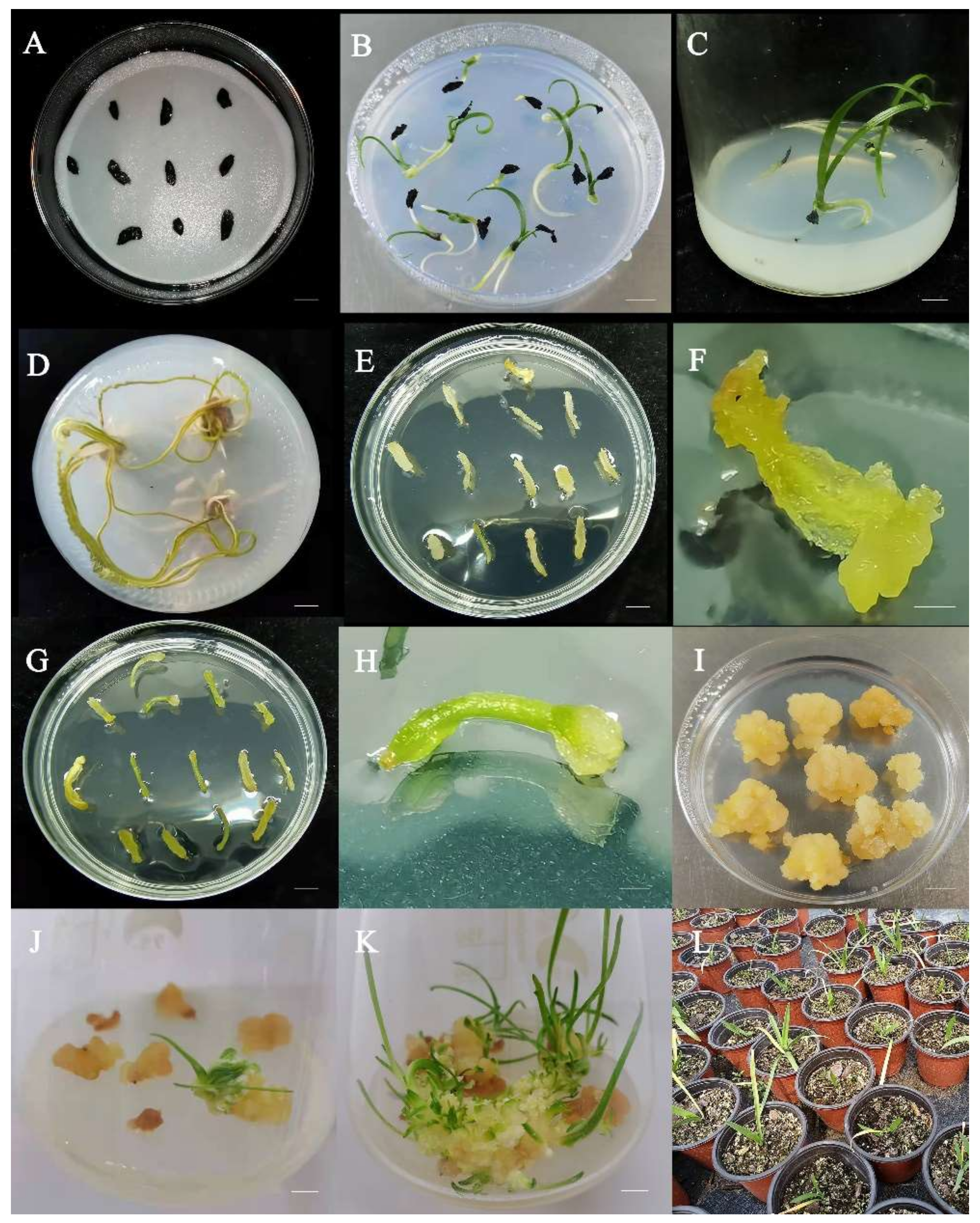
2.1. Plant Material Collection

In late September 2020, mature seeds of evergreen A. praecox ssp. Orientalis ‘Big Blue’ grown in the open field of Kunming city, Yunnan province, China, were collected. After natural air drying, one hundred seeds were randomly selected with 5 replications, and the average dry weight was calculated according to Chen [23]. The morphological indicators, for instance, the length and thickness of zygotic embryos and sizes of seeds, were measured to evaluate the seed quality. The selected seeds of good quality were used for subsequent sterilization and germination experiments.

2.2. Sterilization and Germination

Due to the large number of endophytes in the seeds of A. praecox, ensuring aseptic operations required complicated disinfection procedures according to the preliminary experiment. The selected A. praecox seeds had their seed wings removed first and were placed in 50 mL centrifuge tubes, then washed with sterile water twice, 20 min each to remove impurities, followed by 75% (v/v) alcohol for 1 min and were shaken continuously. These seeds were then rinsed with sterile water 3 times, and the surface was disinfected with 20% NaClO for 20 min and later rinsed with sterile water 6 times. Finally, sterilized filter paper was used to absorb the residual water off the seeds.
Referring to the method of Fan [1], the sterilized seeds were cultured on an MS medium supplemented with 30 g/L sucrose and 10 g/L agar (pH 5.8, the same below) for germination. Each dish (Φ 9.0 cm) was plated with 9~15 seeds and repeated 3 times. After about 30 days, the seeds germinated into seedlings with flourishing root systems and were ready for the explant collection for callus induction experiments.

2.3. Callus Induction from Root Explants

The roots (1.0~2.0 mm in diameter) of 60-day-old seedlings were directly cut into sections (0.8~1.5 cm in length). Two types of explants, with or without root tips (root segments), were picked and inoculated on callus induction media (CIM). CIM is an MS medium supplemented with 30 g/L sucrose, 3.0 g/L phytagel and three kinds of PGRs with different concentrations, shown in Table 1, designed by an orthogonal experiment.
The morphological change, contamination rate, browning rate and callus induction rate were recorded at 45 days and calculated using the following equation:
% Callus   induction = n N × 100
where “n” is the number of explants capable of inducing calluses and “N” is the total number of explants used in the experiment. The old leaves and residual roots were excised from the seedlings and then transferred onto the rooting medium (RM) with 2.17 g/L MS, 0.15 mg/L IBA, 30 g/L sucrose and 6 g/L agar. The light-green roots regenerated from the seedlings after 7 days, and the explants could be captured repeatedly for callus induction after 14 days.

2.4. Analysis of Endogenous Hormone Content during Callus Induction

The CIM was an MS medium supplemented with 3.0 mg/L PIC, 30 g/L sucrose and 3 g/L phytagel. Repeating the process at Section 2.3, at 0, 5, 10, 15 and 20 days, the root explants (not less than 0.2 g fresh weight (FW), including root tips and root segments respectively) were harvested and wrapped in the sealed foil, and then stored in a −20 °C refrigerator until the sampling at each time point was finished. All samples were taken out at the same time, thawed, and temporarily stored in a 2~8 °C refrigerator. Then, an appropriate amount of phosphate-buffered saline (PBS, pH 7.4) was added to each sample, fully homogenized, and centrifuged at 2000 rpm for 20 min. The supernatant was collected; the contents of IAA, ABA, GA4 and ZT were determined by an enzyme-linked immunosorbent assay (ELISA) according to the instructions of the kit (Shanghai Enzyme Link Biotechnology Co., Ltd., Shanghai, China)

2.5. Callus Proliferation

According to the methods described by Fan [1] and Iraq [24], 0.20 g of a callus cultured for about 45 days was transferred onto the callus proliferation media (CPM) conducted by an orthogonal experimental design, and the PGRs with three concentrations (1.00~2.00 mg/L PIC, 0~1.00 mg/L 6-BA, and 0.10~0.40 mg/L NAA) were added to the MS medium.
At 0, 5, 10, and 15 d cultured on the CPM, fresh samples of the callus were weighed for drawing proliferation curves in proliferation cultures. The proliferation rate was calculated using the equation below:
% Callus   proliferation = m M × 100
where ‘m’ is the total mass of the callus after being cultured at a sampling date point and ‘M’ is the total mass of the callus used at the beginning of the experiment. Meanwhile, using the acetocarmine method described by Yue et al. [25], the cytological observation of the callus at different states in proliferation cultures over 15 days was carried out.

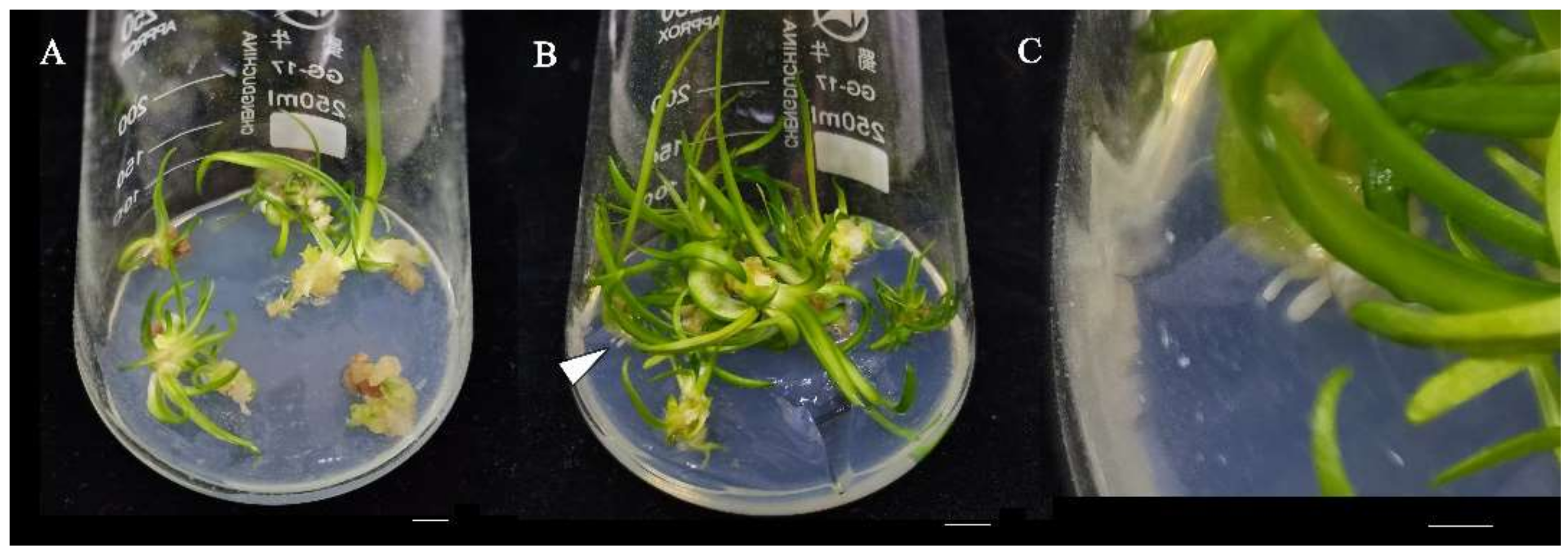
2.6. Adventitious Buds Induction and Regeneration

The callus induced from root explants was cultured on a proliferation medium (MS supplemented with 1.0 mg/L PIC, 1.0 mg/L 6-BA, and 0.4 mg/L NAA) for 5 subcultures. A well-proliferated callus (1.0 g in FW) was picked up and transferred onto the indirect organogenesis medium which contained MS supplemented with 1.5 mg/L 6-BA, 0.3 mg/L NAA, 30 g/L sucrose, and 10 g/L agar. The state and ratio of normal plantlets were observed and recorded after 25 days. The plantlets with well-developed roots were transplanted to the artificial substrates containing peat and vermiculite (v:v, 3:1) for acclimatization under natural light in the culture room. Survival rates of the plantlets were investigated after 30 days.

2.7. Culture Conditions

For callus induction and proliferation, an artificial incubator without light at 25 ± 2 °C and a relative humidity of 45 ± 5% was used. For adventitious buds and plantlet regenerations, the cultures were placed in a growth chamber under light for 14 h with an intensity of 2500~3000 Lx at 25 ± 2 °C, and the relative humidity for buds was 75 ± 10% and 35 ± 5% for plantlets.

2.8. Experimental Design and Statistical Analysis

All the treatments were repeated at least three times. Data were recorded using Excel 2019 (Microsoft Co., Redmond, WA, USA) to calculate the mean value, variance and standard deviation. Data processing, variance analysis (Duncan method, p < 0.05), multiple comparisons (LSD method, p < 0.05) and correlation charts were performed using SPSS 26.0 and GraphPad Prism 7.0.

3. Results

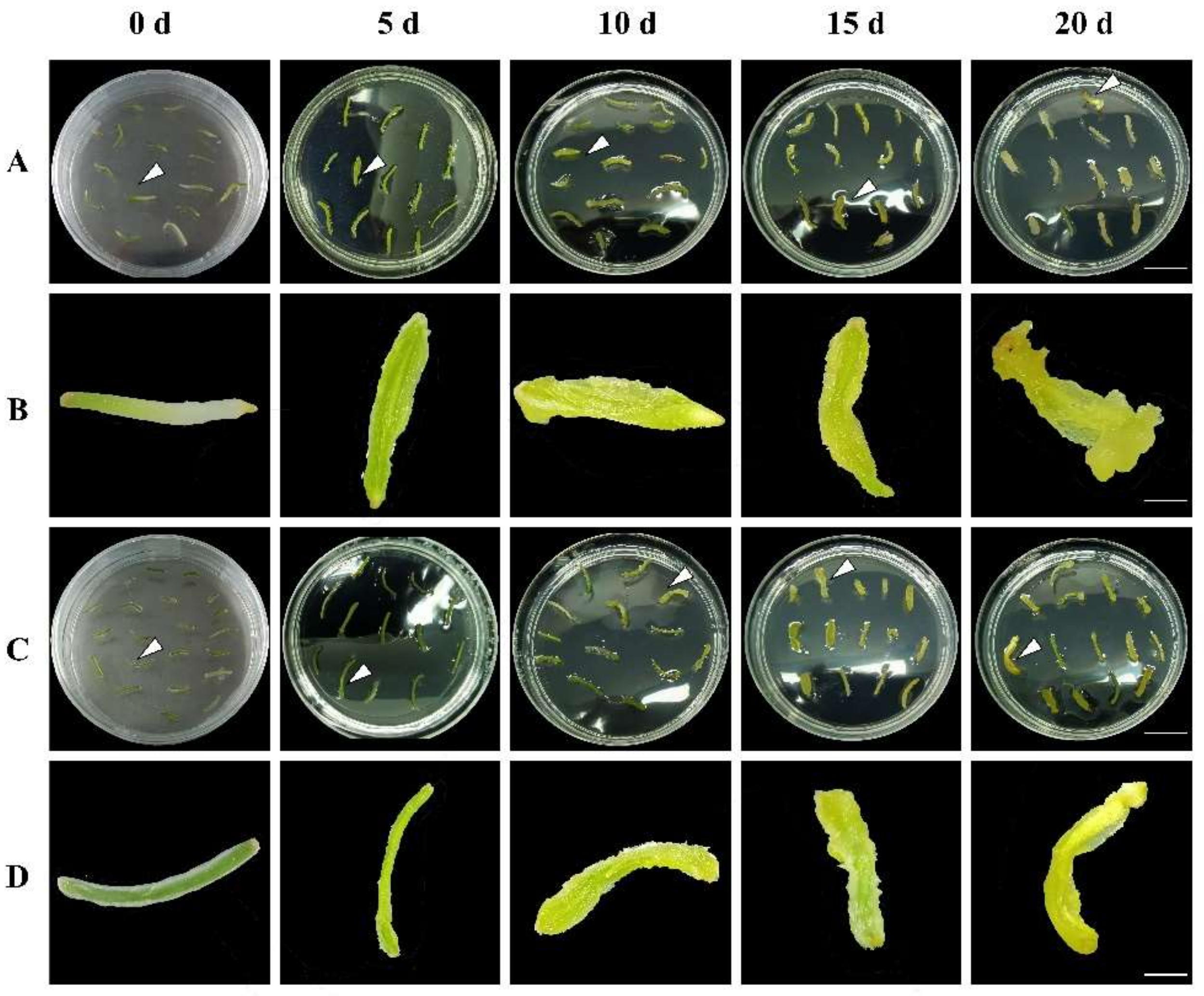
3.1. Effects of Three PGRs on Callus Induction Rate and Morphogenesis

Table 2 shows the effect of adding PIC, 2,4-D and TDZ to the CIM on the callus induction rates of A. praecox roots.
Except for the combination of 3.0 mg/L PIC and 2.0 mg/L 2,4-D, the callus induction rate with root tips as explants was higher than that of the root segment, among which the highest was 76.60%. Among the treatments using root segments as explants, the highest induction rate was only 36.03%, indicating that the root tip was more suitable for callus induction. The R-value shows that 2,4-D and TDZ on callus induction from the root tip and root segment were more effective than that of PIC, up to 32.46 and 26.81, respectively, and of which 2,4-D was the most potent hormone. The optimal PGRs combination was 1.0 mg/L PIC and 0.5 mg/L 2,4-D in an MS medium.
Figure 1 shows the morphogenesis of explant RTs and RMs under optimal culture conditions. It was obvious that the root tips began to swell after 7 days, and some root tips ruptured and dedifferentiated to form calluses at 14 days (Figure 1A,C); however, the morphological changes of the root segments were slightly delayed for 3~5 days (Figure 1B,D), demonstrating that root tips as explants can shorten the time of callus induction.

3.2. Optimization of Three PGRs Combination on Callus Induction from Root Tips

Using the root tip as the best explant material, the effects of different combinations of auxin (PIC, NAA) and cytokinin (KT) on the induction rate of A. praecox calluses were further explored, aiming to promote the induction process. The results were shown in Table 3 and Figure 2.
According to the induction rate, the most optimal PGRs combination was 2.0 mg/L PIC, 1.5 mg/L KT and 0.1 mg/L NAA in MS medium, and the callus induction rate was 100.00%. The induction effects of five treatments were found to be better than the optimized treatment (the highest induction rate was 76.60%) in Table 2. In the range analysis, the R-value of NAA was the largest, followed by KT, and PIC was the smallest, indicating that the effects of the three PGRs on the callus induction rate were in the order of NAA > KT > PIC.
Corresponding to the results in Section 3.1, the root tips began to expand after 7 days of inoculation on the medium, and gradually dedifferentiated into yellowish calluses (Figure 2A,B); when cultured for 20 days, it was obvious that some calluses directly differentiated to produce adventitious buds (Figure 2C).

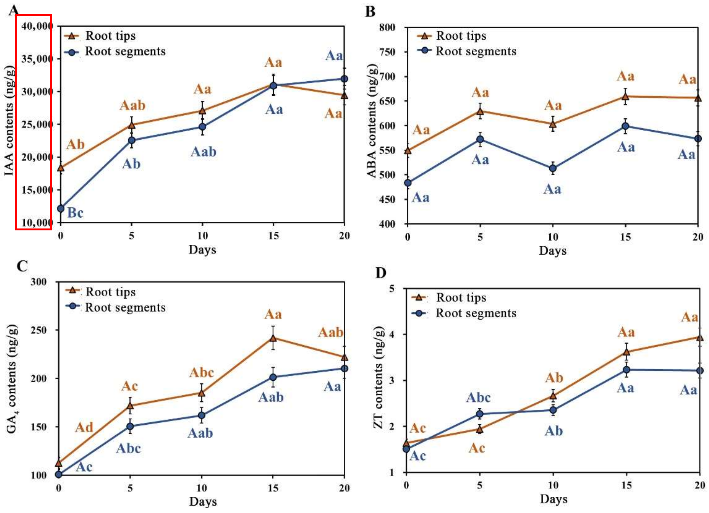
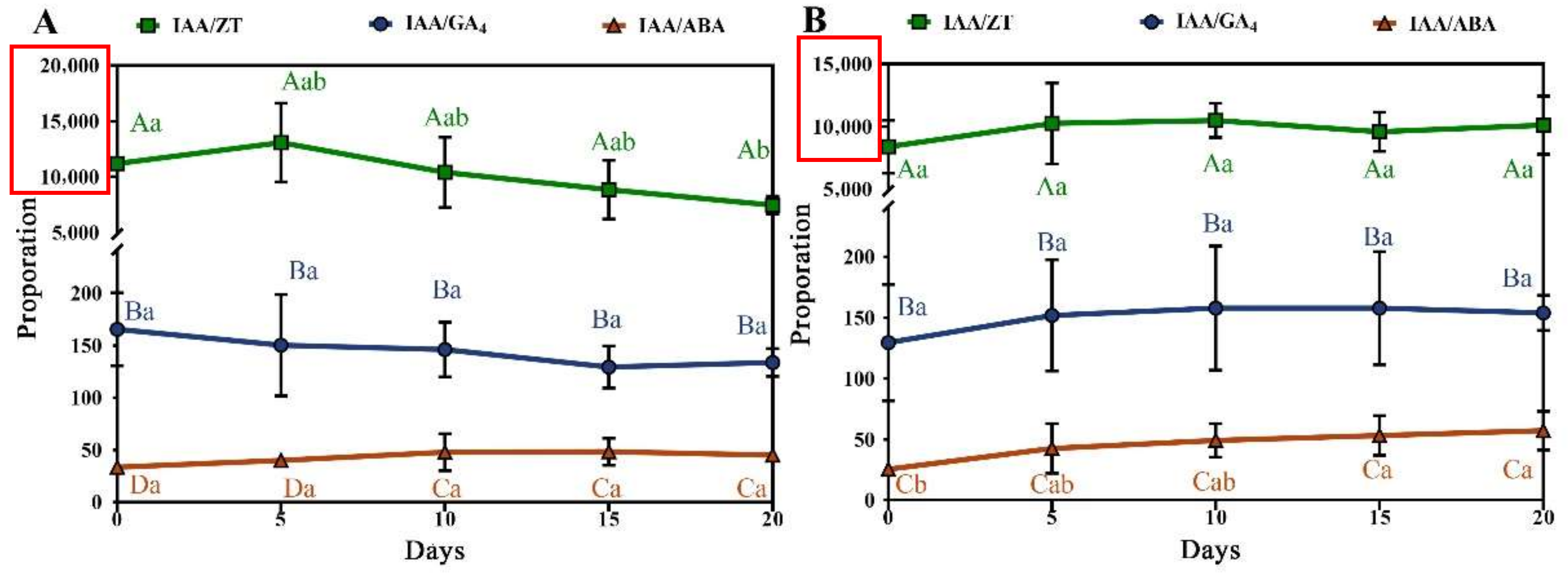
3.3. Role of Four Endogenous Hormones during Callus Induction

The contents of the four endogenous hormones (IAA, ABA, GA4 and ZT) in the explants (root tips and root segments, respectively) were investigated to reveal their functions during the primary induction process. Significant differences were found as shown in Figure 3, and the order from high to low was IAA > ABA > GA4 > ZT, in which the content of IAA was tens to hundreds of times that of ABA and GA4, and tens of thousands of times higher than that of ZT, indicating that IAA may play a crucial role in the process of callus induction.
Furthermore, the IAA content in the root tips of A. praecox was significantly higher than that in the root segments at the initial stage, continued to reach the highest level on the 15th day, and then decreased, but the IAA content in the root segments continued to increase in the entire induction process. On the 15th day, the IAA content was the same as that in the root tip and was higher on the 20th day than that in the root tip, which might be the main reason why the initiation of callus induction in the root segment was delayed. The ABA content had a wave-like change that increased and then decreased two times, but no significant difference between different induction times and two types of explants was observed. The contents of GA4 and ZT in root tips and root segments had an overall upward trend, but there were no significant differences in either.
Figure 4 shows that the ratio of IAA to the other three endogenous hormones contents in A. praecox root tips and root segments changed with the prolongation of induction time, and the IAA/ZT ratio in the root tips increased to a maximum value on the fifth day and then gradually decreased (Figure 4A). However, the proportional relationship between IAA to ZT, GA4 and ABA in the root tips and root segments of plantlets was not obvious (Figure 4B). In addition, IAA/GA4 had no significant difference in the root tip and root segment, similar to IAA/ABA, and tended to be stable in the root tip, while IAA/GA4 gradually increased in the root segment and stabilized after 15 days.

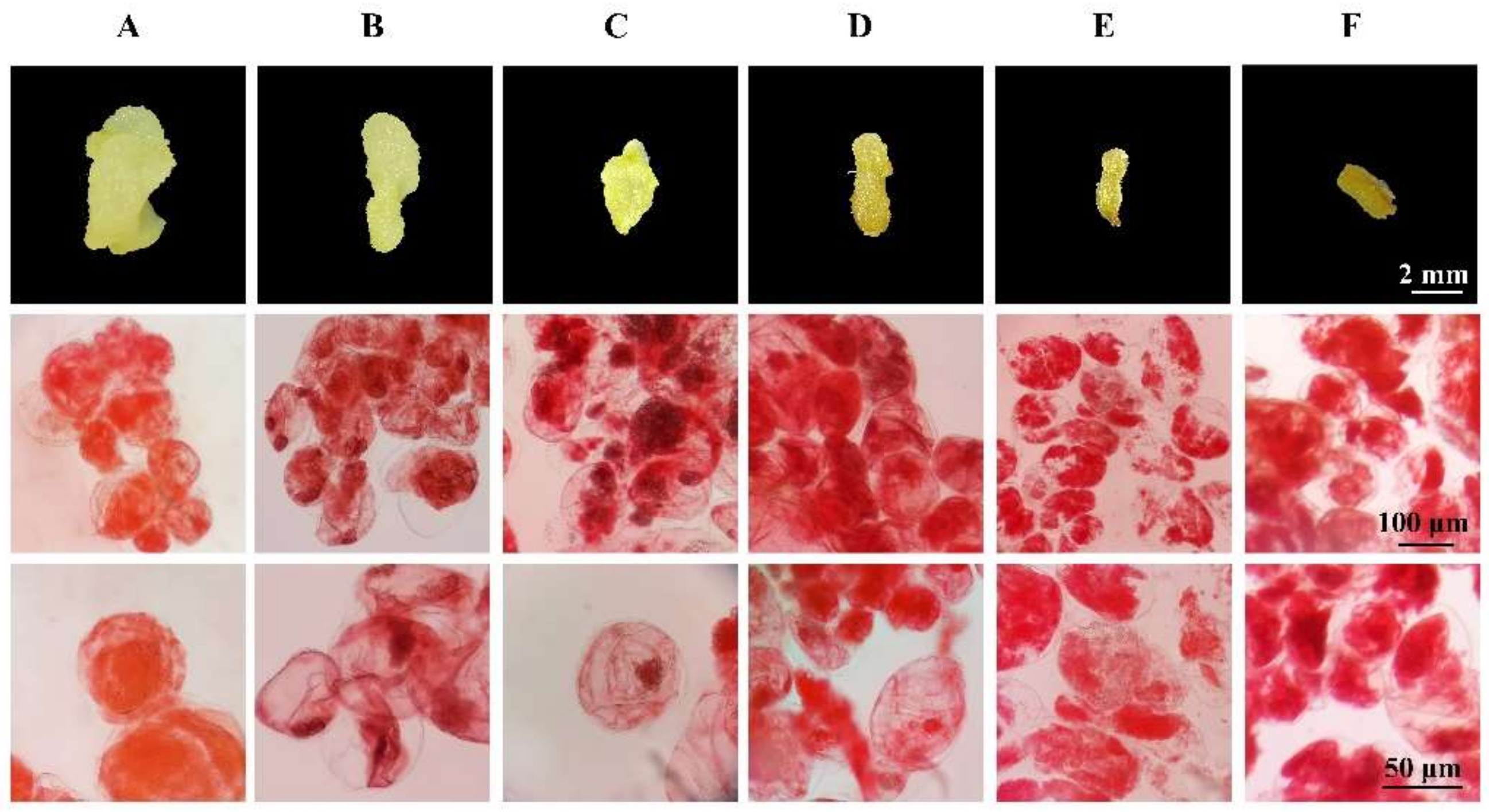
3.4. Effects of Three PGRs on Callus Proliferation Rate and State

There were differences between the proliferation effects of calluses among nine treatments containing three kinds of PGRs shown in Table 4. The maximum FW under the addition of 1.0 mg/L PIC, 1.0 mg/L 6-BA, and 0.4 mg/L NAA reached 345.0 mg, the highest proliferation rate of 72.74% after 15 days, proving to be the most stimulating treatment for proliferation. The R-value analysis shows that the impact of three PGRs in the callus proliferation was in the order of PIC > 6-BA > NAA.
There were also differences in the appearance of calluses cultured on proliferation media after 15 days (Figure 5). Milky white and soft callus cells are spherical, uniform in size and regularly arranged, with large and obvious nuclei, dense cytoplasm, and accumulation of starch granules and other nutrients (Figure 5A,B). The callus with a slightly yellowish and slightly compact structure has an obvious difference in cell size, the nucleus is relatively smaller, and the proportion of cytoplasm in the cell volume is increased (Figure 5C,D). The calluses with browning and indistinct differentiation are oblong, have a chaotic arrangement, a large volume difference, indistinct nuclei and a blurred nucleocytoplasmic boundary, and the cell contents can be observed as scattered in the intercellular space under a microscope, indicating cell death (Figure 5E,F). In general, the states of calluses shown in Figure 5A,B were optimal and suitable for subsequent differentiation experiments.

3.5. Hardening and Transplanting of Seedlings Dedifferentiated from Callus

After the callus was transferred to the organogenesis medium for 25 days, part of the callus differentiated into bright green adventitious buds (Figure 6A). These adventitious buds were then cultivated for 90 days, and the color of the upper part of the leaves changed from green to dark green, while the lower part was light green (Figure 6B), from the base of which some roots regenerated (Figure 6C).
The culture vessel was partially opened for 3 days for hardening, and the plantlet was transplanted to the substrates containing peat and vermiculite (v:v, 3:1) for acclimation. After 30 days, the survival rate of plantlets with a well-developed root system was as high as 100.00% (Figure 7).

4. Discussion

Agapanthus is a native species to South Africa, and its suitable temperature for growth is below 35 °C. Kunming city is located in the south west region of China, and its climate is similar to that of South Africa, which is more conducive to the growth and development of A. praecox.
A. praecox is a perennial herb flower belonging to a monocotyledonous plant with a well-developed root system, which leads us to a new idea of using its roots as explants for the development of tissue culture systems. Chen [26] firstly reported that the induction rates of Lilium spp. with different parts of roots (root tips, root segments without tips) as explants were different in an MS medium, and the induction rate with root tips as explants was as high as 96.67%, which was 987.40% higher than that without root tips (only 8.89%). Previous studies have revealed that auxin is biosynthesized in the root tip and maintains a gradient distribution, then is transported to other organs through polarity to stimulate the division, expansion and differentiation of somatic cells, thereby affecting callus formation. The relevant experimental evidence also indicated that the strongest auxin signals appeared in the root tip quiescent center (QC) using the green fluorescent protein (GFP) labeling method [27]. High callus induction of A. praecox in this study also proved the importance of the root tip’s presence in the root segment as an explant. The callus induction rate on the MS medium containing PIC, KT and NAA reached 100.00%, which is much higher than previous works of Ma (27.71%) [10] and Fan (87.51%) [1]. The expansion of the explant types and the optimization of the induction medium will provide strong technical support for in vitro propagation of Agapanthus plants.
The intrinsic genetics, developmental status of explants and the ratio of plant hormones are the key factors in the process of callus formation that determine the establishment of tissue culture protocol. Existing research data show the types and concentrations and combinations of endogenous PGRs influence the callus induction frequency and callus morphogenesis [28]. In the process of culturing the roots and stem segments of Centaurium erythraea, IAA content is higher than that of cytokinins in the roots, and it is also twice the IAA contents in the stem [29]. These PGRs may significantly promote the expansion of dividing cells in explants and callus initiation. In agreement with the conclusions above, our present study found that IAA content was significantly higher than that of ABA, GA4 and ZT in the root tips of A. praecox, and the contents of four endogenous hormones in the root tips were higher than those in the root segments. In addition, the peak of IAA and GA4 contents in A. praecox root explants appeared on the 15th day, coinciding with morphological changes of the explants, which were consistent with Fu et al. [30]. Further observations confirmed that about two weeks after the initiation of induction culture was the critical point for the morphological changes of explants and the formation of calluses. Similar results were reported by Zayed et al., who found that in immature female inflorescence explants of Phoenix dactylifera, the contents of IAA and GA3 reached the highest value at the beginning of callus formation and then gradually decreased; the subsequent peaks of ZT, IAA and ABA contents appeared after an embryogenic callus was initiated and developed [31].
The ratio of endogenous hormone contents also affects callus induction. A higher ratio of auxin to cytokinins was beneficial to improving the callus initiation rate in rice (Oryza sativa) anther cultures [32]. Zhang et al. [33] found that the ratio of IAA/ABA in the young leaves of Sopatholobus suberechtu increased rapidly on the 14th day of induction, regulating the formation of calluses. In the process of callus induction of A. praecox root tips and root segments, the ratio of endogenous hormones also has a similar change pattern with the prolongation of culture time, that is, when the ratio of GA4/ZT, IAA/ABA is increased, the root tips expanded exceedingly. These results suggest that endogenous hormones play an important role in the dedifferentiation of plant explants to form calluses and exogenous growth regulators should be rationally optimized according to their changing patterns so that the levels of endogenous and exogenous hormones reach a moderate dynamic balance to promote callus formation.
Exogenous additives have a crucial effect on the proliferation of calluses. Previous studies have shown that PIC is a commonly used hormone for callus induction and proliferation culture of A. praecox and other monocotyledonous plants, but the optimal concentration of PIC in various types of explants for callus induction was significantly different. The A. praecox embryonic callus (EC) cultured on the medium containing 1.5 mg/L PIC had uniform morphology and strong embryonic potential in Zou’s research [22]. Yue et al. [25] studied the effect of different concentrations of 6-BA on the proliferation of calluses from A. praecox florets and found that the callus under 1.0 mg/L 6-BA was significantly larger than other treatments. Li et al. [34] subcultured the filamentous callus of Lilium brownie ‘Manissa’ and found that the proliferation was faster when the NAA concentration was higher (0.5 mg/L), which was consistent with the optimal PGRs combination for root callus proliferation in this study (1.0 mg/L PIC, 1.0 mg/L 6-BA, and 0.4 mg/L NAA).

5. Conclusions

Current A. praecox tissue cultures often use small pedicels and young leaves as explants. However, the collection time of small pedicels is limited, and secondary metabolites in leaves with a high degree of differentiation can easily cause browning of explants and calluses, thus reducing the callus induction rate. This study established an in vitro propagation system of A. praecox through indirect organogenesis using its roots as explants. Sterile roots are easy to get and are available without the limitations of collecting seasons, plant growth and development. Abundant calluses and adventitious buds derived from the protocol provide sufficient materials for A. praecox micropropagation, somatic embryogenesis research and further genetic improvement in the new varieties.

6. Patents

The results of this paper have applied for a patent (CN 114532227 A).

Author Contributions

Conceptualization, G.C. and X.S.; methodology, X.G. and Q.T.; software, X.G. and Q.L.; formal analysis, X.G.; investigation, X.G. and H.S.; resources, H.S.; data curation, Q.T.; writing—original draft preparation, Q.T.; writing—review and editing, Y.Z., W.W., G.C. and X.S.; visualization, X.G. and Q.T.; supervision, X.S.; project administration, G.C.; funding acquisition, X.S. All authors have read and agreed to the published version of the manuscript.

Funding

This research was funded by National Natural Science Foundation of China (grant number 31901351 and 31370695) and Science and Technology Commission of Shanghai Municipality (grant number 22ZR1428600).

Institutional Review Board Statement

Not applicable.

Informed Consent Statement

Not applicable.

Data Availability Statement

Not applicable.

Acknowledgments

The authors extend their deep appreciation to Yunwei Zi from Yunnan Lan Rong Landscape Engineering Co., Ltd. for providing seeds of Agapanthus praecox subsp. orientalis ‘Big Blue’.

Conflicts of Interest

The authors declare no conflict of interest.

References

  1. Fan, X. Micropropagation In Vitro of Agapanthus praecox ssp. Orientalis ‘Big Blue’; Shanghai Jiao Tong University: Shanghai, China, 2009; pp. 31–45. [Google Scholar]
  2. Zurita, F.; Belmont, M.A.; De Anda, J.; White, J.R. Seeking a way to promote the use of constructed wetlands for domestic wastewater treatment in developing countries. Water Sci. Technol. 2011, 63, 654–659. [Google Scholar] [CrossRef] [PubMed]
  3. Calheiros, C.; Bessa, V.S.; Mesquita, R.; Brix, H.; Rangel, A.; Castro, P. Constructed wetland with a polyculture of ornamental plants for wastewater treatment at a rural tourism facility. Ecol. Eng. 2015, 79, 1–7. [Google Scholar] [CrossRef]
  4. Mor, Y.; Halevy, A.H.; Kofranek, A.M.; Reid, M.S. Post-harvest handling of lily of the Nile flowers. J. Am. Soc. Hortic. Sci. 1984, 109, 494–497. [Google Scholar]
  5. Chen, X.; Lu, L.; Qian, Y.; Fan, Y. Recent advances in germplasm and landscape application of Agapanthus spp. Chin. Landsc. Archit. 2016, 32, 99–105. [Google Scholar]
  6. Snoeijer, W. Agapanthus: A Revision of the Genus; Timber Press: Portland, OR, USA, 2004. [Google Scholar]
  7. Supaibulwatana, K.; Mii, M. Organogenesis and somatic embryogenesis from young flower buds of Agapanthus africanus Hoffmanns. Plant Biotechnol. 1997, 14, 23–28. [Google Scholar] [CrossRef]
  8. Suzuki, S.; Nakano, M. Agrobacterium-mediated transformation in Liliaceous ornamental plants. Jpn. Agric. Res. Q. 2002, 36, 119–127. [Google Scholar] [CrossRef]
  9. Nakano, M.; Tanaka, S.; Oota, M.; Ookawa, E.; Suzuki, S.; Saito, H. Regeneration of diploid and tetraploid plants from calluse-derived protoplast of Agapanthus praecox ssp. orientalis (Leighton) Leighton. Plant Cell Tissue Organ Cult. 2003, 72, 63–69. [Google Scholar] [CrossRef]
  10. Ma, K. Establishment of Regeneration System Cultured In Vitro of Agapanthus praecox ssp. Orientalis ‘Big Blue’; Northeast Forestry University: Harbin, China, 2007; pp. 24–26. [Google Scholar]
  11. Yaacob, J.S.; Syafawati, J. Tissue Culture, Cellular Behaviour and Pigment Analysis of Agapanthus praecox ssp. minimus; University of Malaya: Kuala Lumpur, Malaysia, 2013. [Google Scholar]
  12. Kang, L. Preliminary report on the experiment of regeneration system of Agpanthus praecox ssp. minimus ‘Storms River’. Contemp. Hortic. 2009, 12, 7–8 + 25. [Google Scholar]
  13. Hu, Z.; He, Y. Study on tissue culture and plantlet regeneration of Agapanthus praecox ssp. orientalis ‘Big Blue’. North. Hortic. 2011, 10, 118–120. [Google Scholar]
  14. Suzuki, S.; Oota, M.; Nakano, M. Embryogenic callus induction from leaf explants of the Liliaceous ornamental plant, Agapanthus praecox ssp. orientalis (Leighton) Leighton—Histological study and response to selective agents. Sci. Hortic. 2002, 95, 123–132. [Google Scholar] [CrossRef]
  15. Suzuki, S.; Supaibulwatana, K.; Mii, M.; Nakano, M. Production of transgenic plants of the Liliaceous ornamental plant Agapanthus praecox ssp. orientalis (Leighton) Leighton via Agrobacterium-mediated transformation of embryogenic calli. Plant Sci. 2001, 161, 89–97. [Google Scholar] [CrossRef]
  16. Yue, J.; Dong, Y.; Wang, X.; Sun, P.; Wang, S.; Zhang, X.; Zhang, Y. A regeneration system for organogenesis and somatic embryogenesis using leaves of Agapanthus praecox as explants. Chin. Bull. Bot. 2020, 55, 588–595. [Google Scholar]
  17. Wang, Y.; Fan, X.; Wang, G.; Zhang, D.; Shen, X. Regeneration of Agapanthus praecox ssp. orientalis ‘Big Blue’ via somatic embryogenesis. Propag. Ornam. Plants 2012, 12, 148–154. [Google Scholar]
  18. Baskaran, P.; Staden, J.V. Rapid in vitro micropropagation of Agapanthus praecox. S. Afr. J. Bot. 2013, 86, 46–50. [Google Scholar] [CrossRef] [Green Version]
  19. Yaacob, J.S.; Taha, R.M.; Mohajer, S. Organogenesis induction and acclimatization of African blue Lily (Agapanthus praecox ssp. minimus). Int. J. Agric. Biol. 2014, 16, 57–64. [Google Scholar]
  20. Yaacob, J.S.; Saleh, A.; Elias, H.; Abdullah, S.; Mahmad, N.; Mohamed, N. In vitro regeneration and acclimatization protocols of selected ornamental plants (Agapanthus praecox, Justicia betonica and Celosia cristata). Sains Malays. 2014, 43, 715–722. [Google Scholar]
  21. Zou, M. Physiological and Biochemical Aspects of Picloram Regulates the Embryonic Potential of Callus in Agapanthus; Shanghai Jiao Tong University: Shanghai, China, 2015. [Google Scholar]
  22. Ren, N.; Zou, M.; Chen, G.; Shen, X. Difference analysis of redox levels in the callus of Agapanthus praecox ssp. orientalis with different embryonic ability. Mol. Plant Breed. 2022, 20, 1281–1288. [Google Scholar]
  23. Chen, X.; Lv, X.; Yin, L.; Feng, J.; Zhang, D. Comparison with morphology and germination of Agapanthus praecox seeds under different cultivation region and environment. Acta Agric. Shanghai 2020, 4, 43–47. [Google Scholar]
  24. Iraqi, D.; Francine, M. Analysis of carbohydrate metabolism enzymes and cellular contents of sugars and proteins during spruce somatic embryogenesis suggests a regulatory role of exogenous sucrose in embryo development. J. Exp. Bot. 2001, 52, 2301–2311. [Google Scholar] [CrossRef] [Green Version]
  25. Yue, J.; Wei, Z.; Li, P.; Wang, Z. Influence on callus induction and physiological characters of callus proliferation in Agapanthus praecox ssp. orientalis by carbon sources. Acta Bot. Boreali Occident. Sin. 2021, 41, 439–449. [Google Scholar]
  26. Chen, M.; Zhang, J.; Zhou, Y.; Gu, J.; Shen, Q.; Zhang, P. Study on factors influence somatic embryogenesis fromoots of Lilium spp. J. Nucl. Agric. Sci. 2015, 29, 1494–1501. [Google Scholar]
  27. Tian, H.; Niu, T.; Yu, Q.; Quan, T.; Ding, Z. Auxin gradient is crucial for the maintenance of root distal stem cell identity in Arabidopsis. Plant Signal. Behav. 2013, 8, e26429. [Google Scholar] [CrossRef] [PubMed] [Green Version]
  28. Zhao, L.; Wang, X.; Zhang, J.; Wang, S. Plant tissue culture and its research advances and application in the forage plants. Pratacult Sci. 2011, 28, 1140–1148. [Google Scholar]
  29. Trifunović-Momčilov, M.; Motyka, V.; Dragićević, I.Č.; Petrić, M.; Jevremović, S.; Malbeck, J.; Holík, J.; Dobrev, P.I.; Subotić, A. Endogenous phytohormones in spontaneously regenerated Centaurium erythraea Rafn. plants grown in vitro. J. Plant Growth Regul. 2016, 35, 543–552. [Google Scholar] [CrossRef]
  30. Fu, F.; Feng, Z.; Qu, B.; Li, W. Relatianships between induction rate of embryonic callus and endogenous hormones content in Maize. J. Nucl. Agric. Sci. 2006, 1, 10–14. [Google Scholar]
  31. Zayed, E.M.M.; Abd Elbar, O.H. Morphogenesis of immature female inflorescences of date palm in vitro. Ann. Agric. Sci. 2015, 60, 113–120. [Google Scholar] [CrossRef] [Green Version]
  32. Wang, X.; Shi, X.; Wu, X.; Wang, X.; Zhou, K. The influence of endogenous hormones on culture capability of different explants in rice. Sci. Agric. Sin. 2004, 12, 1819–1823. [Google Scholar]
  33. Zhang, Y.; He, g.; Rong, G.; Liu, X.; Ni, S. Changes of endogenous hormones during the culture of callus from Sopatholobus suberechtus. Biotechnol. Bull. 2017, 33, 66–70. [Google Scholar]
  34. Li, Y. Callus induction and plant regeneration from floral organ of Lilium ‘Manissa’. Chin. J. Trop. Crops 2013, 34, 1507–1512. [Google Scholar]
Figure 1. Morphological changes of the root tips and segments of A. praecox during callus induction. (A) Root tip. Bars = 1.0 cm; (B) Explants indicated by arrows in (A). Bars = 0.3 cm; (C) Root segments. Bars = 1.0 cm; (D) Explants indicated by arrows in (C). Bars = 0.3 cm.
Horticulturae 08 00715 g001
Figure 2. Callus and adventitious buds from root tips of A. praecox induced by the combination of hormones PIC, NAA and KT for 20 days. (A) The yellowish callus. Bars = 1 cm; (B) callus collected, respectively, from the explants indicated by arrows in A. Bars = 0.2 cm; (C) Adventitious bud. Bars = 0.2 cm.
Horticulturae 08 00715 g002
Figure 3. The dynamic changes of endogenous hormones in the root tips and segments of A. praecox during callus induction. (A) IAA; (B) ABA; (C) GA4; (D) ZT. The different letters indicate significant differences at 5% level by Duncan’s range test.
Horticulturae 08 00715 g003
Figure 4. The changes of endogenous hormone proportion in A. praecox root tips and segments during callus induction. (A): Root tip; (B): Root segments. The different letters indicate significant differences at 5% level by Duncan’s range test.
Horticulturae 08 00715 g004
Figure 5. Morphological differences of calluses from A. praecox roots after 15 days of culture. (A,B) Creamy-white callus; (C,D) Yellowish callus; (E,F) Yellow-brown callus.
Horticulturae 08 00715 g005
Figure 6. Plantlets regeneration from calluses of A. praecox root tips after 25 days and 90 days in organogenesis medium. (A) Adventitious bud after 25 days of differentiation. Bars = 1 cm; (B) plantlets after 90 days of differentiation. Bars = 1 cm; (C) adventitious root indicated by arrows in (B). Bars = 0.2 cm.
Horticulturae 08 00715 g006
Figure 7. Indirect organogenesis tissue culture system of A. praecox root explants. (A) Germinated seeds after inoculation into MS medium; (B,C) Seedlings after germination for 20 days (B) and 40 days (C); (D) Root system of 40-day-old seedlings after germination; (E,F) 7 days after inoculated on the induction medium, most of the root tips began to swell; (G,H) 7 days after inoculated on the induction medium, fewer of root segments began to swell; (I) Callus induced from root tips inoculated on the proliferation medium; (J,K) Adventitious buds produced through dedifferentiation from the callus and well-developed seedlings on the indirect organogenesis medium for 25 days (J) and 90 days (K); (L) Seedlings after hardening and transplanting on the mixed culture medium for 30 days. (AE,G,IK) Bars = 1 cm; (F,H) Bars = 0.2 cm.
Horticulturae 08 00715 g007
Table 1. CIM design for callus induction from root explants of A. praecox seedlings.
Explant TypesConcentration (mg/L)
PIC 1TDZ 22,4-D 3KT 4NAA 5
root tips,
root segments
1.000.000.50//
1.000.401.00//
1.000.602.00//
2.000.001.00//
2.000.402.00//
2.000.600.50//
3.000.002.00//
3.000.400.50//
3.000.601.00//
root tips1.00//0.000.10
1.00//1.000.50
1.00//1.501.00
2.00//0.000.50
2.00//1.001.00
2.00//1.500.10
3.00//0.001.00
3.00//1.000.10
3.00//1.500.50
1 Picloram, bought from Sigma-Aldrich (suitable for plant cell culture). 2 Thiadiazolone, bought from Sangon Biotech (≥95.0%). 3 2,4-Dichlorophenoxyacetic acid, bought from Sigma-Aldrich (≥95%, crystalline). 4 Kinetin, bought from Sangon Biotech (≥98.0%). 5 Naphthalene acetic acid, bought from Shanghai Yuhan Biotech (≥99.5%). The same below.
Table 2. Effects of combination of PIC, TDZ and 2,4-D on callus induction from A. praecox roots.
PIC Concentration (mg/L)TDZ Concentration (mg/L)2,4-D Concentration (mg/L)Induction Rate (%)
RT 1RS 2RTRSRTRSRTRS
1.000.000.5076.6025.57
1.000.401.0033.3311.45
1.000.602.0026.1923.98
2.000.001.0048.5326.40
2.000.402.0020.8325.94
2.000.600.5054.0211.56
3.000.002.0025.4936.03
3.000.400.5039.2923.04
3.000.601.0038.1016.92
k1 345.38 a *17.93 b50.21 a32.77 a56.64 a19.77 a
k2 441.13 a21.82 a31.15 b5.96 b39.99 b16.48 a
k3 534.29 a6.88 c39.43 ab7.89 b24.17 b10.37 b
R 611.0914.9419.0626.8132.469.40
1 Root tips. 2 Root segments. 3 The sum of the numerical values of the test indicators corresponding to the lowest level of concentrations. 4 The sum of the numerical values of the test indicators corresponding to the medium level of concentrations. 5 The sum of the numerical values of the test indicators corresponding to the highest level of concentrations. 6 Range, indicating the magnitude of the factor’s influence on the result. * Means followed by the same letters are not significantly different according to Duncan’s range test at p < 0.05, the same below.
Table 3. Effects of combination of PIC, NAA and KT on callus induction from A. praecox root tips.
PIC Concentration (mg/L)KT Concentration (mg/L)NAA Concentration (mg/L)Induction Rate (%)
1.000.000.1088.89
1.001.000.5061.11
1.001.501.0083.33
2.000.000.5072.22
2.001.001.0066.67
2.001.500.10100.00
3.000.001.0088.89
3.001.000.1088.89
3.001.500.5065.08
k1 177.78 a72.22 a92.59 a
k2 279.63 a82.80 a66.14 b
k3 380.95 a83.33 a79.63 ab
R 43.1711.1126.46
1 The sum of the numerical values of the test indicators corresponding to the lowest level of concentrations. 2 The sum of the numerical values of the test indicators corresponding to the medium level of concentrations. 3 The sum of the numerical values of the test indicators corresponding to the highest level of concentrations. 4 Range, indicating the magnitude of the factor’s influence on the result. Means of k1, k2 and k3 followed by the same letter within a row between different level of harmones are not significantly different at p ≤ 0.05. The same below.
Table 4. Effects of different hormone combinations on proliferation of callus induced from A. praecox root explants.
PIC Concentration (mg/L)6-BA 1 Concentration (mg/L)NAA Concentration (mg/L)Proliferation Rate (%)
1.000.000.1031.69
1.000.400.2043.45
1.001.000.4072.74
1.500.000.2035.43
1.500.400.4023.13
1.501.000.1031.92
2.000.000.4014.87
2.000.400.1043.60
2.001.000.2031.69
k1 20.493 a0.273 b0.357 a
k2 30.302 b0.367 ab0.363 a
k3 40.295 b0.449 a0.369 a
R 50.1970.1760.012
1 6-benzylaminopurine, bought from Sigma-Aldrich (suitable for plant cell culture). 2 The sum of the numerical values of the test indicators corresponding to the lowest level of concentrations. 3 The sum of the numerical values of the test indicators corresponding to the medium level of concentrations. 4 The sum of the numerical values of the test indicators corresponding to the highest level of concentrations. 5 Range, indicating the magnitude of the factor’s influence on the result.
Publisher’s Note: MDPI stays neutral with regard to jurisdictional claims in published maps and institutional affiliations.

