Article

Changes in Carotenoid Concentration and Expression of Carotenoid Biosynthesis Genes in Daucus carota Taproots in Response to Increased Salinity

1
State Key Laboratory of Crop Genetics and Germplasm Enhancement, Nanjing Agricultural University, Nanjing 210095, China
2
Jiangsu Modern Horticultural Engineering Technology Center, Jiangsu Vocational College of Agriculture and Forestry, Zhenjiang 212400, China
*
Author to whom correspondence should be addressed.
These authors contributed equally to this work.
Horticulturae 2022, 8(7), 650; https://doi.org/10.3390/horticulturae8070650
Submission received: 11 June 2022 / Revised: 10 July 2022 / Accepted: 13 July 2022 / Published: 17 July 2022

Abstract

:
Studying the changes of carotenoids in the taproot of carrots under salt treatment is helpful to probe the salt stress response mechanism of carrots. The carotenoid concentration and the expression profiles of 10 carotenoid-related genes were determined in two carrot cultivars with different taproot colors. Under salt stress, the biosynthesis of carotenoids in the taproot of both ‘KRD’ and ‘BHJS’ was activated. RT-qPCR manifested that the expression levels of DcPSY1, DcPSY2, DcZDS1, DcCRT1 and DcCRT2 increased significantly in both ‘KRD’ and BHJS’ under salt stress, but DcCHXE transcripts decreased and DcPDS transcripts maintained a basal level compared to that of the control. In the taproot of ‘KRD’, the expression level of DcLCYB, DcLCYE and DcCHXB1 climbed dramatically. However, there was no significant change in the taproot of ‘BHJS’. The study showed that salt stress can stimulate the biosynthesis of carotenoids. The accumulation of lutein in the taproots of ‘KRD’ and ‘BHJS’ may be mainly attributed to the variation in DcLCYE and DcCHXB1 transcripts. The increase in β-carotene accumulation is speculated to increase salt tolerance.

1. Introduction

The carrot (Daucus carota L.) is the most representative root vegetable of the Apiaceae family, which is grown all over the world [1,2]. Carotenoid accumulation in carrot taproots is a complex regulatory process. The study of carotenoid metabolism and its response to stress in carrot taproots has important guiding significance for carrot production [3]. As a lipophilic molecule, carotenoids play a critical role in photosynthesis [4], photomorphogenesis [5] and plant development [6,7] in carrots. In the past decades, carotenoid biosynthesis pathways in many plants have been studied, such as tomato [8], pepper [9], citrus [10], watermelon [11], carrot [12,13] and celery [14].
Carrots are good model plants for carotenoid research since they are rich in different carotenoids thus their taproot’s color is various [15,16]. Initially, the root of a carrot was colorless ahead of domestication, and numerous breeding works brought up diverse varieties rich in carotenoids, such as anthocyanins, lycopene, α-carotene, lutein, and β-carotene [17]. Carotenoid profiles also experienced remarkable change during the growth period [18,19]. During the early stage, the root is thin and colorless. After two months, the roots begin to thicken and start accumulating carotenoids. Latterly, secondary root growth results in root enlargement and increasing production of carotenoids.
Carotenoids are mainly synthesized in plastids [20] and commenced by geranylgeranyl diphosphate (GGPP) in the methylerythritol 4-phosphate (MEP) pathway [21]. The C40 carotenoid phytoene is formed by condensation of two molecules of the C20 GGPP, produced from isopentenyl pyrophosphate (IPP) and dimethylallyl pyrophosphate (DMAPP), which is catalyzed by phytoene synthase (PSY) [22]. Phytoene is transformed into lycopene via a train of desaturation and isomerization [23]. At this time, linear carotene biosynthesis is completed, and then lycopene is cyclized by lycopene ε-cyclase (LCYE) and lycopene β-Cyclase (LCYB) to produce α-carotene and β-carotene [24]. α-carotene is typically modified to lutein by hydroxylation. β-carotene is converted into assorted volatiles and phytohormones (such as strigolactones and abscisic acid) catalyzed by cross-reactive carbohydrate determinants (CCDs) and 9-cis-epoxycarotenoid dioxygenases (NCEDs) produces [21,25].
Salt stress can exert numerous negative impacts on plants and provoke a serious reduction in crop growth and yield. If plants suffer from inordinate amounts of salt, this will lead to various metabolic perturbations, cutback in water potential (ψw), disordered membrane potential, weaken photosynthesis and diminish nitrogen assimilation [26,27,28]. However, plants have evolved many adaptative mechanisms in response to the condition of high salinity, which involves interacting physiological traits, biochemical or metabolic pathways, and molecular mechanisms [29]. Among them, carotenoids are proved to respond to salt stress [30,31]. Carrots are among the vegetable crops that have a very low salinity threshold [32] and are rich in carotenoids, but the specific salt response mechanism is not clear so far. Hence, it is necessary to investigate the dynamic changes and mechanisms of carotenoids under salt stress in carrots.
In this study, two carrot cultivars with different taproot colors were used as models. The concentration of lycopene, lutein, α-carotene, β-carotene and the total carotenoids, besides the expression of carotenoid biosynthesis-related genes were detected and analyzed. Under salt stress, the biosynthesis of carotenoids in the taproot of both two carrot cultivars was activated and dynamic changes in the carotenoid synthetic pathway were also observed. This study provided a potential theoretical basis for the mechanism of carotenoid biosynthesis response to salt stress in carrots.

2. Materials and Methods

2.1. Plant Materials, Growth Conditions and Stress Treatments

Two carrot cultivars were selected based on their different taproot colors: ‘Kurodagosun (KRD)’ (orange) and ‘Benhongjinshi (BHJS)’ (red). Carrot seeds were preserved in the State Key Laboratory of Crop Genetics and Germplasm Enhancement of Nanjing Agricultural University. The carrot seeds were germinated on filter paper for 5 days, and then transferred into containers (plastic, 32 cm in diameter and 25 cm in height) filled with organic substrate and vermiculite (1:1; v/v). Carrot seedlings were incubated in the greenhouse at Nanjing Agricultural University (25 °C, 14 h light period and 18 °C, 10 h dark period). Salt stress treatment began 45 days after sowing (DAS). Plants were subjected to salt stress by watering each container with 500 mL of NaCl solution (300 mmol·L−1) or distilled water (for control group). Four days after the first exposure, the treatment was repeated until nine rounds of stress treatment within the same time period. The roots of each carrot cultivar were sampled at 80 DAS and three biological repetitions which comprise a pool of different roots of the same carrot cultivar under the same treatment were arranged. The samples that were separated into fragments were frozen in liquid nitrogen immediately and ground to powder, then stored at −80 °C until further experiments.

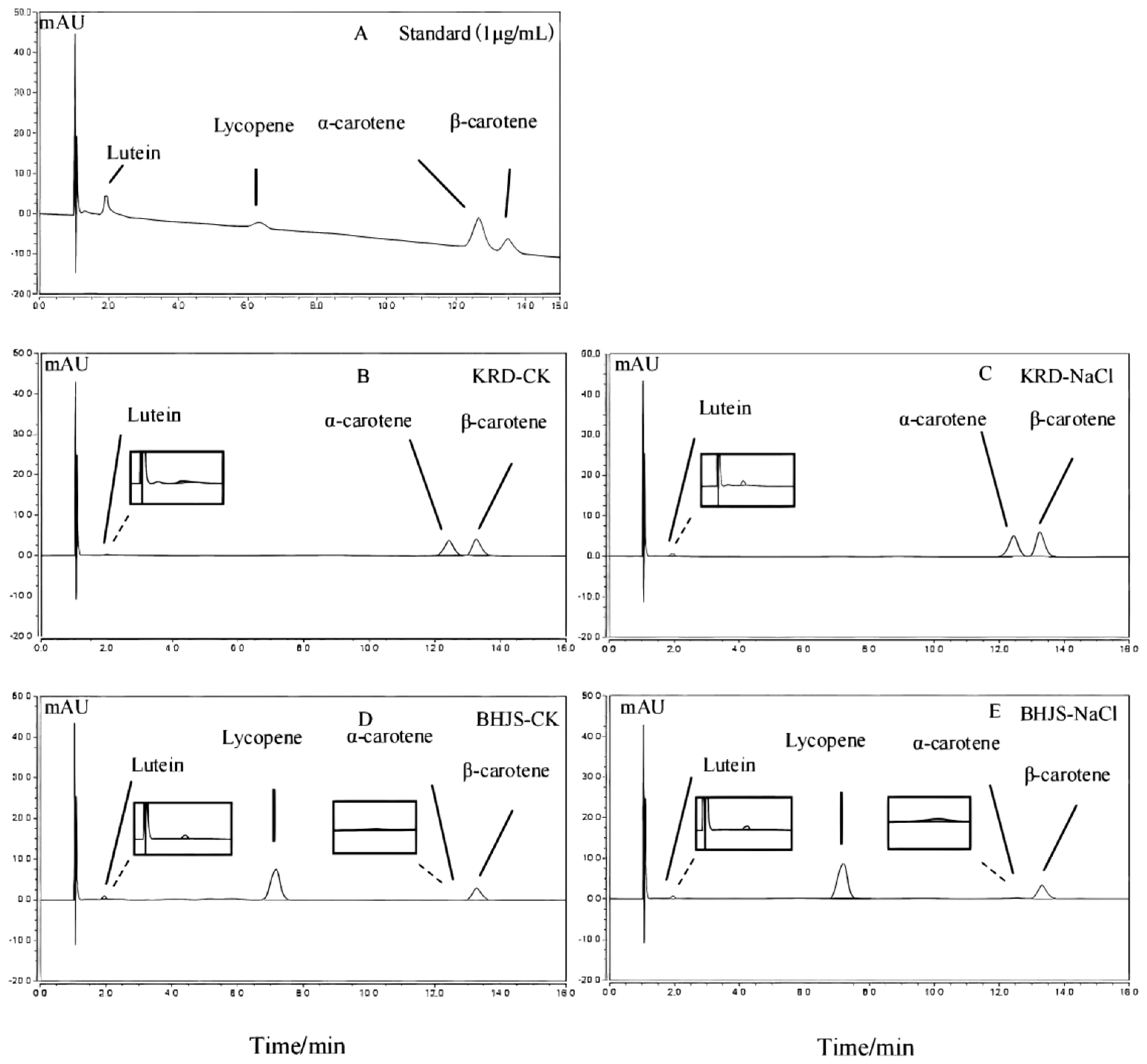
2.2. Determination of Carotenoids

Carotenoids were purified and quantified in accordance with the method of Ma et al. [33]. A total of 50 mg of vacuum freeze-dried powder from each sample was utilized for carotenoid extraction, which was performed by using 2 mL of acetone in a 50 °C water bath each time. Then, the combined extraction supernatants (2 mL) were filtered through 0.45 μm filters and analyzed by HPLC on a Shimadzu LC-20A HPLC System (Shimadzu, Kyoto, Japan), and three biological repetitions were set. Twenty microliters of supernatants were injected into a Hedera ODS-2 C18 analytical column (250 mm × 4.6 mm, 5 μm nominal particle size; Shimadzu) with mobile phase consisting of mixtures of methanol: acetonitrile (90:10, v/v). The flow rate was 1 mL·min−1, and elution was detected with a Shimadzu diode-array detector at 450 nm. All data were quantified on the basis of their standard curves. Standards were derived from Shanghai yuanye Bio-Technology (Shanghai, China). Three biological replicates were set for each assay.

2.3. Total RNA Isolation and Reverse Transcription

Total RNA of carrot was isolated using an RNA simple Total RNA Kit (Tiangen, Beijing, China) according to the instructions. Nanodrop ND-spectrophotometer (NanoDrop Technologies, Waltham, MA, USA) was used to detect the concentration of each RNA sample. Then, the total RNA (1 μg) was reverse transcribed into cDNA using Prime Script RT reagent Kit (Takara, Dalian, China).

2.4. Quantitative Real-Time PCR

Ten carotenoid biosynthesis-related genes (DcPSY1, DcPSY2, DcPDS, DcZDS1, DcCRT1, DcCRT2, DcLCYE, DcLCYB1, DcCHXE, DcCHXB) and reference gene DcActin were selected for gene expression analysis [34,35]. Primers were designed by Premier 6.0 and gene sequences were same as those reported in our previous work [33,34]. RT-qPCR analysis was performed according to the operating instructions of SYBR premix Ex Taq Kit (Takara, Dalian, China) and CFX96 PCR detection system (Bio Rad, CA, USA) with three technical replicates. RT-qPCR reactions were performed in 20 μL volume: SYBR Green I mix 10 μL, ddH2O 7.2 μL, cDNA template 2.0 μL. The forward and reverse primers were 0.4 μL (10 μmol/L) respectively. The gene expression amount is calculated according to the relative quantitative method. The gene expression was calculated by 2ΔΔCt method [36], and the gene expression level was analyzed and mapped using Microsoft Excel. The results were expressed as the mean of three independent biological replicates.

2.5. Data Analysis

SPSS version 25.0 was utilized to statistically analyze data based on one-way ANOVA. Duncan’s multiple range test at p < 0.05 was selected for significance test.

3. Results

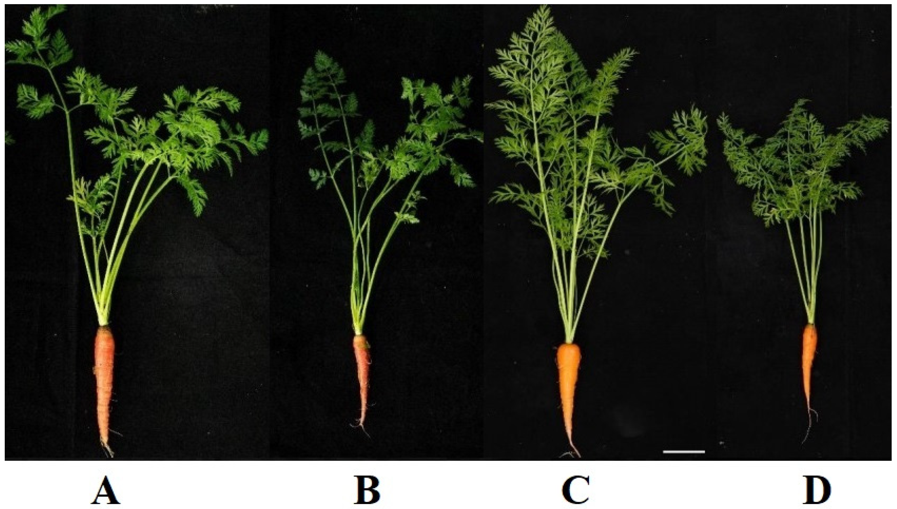
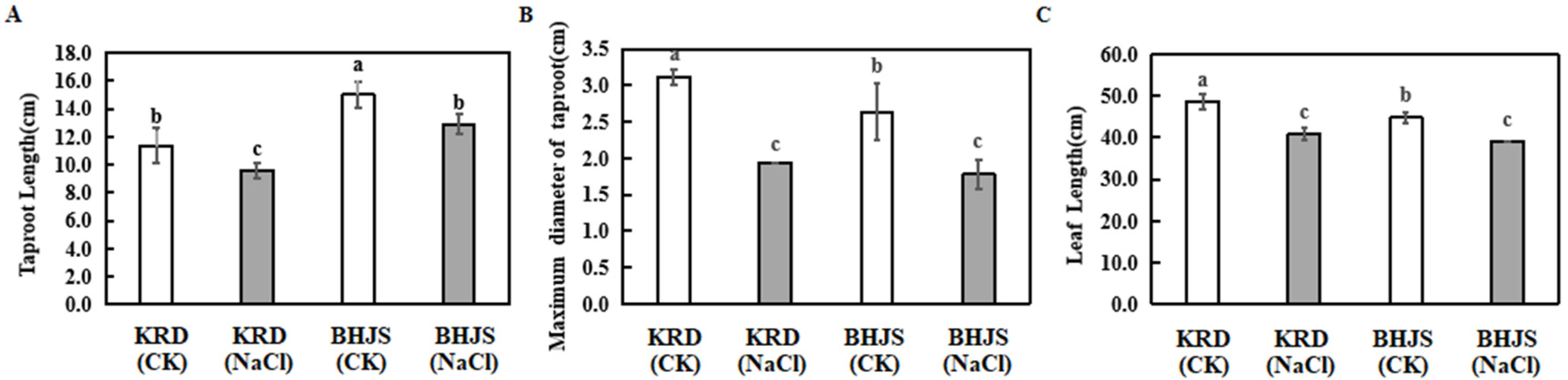
3.1. Growth Analysis of Two Carrot Cultivars under Different Treatments

At 80 DAS, the phenotypes of ‘KRD’ and BHJS’ under different treatments are shown in Figure 1, the taproots of ‘BHJS’ are red and the taproots of ‘KRD’ are orange. The color difference between ‘KRD’ and ‘BHJS’ is caused by the different carotenoid accumulation although the color difference is not very obvious due to the light. Previous studies have shown that high osmotic stress and high ionic stress caused by salt injury led to plant growth inhibition and root biomass reduction in carrots [37]. Consistently, the volume of taproots and the length of petioles of carrots decreased significantly, and there was no significant difference in the number of fibrous roots, petioles, taproots color and leaf color. The taproot length, maximum taproot diameter and leaf length of carrot were decreased due to salt stress. Under salt stress, the average taproot length of ‘KRD’ and ‘BHJS’ decreased by 15.79% and 14.00%, and the maximum diameter of the average taproot decreased by 38.71% and 30.77%, and the average leaf length decreased by 16.22% and 12.5%, respectively (Figure 2).

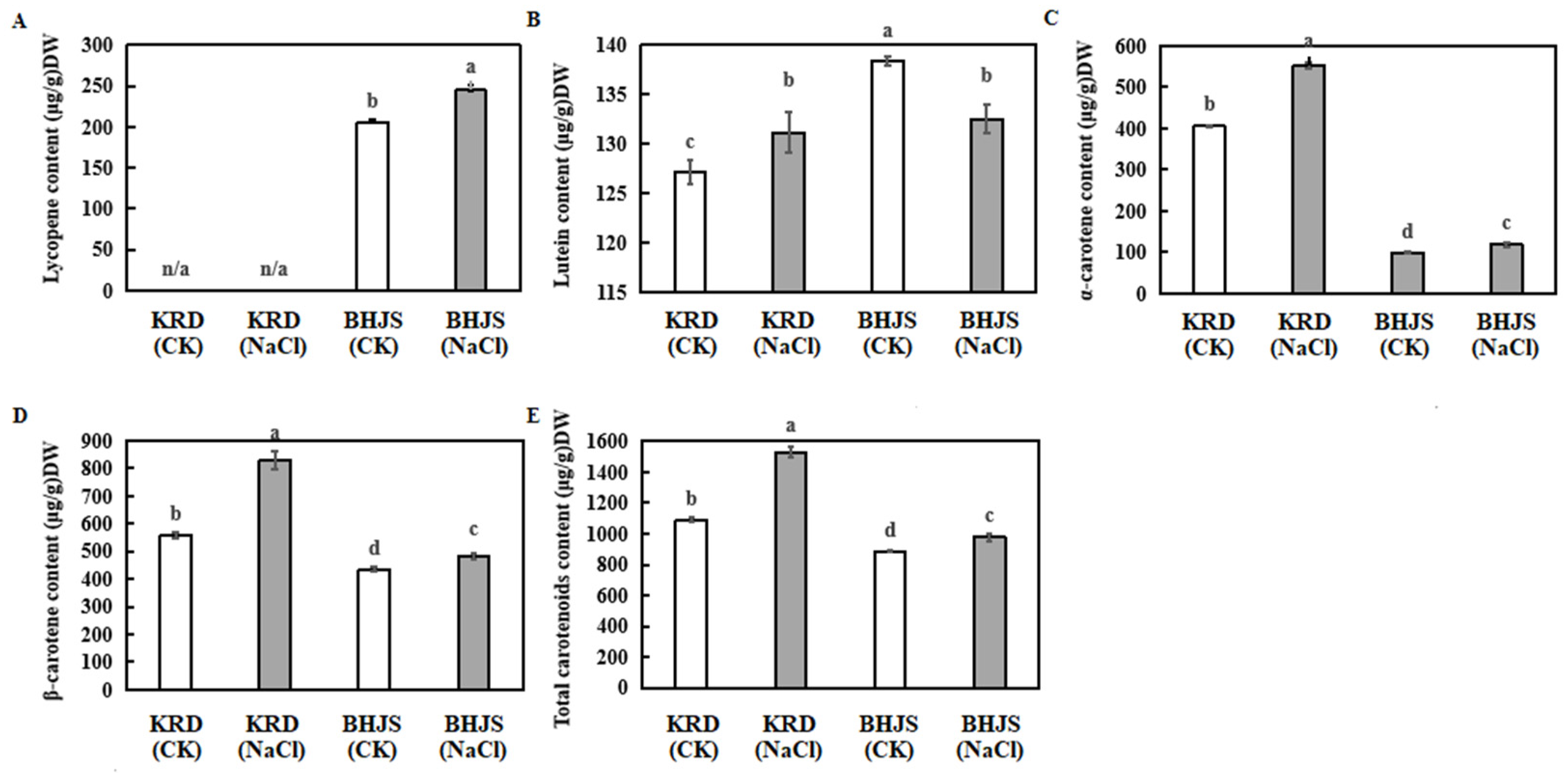
3.2. Effects of Salt Stress on Carotenoids Accumulation in Taproots of Carrot

The accumulation of various carotenoids in the taproots of the ‘KRD’ and ‘BHJS’ under 0 (control) and 300 mmol L−1 salt treatment are shown in Figure 3 and Figure 4. The total carotenoid concentration of ‘KRD’ and ‘BHJS’ under salt treatment was higher than the control. The changes in carotenoid concentration in the taproots under salt stress varied among different cultivars.
The concentration of lutein, α-carotene, β-carotene and total carotenoids in the taproots of ‘KRD’ under salt treatment increased significantly by 4.81%, 33.75%, 48.54% and 38.01%, respectively. The concentration of lycopene, α-carotene, β-carotene and total carotenoids in the taproots of ‘BHJS’ increased significantly by 19.13%, 20.91%, 10.47% and 11.52%, respectively, but the concentration of lutein decreased significantly by 3.29% compared to the control.
The accumulations of α-carotene, β-carotene and total carotenoids in the taproots of ‘KRD’ and ‘BHJS’ were activated under the salt stress treatment. Lycopene was barely detected in the taproot of ‘KRD’ in both the control and the salt treatment group, but a high concentration of lycopene was detected in the control group and salt treatment group of ‘BHJS’, 205.37 μg/g and 244.66 μg/g, respectively, reflecting the difference in lycopene concentration in the taproots of ‘KRD’ and ‘BHJS’. The lutein concentration in the taproots of ‘KRD’ and ‘BHJS’ increased and decreased significantly, respectively, under salt treatment (300 mmol·L−1 NaCl), reflecting the difference in the mechanism of lutein response to salt stress between ‘KRD’ and ‘BHJS’.

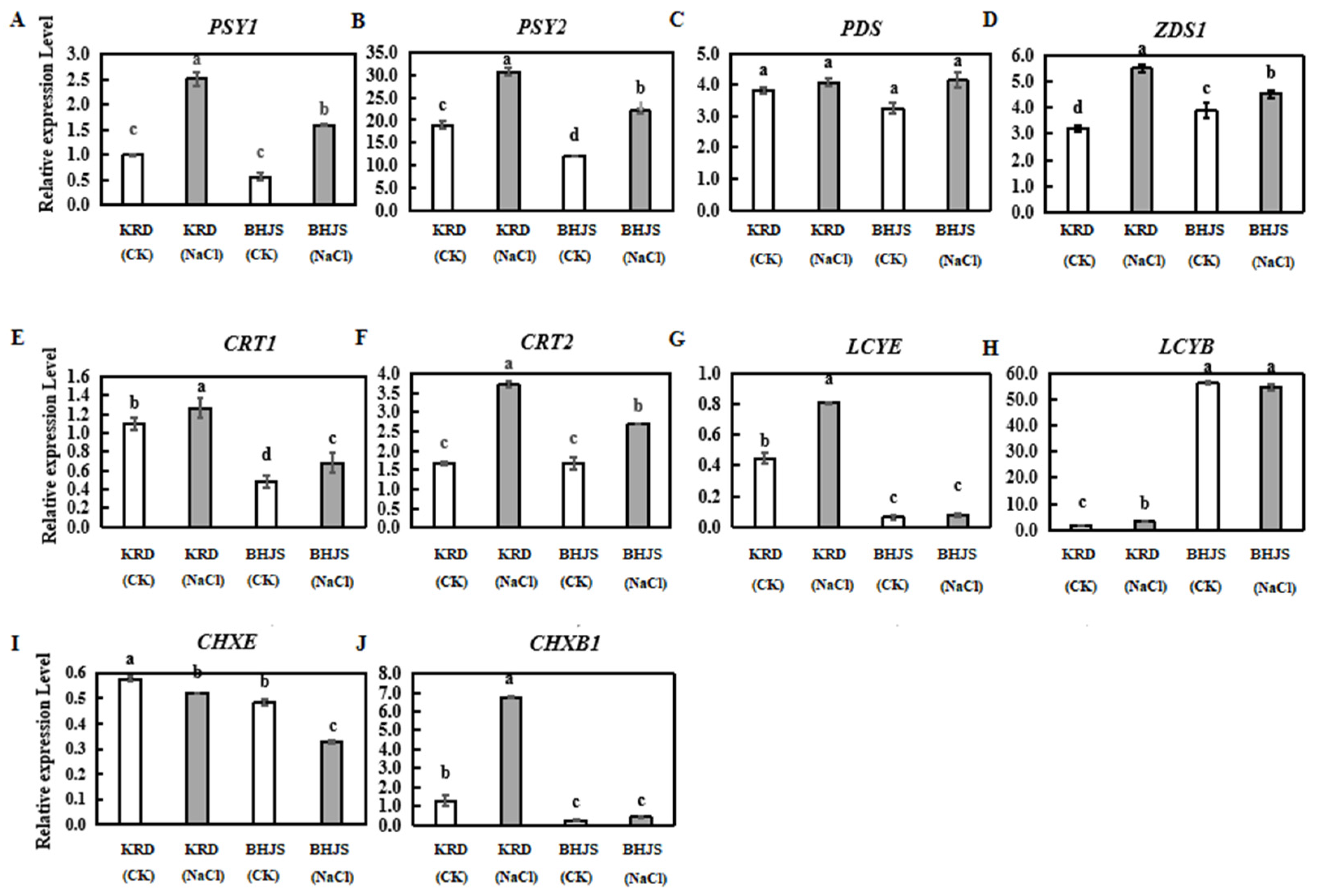
3.3. Expression Profiles of Carotenoid Biosynthesis-Related Genes

RT-qPCR results indicated that most of the 10 carotenoid biosynthesis-related genes were involved in the salt response, and the expression levels were different in ‘KRD’ and ‘BHJS’ (Figure 5). The biosynthesis of carotenoid-related genes in taproots was activated by salt stress. The changes in those selected genes showed consistency. There were also some discrepancies between the two carrot cultivars.
In KRD, in contrast with the control, the expression levels of DcPSY1, DcPSY2, DcZDS1, DcCRT1, DcCRT2, DcLCYE, DcLCYB and DcCHXB1 were significantly increased by 151%, 62%, 71%, 16%, 121%, 84%, 122% and 434%, respectively. DcPDS is basically similar to that of the control, and a 10% down-regulation of DcCHXE was observed.
In BHJS, the expression levels of DcPSY1, DcPSY2, DcZDS1, DcCRT1 and DcCRT2 in the taproots in the salt treatment group significantly increased by 179%, 82%, 11%, 42% and 62%, respectively, in comparison with that of the control group. The expression levels of DcPDS, DcLCYE, DcLCYB and DcCHXB1 sustained steady expression levels similar to that of the control, and the DcCHXE transcript was significantly down-regulated by 31%.
The transcripts of DcPSY1, DcPSY2, DcZDS1, DcCRT1 and DcCRT2 in the taproots of ‘KRD’ and ‘BHJS’ were both promoted by salt stress, and the DcPSY1 transcript was raised by more than 150%. Significantly increased levels of DcLCYE, DcLCYB and DcCHXB1 were observed in the taproots of ‘KRD’ but there was no significant difference in the taproots of ‘BHJS’. The changes in the expression level of DcPDS in the taproots of both ‘KRD’ and ‘BHJS’ are slight. The DcCHXE transcript declined in the taproots of both ‘KRD’ and ‘BHJS’.

4. Discussion

Numerous reports have shown that the increase in carotenoid concentration can enhance the resilience of plants to salt stress [38,39,40,41,42]. Reduced expression of CHXB in sweet potatoes led to an increasing concentration of β-carotene and the total carotene in the cells of transgenic plants, enhancing the antioxidant ability of plants [38]. Shi et al. [43] and Li et al. [41] overexpressed ZDS in sweet potato and tobacco, respectively, which improved the expression level of carotenoids, such as β-carotene and lutein, which confer enhanced salt tolerance to the plant. Salt stress could facilitate the accumulation of carotenoids in the taproots of carrots, since a concurrent increase in DcPSY1, DcPSY2, DcZDS1, DcCRT1 and DcCRT2 transcript levels with carotenoid biosynthesis was observed. A previous study confirmed that DbPSY plays a rate-limiting role in controlling carbon flux into the carotenogenesis pathway [23]. In this study, the expression level of DcPSY1 and DcPSY2 both showed a significant increase in the taproots of carrots under salt stress treatment, which authenticates the important role of DcPSY in prompting carotenogenesis to increase salt resistance. In maize and foxtail millet (Setaria italica), salt stress does not affect PDS [44]. In our study, DcPDS appears to be salt insensitive, which is in keeping with earlier findings. In a previous study, ZDS is characterized to be salt inducible [45,46,47], and Lao et al. [48] demonstrated that DbZDS is hypo-osmotically regulated by its promoter, and HRE (hypo-osmolarity-responsive element) is responsible for the hypo-osmotic response. In this study, the expression level of DcZDS1 increased significantly, which is assumed to facilitate salt resistance. To fulfill the geometrical requirements of the desaturases, carotene isomerase (CRTISO) is employed to transform 9,15,9’-tricis-ζ-carotene into 9,9’-dicis-ζ-carotene, 7,9,9’-tricis-neurosporene into 9-cis-neurosporene and 7,9-dicis-lycopene into all-trans-lycopene [49], and LcCRTISO is able to increase carotenoid content and enhance salt tolerance in higher plants [50]. In this study, DcCRT1 and DcCRT2 were up-regulated under salt stress, which is speculated to improve salt tolerance. In conclusion, DcPSY1, DcPSY2, DcZDS1, DcCRT1 and DcCRT2 collaborated to activate carotenoid biosynthesis to enhance salt tolerance.
The taproot of carrots is colorful and mainly consists of white, yellow, orange, red, and purple, which is decided by accumulated pigment and content [1,51,52,53,54,55]. The white taproot barely has carotenoids, the yellow taproot mainly accumulates lutein, the orange taproot such as ‘KRD’ and ‘Hongxinqicun (HXQC)’ is rich in lutein, α-carotene and β-carotene and the red taproot such as ‘BHJS’ mainly have lutein, α-carotene, β-carotene and lycopene. In this study, lycopene was barely observed in the orange taproots of ‘KRD’, and both α-carotene and β-carotene are relatively higher than ‘BHJS’. Hence, it is speculated that most of the lycopene in ‘KRD’ is used for the biosynthesis of α-carotene and β-carotene downstream. Higher content of lycopene was detected in the taproots of ‘BHJS’ in the salt treatment group in contrast with the control. Li et al. [50] overexpressed PDS, ZDS and CRT in tobacco, leading to the increased concentration of lycopene. Kong et al. [56] found that gourd and wild watermelon rootstocks boost lycopene accumulation in grafted watermelon fruit by up-regulating PSY and ZDS transcripts. Pola et al. [57] found that PSY in green pepper (Capsicum annuum L.) was significantly up-regulated at 30 °C, promoting the accumulation of lycopene. These studies show that the expression of lycopene upstream genes may exert a great impact on the changes in lycopene concentration. In this work, the lycopene concentration in the taproots of ‘BHJS’ under salt stress increased significantly compared to that of the control, which is assumed to be attributed to the up-regulation of carotenoid biosynthesis-related genes upstream. The taproots of ‘KRD’ did not accumulate lycopene, and the mechanism of lycopene accumulation in the taproots of carrots with different colors needs to be further studied.
Lycopene can be taken as a substrate to yield α-carotene and β-carotene. In the ε-branches, lycopene is catalyzed by DcLCYE to produce δ-carotene, and then formed under the catalysis of DcLCYB to produce α-carotene, and finally, lutein is synthesized with the catalysis of DcCHXB and DcCHXE [25]. A significant increase in lutein concentration in the taproots of ‘KRD’ was observed and it is speculated that the accumulation of lutein concentration was attributed to the increasing transcripts of DcLCYE and DcCHXB in this branch. However, the lutein concentration in the taproots of ‘BHJS’ decreased significantly, which may be caused by the dramatic decrease in DcCHXE transcript.
The increase in β-carotene concentration can enhance the salt tolerance of plants [41,43]. Kang et al. [40] inhibited the expression of CHXB in sweet potatoes, thus increasing the concentration of β-carotene in the root, and enhancing the salt tolerance of sweet potatoes. In this study, the concentration of β-carotene in the taproots of ‘KRD’ and ‘BHJS’ increased significantly under salt stress, which can partly explain the carrot responsive mechanism to salt stress.
ABA is a hormone-reinforcing plant abiotic stress response [58] and can be produced in β-branches of carotenoids biosynthesis [59]. A previous study showed that abiotic stress can induce carotenoid synthesis to produce ABA in Arabidopsis to increase resistance [58,60,61]. This indicates that there is a potential relationship between carotenoids and ABA, and it can be reasonably inferred that the activation of carotenoid biosynthesis is related to ABA to boost plant salt tolerance and is worth further exploration.

5. Conclusions

In conclusion, salt stress can facilitate carotenoid biosynthesis and increase carotenoid concentration in the taproots of carrots. The carotenoid compositions in taproots are various due to different carrot cultivars with different colors of the taproot. In the ε-branch, the difference in lutein accumulation in the taproots of carrots with different colors maybe relates to the discrepancy in the expression levels of DcLCYE and DcCHXB. In the β-branch, the significant increase in β-carotene concentration is one of the salt resistance mechanisms of carrots. This study is conducive to the investigation of dynamic changes of carotenoids in carrot taproots under salt stress.

Author Contributions

Conceptualization, A.-S.X., Y.-H.Z. and Y.-J.D.; methodology, Y.-H.Z. and Y.-J.D.; validation, A.-S.X. and Y.-J.D.; formal analysis, Y.-H.Z., Y.-J.D., Y.-H.W., Y.-R.L., L.-F.H., H.L., T.L., Z.-M.Y. and J.Z.; investigation, Y.-H.Z., Y.-R.L. and L.-F.H.; resources, A.-S.X.; writing—original draft preparation, Y.-H.Z.; writing—review and editing, A.-S.X. and Y.-J.D.; visualization, Y.-H.Z.; supervision, A.-S.X.; project administration, A.-S.X.; funding acquisition, A.-S.X.; All authors have read and agreed to the published version of the manuscript.

Funding

This research was funded by the National Natural Science Foundation of China, grant number 31872098; the National Undergraduate Innovation and Entrepreneurship Training Program under grant, 202010307013z; and the Priority Academic Program Development of Jiangsu Higher Education Institutions Project (PAPD).

Institutional Review Board Statement

Not applicable.

Informed Consent Statement

Not applicable.

Data Availability Statement

In this section, transcriptional data, physiological and anatomic metabolic data were measured by the authors themselves.

Conflicts of Interest

The authors declare no conflict of interest.

References

  1. Que, F.; Hou, X.L.; Wang, G.L.; Xu, Z.S.; Tan, G.F.; Li, T.; Wang, Y.H.; Khadr, A.; Xiong, A.S. Advances in research on the carrot, an important root vegetable in the Apiaceae family. Hortic. Res. 2019, 6, 69. [Google Scholar] [CrossRef] [PubMed] [Green Version]
  2. Liu, J.X.; Jiang, Q.; Tao, J.P.; Feng, K.; Li, T.; Duan, A.Q.; Wang, H.; Xu, Z.S.; Liu, H.; Xiong, A.S. Integrative genome, transcriptome, microRNA, and degradome analysis of water dropwort (Oenanthe javanica) in response to water stress. Hortic. Res. 2021, 8, 262. [Google Scholar] [CrossRef] [PubMed]
  3. Wang, Y.H.; Li, T.; Zhang, R.R.; Khadr, A.; Tian, Y.S.; Xu, Z.S.; Xiong, A.S. Transcript profiling of genes involved in carotenoid biosynthesis among three carrot cultivars with various taproot colors. Protoplasma 2020, 257, 949–963. [Google Scholar] [CrossRef] [PubMed]
  4. Zhou, H.; Yang, M.; Zhao, L.; Zhu, Z.; Liu, F.; Sun, H.; Sun, C.; Tan, L. HIGH-TILLERING AND DWARF 12 modulates photosynthesis and plant architecture by affecting carotenoid biosynthesis in rice. J. Exp. Bot. 2021, 72, 1212–1224. [Google Scholar] [CrossRef]
  5. Park, H.; Kreunen, S.S.; Cuttriss, A.J.; DellaPenna, D.; Pogson, B.J. Identification of the Carotenoid Isomerase Provides Insight into Carotenoid Biosynthesis, Prolamellar Body Formation, and Photomorphogenesis. Plant Cell 2002, 14, 321–332. [Google Scholar] [CrossRef] [Green Version]
  6. Tian, L. Recent advances in understanding carotenoid-derived signaling molecules in regulating plant growth and development. Front. Plant Sci. 2015, 6, 790. [Google Scholar] [CrossRef] [Green Version]
  7. Dhar, M.K.; Mishra, S.; Bhat, A.; Chib, S.; Kaul, S. Plant carotenoid cleavage oxygenases: Structure–function relationships and role in development and metabolism. Brief. Funct. Genom. 2020, 19, 1–9. [Google Scholar] [CrossRef]
  8. Lois, L.M.; Rodriguez-Concepcion, M.; Gallego, F.; Campos, N.; Boronat, A. Carotenoid biosynthesis during tomato fruit development: Regulatory role of 1-deoxy-D-xylulose 5-phosphate synthase. Plant J. 2000, 22, 503–513. [Google Scholar] [CrossRef]
  9. Hornero-Mendez, D.; Gomez-Ladron De Guevara, R.; Minguez-Mosquera, M.I. Carotenoid biosynthesis changes in five red pepper (Capsicum annuum L.) cultivars during ripening. Cultivar selection for breeding. J. Agric. Food Chem. 2000, 48, 3857–3864. [Google Scholar] [CrossRef]
  10. Liu, Q.; Xu, J.; Liu, Y.; Zhao, X.; Deng, X.; Guo, L.; Gu, J. A novel bud mutation that confers abnormal patterns of lycopene accumulation in sweet orange fruit (Citrus sinensis L. Osbeck). J. Exp. Bot. 2007, 58, 4161–4171. [Google Scholar] [CrossRef]
  11. Liu, G.; Yang, X.; Xu, J.; Zhang, M.; Hou, Q.; Zhu, L.; Huang, Y.; Xiong, A. Morphological observation, RNA-Seq quantification, and expression profiling: Novel insight into grafting-responsive carotenoid biosynthesis in watermelon grafted onto pumpkin rootstock. Acta Biochim. Biophys. Sin. 2017, 49, 216–227. [Google Scholar] [CrossRef] [PubMed] [Green Version]
  12. Zhang, R.R.; Wang, Y.H.; Li, T.; Tan, G.F.; Tao, J.P.; Su, X.J.; Xu, Z.S.; Tian, Y.S.; Xiong, A.S. Effects of simulated drought stress on carotenoid contents and expression of related genes in carrot taproots. Protoplasma 2021, 258, 379–390. [Google Scholar] [CrossRef] [PubMed]
  13. Li, T.; Deng, Y.J.; Liu, J.X.; Duan, A.Q.; Liu, H.; Xiong, A.S. DcCCD4 catalyzes the degradation of alpha-carotene and beta-carotene to affect carotenoid accumulation and taproot color in carrot. Plant J. 2021, 108, 1116–1130. [Google Scholar] [CrossRef] [PubMed]
  14. Ding, X.; Jia, L.L.; Xing, G.M.; Tao, J.P.; Sun, S.; Tan, G.F.; Li, S.; Liu, J.X.; Duan, A.Q.; Wang, H.; et al. The Accumulation of Lutein and beta-Carotene and Transcript Profiling of Genes Related to Carotenoids Biosynthesis in Yellow Celery. Mol. Biotechnol. 2021, 63, 638–649. [Google Scholar] [CrossRef]
  15. Wang, G.L.; Xiong, F.; Que, F.; Xu, Z.S.; Wang, F.; Xiong, A.S. Morphological characteristics, anatomical structure, and gene expression: Novel insights into gibberellin biosynthesis and perception during carrot growth and development. Hortic. Res. 2015, 2, 15028. [Google Scholar] [CrossRef] [Green Version]
  16. Cavagnaro, P.F.; Chung, S.M.; Manin, S.; Yildiz, M.; Ali, A.; Alessandro, M.S.; Iorizzo, M.; Senalik, D.A.; Simon, P.W. Microsatellite isolation and marker development in carrot-genomic distribution, linkage mapping, genetic diversity analysis and marker transferability across Apiaceae. BMC Genom. 2011, 12, 386. [Google Scholar] [CrossRef] [PubMed] [Green Version]
  17. Rodriguez-Concepcion, M.; Stange, C. Biosynthesis of carotenoids in carrot: An underground story comes to light. Arch. Biochem. Biophys. 2013, 539, 110–116. [Google Scholar] [CrossRef]
  18. Fuentes, P.; Pizarro, L.; Moreno, J.C.; Handford, M.; Rodriguez-Concepcion, M.; Stange, C. Light-dependent changes in plastid differentiation influence carotenoid gene expression and accumulation in carrot roots. Plant Mol. Biol. 2012, 79, 47–59. [Google Scholar] [CrossRef]
  19. Stange, C.; Fuentes, P.; Handford, M.; Pizarro, L. Daucus carota as a novel model to evaluate the effect of light on carotenogenic gene expression. Biol. Res. 2008, 41, 289–301. [Google Scholar] [CrossRef] [Green Version]
  20. Cunningham, F.X.; Gantt, E., Jr. Genes and enzymes of carotenoid biosynthesis in plants. Annu. Rev. Plant Physiol. Plant Mol. Biol. 1998, 49, 557–583. [Google Scholar] [CrossRef]
  21. Nisar, N.; Li, L.; Lu, S.; Khin, N.C.; Pogson, B.J. Carotenoid Metabolism in Plants. Mol. Plant 2015, 8, 68–82. [Google Scholar] [CrossRef] [PubMed] [Green Version]
  22. Wang, Q.; Huang, X.Q.; Cao, T.J.; Zhuang, Z.; Wang, R.; Lu, S. Heteromeric Geranylgeranyl Diphosphate Synthase Contributes to Carotenoid Biosynthesis in Ripening Fruits of Red Pepper (Capsicum annuum var. conoides). J. Agric. Food Chem. 2018, 66, 11691–11700. [Google Scholar] [CrossRef] [PubMed]
  23. Maass, D.; Arango, J.; Wüst, F.; Beyer, P.; Welsch, R. Carotenoid Crystal Formation in Arabidopsis and Carrot Roots Caused by Increased Phytoene Synthase Protein Levels. PLoS ONE 2009, 4, e6373. [Google Scholar] [CrossRef] [PubMed] [Green Version]
  24. Moreno, J.C.; Pizarro, L.; Fuentes, P.; Handford, M.; Cifuentes, V.; Stange, C. Levels of lycopene beta-cyclase 1 modulate carotenoid gene expression and accumulation in Daucus carota. PLoS ONE 2013, 8, e58144. [Google Scholar] [CrossRef] [PubMed] [Green Version]
  25. Sandmann, G.; Römer, S.; Fraser, P.D. Understanding carotenoid metabolism as a necessity for genetic engineering of crop plants. Metab. Eng. 2006, 8, 291–302. [Google Scholar] [CrossRef] [PubMed]
  26. Munns, R. Comparative physiology of salt and water stress. Plant Cell Environ. 2002, 25, 239–250. [Google Scholar] [CrossRef]
  27. Zhu, J.K. Abiotic Stress Signaling and Responses in Plants. Cell 2016, 167, 313–324. [Google Scholar] [CrossRef] [Green Version]
  28. Ahmad, A.; Blasco, B.; Martos, V. Combating Salinity Through Natural Plant Extracts Based Biostimulants: A Review. Front. Plant Sci. 2022, 13, 862034. [Google Scholar] [CrossRef]
  29. Ullah, M.A.; Abdullah-Zawawi, M.-R.; Zainal-Abidin, R.-A.; Sukiran, N.L.; Uddin, I.; Zainal, Z. A Review of Integrative Omic Approaches for Understanding Rice Salt Response Mechanisms. Plants 2022, 11, 1430. [Google Scholar] [CrossRef]
  30. Ren, Y.; Sun, H.; Deng, J.; Huang, J.; Chen, F. Carotenoid Production from Microalgae: Biosynthesis, Salinity Responses and Novel Biotechnologies. Mar. Drugs 2021, 19, 713. [Google Scholar] [CrossRef]
  31. Piña, F.; Contreras-Porcia, L. Enhancement of Xanthophyll Synthesis in Porphyra/Pyropia Species (Rhodophyta, Bangiales) by Controlled Abiotic Factors: A Systematic Review and Meta-Analysis. Mar. Drugs 2021, 19, 221. [Google Scholar] [CrossRef] [PubMed]
  32. Behera, T.K.; Krishna, R.; Ansari, W.A.; Aamir, M.; Kumar, P.; Kashyap, S.P.; Pandey, S.; Kole, C. Approaches Involved in the Vegetable Crops Salt Stress Tolerance Improvement: Present Status and Way Ahead. Front. Plant Sci. 2022, 12, 787292. [Google Scholar] [CrossRef]
  33. Ma, J.; Xu, Z.; Tan, G.; Wang, F.; Xiong, A. Distinct transcription profile of genes involved in carotenoid biosynthesis among six different color carrot (Daucus carota L.) cultivars. Acta Biochim. Biophys. Sin. 2017, 49, 817–826. [Google Scholar] [CrossRef] [PubMed] [Green Version]
  34. Ma, J.; Li, J.; Xu, Z.; Wang, F.; Xiong, A. Transcriptome profiling of genes involving in carotenoid biosynthesis and accumulation between leaf and root of carrot (Daucus carota L.). Acta Biochim. Biophys. Sin. 2018, 50, 481–490. [Google Scholar] [CrossRef] [PubMed] [Green Version]
  35. Tian, C.; Jiang, Q.; Wang, F.; Wang, G.-L.; Xu, Z.-S.; Xiong, A.-S. Selection of Suitable Reference Genes for qPCR Normalization under Abiotic Stresses and Hormone Stimuli in Carrot Leaves. PLoS ONE 2015, 10, e0117569. [Google Scholar] [CrossRef] [Green Version]
  36. Pfaffl, M.W. A new mathematical model for relative quantification in real-time RT-PCR. Nucleic Acids Res. 2001, 29, e45. [Google Scholar] [CrossRef]
  37. Tang, X.; Mu, X.; Shao, H.; Wang, H.; Brestic, M. Global plant-responding mechanisms to salt stress: Physiological and molecular levels and implications in biotechnology. Crit. Rev. Biotechnol. 2015, 35, 425–437. [Google Scholar] [CrossRef]
  38. Kim, S.H.; Ahn, Y.O.; Ahn, M.J.; Lee, H.S.; Kwak, S.S. Down-regulation of beta-carotene hydroxylase increases beta-carotene and total carotenoids enhancing salt stress tolerance in transgenic cultured cells of sweetpotato. Phytochemistry 2012, 74, 69–78. [Google Scholar] [CrossRef]
  39. Kim, S.H.; Jeong, J.C.; Park, S.; Bae, J.Y.; Ahn, M.J.; Lee, H.S.; Kwak, S.S. Down-regulation of sweetpotato lycopene beta-cyclase gene enhances tolerance to abiotic stress in transgenic calli. Mol. Biol. Rep. 2014, 41, 8137–8148. [Google Scholar] [CrossRef]
  40. Kang, L.; Park, S.C.; Ji, C.Y.; Kim, H.S.; Lee, H.S.; Kwak, S.-S. Metabolic engineering of carotenoids in transgenic sweetpotato. Breed. Sci. 2017, 67, 27–34. [Google Scholar] [CrossRef] [Green Version]
  41. Li, R.; Kang, C.; Song, X.; Yu, L.; Liu, D.; He, S.; Zhai, H.; Liu, Q. A zeta-carotene desaturase gene, IbZDS, increases beta-carotene and lutein contents and enhances salt tolerance in transgenic sweetpotato. Plant Sci. 2017, 262, 39–51. [Google Scholar] [CrossRef] [PubMed]
  42. Kang, C.; Zhai, H.; Xue, L.; Zhao, N.; He, S.; Liu, Q. A lycopene β-cyclase gene, IbLCYB2, enhances carotenoid contents and abiotic stress tolerance in transgenic sweetpotato. Plant Sci. 2018, 272, 243–254. [Google Scholar] [CrossRef] [PubMed]
  43. Shi, Y.; Guo, J.; Zhang, W.; Jin, L.; Liu, P.; Chen, X.; Li, F.; Wei, P.; Li, Z.; Li, W.; et al. Cloning of the Lycopene beta-cyclase Gene in Nicotiana tabacum and Its Overexpression Confers Salt and Drought Tolerance. Int. J. Mol. Sci. 2015, 16, 30438–30457. [Google Scholar] [CrossRef]
  44. Li, F.; Vallabhaneni, R.; Wurtzel, E.T. PSY3, a New Member of the Phytoene Synthase Gene Family Conserved in the Poaceae and Regulator of Abiotic Stress-Induced Root Carotenogenesis. Plant Physiol. 2008, 146, 1333–1345. [Google Scholar] [CrossRef] [Green Version]
  45. Kim, S.H.; Kim, Y.H.; Ahn, Y.O.; Ahn, M.J.; Jeong, J.C.; Lee, H.S.; Kwak, S.S. Downregulation of the lycopene epsilon-cyclase gene increases carotenoid synthesis via the beta-branch-specific pathway and enhances salt-stress tolerance in sweetpotato transgenic calli. Physiol. Plant 2013, 147, 432–442. [Google Scholar] [CrossRef] [PubMed]
  46. Babu, M.; Singh, D.; Kodiveri Muthukaliannan, G. Effect of salt stress on expression of carotenoid pathway genes in tomato. J. Stress Physiol. Biochem. 2011, 7, 87–94. [Google Scholar]
  47. Tuan, P.A.; Kim, J.K.; Lee, S.; Chae, S.C.; Park, S.U. Molecular Characterization of Carotenoid Cleavage Dioxygenases and the Effect of Gibberellin, Abscisic Acid, and Sodium Chloride on the Expression of Genes Involved in the Carotenoid Biosynthetic Pathway and Carotenoid Accumulation in the Callus of Scutellaria baicalensis Georgi. J. Agric. Food Chem. 2013, 61, 5565–5572. [Google Scholar] [CrossRef]
  48. Lao, Y.M.; Xiao, L.; Luo, L.X.; Jiang, J.G. Hypoosmotic expression of Dunaliella bardawil zeta-carotene desaturase is attributed to a hypoosmolarity-responsive element different from other key carotenogenic genes. Plant Physiol. 2014, 165, 359–372. [Google Scholar] [CrossRef] [Green Version]
  49. Yu, Q.; Ghisla, S.; Hirschberg, J.; Mann, V.; Beyer, P. Plant carotene cis-trans isomerase CRTISO: A new member of the FAD(RED)-dependent flavoproteins catalyzing non-redox reactions. J. Biol. Chem. 2011, 286, 8666–8676. [Google Scholar] [CrossRef] [Green Version]
  50. Li, C.; Ji, J.; Wang, G.; Li, Z.; Wang, Y.; Fan, Y. Over-Expression of LcPDS, LcZDS, and LcCRTISO, Genes From Wolfberry for Carotenoid Biosynthesis, Enhanced Carotenoid Accumulation, and Salt Tolerance in Tobacco. Front. Plant Sci. 2020, 11, 119. [Google Scholar] [CrossRef]
  51. Iorizzo, M.; Senalik, D.A.; Ellison, S.L.; Grzebelus, D.; Cavagnaro, P.F.; Allender, C.; Brunet, J.; Spooner, D.M.; Van Deynze, A.; Simon, P.W. Genetic structure and domestication of carrot (Daucus carota subsp. sativus) (Apiaceae). Am. J. Bot. 2013, 100, 930–938. [Google Scholar] [CrossRef] [PubMed]
  52. Simon, P.W.; Hamrick, J. Inheritance and Expression of Purple and Yellow Storage Root Color in Carrot. J. Hered. 1996, 87, 63–66. [Google Scholar] [CrossRef] [Green Version]
  53. Wang, X.J.; Luo, Q.; Li, T.; Meng, P.-H.; Pu, Y.; Liu, J.X.; Zhang, J.; Liu, H.; Tan, G.; Xiong, A.S. Origin, evolution, breeding and omics of Apiaceae: A family of vegetables and medicinal plants. Hortic. Res. 2022, 9, uhac076. [Google Scholar] [CrossRef]
  54. Xu, Z.S.; Yang, Q.Q.; Feng, K.; Xiong, A.S. Changing Carrot Color: Insertions in DcMYB7 Alter the Regulation of Anthocyanin Biosynthesis and Modification. Plant Physiol. 2019, 181, 195–207. [Google Scholar] [CrossRef]
  55. Xu, Z.-S.; Yang, Q.Q.; Feng, K.; Yu, X.; Xiong, A.S. DcMYB113, a root-specific R2R3-MYB, conditions anthocyanin biosynthesis and modification in carrot. Plant Biotechnol. J. 2020, 18, 1585–1597. [Google Scholar] [CrossRef] [Green Version]
  56. Kong, Q.; Yuan, J.; Gao, L.; Liu, P.; Cao, L.; Huang, Y.; Zhao, L.; Lv, H.; Bie, Z. Transcriptional regulation of lycopene metabolism mediated by rootstock during the ripening of grafted watermelons. Food Chem. 2017, 214, 406–411. [Google Scholar] [CrossRef]
  57. Pola, W.; Sugaya, S.; Photchanachai, S. Influence of Postharvest Temperatures on Carotenoid Biosynthesis and Phytochemicals in Mature Green Chili (Capsicum annuum L.). Antioxidants 2020, 9, 203. [Google Scholar] [CrossRef] [Green Version]
  58. Ruiz-Sola, M.; Arbona, V.; Gómez-Cadenas, A.; Rodriguez-Concepcion, M.; Rodríguez-Villalón, A. A Root Specific Induction of Carotenoid Biosynthesis Contributes to ABA Production upon Salt Stress in Arabidopsis. PLoS ONE 2014, 9, e90765. [Google Scholar] [CrossRef]
  59. Frey, A.; Boutin, J.P.; Sotta, B.; Mercier, R.; Marion-Poll, A. Regulation of carotenoid and ABA accumulation during the development and germination of Nicotiana plumbaginifolia seeds. Planta 2006, 224, 622–632. [Google Scholar] [CrossRef]
  60. Li, T.; Liu, J.X.; Deng, Y.J.; Xu, Z.S.; Xiong, A.S. Overexpression of a carrot BCH gene, DcBCH1, improves tolerance to drought in Arabidopsis thaliana. BMC Plant Biol. 2021, 21, 475. [Google Scholar] [CrossRef]
  61. Wang, Y.H.; Que, F.; Li, T.; Zhang, R.R.; Khadr, A.; Xu, Z.S.; Tian, Y.S.; Xiong, A.S. DcABF3, an ABF transcription factor from carrot, alters stomatal density and reduces ABA sensitivity in transgenic Arabidopsis. Plant Sci. 2020, 302, 110699. [Google Scholar] [CrossRef] [PubMed]
Figure 1. Phenotype of carrot under salt stress. (A) ‘BHJS’ CK; (B) ‘BHJS’ under salt stress; (C) ‘KRD’ CK; (D) ‘KRD’ under salt stress. Bar = 5 cm. Salt Stress: NaCl solution (300 mmol·L−1). CK: Distilled water.
Horticulturae 08 00650 g001
Figure 2. Morphological parameters of carrot under salt stress. (A) Taproot length; (B) Maximum diameter of taproot; (C) Leaf length. Different lowercase letters indicate significant differences among different carrot cultivars and treatment at the p < 0.05 level.
Horticulturae 08 00650 g002
Figure 3. UPLC chromatograms of carotenoids in different carrot cultivars and treatments. (A) Standard UPLC chromatograms of carotenoids; (B) UPLC chromatograms of carotenoids in ‘KRD’ of control group; (C) UPLC chromatograms of carotenoids in ‘KRD’ of salt treatment group; (D) UPLC chromatograms of carotenoids in ‘BHJS’ of control group; (E) UPLC chromatograms of carotenoids in ‘BHJS’ of salt treatment group. Cultivar abbreviations: BHJS, Benhongjinshi; KRD, Kurodagosun.
Horticulturae 08 00650 g003
Figure 4. Carotenoids concentration in different carrot cultivars and treatments. (A) Lycopene concentration in the roots of ‘BHJS’ and ‘KRD’ under different treatment; (B) Lutein concentration; (C) α-carotene concentration; (D) β-carotene concentration; (E) Total carotenoids concentration. Cultivar abbreviations: BHJS, Benhongjinshi; KRD, Kurodagosun. “n/a” represents no concentration was detected. Different lowercase letters indicate significant differences among different carrot cultivars and treatment at the p < 0.05 level.
Horticulturae 08 00650 g004
Figure 5. Relative expression levels of carotenoid biosynthesis-related genes under salt stress. (A) Relative expression level of PSY1 in the roots of ‘BHJS’ and ‘KRD’ under different treatment; (B) PSY2; (C) PDS; (D) ZDS1; (E) CRT1; (F) CRT2; (G) LCYE; (H) LCYB; (I) CHXE; (J) CHXB1. Different lowercase letters indicate significant differences among different carrot cultivars and treatment at the p < 0.05 level.
Horticulturae 08 00650 g005
Publisher’s Note: MDPI stays neutral with regard to jurisdictional claims in published maps and institutional affiliations.

