Article

Surface Moisture Induces Microcracks and Increases Water Vapor Permeance of Fruit Skins of Mango cv. Apple

1
Institute of Horticultural Production Systems, Leibniz University Hannover, Herrenhäuser Straße 2, 30419 Hannover, Germany
2
School of Food and Nutritional Sciences (SOFNUS), Jomo Kenyatta University of Agriculture and Technology, P.O. Box 62 000, Nairobi 00200, Kenya
*
Author to whom correspondence should be addressed.
Horticulturae 2022, 8(6), 545; https://doi.org/10.3390/horticulturae8060545
Submission received: 24 May 2022 / Revised: 14 June 2022 / Accepted: 15 June 2022 / Published: 18 June 2022

Abstract

:
Exposure to surface moisture triggers cuticular microcracking of the fruit skin. In mango fruit cv. apple, microcracking compromises postharvest performance by increasing moisture loss and infections with pathogens. This study reports the effects of exposing the fruit’s skin to surface moisture on the incidence of microcracking and on water vapor permeance. Microcracking was quantified microscopically following infiltration with a fluorescent tracer. Water mass loss was determined gravimetrically. Moisture exposure increased cuticular microcracking and permeance. During moisture exposure, permeance increased over the first 4 d, remained constant up to approximately 8 d, then decreased for longer exposure times. Fruit development followed a sigmoid growth pattern. The growth rate peaked approximately 103 days after full bloom. This coincided with the peak in moisture-induced microcracking. There were no increases in water vapor permeance or in microcracking in control fruit that remained dry. When experimental moisture exposure was terminated, microcracking and water vapor permeance decreased. This suggests a repair process restoring the barrier properties of the fruit skin. Histological analyses reveal a periderm forms in the hypodermis beneath a microcrack. Our study demonstrates that surface moisture induces microcracking in mango cv. apple that increases the skin’s water vapor permeance and induces russeting.

1. Introduction

Mango cv. apple (Mangifera indica L.) is an important commercial fruit crop in Kenya but is prone to russeting. The russeted skins of cv. apple mangoes are typically brown, rough and cracked, rendering them less attractive and thus worth less at point of sale, despite their still excellent flavor [1]. Apart from mango, russeting also occurs in a wide range of other fruit crop species including in Malus apples (Malus × domestica Borkh.) [2,3,4], pears (Pyrus communis L.) [5,6], prunes (Prunus × domestica L.) [7], citrus (Citrus reticulata Blanco × (C. paradisi Macf. × C. reticulate) [8,9], loquats (Eriobotrya japonica (Thunb.) Lindl.) [10], tomatoes (Solanum lycopersicum L.) [11] and melons (Cucumis melo L.) [12,13]. In anatomical terms, russet represents the formation of a ‘secondary skin’ in the hypodermis, just below the epidermis. This secondary skin—a periderm—comprises phellem, phellogen and phelloderm [14].
A large number of factors can induce russeting in fruit. These include excessive rates of growth strain [15,16], exposure to surface moisture or even simply to high humidity [17,18,19,20], extreme temperatures [10,21,22], excessive sunlight (duration × intensity) [23], mechanical abrasion [7,12], some agrochemicals [16,24,25] and some pests and diseases [8,26,27].
Russeting has been studied extensively in Malus apple. The majority of these studies have concluded that microcracks in the cuticle are the first visible signs of insipient russeting [28,29]. Microcracks are minute fissures in the cuticle that are invisible to the naked eye. They are limited to the cuticle and are sufficiently shallow that they rarely penetrate to the outer cellulosic cell walls of the epidermis [30]. Nevertheless, these microcracks do significantly impair the barrier functions of the cuticle. In this way they facilitate invasion by pathogens [31,32] and greatly increase rates of evaporative water loss [17,33].
In our initial studies on mango cv. apple, marked differences in the incidence of russet were observed between different growing sites across Kenya [1]. Correlation analyses revealed that the number of rainy days, temperatures (average, minimum and maximum) and the number of cold nights were particularly conducive to russeting [1]. There was essentially no russeting on fruit cultivated under warm, dry conditions. However, significant russeting occurred in cooler and wetter climates [1]. The most likely explanation for these observations is that moisture induces microcracking of the cuticle of mango cv. apple with the microcracking being followed by the formation of a periderm.
The objective of this study was to establish the role of surface moisture in triggering microcracking in the fruit skins of mango cv. apple. As mango is susceptible to another skin disorder associated with increased rates of water loss, ‘shrivel’, we also quantified changes in the water vapor permeance of the fruit skin following exposure to surface moisture.

2. Materials and Methods

2.1. Plant Materials

Mango fruits (Mangifera indica L.), cv. apple, grafted on an unclassified rootstock, were obtained from a commercial orchard in Mwala, Kenya (1°19′ S, 37°26′ E). Fruit was grown according to standard local practice.

2.2. Moisture Treatment

Developing fruits free of visual defects and selected for uniformity of size and color were tagged. The fruit selected were thus representative for the population of fruit on the tree. Moisture was applied locally to the surface of the fruit. Briefly, conical polyethylene (PE) tubes (8 mm inside diameter) were cut from the tips of microcentrifuge tubes (Sarstedt, Nümbrecht, Germany) and glued to the cheek of the fruit using a non-phytotoxic, fast-curing silicone rubber (Dowsil™ SE 9186 clear sealant; Dow Toray Co., Tokyo, Japan) (Figure S1A) [17]. For the moisture treatment, a tube was filled with distilled water through a hole cut at the tip. The hole was then sealed with silicone rubber. All such tubes were inspected every 2 to 3 days (d) and, if necessary, they were re-sealed to prevent leakage of water. The moisture treatment was eventually terminated by carefully removing the tube. An untreated area of skin on the opposite cheek of the same fruit served as control. In some cases, the control area was enclosed by an identical PE tube but with no water added (Figure S1B).

2.3. Transpiration Assays

Transpiration through the fruit skin was investigated using excised epidermal segments (ES) mounted in stainless steel diffusion cells (7 mm diameter) [34,35]. These ES discs were excised from a moisture-exposed or a control skin area using a biopsy punch (12 mm diameter; Kai Europe, Solingen, Germany). The cut (inner) surface of the ES was blotted dry and immediately mounted in the diffusion cell using high-vacuum grease (Korasilon-Paste; Kurt Obermeier, Bad Berleburg, Germany). Distilled water was injected through the orifice in the base of the cell using a disposable syringe. Water loss from the diffusion cell was restricted to the exposed top (outer) surface of the ES by sealing the orifice in the base and the gap between the lid and the bottom part of the diffusion cell using clear transparent tape (Tesa Film®; Tesa-Werke Offenburg, Offenburg, Germany). The cells were turned upside down and held overnight to stabilize. The next morning, the cell was placed on a metal grid above dry silica gel inside a PE box such that the exposed (outer) ES faced the silica gel and the whole assembly was allowed to equilibrate for at least one hour. Water loss was then quantified gravimetrically by repeated weighing of the cell on an analytical balance (AUW220D; Shimadzu Corporation, Kyoto, Japan) every 2 h over a 6–8 h period. The rate of water loss (F, g h−1) was estimated from the slope of a linear regression through a plot of mass against time. The permeance (P, m s−1) of the ES was derived from Equation (1).
P = F A × Δ C
In this equation, A is the area of the ES exposed in the diffusion cell (3.8 × 10⁻⁵ m2) and ΔC the difference in water vapor concentration between the water vapor saturated atmosphere inside the diffusion cell (23.07 g m−3 at 25 °C [36]) and the dry atmosphere above the silica gel [34].

2.4. Microcracking

Microcracking was studied using the fluorescent tracer acridine orange [30]. The cheek of the mango including the moisture-exposed or the untreated control area of skin was dipped in 0.1% (w/v) aqueous acridine orange (Carl Roth, Karlsruhe, Germany) for 10 min. Following rinsing with distilled water (5–6 s) and blotting using soft tissue paper, the fruit surface was viewed under a stereo microscope (MZ10F; Leica Microsystems, Wetzlar, Germany) under incident fluorescent light (GFP LP filter, 480–440 nm excitation, ≥510 nm emission wavelength). Calibrated images of the moisture-exposed or control skin areas were prepared (Camera DFC7000T; Leica Microsystems). Four images per fruit, per treatment were taken from a total of 8 to 10 fruit. The area infiltrated by the dye as indexed by the area exhibiting yellow-green fluorescence was measured by image analysis (ImageJ 1.53F51; National Health Institute, Bethesda, MD, USA). All images were processed using the same settings and color thresholds.

2.5. Experiments

The time course of microcracking was established using detached fruit. The portion of the fruit surface that was earlier inspected for microcracks was immersed in distilled water for 0, 6, 24 or 48 h to induce microcracking. Fruit used as controls remained dry. Thereafter, microcracking was quantified as described above.
The time course of microcracking was determined on fruit that remained attached to the tree. At 53 days after full bloom (DAFB), a small area of the fruit surface was exposed to moisture for 0, 4, 8, 12 or 16 days. The untreated opposite side of the same fruit served as the control. Microcracking and water vapor permeance were quantified after moisture removal.
The developmental time course of change in fruit mass, fruit surface area, moisture-induced microcracking and permeance to water vapor was investigated. Fruits were harvested at different stages of development in the mornings and processed on the same day. Fruit mass (TX420L; Shimadzu), height and the two orthogonal diameters in the equatorial plane were measured (digital caliper CD-20PKX; Mitutoyo, Kawasaki/Kanagawa, Japan). Fruit surface area was calculated from the measured dimensions assuming a spherical shape. A sigmoid regression curve was fitted through a plot of fruit surface area against time. The surface growth rate was calculated from the first derivative of the regression model. The effects of moisture exposure on microcracking and on water vapor permeance were studied following 8 d of exposure to moisture starting at 33 DAFB. Microcracking and water vapor permeance were quantified as described above. The opposite side of the same fruit was left untreated and served as control.
The time course of change in microcracking and water vapor permeance after termination of moisture exposure was investigated. The fruit surface was exposed to moisture for 8 d at 72 DAFB. Fruit were harvested at 0, 7, 15 or 30 days after moisture exposure had been terminated (DAT). Microcracking and skin permeance were quantified as described above. The opposite side of the same fruit was left untreated and served as control.
To distinguish any effects of mounting a PE tube on the fruit surface from those of moisture exposure, an experiment was established that compared the following treatments: ‘untreated’ control (no tube, no moisture), ‘open tube’ (tube, no moisture) and ‘moisture treatment’ (tube, moisture). For the ‘open tube’ treatment a cylindrical tube that had the same diameter as the one used for moisture exposure was used. The tube was short to limit any build-up of moisture or high humidity inside the tube. The tubes in the latter two treatments were removed from the surface after 16 days and the treated areas of skin marked using a permanent marker. At maturity, the surface was photographed. Tissue blocks were excised from the treated areas and fixed in Karnovsky solution for histological analysis [37]. Using image analysis (Image J), the russeted area was calculated as a percentage of the tube footprint area exposed to moisture. For the controls without a tube, the average area of the tube footprint was used as an estimate of the exposed area.

2.6. Histology

Tissue blocks previously fixed in Karnovsky solution [37] were rinsed with deionized water. Small sections (5 mm × 2 mm × 2 mm) of these blocks were cut by hand using a razor blade and kept overnight in 70% ethanol at 4 °C. The blocks were then vacuum infiltrated in a series of alcohol solutions of increasing concentration (70, 80, 90, and 96% v/v ethanol and then 100% isopropanol). The blocks were then treated with xylene substitute (AppliClear; PanReac Applichem, Münster, Germany) and thereafter with a 1:1 xylene-substitute: paraffin mixture before embedding in melted paraffin. Embedded blocks were cooled above ice and stored at 4 °C until use. Cross-sections of 10 µm thickness were cut using a microtome (Hyrax M55; Carl Zeiss, Jena, Germany), mounted on a microscope slide over a water bath at 40 °C and oven-dried overnight at 40 °C. Prior to staining, the sections were washed in xylene substitute before rehydrating in a series of decreasing ethanol concentration (96, 80, 70, 60% v/v ethanol and deionized water). Staining was done for a minimum of 1 h using 0.005% (w/v) fluorol yellow 088 dye (Santa Cruz Biotechnology, Dallas, TX, USA) made up by dissolving in a hot mixture of polyethylene glycol (PEG) 4000 (50% w/v; SERVA Electrophoresis; Heidelberg, Germany) and 45% v/v glycerol and 5% deionized water. Following rinsing with deionized water, the microscope slides were placed on the stage of a fluorescence microscope (BX-60; Olympus Europa, Hamburg, Germany), examined under incident bright and fluorescent light (U-MWU filter, 330–385 nm excitation, ≥420 nm emission wavelength; Olympus) and photographed (DP73; Olympus).

2.7. Data Analyses

Data symbols in figures represent the means ± standard errors. Analyses of variance and correlation analyses were conducted using the statistical software package SAS (Version 9.1.3; SAS Institute, Cary, NC, USA).

3. Results

Moisture exposure increased microcracking of the cuticle as indexed by the increase in area infiltrated with acridine orange compared to that of the untreated control. The fluorescing area was initially low but then increased rapidly, reaching an asymptote within 48 h of moisture exposure. There were no changes in the fluorescing areas of the untreated controls (Figure 1).
Quantitatively similar data were obtained for longer exposures to moisture. The percentage infiltrated area was significantly larger following moisture exposure for 8 to 16 d compared to the unexposed controls. There were no significant changes in infiltrated area in the controls (Figure 2A). The amount of water transpired increased linearly with time and was significantly higher in the moisture-exposed areas than in the untreated control areas (Figure 2B). The permeance of the fruit skin to water vapor increased until 4 d of moisture exposure, then remained approximately constant up to 8 d. For longer exposure times skin permeance decreased (Figure 2C). Skin permeance was significantly higher for the water-exposed areas than for the control areas at all times. There were no significant changes in skin permeance in the untreated controls.
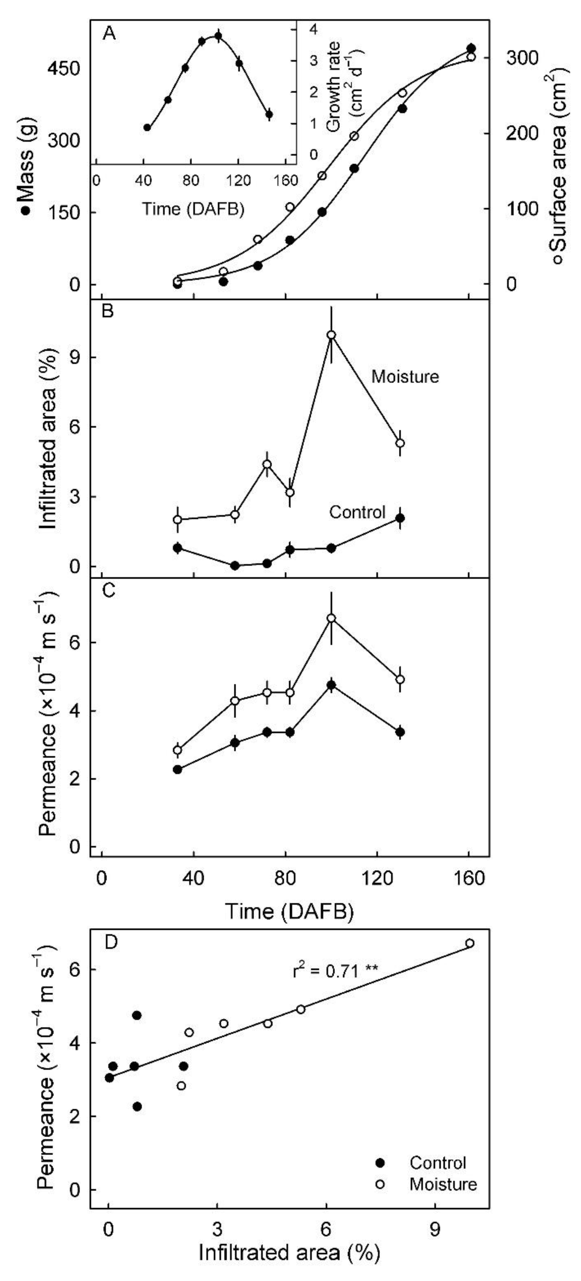
The increase in mass and surface area of developing fruit of mango cv. apple followed a sigmoid pattern (Figure 3A). The growth rate in surface area peaked at approximately 103 DAFB (Figure 3A, inset). The peak in growth rate at 103 DAFB coincided with a peak in microcracking, as indexed by the areas infiltrated with the fluorescent tracer (Figure 3B). There was only a slight increase in infiltrated surface area in the untreated control fruit during fruit development (Figure 3B). The change in permeance to water vapor essentially paralleled the change in tracer infiltrated area (Figure 3C). Permeance was higher in moisture-treated ES compared to control ES, irrespective of developmental stage (Figure 3C). There was a significant and positive linear correlation between the permeance of the fruit skin to water vapor and the percentage of surface area infiltrated with acridine orange (Figure 3D).
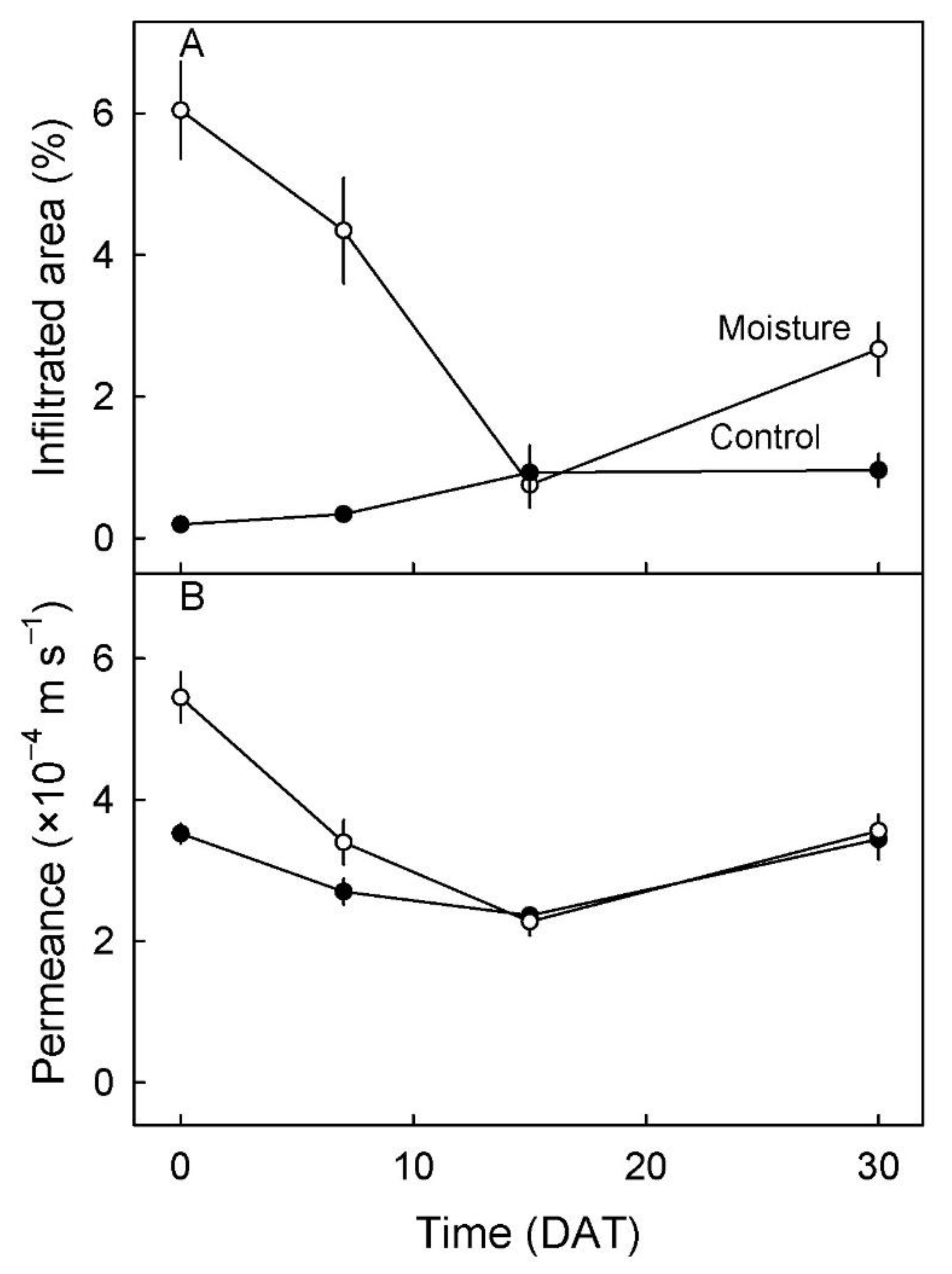
The time course of change in infiltrated area and in skin permeance after moisture exposure was terminated revealed a decrease in both infiltrated area and in permeance. The area infiltrated and the permeance were both higher in moisture-exposed areas than in unexposed control areas (Figure 4).
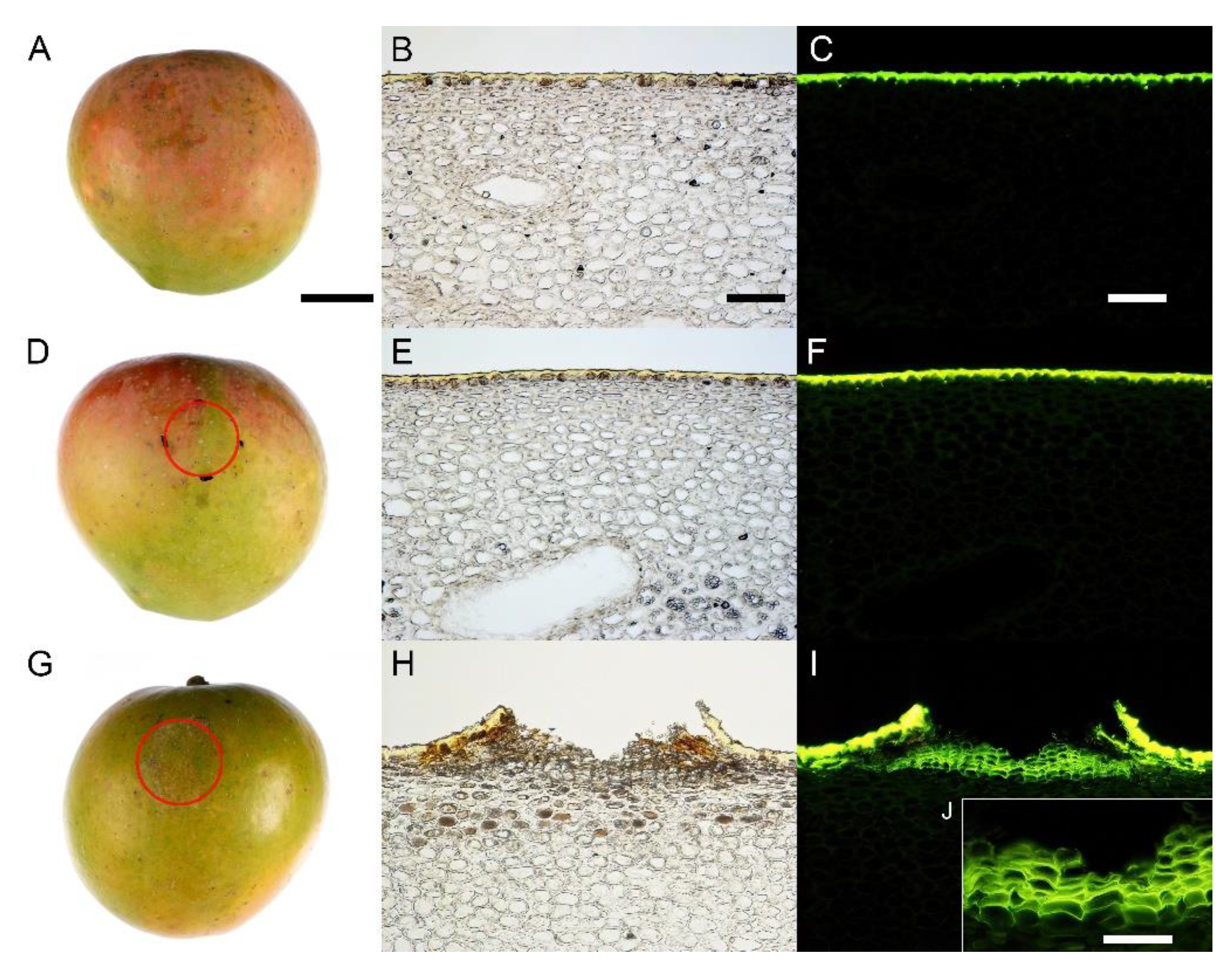
Histological analyses revealed that the brownish appearance of the skin was due to the formation of a periderm in the hypodermis of the moisture-exposed areas of skin (Figure 5I,J). There was no evident periderm in the unexposed skin areas (Figure 5A–F). Moisture-exposed skin areas developed a rough, brownish appearance (Figure 5G).
The areas affected by periderm were consistently and significantly larger in moisture-exposed areas of skin than in unexposed (control) areas and were unaffected by merely attaching a tube to the skin surface (i.e., with no added water) (Table 1).

4. Discussion

In our discussion, we focus on (1) the effect of surface moisture on microcracking and russeting in the fruit of mango cv. apple and (2) the increase in skin permeance to water vapor in association with this microcracking.

4.1. Surface Moisture Induces Microcracking and Russeting in Mango cv. Apple

We observed increases in microcracking and russeting following exposure of the fruit surface to moisture. This observation is consistent with reports for many fruit crop species [16,17,19,38]. It is also consistent with reports where surface moisture is absent due to the fruit growing in a dry climate [1,39,40,41] or in a protected environment [42] or in a bag [43,44] installed to prevent russeting. The level of microcracking in mango cv. apple was, however, low compared with the levels in many other fruit crop species. Nevertheless, levels of microcracking were sufficient to trigger localized russeting in the moisture-exposed areas of the skin. The process of russeting in mango cv. apple is therefore generally similar to that described for Malus apples [17,19,38].
Just why surface moisture induces microcracking is not clear. Several factors are likely involved. First, the rheological properties of a cuticle are affected by hydration. For hydrated cuticle, the strain at fracture increases but stiffness and fracture force both decrease [45,46,47]. The net effect of these rheological changes is a weaker cuticle.
Second, exposure to moisture may cause swelling of cell walls in the fruit skin. This reduces cell-to-cell adhesion [48]. It is the epidermal and hypodermal cell layers, not the cuticle, that represent the main structural component of a fruit skin [46,49]. Hence, cell wall swelling renders a hydrated skin mechanically weaker. The appearance of cuticular microcracking is the first sign that the skin is about to fail [49].
Third, reduced rates of cutin and wax deposition have recently been reported in moisture-exposed skins [19,38]. This reduction has been attributed to a down-regulation of the genes involved in cutin and wax synthesis and deposition. Because wax acts as a filler that ‘fixes’ elastic strain in an expanding cutin matrix [50,51], decreased wax deposition makes cuticle failure more likely, i.e., more elastic strain builds up during expansion growth because it remains un-fixed. The opposite is also true, namely that the deposition of cutin in the cuticle’s inner surface (abutting the cell wall) fixes the elastic strain thereby decreasing the likelihood of failure [52].
Fourth, growth causes the stretching (strain) that predisposes the cuticle to failure (microcracking). In pear, growth strain has been shown to trigger russeting [5] and this russeting is also preceded by microcracking [16,28,29]. Growth strain also seems to be involved in microcracking in mango cv. apple, with peak surface growth rate, coinciding with peak microcracking. A positive correlation between the severity of cuticular microcracking and fruit size has also been demonstrated in peach—particularly during the rapid growth phase [53]. In pear, the occurrence and severity of microcracking depend on the relative growth rate. The strongly tapered pear fruits showing more severe microcracking in the cheek region (larger diameter) than in the neck region (smaller diameter) [5,54]. Finally, the occurrence of microcracks immediately above the anticlinal walls of the epidermal cells of the fruit skin in Malus apple has been attributed to the high stress concentrations in this region [55].
Taken together, all these arguments demonstrate that mango cv. apple responds to the presence of moisture on the skin surface in a manner very similar to that already determined for Malus apple but at a somewhat reduced level. It is interesting that, after termination of moisture exposure, the infiltration of microcracks by acridine orange gradually decreased to control levels. The most plausible interpretation of this observation is that microcracks successfully ‘healed’. We suggest that this is likely the result of the deposition of new wax material in these microcracks [17,19]. The filling of microcracks restored the barrier properties of the cuticle. The mechanical properties of a microcracked cuticle are likely to remain unaffected by the filling. Direct visual evidence for wax filling of microcracks has been published for Malus apples [56,57]. In addition, in Malus apple, a periderm may be formed following microcracking. This apparently was also the case in mango cv. apple at maturity. Like in Malus apple, a surface with periderm (russet) has a higher permeance to water vapor in mango cv. apple than a non-russeted surface (no periderm) [1].
In Malus apple, the complex growth processes involved in the formation of this new cellular tissue layer are possibly triggered by an increase in the partial pressure of O2 just inside the skin [58,59]. The periderm thus formed partially restores the barrier properties of the microcracked skin, so hindering penetration by the fluorescent dye [15] and reducing the skin’s permeance to water vapor [17]. Malus apple and mango cv. apple are similar in this respect.

4.2. Microcracking Increased Skin Permeance to Water Vapor

Microcracks compromise the barrier functions of the cuticle. Throughout development, mango cv. apple fruit skins exposed to moisture had increased permeance compared to control fruit. This observation is also consistent with earlier findings for Malus apple [17,54]. A microcrack represents an enhanced pathway for water diffusion that is in parallel with those through nearby stomata, lenticels and through the undamaged cuticle itself. It was not possible to quantify the relative contributions of these parallel pathways to total skin permeance in mango cv. apple for two reasons: (1) the water vapor permeance of the fruit skin was highly variable both from fruit to fruit and also from area to area on the surface of a fruit and (2) the limited range in variability of stomatal and lenticellular density (numbers per unit area).

5. Conclusions

The fruit of mango cv. apple responds to exposure to surface moisture by microcracking and russeting in much the same way as has already been established for the fruit of Malus apple. However, compared with Malus apple, both microcracking and dye infiltration are reduced. Based on the behavioral similarity of the responses of these two fruits to surface moisture, it is considered likely that measures that hinder the deposition of surface moisture on the fruit surface will be effective in reducing the incidence of microcracking and, hence, russeting. These measures include the bagging of fruit using special bags that are sufficiently permeable to water vapor as to prevent the build-up of a high humidity within, while also preventing direct contact with liquid water from outside (rain, dew, mist, condensation). Similarly, the application of gibberellins which has been shown effective in reducing russeting in Malus apples [41,60]. Given the importance of mango cv. apple to the Kenyan market, both these two potential avenues for russet mitigation merit further study.

Supplementary Materials

The following supporting information can be downloaded at: https://www.mdpi.com/article/10.3390/horticulturae8060545/s1, Figure S1: (A) Illustration of moisture application to the surface of a developing mango fruit. A polyethylene (PE) tube was mounted on the surface using silicone rubber and filled with distilled water. (B) Empty open PE tube or surface area on the opposite side that were used as controls.

Author Contributions

Conceptualization, M.K. and T.O.A.; funding acquisition, M.K. and W.O.O.; investigation, T.O.A. and A.W.; methodology, T.O.A., A.W., W.O.O. and M.K.; supervision, M.K. and W.O.O.; visualization, T.O.A. and A.W.; writing—original draft, T.O.A. and M.K.; writing—review and editing, T.O.A., A.W., W.O.O. and M.K. All authors have read and agreed to the published version of the manuscript.

Funding

This study was funded by a grant (KN 402/21-1 from the Deutsche Forschungsgemeinschaft) to M.K. and a stipend from the German Academic Exchange Service (DAAD) to T.O.A. The publication of this article was funded by the Open Access fund of Leibniz Universität Hannover.

Institutional Review Board Statement

Not applicable.

Informed Consent Statement

Not applicable.

Data Availability Statement

The data that support the findings of this study are available from the corresponding author upon reasonable request.

Acknowledgments

We thank Gaston Odiwuor, David Votha, Simon Sitzenstock, Yun-Hao Chen and Bishnu P. Khanal for technical help, Patrick and Stellamaries Musyoka for their permission to sample fruit in their orchard and Sandy Lang for useful comments on an earlier version of this manuscript.

Conflicts of Interest

The authors declare no conflict of interest.

References

  1. Athoo, T.O.; Winkler, A.; Knoche, M. Russeting in ‘Apple’ mango: Triggers and mechanisms. Plants 2020, 9, 898. [Google Scholar] [CrossRef] [PubMed]
  2. Tukey, L.D. Observations on the russeting of apples growing in plastic bags. Proc. Amer. Soc, Hort. Sci. 1969, 74, 30–39. [Google Scholar]
  3. Skene, D.S. The development of russet, rough russet and cracks on the fruit of the apple Cox’s Orange Pippin during the course of the season. J. Hortic. Sci. 1982, 57, 165–174. [Google Scholar] [CrossRef]
  4. Bell, H.P. the origin of russeting in the Golden Russet apple. Can. J. Res. 1937, 15c, 560–566. [Google Scholar] [CrossRef]
  5. Scharwies, J.D.; Grimm, E.; Knoche, M. Russeting and relative growth rate are positively related in “Conference” and “Condo” pear. HortScience 2014, 49, 746–749. [Google Scholar] [CrossRef] [Green Version]
  6. Macnee, N.C.; Rebstock, R.; Hallett, I.C.; Schaffer, R.J.; Bulley, S.M. A review of current knowledge about the formation of native peridermal exocarp in fruit. Funct. Plant Biol. 2020, 47, 1019–1031. [Google Scholar] [CrossRef] [PubMed]
  7. Michailides, T.J. Russeting and russet scab of prune, an environmentally induced fruit disorder: Symptomatology, induction, and control. Plant Dis. 1991, 75, 1114. [Google Scholar] [CrossRef]
  8. McCoy, C.W. Stylar feeding injury and control of eriophyoid mites in citrus. In World Crop Pests; Lindquist, E.E., Sabelis, M.W., Bruin, J., Eds.; Elsevier B.V.: Amsterdam, The Netherlands, 1996. [Google Scholar]
  9. Achor, D.S.; Albrigo, L.G.; McCoy, C.W. Developmental anatomy of lesions on ‘Sunburst’ mandarin leaves initiated by citrus rust mite feeding. J. Am. Soc. Hortic. Sci. 1991, 116, 663–668. [Google Scholar] [CrossRef] [Green Version]
  10. Avidan, B.; Klein, I. Physiological disorders in loquat (Eriobotrya japonica Lindl.). I. Russeting. Adv. Hortic. Sci. 1998, 12, 190–195. [Google Scholar]
  11. Bakker, J.C. Russeting (cuticle cracking) in glasshouse tomatoes in relation to fruit growth. J. Hortic. Sci. 1988, 63, 459–463. [Google Scholar] [CrossRef]
  12. Gerchikov, N.; Keren-Keiserman, A.; Perl-Treves, R.; Ginzberg, I. Wounding of melon fruits as a model system to study rind netting. Sci. Hortic. 2008, 117, 115–122. [Google Scholar] [CrossRef]
  13. Cohen, H.; Dong, Y.; Szymanski, J.; Lashbrooke, J.; Meir, S.; Almekias-Siegl, E.; Zeisler-Diehl, V.V.; Schreiber, L.; Aharoni, A. A multilevel study of melon fruit reticulation provides insight into skin ligno-suberization hallmarks. Plant Physiol. 2019, 179, 1486–1501. [Google Scholar] [CrossRef] [Green Version]
  14. Evert, R.F. Esau’s Plant Anatomy, 3rd ed.; Evert, R.F., Ed.; John Wiley & Sons, Inc.: Hoboken, NJ, USA, 2006; ISBN 9780470047385. [Google Scholar]
  15. Knoche, M.; Lang, A. Ongoing growth challenges fruit skin integrity. CRC. Crit. Rev. Plant Sci. 2017, 36, 190–215. [Google Scholar] [CrossRef]
  16. Winkler, A.; Athoo, T.; Knoche, M. Russeting of fruits: Etiology and management. Horticulturae 2022, 8, 231. [Google Scholar] [CrossRef]
  17. Khanal, B.P.; Imoro, Y.; Chen, Y.H.; Straube, J.; Knoche, M. Surface moisture increases microcracking and water vapour permeance of apple fruit skin. Plant Biol. 2020, 23, 74–82. [Google Scholar] [CrossRef]
  18. Sugar, D.; Villardel, P.; Asin, L. Relationship of weather factors to russet incidence in “comice” and “bosc” pear fruit. Acta Hortic. 2015, 1094, 533–538. [Google Scholar] [CrossRef]
  19. Chen, Y.H.; Straube, J.; Khanal, B.P.; Knoche, M.; Debener, T. Russeting in apple is initiated after exposure to moisture ends—i. Histological evidence. Plants 2020, 9, 1293. [Google Scholar] [CrossRef] [PubMed]
  20. Asín, L.; Torres, E.; Vilardell, P. Orchard cooling with overtree microsprinkler irrigation to increase fruit russet on ‘Conference’ pear. Acta Hortic. 2011, 909, 557–564. [Google Scholar] [CrossRef]
  21. Simons, R.K.; Chu, M.C. Periderm morphology of mature “Golden Delicious” apple with special reference to russeting. Sci. Hortic. 1978, 8, 333–340. [Google Scholar] [CrossRef]
  22. Joshi, M.; Schmilovitch, Z.; Ginzberg, I. Pomegranate fruit growth and skin characteristics in hot and dry climate. Front. Plant Sci. 2021, 12, 725479. [Google Scholar] [CrossRef]
  23. Noè, N.; Eccher, T. ‘Golden Delicious’ apple fruit shape and russeting are affected by light conditions. Sci. Hortic. 1996, 65, 209–213. [Google Scholar] [CrossRef]
  24. Brown, G.S.; Kitchener, A.E.; Barnes, S. Calcium hydroxide sprays for the control of black spot on apples—Treatment effects on fruit quality. Acta Hortic. 1998, 513, 47–52. [Google Scholar] [CrossRef]
  25. Palmer, J.W.; Davies, S.B.; Shaw, P.W.; Wünsche, J.N. Growth and fruit quality of ‘Braeburn’ apple (Malus domestica) trees as influenced by fungicide programmes suitable for organic production. New Zeal. J. Crop Hortic. Sci. 2003, 31, 169–177. [Google Scholar] [CrossRef] [Green Version]
  26. Johnson, R.B.; King, J.R.; Mcbride, J.J. Zineb controls citrus rust mites. Proc. Florida State Hortic. Soc. 1957, 70, 38–47. [Google Scholar]
  27. Lindow, S.E.; Desurmont, C.; Elkins, R.; McGourty, G.; Clark, E.; Brandl, M.T. Occurrence of indole-3-acetic acid-producing bacteria on pear trees and their association with fruit russet. Phytopathology 1998, 88, 1149–1157. [Google Scholar] [CrossRef] [Green Version]
  28. Faust, M.; Shear, C.B. Russeting of apples, an interpretive review. HortScience 1972, 7, 233–235. [Google Scholar]
  29. Faust, M.; Shear, C.B. Fine structure of the fruit surface of three apple cultivars. J. Am. Soc. Hortic. Sci. 1972, 97, 351–355. [Google Scholar]
  30. Peschel, S.; Knoche, M. Characterization of microcracks in the cuticle of developing sweet cherry fruit. J. Am. Soc. Hortic. Sci. 2005, 130, 487–495. [Google Scholar] [CrossRef] [Green Version]
  31. Borve, J.; Sekse, L.; Stensvand, A. Cuticular fractures promote postharvest fruit rot in sweet cherries. Plant Dis. 2000, 84, 1180–1184. [Google Scholar] [CrossRef] [Green Version]
  32. Nguyen-the, C. Structure of epidermis wall, cuticle and cuticular microcracks in nectarine fruit. Agronomie 1991, 11, 909–920. [Google Scholar] [CrossRef]
  33. Knoche, M.; Grimm, E.; Winkler, A.; Alkio, M.; Lorenz, J. Characterizing neck shrivel in European plum. J. Am. Soc. Hortic. Sci. 2019, 144, 38–44. [Google Scholar] [CrossRef] [Green Version]
  34. Geyer, U.; Schönherr, J. In vitro test for effects of surfactants and formulations on permeability of plant cuticles. In Pesticide Formulations: Innovations and Developments; Cross, B., Scher, H.B., Eds.; American Chemical Society: Washington, DC, USA, 1988; pp. 22–33. [Google Scholar]
  35. Knoche, M.; Peschel, S.; Hinz, M.; Bukovac, M.J. Studies on water transport through the sweet cherry fruit surface: Characterizing conductance of the cuticular membrane using pericarp segments. Planta 2000, 212, 127–135. [Google Scholar] [CrossRef] [PubMed]
  36. Nobel, P.S. Physicochemical and Environmental Plant Physiology, 5th ed.; Academic Press: San Diego, CA, USA, 2020; ISBN 978-0-12-819146-0. [Google Scholar]
  37. Karnovsky, M.J. A formaldehyde-glutaraldehyde fixative of high osmolality for use in electron microscopy. J. Cell Biol. 1965, 27, 137A–138A. [Google Scholar]
  38. Straube, J.; Chen, Y.-H.; Khanal, B.P.; Shumbusho, A.; Zeisler-Diehl, V.; Suresh, K.; Schreiber, L.; Knoche, M.; Debener, T. Russeting in apple is initiated after exposure to moisture ends: Molecular and biochemical evidence. Plants 2020, 10, 65. [Google Scholar] [CrossRef] [PubMed]
  39. Sugar, D.; Basile, S.R. Russet induction in “Beurr’e Bosc” and “Taylor’s Gold” pears. Acta Hortic. 2008, 800, 257–261. [Google Scholar] [CrossRef]
  40. Creasy, L.L. The correlation of weather parameters with russet of “Golden Delicious” apples under orchard conditions. J. Am. Soc. Hortic. Sci. 1980, 105, 735–738. [Google Scholar]
  41. Knoche, M.; Khanal, B.P.; Stopar, M. Russeting and microcracking of “Golden Delicious” apple fruit concomitantly decline due to gibberellin a4+7 application. J. Am. Soc. Hortic. Sci. 2011, 136, 159–164. [Google Scholar] [CrossRef]
  42. Shi, C.; Qi, B.; Wang, X.; Shen, L.; Luo, J.; Zhang, Y. Proteomic analysis of the key mechanism of exocarp russet pigmentation of semi-russet pear under rainwater condition. Sci. Hortic. 2019, 254, 178–186. [Google Scholar] [CrossRef]
  43. Yuan, G.; Bian, S.; Han, X.; He, S.; Liu, K.; Zhang, C.; Cong, P. An integrated transcriptome and proteome analysis reveals new insights into russeting of bagging and non-bagging “Golden Delicious” apple. Int. J. Mol. Sci. 2019, 20, 4462. [Google Scholar] [CrossRef] [Green Version]
  44. Mathooko, F.M.; Kahangi, E.M.; Runkua, J.M.; Onyango, C.A.; Owino, W.O. Preharvest mango (Mangifera indica L. ’Apple’) fruit bagging controls lenticel discolouration and improves postharvest quality. Acta Hortic. 2011, 906, 55–62. [Google Scholar] [CrossRef]
  45. Edelmann, H.G.; Neinhuis, C.; Bargel, H. Influence of hydration and temperature on the rheological properties of plant cuticles and their impact on plant organ integrity. J. Plant Growth Regul. 2005, 24, 116–126. [Google Scholar] [CrossRef]
  46. Khanal, B.P.; Knoche, M. Mechanical properties of cuticles and their primary determinants. J. Exp. Bot. 2017, 68, 5351–5367. [Google Scholar] [CrossRef] [PubMed]
  47. Knoche, M.; Peschel, S. Water on the surface aggravates microscopic cracking of the sweet cherry fruit cuticle. J. Am. Soc. Hortic. Sci. 2006, 131, 192–200. [Google Scholar] [CrossRef] [Green Version]
  48. Brüggenwirth, M.; Knoche, M. Cell wall swelling, fracture mode, and the mechanical properties of cherry fruit skins are closely related. Planta 2017, 245, 765–777. [Google Scholar] [CrossRef] [PubMed]
  49. Khanal, B.P.; Knoche, M. Mechanical properties of apple skin are determined by epidermis and hypodermis. J. Am. Soc. Hortic. Sci. 2014, 139, 139–147. [Google Scholar] [CrossRef] [Green Version]
  50. Khanal, B.P.; Grimm, E.; Knoche, M. Russeting in apple and pear: A plastic periderm replaces a stiff cuticle. AoB Plants 2013, 5, 1–12. [Google Scholar] [CrossRef] [Green Version]
  51. Petracek, P.D.; Bukovac, M.J. Rheological properties of enzymatically isolated tomato fruit cuticle. Plant Physiol. 1995, 109, 675–679. [Google Scholar] [CrossRef] [Green Version]
  52. Khanal, B.P.; Knoche, M.; Bußler, S.; Schlüter, O. Evidence for a radial strain gradient in apple fruit cuticles. Planta 2014, 240, 891–897. [Google Scholar] [CrossRef]
  53. Gibert, C.; Chadœuf, J.; Vercambre, G.; Génard, M.; Lescourret, F. Cuticular cracking on nectarine fruit surface: Spatial distribution and development in relation to irrigation and thinning. J. Am. Soc. Hortic. Sci. 2007, 132, 583–591. [Google Scholar] [CrossRef] [Green Version]
  54. Knoche, M.; Grimm, E. Surface moisture induces microcracks in the cuticle of “Golden Delicious” apple. HortScience 2008, 43, 1929–1931. [Google Scholar] [CrossRef] [Green Version]
  55. Knoche, M.; Khanal, B.P.; Brüggenwirth, M.; Thapa, S. Patterns of microcracking in apple fruit skin reflect those of the cuticular ridges and of the epidermal cell walls. Planta 2018, 248, 293–306. [Google Scholar] [CrossRef] [PubMed]
  56. Curry, E.; Arey, B. Apple cuticle: The perfect interface. Scanning Microsc. 2010, 7729, 77291P. [Google Scholar] [CrossRef]
  57. Curry, E.A. Growth-induced microcracking and repair mechanisms of fruit cuticles. In Proceedings of the SEM Annual Conference, Albuquerque, NM, USA, 1–4 June 2009. [Google Scholar]
  58. Lipton, W.J. Some effects of low-oxygen atmospheres on potato tubers. Am. Potato J. 1967, 44, 292–299. [Google Scholar] [CrossRef]
  59. Wei, X.; Mao, L.; Han, X.; Lu, W.; Xie, D.; Ren, X.; Zhao, Y. High oxygen facilitates wound induction of suberin polyphenolics in kiwifruit. J. Sci. Food Agric. 2018, 98, 2223–2230. [Google Scholar] [CrossRef] [PubMed]
  60. Wertheim, S.J. Fruit russeting in apple as affected by various gibberellins. J. Hortic. Sci. 1982, 57, 283–288. [Google Scholar] [CrossRef]
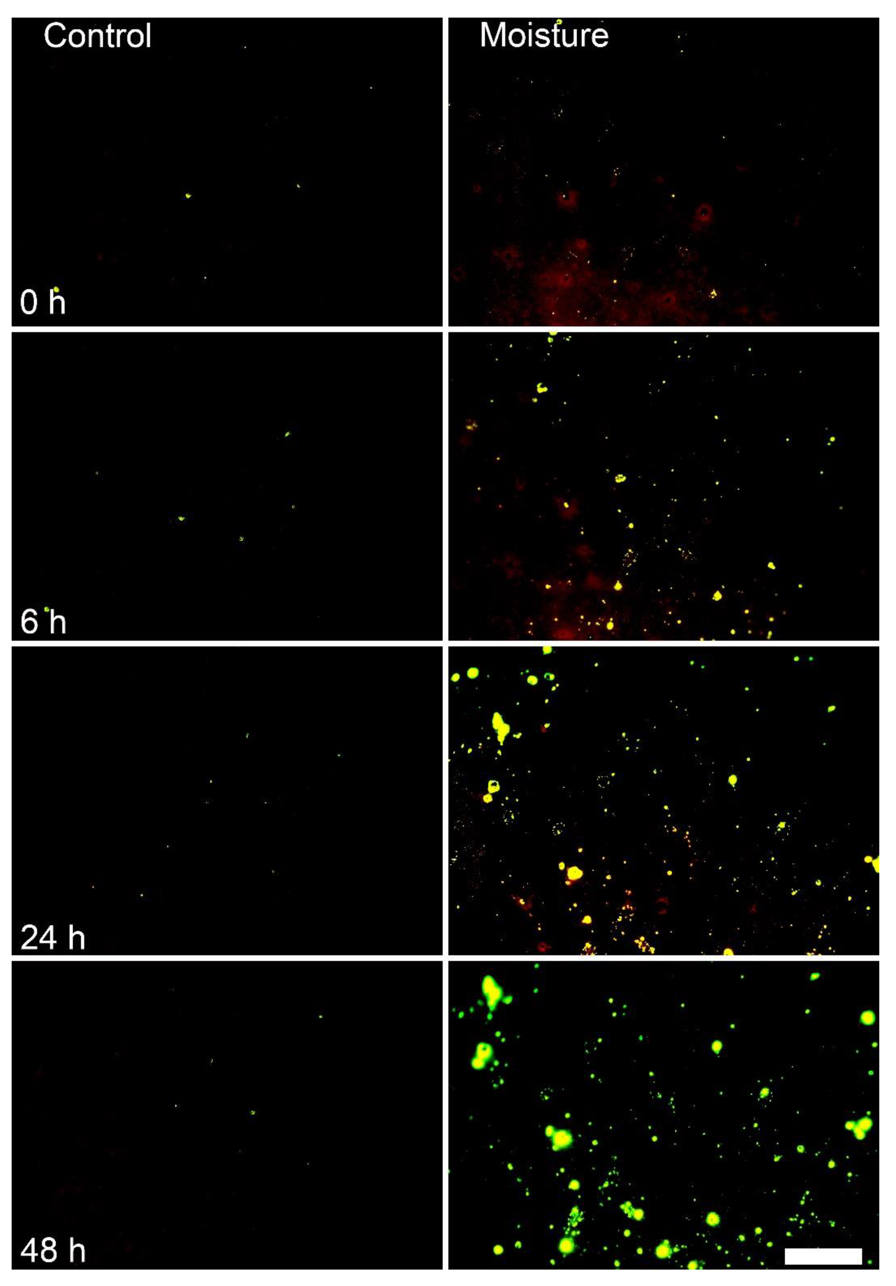
Figure 1. Effect of moisture exposure of the surface of fruit of mango cv. apple on microcracking of the cuticle. The fruit surface was exposed to surface moisture for 0, 6, 24 or 48 h. An untreated surface remained dry and served as the ‘control’. Microcracking was visualized by infiltration with aqueous acridine orange. N = 10. Scale bar 1 mm.
Horticulturae 08 00545 g001
Figure 2. (A) Time course of change in moisture-induced cuticular microcracking of fruit of mango cv. apple. (B) Time course of water loss through moisture-exposed fruit skins. Skins not exposed to moisture served as controls. The fruits were exposed to moisture for 8 d beginning at 53 days after full bloom (DAFB). (C) Time course of change in the water vapor permeance of the fruit skin during moisture exposure. Fruit was exposed to moisture for 4, 8, 12 and 16 d. Microcracking was indexed by quantifying the area infiltrated with aqueous acridine orange. Fruit surfaces that remained dry served as controls. N = 40 (microcracks) and 13–15 (permeance). Data represent the means ± SE.
Horticulturae 08 00545 g002
Figure 3. Developmental time course of change in fruit mass and surface area of mango cv. apple (A), microcracking following moisture exposure (B) and permeance to water vapor (C) following 8 days exposure to surface moisture. (D) Relationship between permeance to water vapor and microcracking (D). Untreated surfaces served as controls. Microcracking was indexed by quantifying the area infiltrated with acridine orange. X-axis scale in A, B and C is days after full bloom (DAFB). N = 20 (mass and surface area), 40 (microcracking) and 13–15 (permeance). Data represent the means ± SE. Significance of coefficients of determination (r2) at p ≤ 0.01 is indicated by **.
Horticulturae 08 00545 g003
Figure 4. Time course of changes in microcracking (A) and permeance to water vapor (B) of fruit skin of mango cv. apple after 8 days exposure to moisture. X-axis scale in days after treatment (DAT). At 72 days after full bloom, the cheek of the developing fruit was exposed to moisture for 8 d. Thereafter, the time course of change in microcracking and permeance after moisture termination was followed. Unexposed fruit surfaces served as controls. N = 40 (microcracking) and 12 (permeance). Data represent the means ± SE.
Horticulturae 08 00545 g004
Figure 5. Images of mature fruits of mango cv. apple (A,D,G) and histological micrographs of moisture-treated and control fruit skin taken under bright (B,E,H) and incident fluorescent light (C,F,I) as affected by a surface moisture treatment (see text for details). Control fruit (no tube attached, not exposed to moisture) (A,B,C), open tube (tube attached but not exposed to moisture) (D,E,F) and moisture-treated fruits (tube attached and exposed to moisture) (G,H,I) and enlarged micrograph of moisture-treated skin (J). Fruit was exposed to surface moisture by attaching a tube filled with distilled water to the fruit surface for 16 days at 53 days after full bloom (DAFB). Untreated fruit and fruit with an open tube attached that remained dry served as controls. At the end of the treatment period, tubes were removed and fruits were left to grow to maturity. The red circle identifies the area previously enclosed by the plastic tube. N = 18–20 (whole fruits) and 3 (histology). The scale bars are 20 mm (A,D,G), 100 µm (B,C,E,F,H,I) and 50 µm (J).
Horticulturae 08 00545 g005
Table 1. Effect of exposure to surface moisture on the russeting of fruit of mango cv. apple. Fruit was exposed to surface moisture by attaching a short plastic tube filled with distilled water to the fruit surface for 16 d at 53 days after full bloom. Untreated fruit (control) and fruit with an open tube attached (open tube, no water) served as controls. Tubes were removed after 16 d and the fruits left to grow to maturity. Russeting was estimated by expressing the area russeted as a percentage of the area of the footprint of the tube. N = 18–20.
TreatmentArea Russeted (%)
Untreated fruit1.3 ± 0.2 a
Open tube, no moisture1.3 ± 0.2 a
Closed tube, moisture20.2 ± 3.9 b
Mean separation by Tukey Studentized range test, p ≤ 0.05. Means followed by the same letter are not significantly different. Data represent the means ± SE.
Publisher’s Note: MDPI stays neutral with regard to jurisdictional claims in published maps and institutional affiliations.