Article

Self-Incompatibility of Camellia weiningensis Y.K. Li.

Institute for Forest Resources and Environment of Guizhou, Key Laboratory of Forest Cultivation in Plateau Mountain of Guizhou Province, College of Forestry, Guizhou University, Guiyang 550025, China
*
Author to whom correspondence should be addressed.
Horticulturae 2022, 8(4), 297; https://doi.org/10.3390/horticulturae8040297
Submission received: 27 February 2022 / Revised: 22 March 2022 / Accepted: 28 March 2022 / Published: 31 March 2022
(This article belongs to the Section Genetics, Genomics, Breeding, and Biotechnology (G2B2))

Abstract

:
This study compared the pollen tube growth, fruit setting, and seed setting characteristics of Camellia weiningensis Y.K. Li. under self- and cross-pollination to identify its self-incompatibility characteristics and types. C. weiningensis pollen tube growth was observed by fluorescence and scanning electron microscopy, and a field experiment with manual pollination verified fruit and seed setting characteristics. Both self- and cross-pollinated pollen germinated from the stigma. At 72 h after cross-pollination, the pollen tube reached the style base, with tube growth showing a slow-fast-slow pattern. The tube growth speed was maximal, 343.36 μm·h−1, at 12–24 h after pollination. For self-pollination, the pollen did not germinate on the stigma 4 h before pollination. At 12–24 h after pollination, the growth rate was maximal at 263.36 μm·h−1. At 96 h, a small amount of pollen reached the style base and stagnated. The pollen tube end showed callose reactions, such as abnormal swelling, distortion, and brightness. In the field experiment, the fruit setting rate under cross-pollination was 68.5%, while that under self-pollination was 15.3%. When the fruit grew to maturity, the growth dynamics of the transverse and longitudinal diameters showed a “slow-fast-slow”, S-shaped curve. The number of aborted selfed and outcrossed seeds was 13.9 and 4.7, respectively. Thus, C. weiningensis showed self-incompatibility. The self-incompatibility reaction occurred at the style base and represented prezygotic self-incompatibility. The self-incompatibility of C. weiningensis is one of the main reasons for its low seed setting rate, which should be fully considered in cross breeding.

1. Introduction

Self-incompatibility is an evolutionary mechanism widely existing in most angiosperms that inhibit inbreeding and promote high heterozygosity of the offspring [1,2,3]. According to its genetic mechanism, the patterns of self-incompatibility are usually divided into sporophyte self-incompatibility (SSI) and gametophyte self-incompatibility (GSI) [4,5,6]. Self-pollinated pollen with the same genotype (including asexual reproductive materials) does not germinate on the stigma, or the germinated pollen tube cannot grow normally in the pistil, with the consequence that the fruit setting rate under self-pollination (the same genotype) is much lower than that under cross-pollination (different genotypes) [7,8,9]. In the cytological study of self-incompatibility reactions, fluorescence microscopy serves as the most efficient, most straightforward, and most convenient experimental means for observing pollen germination and pollen tube growth [10,11]. According to observations of pollen tube growth in the stigma and style of Camellia plants under a fluorescence microscope, when the pollen tube germinates in the stigma but cannot reach the base of the style, this is associated with the incompatibility of the pollen with the style canal [12]. With outcrossing, the pollen tubes of Camellia continued to grow into the ovary in the hollow axial placenta; in contrast, for self-pollinated pollen tubes, only a small amount could enter the ovary, and they stopped growing in the ovary and consequently could not reach the ovule, resulting in serious ovule abortion [13,14]. Similarly, comparative studies of fruit setting and seed setting after self-pollination and cross-pollination have also provided powerful field evidence to verify self-incompatibility. Hazelnut self-pollination reduces the quality and weight of nuts [15]; the podding rate of Acacia species is very low (or even close to 0) after self-pollination, indicating self-incompatibility characteristics in these species [16,17,18].
Camellia weiningensis is an important species of Camellia in the Theaceae family. It is distributed in alpine habitats within an altitude range of 1800–2700 m in Guizhou Province, China; this endemic species is only distributed in this area of the world [19]. Camellia weiningensis has the advantages of strong adaptability, resistance to barren landscapes, a deep root system, being evergreen in all four seasons and having highly nutritious oil, providing important application value in maintaining species stability, preventing and controlling soil erosion and producing edible oil in high-altitude mountainous areas. Camellia is an economically important plant group in general; tea [20,21,22], tea oil, and other products widely loved by people all over the world originate from this genus. However, the efficient utilization of Camellia is confronted with a series of production problems, such as cultivar improvement [2,23], cross breeding [24,25], and pollination configuration [14], and these problems are all related to the understanding of its reproductive and developmental characteristics. Camellia sinensis and Camellia oleifera (two other Camellia species) have been proven to have self-incompatibility [20,26,27,28,29,30], and studies on their self-incompatibility mainly focus on gametophyte self-incompatibility [13,31,32]. Particularly in recent years, the discovery of late self-incompatibility in these two species of Camellia [33,34,35] has further expanded the basic data on the reproductive biology of Camellia.
In actual production practice, our research team found that a severe fruit setting problem existed in C. weiningensis, despite of its normal flowering. Additionally, our team found that the seed setting rate of self-pollinated C. weiningensis was much lower than that with cross-pollination in a previous pollination experiment, and we preliminarily assumed the existence of self-incompatibility. Despite that, as a species of Camellia with an important niche, the reproductive biology of C. weiningensis has attracted increasing attention in recent years [36]; no studies involving pollination of C. weiningensis have as yet been reported, to the best of our knowledge. An in-depth understanding of the reproductive biology of C. weiningensis is an important premise for breeding and improvement, which can, in particular, provide guidance for interspecific and intraspecific artificial hybridization breeding or distant hybrid breeding to create new germplasms.
Based on the aforementioned information, in this study, we investigated the self-incompatibility of Camellia weiningensis. To achieve this goal, we observed the growth of pollen tubes in pistils under artificial self-pollination and cross-pollination using fluorescence microscopy and scanning electron microscopy, and performed a field pollination test to verify the characteristics of self-pollination, fruit setting, and seed setting. The following questions were at issue: (1) What were the pistil structural characteristics of C. weiningensis? (2) What were the characteristics of pollen tube germination and growth under different pollination conditions? (3) What would be the impact of different pollination modes upon fruit growth? The results of this study should enrich the research data on Camellia and provide a theoretical basis for expanding production and exploiting the reproductive development of Camellia species in high-altitude areas worldwide.

2. Materials and Methods

2.1. Experimental Materials

The experimental site was located in the Camellia weiningensis Scientific Research Base of Guizhou University, Weining County, Bijie City, Guizhou Province, China (lat. 27°11′53″ N, long. 104°7′53″ E). The altitude of the site is 2200 m, with an annual average precipitation of 909 mm. The annual average temperature is 11.5 °C, the annual average maximum temperature in summer is 18 °C, the annual average frost-free period is 206 days, and the annual average sunshine hours are 1812 h. In this region, the light and heat conditions are sufficient, the annual temperature difference is small, and the daily temperature difference is large, with a subtropical monsoon humid climate and yellow brown soil. Three clones (designated ‘GW1’, ‘GW2’ and ‘GW3’) that had been observed for a long time prior to this study and could blossom and bear fruit normally with satisfactory characteristics were selected. The age of the trees was 15 years, and the tree size was medium. All of the trees were healthy without diseases and pests. The trees were maintained under normal water and fertilizer management (watering once a month with organic fertilizer application once a year).

2.2. Methods

2.2.1. Self- and Cross-Pollination

From 2019 to 2021, the pollen of clones of ‘GW1’, ‘GW2’ and ‘GW3’ during the full flowering period was separately collected from male parents (flower buds that were just about to open were selected to avoid possible errors caused by pollen pollution). Similarly, during pollination, the clone flower buds that were about to open were selected as female parents. The stamens were removed manually. According to the pollination combination, the pollen was collected with a pollination rod and applied onto the stigma for pollination (ideally, the stigma was covered completely with yellow pollen grains). The pollinations were performed from 9:00–12:00 a.m. on sunny days. After pollination, the pollinated flower was bagged with sulfuric acid paper and labeled, and the bag was removed after 7 days. The designed pollination combinations are summarized in Table 1.

2.2.2. Fluorescence Microscopy

According to the results of the pre-experiment, the pistils at 4, 12, 24, 48, 72, and 96 h after pollination were collected (pistils that did not receive pollination treatment were used as the control). After collection, the materials were fixed in a centrifuge tube containing Carnot’s fixed solution (Vabsolute ethanol: Vglacial acetic acid = 3:1) for 1 day, transferred into 70% ethanol and stored in a 4 °C refrigerator until use. The style was cut off from the base and then rehydrated with 70%, 50%, and 30% alcohol gradients for 30 min. The sample was washed three times with distilled water (10 min per wash). Afterward, the sample was transferred to 8 mol·L−1 NaOH for 3 h of softening. The sample was rinsed thrice with distilled water, soaked for 30 min, and then dyed in 0.1% aniline blue staining solution for 5 h. The style was gently flattened and then pressed onto a slide. Dye was applied by a dropper, and another slide was used to cover the material (the slide did not need to be pressed). The germination of the pollen on the stigma and the growth of the pollen tube in the style were observed under a fluorescence microscope (Leica DM3000, Weztlar, Germany).

2.2.3. Scanning Electron Microscopy

The flower buds at the pistil and stamen formation stage, mature styles, and stigmas when anther powder was dispersed, stigmas where pollen germinated, and pollen tubes in the style were selected. The samples were fixed onto the sample table with conductive tape and plated with gold in an ion sputtering instrument (MSP-mini; IXRF, Austin, TX, USA) for 30 s. Then, the samples were observed with a scanning electron microscope (Hitachi TM4000 plus, Tokyo, Japan) under a 10 KV voltage, high vacuum, and BSE mode. Photos were taken.

2.2.4. Stereoscopy

Complete pistils were selected, and the stigma, style, and ovary were observed using stereoscopy (Leica KL300 LED, Berlin, Germany). For each treatment group, three plants were selected and 10 flowers were collected from each plant. The petal length and width, corolla diameter, filament length, and style length were measured with a vernier caliper. In the meantime, the number of stamens and ovules were counted.

2.2.5. Field Pollination Experiment

During the full flowering period, artificial pollination was performed after emasculation (self-pollination, ‘GW1’ × ‘GW1’, ‘GW2’ × ‘GW2’, ‘GW3’ × ‘GW3’; cross-pollination, ‘GW3’ × ‘GW1’, ‘GW1’ × ‘GW2’, ‘GW2’ × ‘GW3’). For each combination, 100 flowers were used. The experiment was repeated three times. One week after pollination, the bags were removed. Fruit setting was observed from 1 month after pollination to the fruit maturation stage. Additionally, the fruit set number and seed set number were counted at the time of fruit maturation.

2.3. Statistical Analysis

Excel 2019 and SPSS 26.0 were used for data analysis. Data are presented as the mean ± deviation. The Duncan test was performed for multiple comparisons, and a difference of p < 0.05 was considered to be statistically significant.

3. Results

3.1. Structural Characteristics of Flower Organs

The flower organ of C. weiningensis was composed of a petiole, receptacle, calyx, corolla, stamen, and pistil, and the petals were pink (Figure 1A–C). The pistil consisted of consisted of three parts, namely, the stigma, style, and ovary (Figure 1D–F). The stamen consisted of anthers and filaments. In all clones, the pistils (the styles) were shorter than the stamens. The flower organs differed slightly in number and parameters between the studied clones (Table 2): In ‘GW1’, the average diameter of the corolla was 71.18 ± 6.17 mm, and the length and width of the petal were 41.14 ± 3.71 mm and 29.24 ± 4.55 mm, respectively. The average number of stamens was 78 ± 8.71, the average number of ovules was 9 ± 2.07, and the length of the style was 16.58 ± 1.54 mm. In ‘GW2’, the average diameter of the corolla was 75.65 ± 4.74 mm, with a length and width of 40.59 ± 2.15 mm and 29.57 ± 1.88 mm, respectively. The average number of stamens was 77 ± 8.10, the average number of ovules was 8 ± 1.69, and the average length of the style was 16.02 ± 0.51 mm. In ‘GW3’, the average diameter of the corolla was 79.02 ± 4.34 mm, with a length and width of 46.86 ± 1.92 mm and 31.31 ± 1.63 mm, respectively. The average number of stamens was 81 ± 7.98, the average number of ovules was 9 ± 1.57, and the average length of the style was 17.93 ± 1.48 mm. During pistil formation, young mastoid cells could be observed on the surface of the stigma. They were consistent in size and closely arranged (Figure 1G,J). When the stigma was mature, the mastoid cells on the contact surface were narrow, striped, and loosely arranged, and the stigma belonged to the wet type with secretions to capture pollen as much as possible (Figure 1H,K) and to accept pollen grains during the flowering period (Figure 1I). The pollen grains germinated on the stigma and grew pollen tubes (Figure 1L).

3.2. Pollen Germination

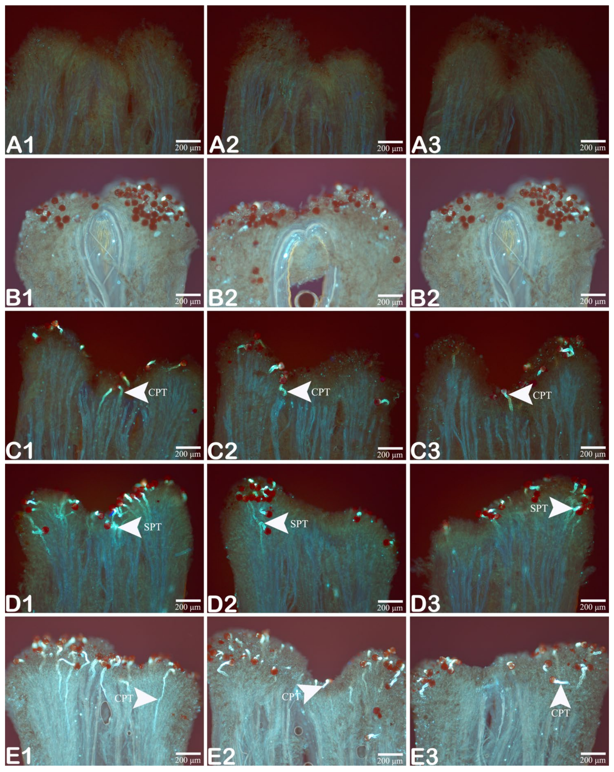
After water-soluble aniline blue dye application, the pollen grains normally germinate and the pollen tubes can display strong blue and green fluorescence reactions. As shown in Figure 2, the pollen of the self-pollinated ‘GW1’, ‘GW2’ and ‘GW3’ did not germinate at 4 h after pollination compared with the control (Figure 2A1–A3), and no pollen tubes were observed on the stigma (Figure 2B1–B3). In the cross-pollination group, noticeable differences could be observed at 4 h compared with the self-pollination group: most of the pollen began to germinate, and the germinated pollen tubes were tadpole-shaped; the pollen tube that germinated from the germination pore entered the stigma through mastoid cells. In the ‘GW3’ × ‘GW1’ group, 82.3% of the pollen germinated on the stigma (Figure 2C1) and the germination rates of the ‘GW1’ × ‘GW2’ and ‘GW2’ × ‘GW3’ groups were 73.6% and 64.8%, respectively (Figure 2C2,C3). At 12 h after self-pollination, a large amount of pollen germinated on the stigma, and the germination rates of ‘GW1’ × ‘GW1’, ‘GW2’ × ‘GW2’, and ‘GW3’ × ‘GW3’ were 92.3%, 80.4%, and 85.6%, respectively (Figure 2D1–D3). At 12 h after cross-pollination, the extension of the pollen tube became noticeable. In ‘GW3’ × ‘GW1’, a small number of the pollen tubes had already passed through the stigma completely and entered the style canal (Figure 2E1). In ‘GW1’ × ‘GW2’ (Figure 2E2) and ‘GW2’ × ‘GW3’ (Figure 2E3), the number of germinated pollen grains increased, and the pollen tubes continued to extend within the style canal.

3.3. Growth Behavior of the Pollen Tube

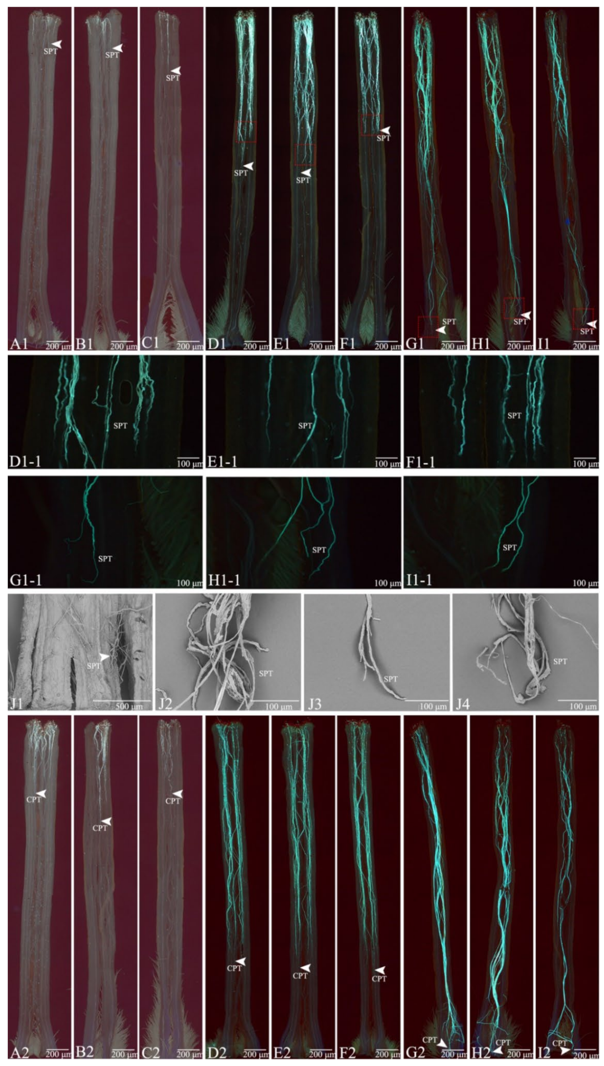
At 24 h after pollination, both selfed and crossed pollen tubes grew along the hollow style canal, and all selfed pollen tubes were shorter than their counterparts. At 24 h after pollination, the crossed pollen tube reached approximately 1/3 of the style (Figure 3A1,B1,C1)while the selfed pollen tube reached approximately 1/4 of the style (Figure 3A2,B2,C2). At 48 h, the crossed pollen tube grew to 2/3 of the style (Figure 3D1,E1,F1)while the selfed pollen tube reached approximately 1/3 of the style (Figure 3D2,E2,F2). At this moment, the growth of the pollen tubes was normal, displaying a strong growth trend toward the style base (Figure 3D1-1,E1-1,F1-1). At 72 h, a large amount of the crossed pollen tubes extended to the style base and grew downward from the ovary; during the whole growth process, the tubes were normal, and no abnormal growth was observed (Figure 3G1,H1,I1). At 96 h, most of the selfed pollen tubes stagnated at the halfway point of the style, with only a small proportion reaching the style base; the growth of the pollen tubes reaching the style base showed abnormalities and exhibited callose reactions, such as abnormal swelling, distortion, and brightness (Figure 3G2,G1-1,H2,H1-1,I2,I1-1). The selfed pollen tubes were removed and then observed under a scanning electron microscope. A small number of pollen tubes reached the base of the style, and the pollen tubes reaching the base showed various incompatibility reactions and failed to enter the ovary to reach the ovule (Figure 3J1,J2,J4). Only a very small proportion of the tubes was upright and continued to grow downward (Figure 3J3).

3.4. Comparison of Pollen Tube Growth between the Selfed and Crossed Groups

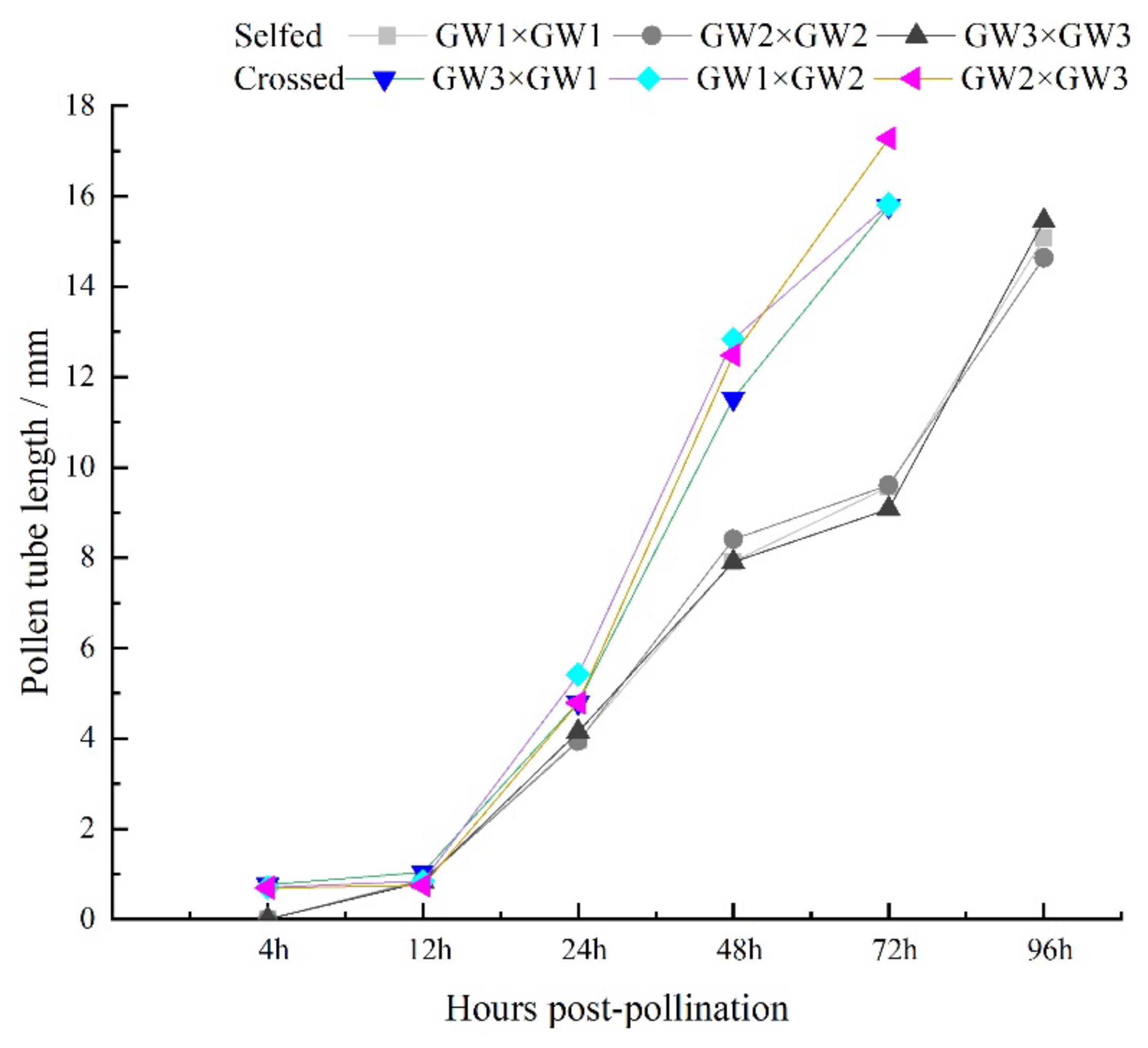
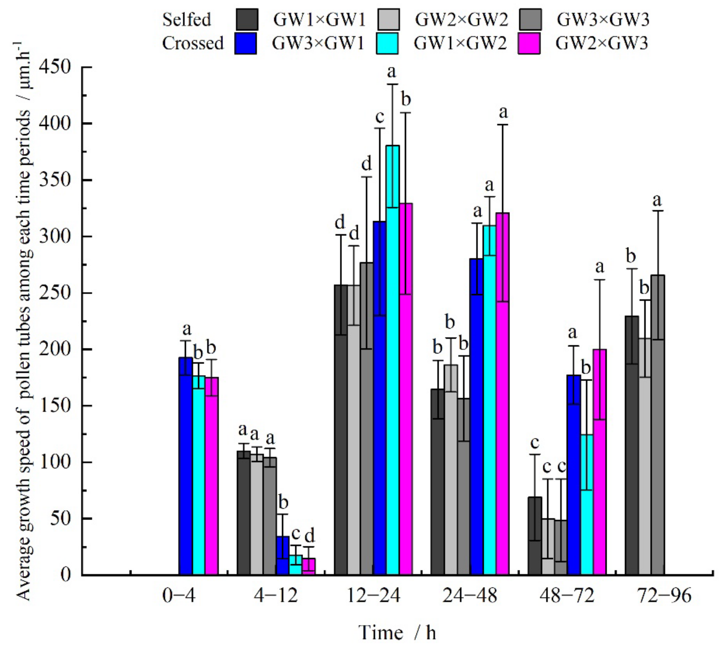
Based on the growth (in length) of the pollen tube (Table 3 and Figure 4), the growth speeds of the selfed and crossed pollen tubes showed a significant difference. The crossed pollen tube reached the base of the style earlier, at 72 h after pollination, than the selfed pollen tube. In contrast, the selfed pollen tube did not reach the style base until 96 h after pollination, which was 24 h later than its counterpart. At all time intervals, i.e., 0–4 h, 4–12 h, 12–24 h, 24–48 h, and 48–72 h, the length of the crossed pollen tube was significantly longer than that of the selfed pollen tube. The selfed and crossed pollen tubes also differed greatly in growth speed within any of the time intervals (Figure 5). Within 4 h after pollination, the crossed pollen tube germinated at an average speed of 181.33 μm·h−1, whereas the selfed pollen did not germinate and, therefore, no pollen tube was observed. Although at 4–12 h, the growth speed of the selfed pollen tube was faster than that of the counterpart (107.04 μm·h−1 vs. 19.25 μm·h−1) a fast growth speed was observed in the crossed pollen tubes within the remaining time intervals. At 12–24 h, both the selfed and crossed pollen tubes grew rapidly, at rates of 263.36 μm·h−1 and 343.36 μm·h−1, respectively. At 48–72 h, the crossed pollen tube reached the style base, and its growth speed was three times that of the selfed pollen tube. At 72–96 h, the selfed pollen tube reached the style base, while the crossed pollen tube continued to grow toward the ovary.

3.5. Field Experiment: Fruit Setting Rate

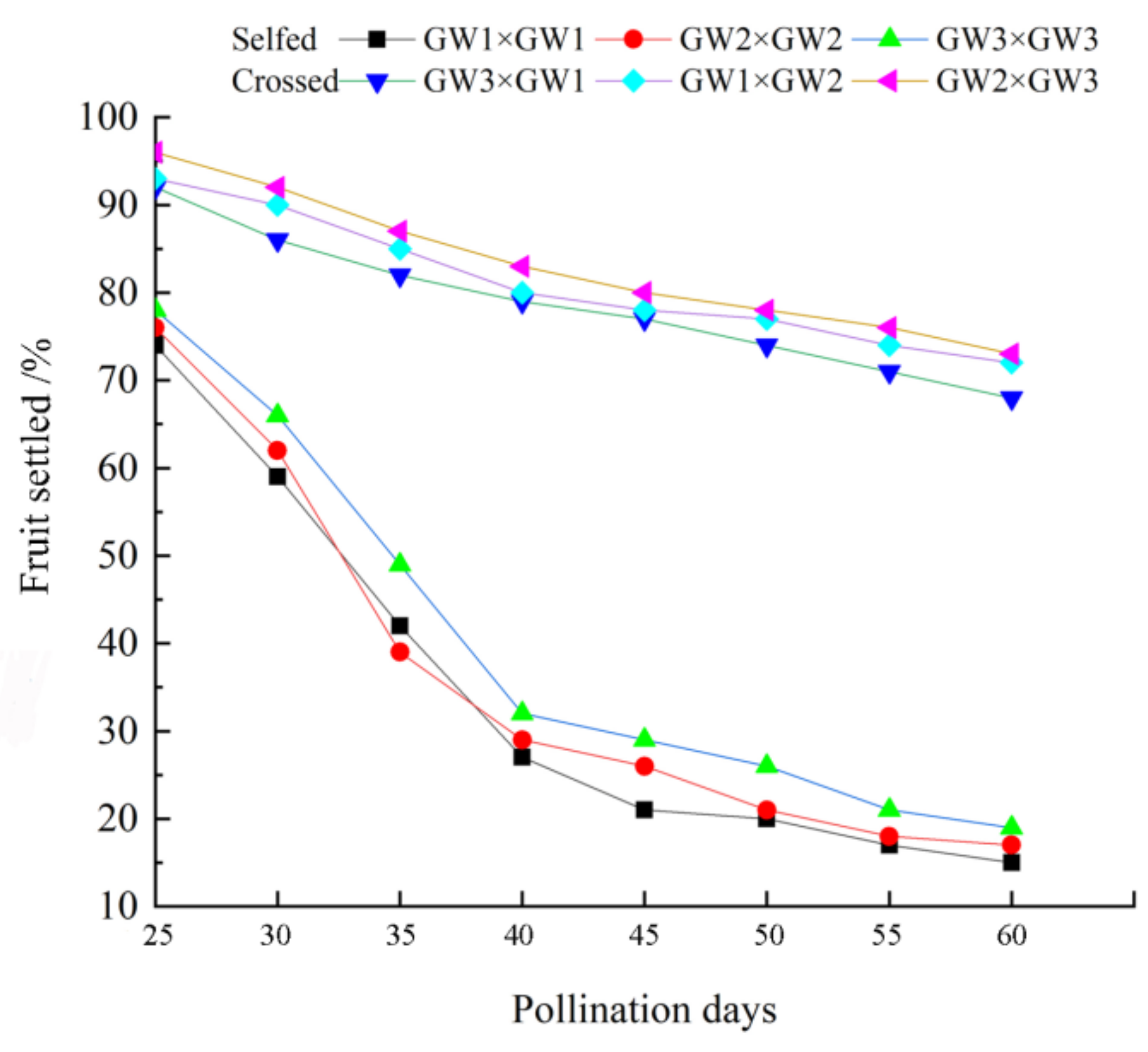
Based on observations as well as the outcomes of the pre-experiment, the duration of single flowers of C. weiningensis was 12–13 days, and the effective pollination period was 1–5 days after flowering, with the highest receptivity at 2 and 3 days after flowering, which was followed by a gradual decrease until receptivity ceased. Through the field experiment of self- and cross pollination of the three clones of C. weiningensis, the fruit setting dynamics in the initial stages of self- and cross-pollination were investigated (Figure 6). The outcrossing combinations ‘GW3’ × ‘GW1’, ‘GW1’ × ‘GW2’ and ‘GW2’ × ‘GW3’ outperformed the selfed combinations ‘GW1’ × ‘GW1’, ‘GW2’ × ‘GW2’, and ‘GW3’ × ‘GW3’ in fruit setting rate at any time period after pollination. The fruit setting rates and fruit drop of the selfed ‘GW1’ × ‘GW1’, ‘GW2’ × ‘GW2’ and ‘GW3’ × ‘GW3’ combinations showed basically consistent decreasing tendencies compared with the crossed ‘GW3’ × ‘GW1’, ‘GW1’ × ‘GW2’ and ‘GW2’ × ‘GW3’. Both the selfed and crossed groups showed the highest fruit setting rate at 25 days after pollination. With the passage of time, the fruit setting rate decreased gradually, and the number of dropped fruits increased. The highest fruit drop rate of both the selfed and crossed groups was at 30–40 days after pollination. At 40 days after pollination, the fruit drop rate of the crossed group became comparatively stable. At 60 days after pollination, the fruit setting rate of this group remained above 68%, and this rate remained almost unchanged until fruit ripening.
In the selfed group, although the fruit drop rate slowed down at 40 days after pollination, fruit drop continued to occur. It was not until 50 days after pollination that the fruit drop rate began to stabilize. At the time of fruit maturation, the fruit setting rate of the selfed group was only approximately 15%. The fruit setting rate of the crossed group was four times that of the selfed group.
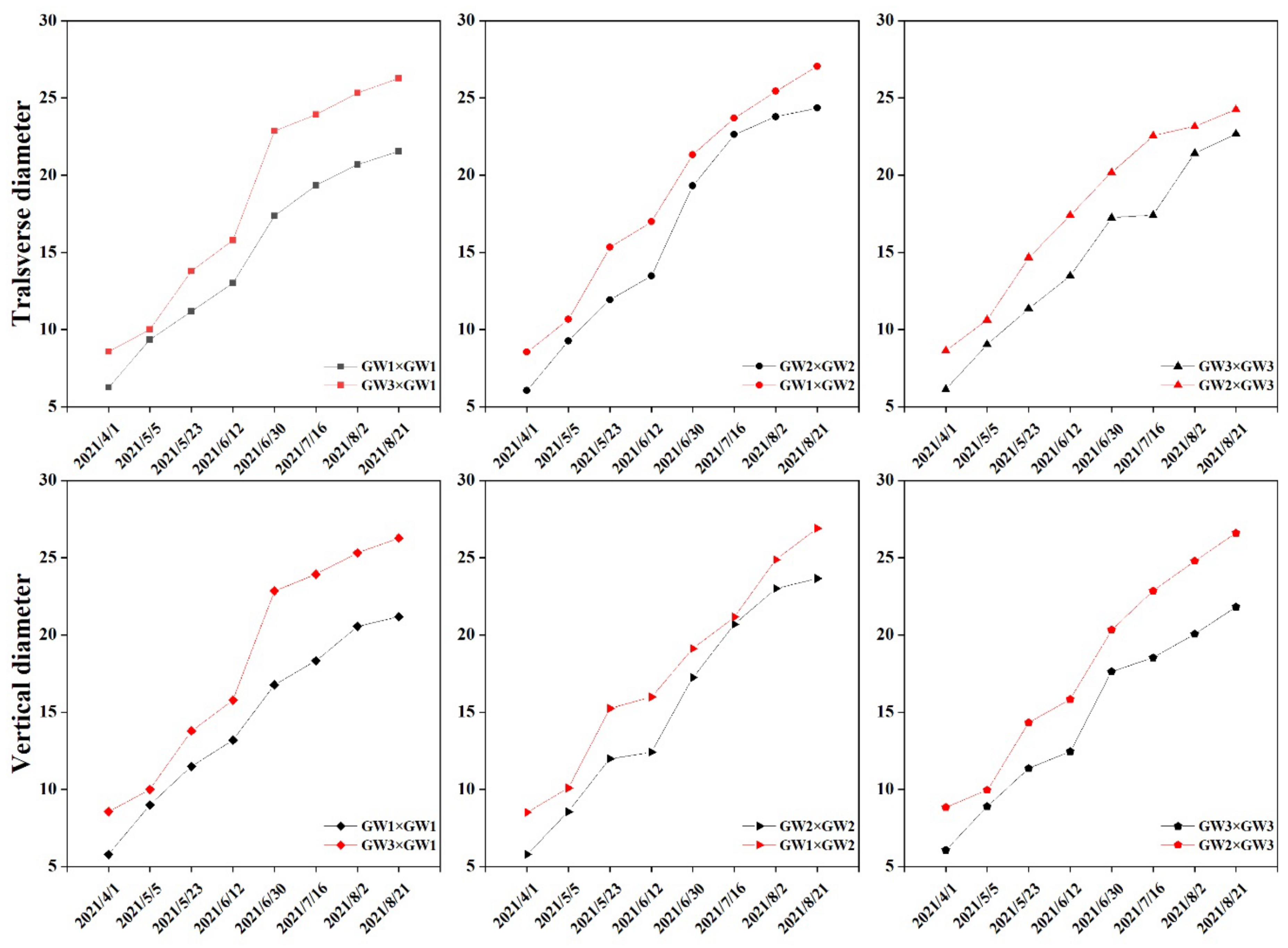
The growth dynamics of the transverse and longitudinal diameters of the fruit after self-pollination and cross-pollination showed the same increasing trend (Figure 7). From April to May, the growth and development of young fruit showed a constant upward trend, and the growth accelerated rapidly in June. When fruits were about to mature in August, the horizontal and vertical diameters tended to become stable. The growth process showed a “slow-fast-slow”, S-shaped curve. The transverse diameter of fruits was directly proportional to the longitudinal diameter. However, the transverse and vertical diameter curves of the crossed group were always above those of the selfed group. Therefore, the transverse and longitudinal diameters of fruit always maintained the growth characteristics confirming that outcrossed fruits outperformed selfed fruits.
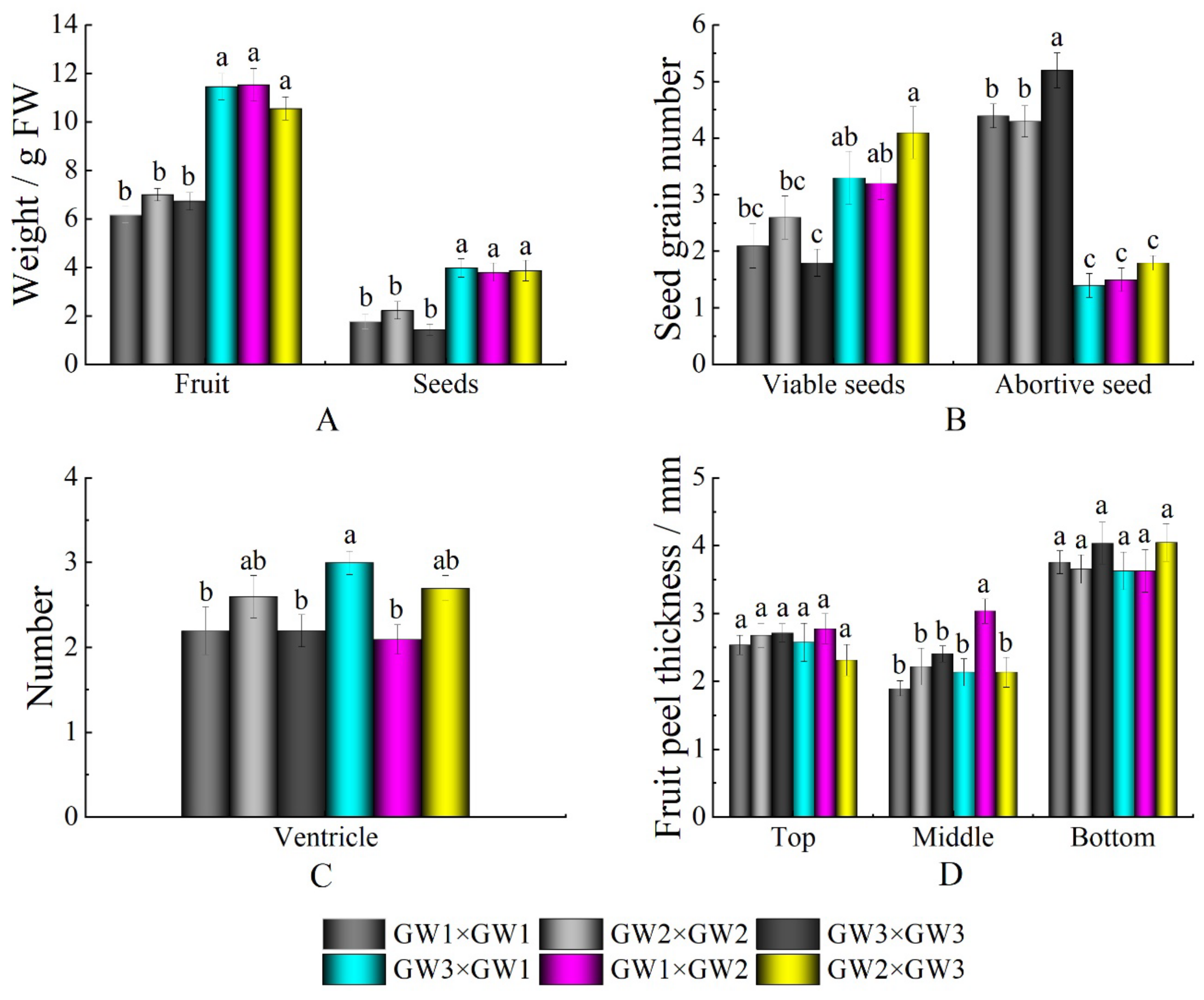
At the end of August of the current year, the fruit ripened, which took approximately six months from pollination. As shown in Figure 8, in terms of fruit volume, the selfed fruit was significantly smaller than the crossed fruit, and the fruit weight was directly proportional to the volume. The mature fruit was peeled and then measured and observed. The fresh seed weight under cross-pollination was twice that under self-pollination. The fruits mostly exhibited three ventricles. The peel thickness was comparable between the crossed and selfed groups. The numbers of aborted seeds of ‘GW1’, ‘GW2’, and ‘GW3’ were 4.4, 4.3, and 5.2, respectively, while those of ‘GW3’ × ‘GW1’, ‘GW1’ × ‘GW2’, and ‘GW2’ × ‘GW3’ were 1.4, 1.5, and 1.8, respectively. The total number of aborted seeds in the selfed group was 13.9, while that in the crossed group was 4.7. The abortion rate under selfed pollination was 2.95 times that under cross-pollination.

4. Discussion

The complete pistil of C. weiningensis is composed of the stigma, style and ovary, and serves as an important place for pollination and fertilization. The stigma is located at the top of the pistil and receives pollen. It generally maintains a certain receptivity at 1–4 days after flowering that begins to decline from 6 days. In nature, according to whether there is mucus on the surface of the pistil stigma when the species is fully mature, stigmas are divided into the wet type and the dry type. During pollination, the mucus secreted by the wet stigma not only reduces water loss but also increases the probability of pollen adhering to the stigma and promotes pollen germination [37].
In addition, research on the structural characteristics of the stigma and style in C. oleifera shows that the surface of the C. oleifera stigma is densely covered with mastoid cells, and the mastoid cells change from a single cell of cuticle membrane to a double-layered structure closely arranged with cellulose with electron clouds. The gap and electrification between protrusions are conducive to the attachment of pollen [38]. After flowering, the structure expands in an outward rolling shape to form the largest area, which is conducive to receiving as much pollen as possible. The stigma is covered with secretions, which are conducive to pollen adhesion and germination; moreover, this characteristic also plays an important role in the process of pollen identification by the stigma [39,40,41]. The style structure of C. oleifera is hollow, and there is often a layer of specialized inner epidermal cells around the style canal that can secrete substances into the style canal, provide nutrients for the growth of pollen tubes and guide the growth of pollen tubes [42]. The above anatomical evidence lays the biological foundation for the receptivity of the C. weiningensis stigma [36].
In this study, the pistil of C. weiningensis was anatomically observed. There was a large amount of mucus secretion on the surface of its stigma, indicating a typical wet stigma. The mastoid cells that recognize pollen covered a large area on the surface of the C. weiningensis pistil. In the process of flower opening, the stigmas exposed to the air generally were conducive to pollen adhesion. At the same time, the stigma rolled outward for maximum area to receive as much pollen as possible.
In flowering plants, pollination is very important for fruit development and affects the quantity and quality of fruits [43,44]. In the process of pollination and fertilization, pollen needs to undergo three processes: the identification of pollen on the stigma, positioning of the pollen tube in the style canal, and extension of the pollen tube to the ovary. If any of these processes are blocked, it will lead to abnormal pollination and fertilization and thus affect the seed setting rate. In this study, fluorescence staining microscopy was used to systematically observe the germination of pollen on the stigma and the growth process of the pollen tube in the style of selfed and crossed C. weiningensis. Both selfed and crossed C. weiningensis pollen could adhere to and germinate on the stigma, and the pollen tube grew to the base of the style in the style canal. Within the period of 0–4 h after pollination, the crossed pollen tube germinated on the stigma, but no fluorescence was observed in the selfed pollen. After 12 h, the selfed pollen tube began to germinate on the stigma. The growth rates of the pollen tubes under the cross- and self-pollination treatments in the early stage (0–48 h) were higher than those in the late stage (48–72 h). This phenomenon was consistent with the growth phenomenon of the binucleate pollen tube [45]. At 72 h, the pollen tube under the crossed treatment reached the base of the style, and the pollen tube generally showed a “slow-fast-slow” growth trend. In the selfed group, however, most of the pollen tubes stagnated at halfway point of the style canal, and then a small portion continued to grow. At 96 h, the pollen tubes reached the base of the style, but the end of the pollen tube reaching the base of the style showed swelling, curling, and bifurcation. This location is the site of self-incompatibility in C. weiningensis. After pollination, the germination and growth of the pollen tube in C. weiningensis lagged significantly behind those of ordinary C. oleifera [32,39]. This phenomenon might have been related to the growth environment of the species: Camellia weiningensis grows in alpine areas above 1800 m above sea level and shows strong cold resistance; low temperature leads to slow growth of the species, and, therefore, it may lead to the phenomenon of late germination during field pollination.
Self-incompatibility is a common genetic control mechanism of flowering plants in nature. It is a process that prevents inbreeding and ensures the long-term evolution of genetic diversity [46,47], especially in the process of cross breeding of fruit trees. Based on the seed setting rate, the compatibility of selfing and crossing can be estimated for most plants [48]. In this study, the effects of the different pollination treatments on the fruit set of C. weiningensis in the field were significantly different. The fruit setting rate of the crossed treatment group was five times that under the self-pollination treatment, and the abortion rate under the self-pollination treatment was high. Camellia weiningensis has strong self-incompatibility and is a typical outcrossing plant. Artificial pollination can improve the fruit setting rate. Similar findings have also been reported in C. oleifera, a well-researched Camellia species, whose yield can be improved by promoting outcrossing [14]. In addition, the endogenous quality of fruits under different pollination treatments was not measured in this experiment. The quality of fruits with different pollination treatments needs to be investigated further.

5. Conclusions

Through field controlled-pollination experiments, observation of C. weiningensis pollen tube growth, and determination of the fruit setting rate in the field, this experiment demonstrated that self-incompatibility is the main reason for more flowers and fewer fruits and serious flower and fruit drop in Camellia weiningensis. Therefore, planting methods employing a single variety (clone) should be changed in future plantings of C. weiningensis, and multiple cultivars should be selected for mixed planting to ensure their normal fruit setting and fruit quality and achieving satisfactory economic benefits. However, the degree of cross-compatibility between the selected strains must be considered in advance in the selection of mixed varieties.

Author Contributions

Conceptualization, C.G.; methodology, C.G.; software, H.W.; validation, L.Y.; formal analysis, L.L.; investigation, J.Q. and L.L.; resources, C.G.; data curation, L.Y.; writing—original draft preparation, H.W.; writing—review and editing, C.G.; visualization, J.Q.; supervision, C.G.; project administration, C.G.; funding acquisition, C.G. All authors have read and agreed to the published version of the manuscript.

Funding

This work was supported by the National Natural Science Foundation of China (32060331) Science and Technology Plan Project of Guizhou Province of China (Qian Ke He Zhi Cheng [2019]2310), the Science and Technology Project of Guizhou Education Department (Qian Jiao He KY [2018]097), the Cultivation Project of Guizhou University ([2019]35), and the Research Project of Introducing Talents in Guizhou University ([2017]41).

Institutional Review Board Statement

Not applicable.

Informed Consent Statement

Not applicable.

Data Availability Statement

The data used for the analysis in this study are within the article.

Acknowledgments

We thank Jiayin Zhi for his assistance in sampling.

Conflicts of Interest

The authors declare no conflict of interest. The funders had no role in the design of the study; in the collection, analyses, or interpretation of data; in the writing of the manuscript, or in the decision to publish the results.

References

  1. Xavier, V.; Vincent, C. When the genetic architecture matters: Evolutionary and ecological implications of self versus nonself recognition in plant self-incompatibility. New Phytol. 2021, 231, 1304–1307. [Google Scholar]
  2. Chen, X.D. Analysis on the development and improvement of Zhejiang Safflower Camellia resources in Qingtian County. J. Agric. Catastrophol. 2021, 11, 188–189, 192. [Google Scholar]
  3. Sayyad-Amin, P.; Davarynejad, G.; Abedy, B. The effects of self-incompatibility control substances on yield and fruit growth traits of pear Pyrus communis L. Cultivar ‘Williams’. Erwerbs-Obstbau 2021, 63, 117–123. [Google Scholar] [CrossRef]
  4. Hou, S.; Hao, Z.Z.; Li, Y.H. Observation of pollen tubes behavior and early embryogenesis following self and cross-pollination in Chinese jujube. J. Fruit Sci. 2019, 36, 1515–1523. [Google Scholar] [CrossRef]
  5. Nettancourt, D.D. Incompatibility in Angiosperms; Springer: Berlin/Heidelberg, Germany, 1977. [Google Scholar]
  6. Allen, A.M.; Hiscock, S.J.; Franklin-Tong, V.E. Evolution and phylogeny of self-incompatibility systems in angiosperms. In Self-Incompatibility in Flowering Plants; Springer: Berlin/Heidelberg, Germany, 2008; pp. 73–101. [Google Scholar]
  7. Maskromo, I.; Larekeng, S.H.; Novarianto, H.; Sudarsono, S. Xenia negatively affecting kopyor nut yield in Kalianda Tall kopyor and Pati Dwarf kopyor coconuts. Emir. J. Food Agric. 2016, 28, 644–652. [Google Scholar] [CrossRef] [Green Version]
  8. Franklin-Tong, V.E.; Ride, J.P.; Franklin, F.C.H. Recombinant stigmatic self-incompatibility (S-) protein elicits a Ca2+ transient in pollen of Papaver rhoeas. Plant J. 1995, 8, 299–307. [Google Scholar] [CrossRef]
  9. He, Y.F.; Song, Q.Q.; Wu, Y.F.; Ye, S.T. TMT-based quantitative proteomic analysis reveals the crucial biological pathways involved in self-incompatibility responses in Camellia oleifera. Int. J. Mol. Sci. 2020, 21, 1987. [Google Scholar] [CrossRef] [Green Version]
  10. Duarte, M.O.; Oliveira, D.M.T.; Borba, E.L. Two self-incompatibility sites occur simultaneously in the same acianthera species (Orchidaceae, pleurothallidinae). Plants 2020, 9, 1758. [Google Scholar] [CrossRef]
  11. Johnson, S.D.; Butler, H.C.; Robertson, A. Breeding systems in Cyrtanthus (Amaryllidaceae): Variation in self-sterility and potential for ovule discounting. Plant Biol. 2019, 21, 1008–1015. [Google Scholar] [CrossRef]
  12. Wang, Y.; Wang, J.R.; Liu, M.J. Fluorescent microscope observation on pollen germination in different cross combinations of Chinese Jujube. Acta Agric. Boreali-Sin. 2008, 2008, 46–49. [Google Scholar]
  13. Gao, C.; Yuan, D.Y.; Yang, Y. Anatomical characteristics of self-Incompatibility in Camellia Oleifera. Sci. Silvae Sin. 2015, 51, 60–68. [Google Scholar]
  14. Hu, G.X.; Gao, C.; Fan, X.M. Pollination compatibility and xenia in Camellia oleifera. Hortscience 2020, 55, 898–905. [Google Scholar] [CrossRef]
  15. Fattahi, R.; Mohammadzedeh, M.; Khadivi-Khub, A. Influence of different pollen sources on nut and kernel characteristics of hazelnut. Sci. Hortic. 2014, 173, 15–19. [Google Scholar] [CrossRef]
  16. Casiva, P.V.; Vilardi, J.C.; Cialdella, A.M.; Saidman, B.O. Mating system and population structure of Acacia aroma and A. macracantha (Fabaceae). Am. J. Bot. 2004, 91, 58–64. [Google Scholar] [CrossRef] [Green Version]
  17. Kenrick, J. Review of pollen–pistil interactions and their relevance to the reproductive biology of Acacia. Aust. Syst. Bot. 2003, 16, 119–130. [Google Scholar] [CrossRef]
  18. Zhang, R.P.; Zhan, N.; Huang, L.J. Control pollination of Acacia mangium and Acacia auriculiformis. Mol. Plant Breed. 2020, 18, 5877–5882. [Google Scholar]
  19. Geng, J.B.; Xu, J.; Wang, J. Resource Survey on Camellia weiningensis Y.K. Li. sp. nov in Weining County, Guizhou Province and utilization status. J. Anhui Agric. Sci. 2019, 47, 128–131. [Google Scholar]
  20. Mondal, T.K.; Bhattacharya, A.; Laxmikumaran, M.; Ahuja, P.S. Recent advances of tea (Camellia sinensis) biotechnology. Plant Cell Tissue Organ Cult. 2004, 76, 195–254. [Google Scholar] [CrossRef]
  21. Chen, L.; Zhou, Z.X.; Yang, Y.J. Genetic improvement and breeding of tea plant (Camellia sinensis) in China: From individual selection to hybridization and molecular breeding. Euphytica 2007, 154, 239–248. [Google Scholar] [CrossRef]
  22. Wei, K.; Wang, L.Y.; Wu, L.Y. Transcriptome analysis of indole-3-butyric acid-induced adventitious rootformation in nodal cuttings of Camellia sinensis (L.). PLoS ONE 2014, 9, e107201. [Google Scholar] [CrossRef] [Green Version]
  23. Tan, L.Q.; Wang, L.Y.; Wei, K. Floral transcriptome sequencing for SSR markerdevelopment and linkage map construction in the tea plant (Camellia sinensis). PLoS ONE 2013, 8, e81611. [Google Scholar] [CrossRef] [Green Version]
  24. Chen, X.; Liu, T.K.; Hao, S. Differential gene expression analysis of self incompatible lines in tea by cDNA-AFLP. Acad. J. 2011, 10, 1684–5315. [Google Scholar]
  25. Huang, X.N.; Qi, Y.; Ye, P.M. The breeding of two new yellow Camellia hybrid. J. Anhui Agric. Sci. 2021, 49, 54–56, 66. [Google Scholar]
  26. Kaundun, S.S.; Matsumoto, S. Heterologous nuclear and chloroplast microsatellite amplification and variation in tea, Camellia sinensis. Genome 2002, 45, 1041–1048. [Google Scholar] [CrossRef]
  27. Chen, L.; Zhao, L.P.; Gao, Q.K. Generation and analysis of expressed sequence tags from the tender shoots cDNA library of tea plant (Camellia sinensis). Plant Sci. 2005, 168, 359–363. [Google Scholar] [CrossRef]
  28. Zhao, L.P.; Liu, Z.; Chen, L. Generation and characterization of 24 novel EST derived microsatellite from tea plant (Camellia sinensis) and cross-species amplification in its closely related species and varieties. Conserv. Genet. 2008, 9, 1327–1331. [Google Scholar] [CrossRef]
  29. Seth, R.; Bhandawat, A.; Parmar, R.; Singh, P.; Kumar, S.; Sharma, R.K. Global transcriptional insights of pollen-pistil interactions commencing self-incompatibility and fertilization in tea [Camellia sinensis (L.) O. Kuntze]. Int. J. Mol. Sci. 2019, 20, 539. [Google Scholar] [CrossRef] [Green Version]
  30. Zhou, J.Q.; Lu, M.Q.; Yu, S.S.; Liu, Y.Y.; Yang, J.; Tan, X.F. In-depth understanding of Camellia oleifera self-incompatibility by comparative transcriptome, proteome and metabolome. Int. J. Mol. Sci. 2020, 21, 1600. [Google Scholar] [CrossRef] [PubMed] [Green Version]
  31. McClure, B. New views of S-RNase-based self-incompatibility. Curr. Opin. Plant Biol. 2006, 9, 639–646. [Google Scholar] [CrossRef] [PubMed]
  32. Liao, T.; Yuan, D.Y.; Zou, F. Self-sterility in Camellia oleifera may be due to the prezygotic late-acting self-incompatibility. PLoS ONE 2014, 9, e99639. [Google Scholar] [CrossRef] [PubMed]
  33. Chen, X.; Hao, S.; Wang, L. Late-acting self-incompatibility in tea plant (Camellia sinensis). Sect. Bot. 2012, 67, 347–351. [Google Scholar] [CrossRef] [Green Version]
  34. Wachira, F.N.; Kamunya, S.K. Pseudo-self incompatibility in some tea clones (Camellia sinensis (L.) O. Kuntze). J. Hortic. Sci. Biotechnol. 2005, 80, 716–720. [Google Scholar] [CrossRef]
  35. Liao, T.; Yuan, D.Y.; Gao, C. Pollination, fertilization and early embryonic development of Camellia oleifera. Sci. Silvae Sin. 2014, 50, 50–55. [Google Scholar]
  36. Wei, H.L.; Gao, C.; Jie, Q. Flowering biological characteristics of Camellia weiningensis Y.K. Li. HortScience 2021, 56, 1331–1339. [Google Scholar] [CrossRef]
  37. Huo, H.L.; Zhang, R.Q. Stigma receptivity and characteristics of pollen tube growth of Corylus kweichowensis. For. Res. 2014, 27, 403–409. [Google Scholar]
  38. Ciampolini, F.; Cresti, M. The structure and cytochemistry of the stigma-style complex of Corylus avellana L., ‘Tonda Gentile delle Langhe’ (Corylaceae). Ann. Bot. 1998, 81, 513–518. [Google Scholar] [CrossRef] [Green Version]
  39. Gao, Q.G.; Lei, Z.Z.; Liang, Y.F. Screening of Brassica oleracea MLPK-interacting PUB proteins based on gene family analysis. Acta Hortic. Sin. 2019, 46, 714–722. [Google Scholar]
  40. Heslop-Harrison, Y.; Shivanna, K.R. The receptive surface of the angiosperm stigma. Ann. Bot. 1997, 41, 1233–1258. [Google Scholar] [CrossRef] [Green Version]
  41. Hiscock, S.J.; McInnis, S.M. Pollen recognition and rejection duringthe sporophytic self-incompatibility response: Brassica and beyond. Trends Plant Sci. 2003, 8, 606–613. [Google Scholar] [CrossRef]
  42. Chen, Y.; Wang, L.L.; Liu, W.Z. Pollination effects on the structure of tobacco’s (Nicotiana tabacum) transmitting tissue. Acta Bot. Boreali-Occident. Sin. 2016, 36, 486–492. [Google Scholar]
  43. Jahromi, H.A.; Zarei, A.; Mohammadkhani, A. Analysis the effects of pollen grain sources on the fruits set and their characteristics of ‘Clementine’ mandarin using microscopic and molecular approaches. Sci. Hortic. 2019, 249, 347–354. [Google Scholar] [CrossRef]
  44. Selak, G.V.; Cuevas, J.; Ban, S.G.; Perica, S. Pollen tube performance in assessment of compatibility in olive (Olea europaea L.) cultivars. Sci. Hortic. 2014, 165, 36–43. [Google Scholar] [CrossRef]
  45. Tangitcharoen, S.; Owens, J.N. Pollen viability and pollen-tube growth following controlled pollination and their relation to low fruit production in teak (Tectona grandis Linn. f.). Ann. Bot. 1997, 80, 401–410. [Google Scholar] [CrossRef] [Green Version]
  46. Pérez, V.; Herrero, M.; Hormaza, J.I. Self-fertility and preferential cross-fertilization in mango (Mangifera indica). Sci. Hortic. 2016, 213, 373–378. [Google Scholar] [CrossRef] [Green Version]
  47. Wang, G.M.; Gu, C.; Qiao, X.; Zhao, B.Y.; Ke, Y.Q.; Guo, B.B.; Hao, P.P.; Qi, K.J.; Zhang, S.L. Characteristic of pollen tube that grew into self-style in pear cultivar and parent assignment for cross-pollination. Sci. Hortic. 2017, 216, 226–233. [Google Scholar] [CrossRef]
  48. Mhanna, M.A.; Douay, F.; Rajab, M. Identifying the optimal pollinizer for ‘Dermlali’ olive cultivar under syrian coast conditions. Int. J. Fruit Sci. 2020, 20, 1984–1993. [Google Scholar] [CrossRef]
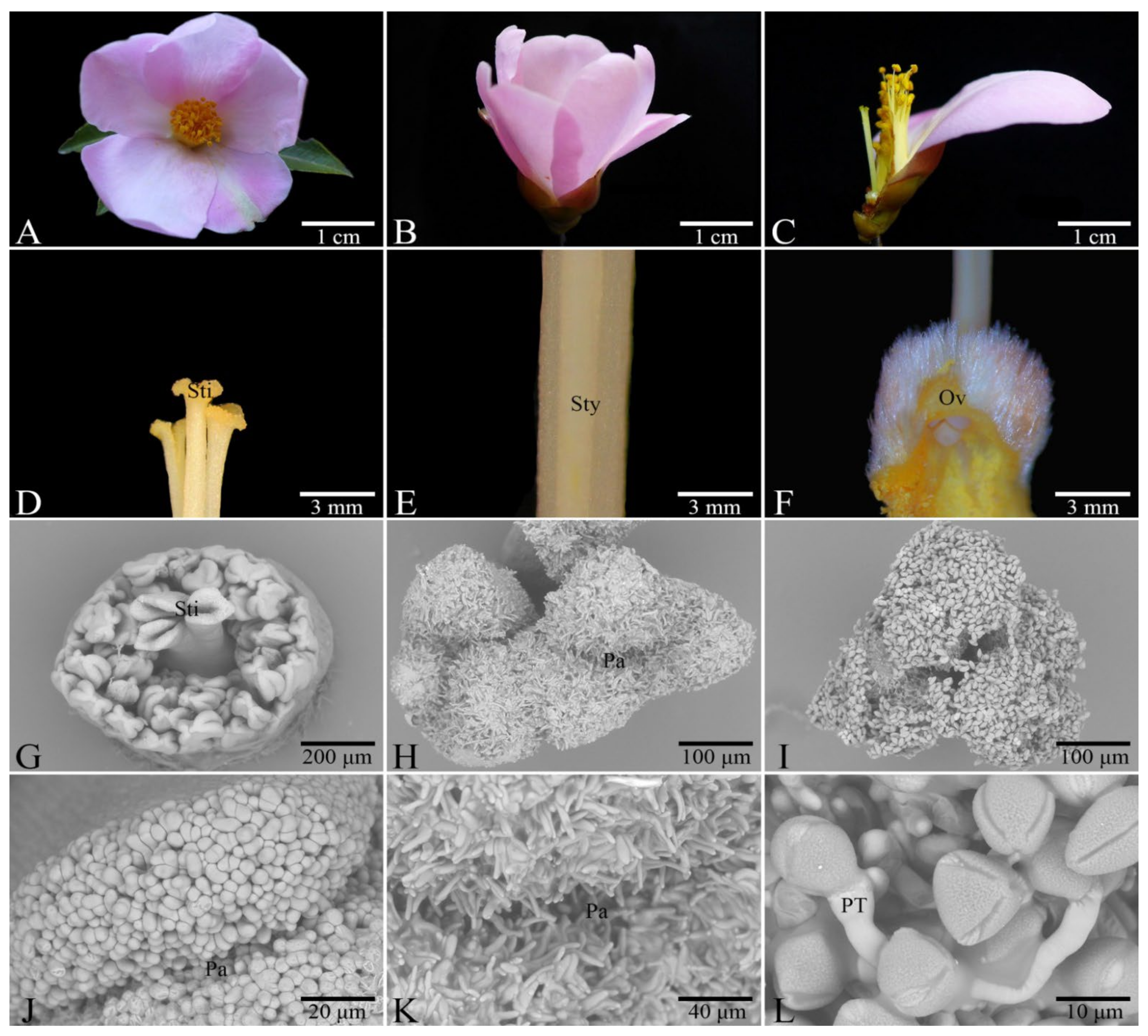
Figure 1. Characteristics of the flower organs of C. weiningensis. (AC), flower morphology; (D) the stigma; (E) the style; (F) the ovary; (G) the pistil and stamen formation stage; (H) the style maturation period; (I) the style covered with pollen after pollination; (J) short mastoid cells of the stigma arranged regularly during pistil formation; (K) mastoid cells formed at the time of stigma maturation; (L) pollen grains grew into pollen tubes after pollination. Sti: stigma; Sty: style; Ov: ovary; Pa: papilla; PT: pollen tube.
Horticulturae 08 00297 g001
Figure 2. Pollen germination on the stigma after self-pollination and cross-pollination. (A1) ‘GW1’ without pollination; (A2) ‘GW2’ without pollination; (A3) ‘GW3’ without pollination; (B1) ‘GW1’ at 4 h after self-pollination; (B2) ‘GW2’ at 4 h after self-pollination; (B3) ‘GW3’ at 4 h after self-pollination; (C1) stigma of ‘GW3’ × ‘GW1’ 4 h after pollination; (C2) stigma of ‘GW1’ × ‘GW2’ 4 h after pollination; (C3) stigma of ‘GW2’ × ‘GW3’ 4 h after pollination; (D1) stigma of ‘GW1’ 12 h after self-pollination; (D2) stigma of ‘GW2’ 12 h after self-pollination; (D3) stigma of ‘GW3’ 12 h after self-pollination; (E1) stigma of ‘GW3’ × ‘GW1’ 12 h after cross-pollination; (E2) stigma of ‘GW1’ × ‘GW2’ 12 h after cross-pollination; (E3) stigma of ‘GW2’ × ‘GW3’ 12 h after cross-pollination. SPT: selfed pollen tube; CPT: crossed pollen tube.
Horticulturae 08 00297 g002
Figure 3. Growth behavior of the selfed and crossed pollen tubes within the style. (A1) style of ‘GW3’ × ‘GW1’ at 24 h after pollination; (B1) style of ‘GW1’ × ‘GW2’ at 24 h after pollination; (C1) style of ‘GW2’ × ‘GW3’ at 24 h after pollination; (D1) style of ‘GW3’ × ‘GW1’ at 48 h after pollination. (E1) style of ‘GW1’ × ‘GW2’ at 24 h after pollination; (F1) style of ‘GW2’ × ‘GW3’ at 24 h after pollination; (G1) style of ‘GW3’ × ‘GW1’ at 72 h after pollination; (H1) style of ‘GW1’ × ‘GW2’ at 72 h after pollination; (I1) style of ‘GW2’ × ‘GW3’ at 72 h after pollination; (A2), style of ‘GW1’ at 24 h after self-pollination; (B2), style of ‘GW2’ at 24 h after self-pollination; (C2), style of ‘GW3’ at 24 h after self-pollination; (D2), style of ‘GW1’ at 48 h after self-pollination; (E2), style of ‘GW2’ at 48 h after self-pollination; (F2), style of ‘GW3’ at 48 h after self-pollination; (G2), style of ‘GW1’ at 96 h after self-pollination; (H2), style of ‘GW2’ at 96 h after self-pollination; (I2), style of ‘GW3’ at 96 h after self-pollination; (D1-1) the enlarged image of the red frame in (D2), which shows the end of the pollen tube; (E1-1) the enlarged image of the red frame in (E2), which shows the end of the pollen tube; (F1-1) the enlarged image of the red frame in (F2), which shows the end of the pollen tube; (G1-1) the enlarged image of the red frame in (G2), which shows the end of the pollen tube; (H1-1) the enlarged image of the red frame in (H2), which shows the end of the pollen tube; (I1-1) the enlarged image of the red frame in (I2), which shows the end of the pollen tube; (J1J4), at 96 h after self-pollination, the base of the self-pollinated style and the end of the pollen tube were observed under a scanning electron microscope. SPT: selfed pollen tube; CPT: crossed pollen tube.
Horticulturae 08 00297 g003
Figure 4. The growth of the selfed and crossed pollen tubes.
Horticulturae 08 00297 g004
Figure 5. The average growth speed of the selfed and crossed pollen tubes within different time periods. A different letter indicates a significant difference at the level of 0.05 between different treatment groups.
Horticulturae 08 00297 g005
Figure 6. Dynamic changes in the early fruit setting rates after self- and cross-pollination.
Horticulturae 08 00297 g006
Figure 7. Change patterns of the transverse and horizontal diameters of the fruits of the selfed and crossed groups.
Horticulturae 08 00297 g007
Figure 8. Fruit characteristics under the self- and cross-pollinations. (A), fresh fruit and fresh seed weight. (B), numbers of fertile and aborted seeds. (C), number of ventricles. (D), peel thickness. Different letters indicate significant differences between different treatment groups (p < 0.05).
Horticulturae 08 00297 g008
Table 1. Self- and cross-pollination combinations of C. weiningensis.
Cross-Pollination CombinationSelf-Pollination Combination
Male parent × Female parentMale parent × Female parent
GW3 × GW1GW1 × GW1
GW1 × GW2GW2 × GW2
GW2 × GW3GW3 × GW3
Table 2. Morphology of the flower organs of C. weiningensis.
MaterialPetal Length (mm)Petal Width (mm)Corolla Diameter (mm)Filament Length (mm)Style Length (mm)Number of StamensNumber of Ovules
GW141.21 ± 0.78b29.24 ± 0.65b71.18 ± 1.24c22.39 ± 0.72b16.58 ± 0.84ab78 ± 0.83b9 ± 0.71
GW240.59 ± 1.88b29.57 ± 0.73b75.65 ± 0.90b19.23 ± 1.09c16.02 ± 0.86b77 ± 1.30b8 ± 0.71
GW346.86 ± 1.18a31.31 ± 0.72a79.02 ± 1.48a23.75 ± 0.83a17.93 ± 0.66a81 ± 0.84a9 ± 1.14
Notes: Data are presented as the mean ± deviation. A different letter in the same column indicates a significant difference (p < 0.05).
Table 3. The growth of the selfed and crossed pollen tubes.
Pollination CombinationPollen Tube Length (μm)
4 h12 h24 h48 h72 h96 h
GW1 × GW10880 ± 5.77b3965 ± 75.067913 ± 46.93c9566 ± 60.48d15,069 ± 39.19ab
GW2 × GW20857 ± 8.82bc3935 ± 70.24c8407 ± 62.74c9605 ± 50.58d14,636 ± 326.67b
GW3 × GW30832 ± 3.33c4150 ± 45.83c7907 ± 58.02c9075 ± 56.86e15,453 ± 318.35a
GW3 × GW1770 ± 8.66a1044 ± 29.19a4800 ± 59.32b11,526 ± 299.82b16,479 ± 263.27b16,479 ± 263.27b
GW1 × GW2706 ± 9.35b848 ± 12.92bc5412 ± 108.10a12,836 ± 470.61a16,019 ± 52.87c16,019 ± 52.87c
GW2 × GW3700 ± 5.77b746 ± 7.37d4787 ± 75.92b12,485 ± 287.33ab17,382 ± 32.52a17,382 ± 32.52a
Notes: Data are presented as the mean ± deviation. A different letter in the same column indicates a significant difference at the level of 0.05.
Publisher’s Note: MDPI stays neutral with regard to jurisdictional claims in published maps and institutional affiliations.