Article

Low CO2 Levels Are Detrimental for In Vitro Plantlets through Disturbance of Photosynthetic Functionality and Accumulation of Reactive Oxygen Species

by
Naser Askari
1,2,*,
Sasan Aliniaeifard
3 and
Richard G. F. Visser
1
1
Plant Breeding, Wageningen University & Research, P.O. Box 386, 6700 AJ Wageningen, The Netherlands
2
Department of Horticultural Science, University of Jiroft, Jiroft 78671-55311, Iran
3
Photosynthesis Laboratory, Department of Horticulture, Aburaihan Campus, University of Tehran, Tehran 33916-53755, Iran
*
Author to whom correspondence should be addressed.
Horticulturae 2022, 8(1), 44; https://doi.org/10.3390/horticulturae8010044
Submission received: 16 November 2021 / Revised: 7 December 2021 / Accepted: 16 December 2021 / Published: 4 January 2022

Abstract

:
Photosynthesis of plantlets in tissue culture containers is not considered important, compared to photosynthesis of ex vitro plants, due to the exogenous source of carbohydrates present in tissue culture media. However, CO2 starvation can generate a burst of reactive oxygen species (ROS). We examined this phenomenon in tissue culture, since CO2 levels may become very low during the light period. The research was carried out with lily scales, regenerating adventitious bulblets, and with Arabidopsis seedlings. CO2 starvation was achieved by placing a small vial of concentrated KOH solution in the culture container. CO2 removal reduced the growth of regenerated lily bulblets by 33% or 23%, with or without sucrose in the medium, respectively. In Arabidopsis seedlings, CO2 removal decreased growth by 50% or 78% in the presence or absence of sucrose in the medium, respectively. Therefore, the addition of sucrose as a replacement for photosynthesis resulted in only partial recovery of growth. Staining with nitroblue tetrazolium (NBT) showed little to no ROS in ex vitro growing seedlings, while abundant ROS were detected in seedlings grown under in vitro CO2 starvation. Seedlings grown under normal tissue culture conditions (no CO2 withdrawal) showed low levels of ROS. In lily tissue culture, CO2 starvation decreased the maximum quantum efficiency of photosystem II (Fv/Fm) from 0.69 to 0.60, and in Arabidopsis from 0.76 to 0.62. Fv/Fm of ex vitro lily and Arabidopsis seedlings was 0.77 and 0.79, respectively. This is indicative of a disturbance in photosynthesis functionality and the occurrence of in vitro stress under reduced CO2 concentrations. We conclude that poor growth, in the absence of CO2, was partly due to strongly reduced photosynthesis, while the detrimental effects were most likely due to a burst of ROS.

1. Introduction

Although plants are dependent on photosynthesis to absorb light energy and obtain carbon and energy for development, reports on the evaluation of photosynthesis of in vitro plantlets are relatively rare [1]. The microenvironment in tissue culture containers is very different from the ex vitro environment in which plants normally grow. Apart from organic nutrition (sucrose), high external levels of plant growth regulators, and low light intensity, the tissue culture environment is characterized by a very unusual atmosphere: extremely high relative humidity (RH, 98–99.5%), high levels of organic gases, such as ethylene, and fluctuating high and low levels of CO2 and O2 [2,3]. During the light period, the CO2 concentration in an air-tight vessel with green plantlets is often lower than 100 µL L−1 which is much lower than the normal atmospheric CO2 concentration of 400 µL L−1, and may even reach 50 µL L−1 [4,5]. Even in loosely capped vessels, or vessels capped with gas permeable film, the concentration is often lower than 200 µL L−1 [6]. During the dark period, CO2 concentration increases up to 3000–9000 µL L−1. The unusual atmosphere in the headspace of culture vessels imposes several disorders, such as stomatal malfunctioning and photosynthesis disturbance, in the plant samples growing in vitro [7]. Generally, it is expected that the low CO2 concentration during the light period, along with the low light intensity, restrain photosynthesis. Poor photosynthesis in vitro may also be caused by the addition of sugar to the medium [8]. A reduction of sucrose enhances net photosynthesis, e.g., in coconut [9], and rain tree [10] plants. However, the addition of sugar is necessary for successful tissue culture. Despite the increase of photosynthesis, the omission of sucrose reduces the growth of plantlets dramatically, e.g., in grape [11] and Mountain ash [12] plantlets. Expectations are that a very low CO2 level during exposure to light is deleterious for plants, due to the lack of electron acceptors in the photosynthetic electron transport chain [13]. Furthermore, there is a general belief that, due to the presence of sugar in the medium, there is no need for photosynthesis in vitro. However, it has been confirmed that removal of the two main inputs of photosynthesis (light and CO2) are detrimental for production of plants in vitro [6]. In accordance, CO2 enrichment in vitro increased the shoot growth and multiplication rate in Celastrus paniculatus [14]. In the course of CO2 starvation, all endogenous electron acceptors become reduced, leaving oxygen as the main available electron acceptor. Oxygen can serve as an electron acceptor in the Mehler reaction [15]. Products of the Mehler reaction, such as superoxide, hydroxyl radicals, and hydrogen peroxide (reactive oxygen species, ROS) are toxic, attack vulnerable macromolecules, including chlorophyll, and bring about bleaching [16]. In the present study, we aimed to investigate growth, photosynthetic performance, and the occurrence of oxidative stress in tissue cultures of Arabidopsis seedlings (as a model plant, it is easy growing both in vitro and ex vitro) and lily bulblets, regenerating from scale explants under in vitro CO2 starvation. For this aim, we have used chlorophyll-a fluorescence to detect maximum quantum efficiency of photosystem II (Fv/Fm), as an indicator of the damage to the photosynthetic apparatus, and nitroblue tetrazolium (NBT) staining for detection of ROS.

2. Materials and Methods

2.1. Growth Conditions

Field-grown bulbs (circumference 18–20 cm) of Lilium cv. Santander were harvested, cold-treated to break dormancy, and stored at −1.0 °C until use. The procedure for lily tissue culture was performed according to [17]. Scales were surface-sterilized for 30 min in 1% (w/v) sodium hypochlorite (NaClO), rinsed for 1, 3, and 10 min with sterile water, and stored until use in sterile water (on average for 1–2 h). Two explants of 7 × 7 mm were cut from the scales and placed with the abaxial side on 30 mL solidified medium in plastic culture tubes (6.5 cm diameter). The medium was composed of macro- and microelements, as according to Murashige and Skoog [18]: 30 g L−1 sucrose, or no sucrose; 0.4 mg L−1 thiamine; 100 mg L−1 myo-inositol; 7 g L−1 Microagar; and 0.05 mg L−1 NAA (α-naphthaleneacetic acid). All chemicals were obtained from Duchefa (Haarlem, The Netherlands). Thirty scale explants per treatment were cultured for 12 weeks at 25 °C and 30 μmol m−2 s−1 light (Philips TL 33) for 16 h per day.

2.2. Arabidopsis Seedling Growth

Arabidopsis thaliana (Col-0) seeds were sterilized with 70% (v/v) ethanol for 1 min, and in 2% (w/v) sodium hypochlorite for 15 min. They were subsequently rinsed three times for 10 min with sterilized distilled water. Sterile seeds were transferred to ten Petri dishes with half-strength Murashige and Skoog salt mixture, including vitamins [18], supplemented with 30 g L−1 sucrose and solidified with 7 g L−1 Microagar. Seeds were stratified in the dark for 3 d at 4 °C, after which they were cultured in a growth chamber with 16 h light/ 8 h dark (30 μmol m–2 s–1, Philips TL33) at 21 °C. After 7 days, the seedlings were transferred to fresh medium with 30 g L−1 sucrose, or without sucrose, and then incubated for two weeks in a growth chamber with 16 h light/8 h dark (30 μmol m–2 s–1, Philips TL33) at 21 °C. Thirty Arabidopsis seedlings were used per treatment.

2.3. Removal of CO2 from the Headspace

A small vial was placed on the medium next to the explants (Figure 1). This vial contained 3 mL 20% KOH and a piece of filter paper, standing vertically, to increase the contact surface between the KOH solution and the atmosphere in the headspace. CO2 was removed from the headspace according to the reaction KOH + CO2→KHCO3. The concentrated KOH solution also reduced the relative humidity [19]. Therefore, a saturated KCl solution [20] was used in the control. Both the 20% KOH and saturated KCl resulted in ca. 85% relative humidity (RH). Thirty lily scale explants and thirty Arabidopsis seedlings were used per treatment.

2.4. Mapping of Maximum Quantum Efficiency of Photosystem II Using Chlorophyll Fluorescence

Lily in vitro plantlets and Arabidopsis in vitro seedlings, obtained as described above, were cultured ex vitro in 8 cm pots filled with potting soil, or in tissue culture containers as described above, under control and low CO2 conditions, in media with 3% sucrose. After ex vitro and in vitro growth (12 weeks for lily and 3 weeks for Arabidopsis), the leaves were used for measurements in a chlorophyll fluorescence imaging system (FluorCam, Photon System Instruments, Brno, Czech Republic). Intact leaves, still attached to the plants in tissue culture containers or pots, were dark-adapted for 20 min. After dark adaptation, intact plants (in vitro or ex vitro) were immediately used to measure Fv/Fm. In the FluorCam imaging system, an 8 bit, 5123512 pixel CCD camera, equipped with an F1.2/2.8–6 mm objective, was used to record fluorescence images. Fv/Fm was calculated using a custom-made protocol. Images were recorded during short measuring flashes in darkness. These flashes were applied using two panels, each containing 345 orange light-emitting diodes. At the end of the short measuring flashes, a 250 W halogen lamp produced a 1s saturating light pulse, with an intensity of 2500 µmol m−2 s−1, causing a transitory saturation of photochemistry. After reaching steady-state, two successive records of fluorescence data were digitized and averaged, one during short flashes during darkness (F0), and the other (Fm) during the saturating light pulse. From these two images, Fv (Fv = Fm − F0) and Fv/Fm were calculated. The average values of Fv/Fm per image were obtained using version five FluorCam software [21].

2.5. Visualization of Superoxide Radicals

Arabidopsis in vitro seedlings were cultured ex vitro in 8-cm pots filled with potting soil, or in tissue culture containers, as described above, in control and low CO2 conditions, in media with 3% sucrose. Superoxide radicals were visualized by staining 3 week old Arabidopsis seedlings, ca. 8h after the start of the light period, with nitroblue tetrazolium (NBT) solution according to Van Den Dries et al. [22], with minor modifications. Seedlings with roots were transferred into a 0.1% (w/v) NBT solution containing 50 mM phosphate buffer (pH 7.8) and 10 mM NaN3. The seedlings were vacuum-infiltrated for 5 min and kept for 45 min in the dark at room temperature. Stained seedlings were then bleached in acetic acid—ethanol 80% (1/4) (v/v) at 100 °C for 30 min, and stored in 50% ethanol until photographs were taken.

2.6. Statistical Analysis

Fresh weight (FW) was recorded after 12 weeks (scale explant culture; FW bulblets, FW leaves, FW roots, and FW scale explant), or after 3 weeks (Arabidopsis seedling culture; FW shoots and FW roots). Thirty explants were used for each observation. In the figures, the means are shown ± SE. Differences were evaluated with the Student t-test: *, p ≤ 0.05 **, p ≤ 0.01; and ***, p ≤ 0.001.

3. Results

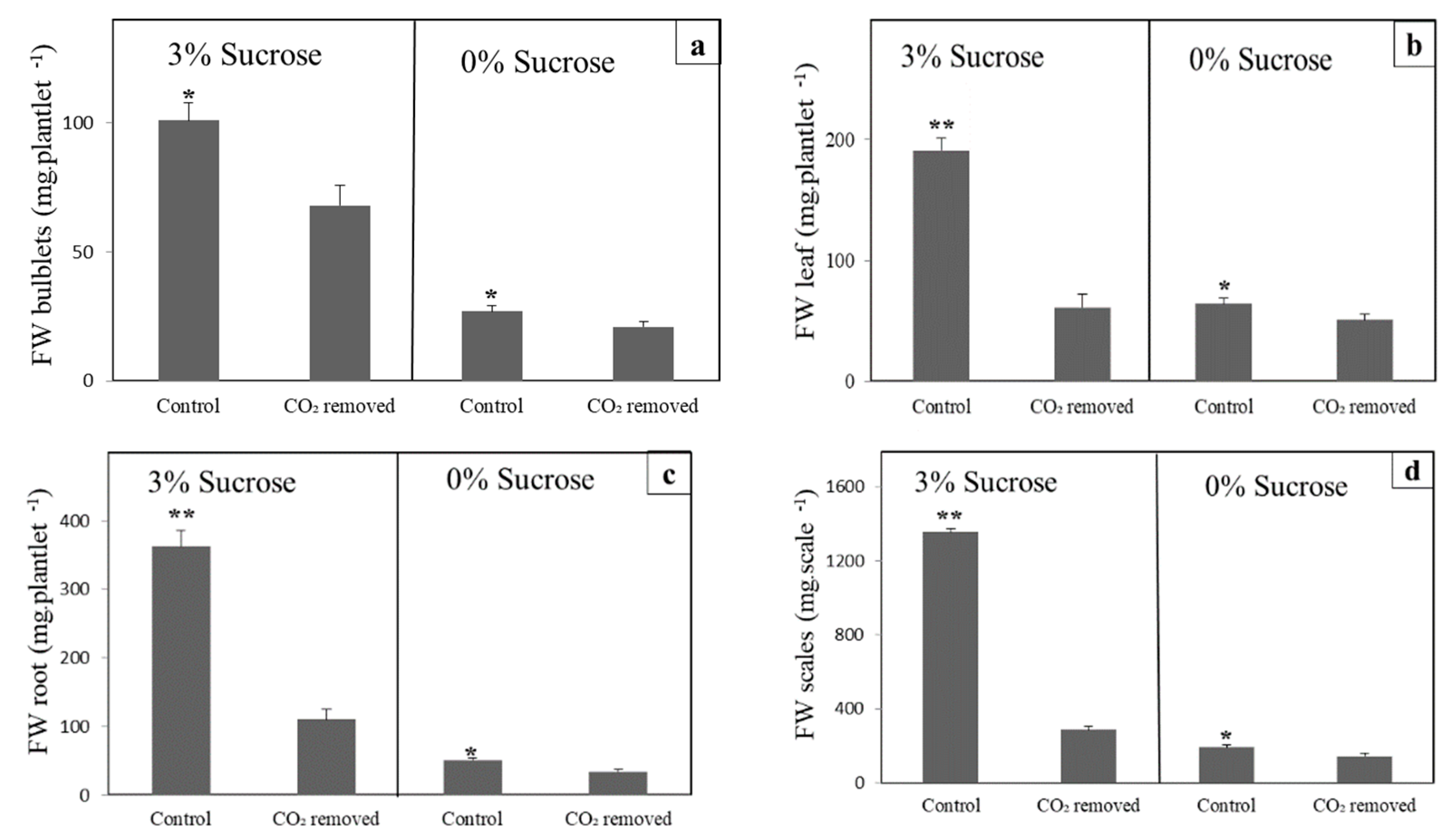
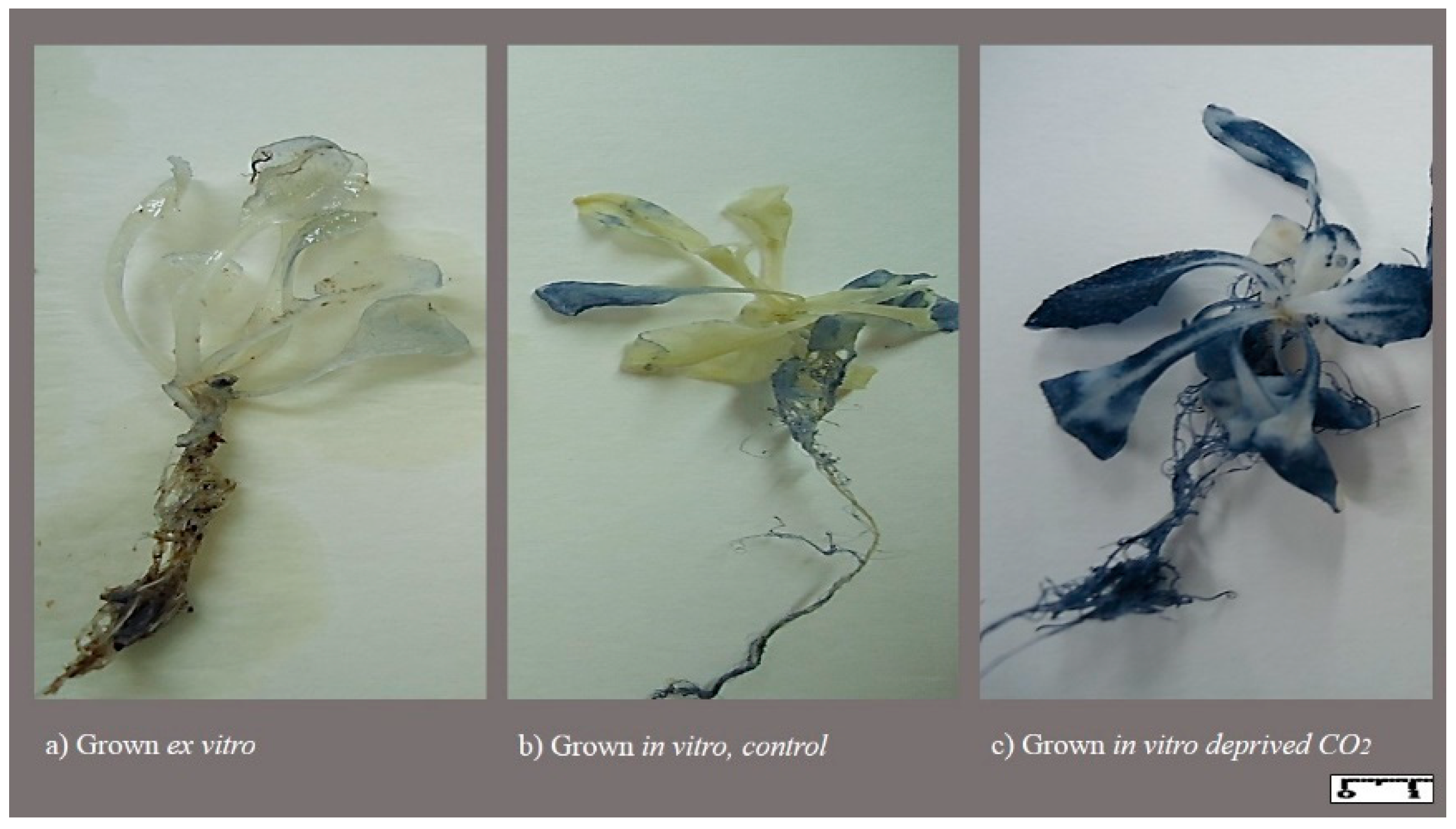
3.1. CO2 Removal Reduces Growth in Lily and Arabidopsis

Lily scale fragments cultured in vitro on nutrient medium display different kinds of growth. They regenerate bulblets with scales that may or may not carry leaves. In addition, roots, which are usually not connected to the shoots, regenerate. Finally, the scale explant may itself increase in size, and may generate callus tissue. We measured the FW of all four tissues: bulblets, leaves, roots, and scales. The addition of sucrose to the nutrient medium strongly increased the growth of all tissues. Removal of CO2, when no sucrose had been added, reduced growth (Figure 2) by: 23% in lily bulblets, 21% in leaves, 34% in roots, and 25% in scale explants. Removal of CO2 caused a larger reduction in growth when 3% sucrose had been added.
Bulblet fresh weight (FW) decreased by 33%, leaf FW by 68%, root FW by 70%, and scale explant FW by 79% as a consequence of CO2 removal. Removal of CO2 has a detrimental effect in lilies. The tissue culture system of lilies, however, differs from most other systems, since scale explants may serve as a rich source of nutrients and as a ‘buffer’ for the regenerating plantlet. We therefore performed a similar experiment with Arabidopsis seedlings. We obtained comparable results to the lily experiments, though values were more extreme (Figure 3 and Figure 4). It should also be noted that the seedlings bleached when CO2 was removed from the headspace (Figure 4a,b), and that they died soon after the period of observation.

3.2. CO2-Low Conditions Disturbs the Physiology of the In Vitro Plants

Arabidopsis seedlings cultured on medium with sucrose, but without CO2 in the headspace, were tiny and had bleached leaves (Figure 4). The bleaching indicates oxidative stress. The occurrence of ROS was visualized by NBT staining (Figure 5). When CO2 was removed, the seedlings colored deep blue, showing abundant formation of ROS, whereas the in vitro seedlings with CO2 showed very little blue staining. Arabidopsis seedlings grown in ex vitro conditions showed only faint and sparse blue staining.
ROS also damages the photosynthetic system. Photosynthetic performance can be properly evaluated by determining Fv/Fm. In general, a higher Fv/Fm was observed in Arabidopsis compared to lily plants (Figure 6). In both plants, in vitro plants had a lower Fv/Fm than ex vitro plants (Figure 6). Furthermore, in vitro Fv/Fm strongly decreased in CO2-poor conditions, in both lily and Arabidopsis plants (Figure 6 and Figure 7).

4. Discussion

When most of the CO2 was removed from the headspace by placing a vial of concentrated KOH solution in the container, we observed a strong inhibition of growth. In the case of a nutrient medium with 3% sucrose, this was unexpected, as the assumption was that sucrose in the medium supplies organic nutrition, and should support adequate growth. Since it has been reported that CO2 starvation brings about an ROS burst [13,23], we presumed that the inhibition of growth was, at least to some extent, caused by this detrimental effect of CO2 withdrawal. When CO2 is not available, other electron acceptors become reduced; particularly oxygen, resulting in toxic, activated oxygen species (ROS). In agreement with this, CO2 removal from the headspace caused severe bleaching of Arabidopsis leaves and reduced the photosynthetic functionality. Growth declined drastically as a result of oxidative stress. In Arabidopsis thaliana cultured in CO2 deprived conditions, in vitro plantlet growth was 3.4 times lower than in aerated cultures [24]. In lilies, bleaching was less pronounced (not shown), though fluorescence dropped even more than in Arabidopsis (Figure 6). Growth reduction in lily plants was more severe in leaves than in bulblets. This may be because the leaves are a site of abundant chlorophyll, while bulblets contain less chlorophyll (Figure 2; also see Arabidopsis in Figure 4). Moreover, staining with NBT showed abundant ROS formation when CO2 had been removed by a concentrated KOH solution.
Tissue culture is usually carried out in the light, and the plant material in question is chlorophyllous. Under these conditions, photosynthesis inevitably occurs, although at a reduced rate. Indeed, in standard tissue culture conditions, the expected curves for CO2 and O2 during a daily regime of light and dark have been observed: high CO2/low O2 levels in the dark and low CO2/high O2 levels during the day [4]. It might often be that CO2 levels are so low that O2 is reduced, leading to ROS production. Indeed, we observed a slight ROS presence in tissue-cultured plants under normal CO2 environment. Here, it should be noted that the amount of plant material in the container (6 small seedlings) was much less than in common tissue culture practices, indicating that in the practice of tissue culture, the detrimental effect is probably much larger.
A reduced electron transport flux through the photosynthetic electron chain, and subsequently ROS formation, has also been reported in the presence of high sugar in the medium, even under normal light conditions [25]. Regarding high sucrose concentration, contradictory effects on photosynthesis activity and ROS formation have been reported. Several authors report that high sucrose concentrations in the medium are deleterious for photosynthetic activity [5,26]. However, sucrose has also been reported to stimulate photosynthesis in in vitro plantlets [27,28]. ROS formation can also be stimulated [29] or decreased [30] by sugar.
A study on gene expression in in vitro and ex vitro tomato leaf tissues showed that stress-related genes and ROS scavenging enzymes were up-regulated in in vitro tomato plants. Superoxide dismutase (SOD), ascorbate peroxidase (APX), and glutathione reductase (GR) enzymes eliminate toxic superoxide and hydrogen peroxide by conversion to water. The expression of APX and GR were higher in tomato leaf tissues in vitro, compared to ex vitro, which leads to adaptation and acclimatization to in vitro conditions [31]. Up-regulation of ROS scavenging enzymes in tomato leaf tissues, grown in vitro, showed that plants react to overcome stressful in vitro conditions by removing detrimental free radicals. Furthermore, upregulation of SOD1 and SOD2 were shown to occur in Arabidopsis thaliana, cultured in vitro in CO2 deprivation conditions, which indicated the accumulation of ROS compared to aerated cultures [25]. In a severe stressful condition of an almost CO2-free condition in Arabidopsis, this leads to an increased and high amount of ROS formation (Figure 5c), extremely damaged Arabidopsis seedlings (Figure 4a,b), and, after a few weeks, death of the seedlings. A higher growth rate of ex vitro plants, compared with in vitro plantlets, has been also observed, e.g., in lily [32] and walnut [20] plants. The lesser growth of in vitro plantlets might be related to the damaging effect of ROS on the growth of in vitro plantlets compared with ex vitro plants.
Connecting all the negative effects of CO2 deprivation directly to oxidative burst may be deceptive. For instance, ethylene can also induce an oxidative burst, leading to acceleration of senescence [33]. It has been reported that CO2, through up-regulation of ACC (precursor of ethylene)-synthase, can enhance ethylene production [34]; on the other hand, it is well-known that CO2 inhibits ethylene action, and due to this inhibitory effect CO2 is widely used in the horticultural industry to maintain the quality of ethylene-sensitive products [33]. The inhibitory effects of ethylene on growth of in vitro plants has also been reported [33,35]. Therefore, it is rational to construe that, due to CO2 deprivation in the present study, no inhibitory obstacles existed to retard the ethylene action, which resulted in oxidative burst and disturbance of photosynthesis. Further study is needed to determine the concentrations of ethylene and CO2 in the headspace of culture vessels during in vitro growth of plantlets, and to elucidate the role of ethylene under CO2 deprivation.

5. Conclusions

In the present study, we confirmed the detrimental effects of CO2 deprivation on production of plantlets in vitro. Disturbance in photosynthetic functionality occurred by in vitro CO2 deprivation in both lily and Arabidopsis plantlets. However, it is not known whether the contribution of photosynthesis is substantial or marginal for plantlets under in vitro conditions. Staining with NBT confirmed higher ROS production in Arabidopsis seedlings under in vitro conditions, and an oxidative burst by CO2 deprivation under the same conditions, when compared to the level of ROS in seedlings grown ex vitro. Therefore, poor growth due to CO2 deprivation in vitro can be, to some extent, due to disturbances in photosynthesis and to a larger extent to a ROS burst.

Author Contributions

N.A. made substantial contributions to conception and design, carried out the experiments, performed the statistical analysis, and drafted the manuscript. S.A. measured the photosynthetic performance, performed statistical analysis, and carried out revision of the manuscript. R.G.F.V. supervised the research, contributed to the conception and design of the experiment, as well as the scientific discussion, and performed critical revision of the final manuscript. All authors have read and agreed to the published version of the manuscript.

Funding

This research received no external funding.

Institutional Review Board Statement

Not applicable.

Informed Consent Statement

Not applicable.

Data Availability Statement

Raw data are available upon request from the corresponding author.

Acknowledgments

Thank you to the University of Jiroft and Wageningen University for funding the project. We want to thank Geert Jan de Klerk (retired researcher from Plant Breeding WUR) for his help in the PhD research, design, and execution of experiments. Thanks to Reza Ghahramani for his input on image illustrations. This manuscript was part of the PhD thesis of the first author N. Askari entitled: ‘Aspects of bulblet growth of lily in vitro’, 130 pages. Wageningen University, Wageningen, NL.

Conflicts of Interest

The authors declare no conflict of interest.

References

  1. Neumann, K.-H.; Kumar, A.; Imani, J. Historical Developments of Cell and Tissue Culture Techniques. In Plant Cell and Tissue Culture—A Tool in Biotechnology; Springer: Cham, Germany, 2020; pp. 13–24. [Google Scholar]
  2. Maleki Asayesh, Z.; Vahdati, K.; Aliniaeifard, S. Investigation of physiological components involved in low water conservation capacity of in vitro Walnut plants. Sci. Hortic. 2017, 224, 1–7. [Google Scholar] [CrossRef]
  3. Vahdati, K.; Maleki Asayesh, Z.; Aliniaeifard, S.; Leslie, C. Improvement of ex vitro desiccation through elevation of CO2 concentration in the atmosphere of culture vessels during in vitro growth. HortScience 2017, 52, 1006–1012. [Google Scholar] [CrossRef]
  4. Chen, C. In situ measurement of microclimate for the plantlets cultured in vitro. Biosyst. Eng. 2006, 95, 413–423. [Google Scholar] [CrossRef]
  5. Chen, C. Humidity in plant tissue culture vessels. Biosyst. Eng. 2004, 88, 231–241. [Google Scholar] [CrossRef]
  6. Kozai, T. Photoautotrophic micropropagation. Vitr. Cell. Dev. Biol. Plant. 1991, 27, 47–51. [Google Scholar] [CrossRef]
  7. Aliniaeifard, S.; Maleki Asayesh, Z.; Driver, J.; Vahdati, K. Stomatal features and desiccation responses of Persian walnut leaf as caused by in vitro stimuli aimed at stomatal closure. Trees Struct. Funct. 2020, 34, 1219–1232. [Google Scholar] [CrossRef]
  8. Chen, L.; Lu, Y.; Hu, Y.; Xue, X. RNA-Seq reveals that sucrose-free medium improves the growth of potato (Solanum tuberosum L.) plantlets cultured in vitro. Plant Cell. Tissue Organ Cult. 2020, 140, 505–521. [Google Scholar] [CrossRef]
  9. Fuentes, G.; Talavera, C.; Desjardins, Y.; Santamaría, J.M. Low exogenous sucrose improves ex vitro growth and photosynthesis in coconut in vitro plantlets if grown in vitro under high light. Acta Hortic. 2007, 748, 151–155. [Google Scholar] [CrossRef]
  10. Mosaleeyanon, K.; Cha-Um, S.; Kirdmanee, C. Enhanced growth and photosynthesis of rain tree (Samanea saman Merr.) plantlets in vitro under a CO 2-enriched condition with decreased sucrose concentrations in the medium. Sci. Hortic. 2004, 103, 51–63. [Google Scholar] [CrossRef]
  11. Zhou, Q.; Wang, Y.; Zhao, X.; Han, L.N.; Yang, S.J.; Li, W.F.; Mao, J.; Ma, Z.H.; Chen, B.H. Effects of CO2 on transplantation of grape plantlets cultured in vitro by promoting photosynthesis. Sci. Hortic. 2021, 287, 110286. [Google Scholar] [CrossRef]
  12. Park, J.E.; Park, Y.G.; Thi, L.T.; Soundararajan, P.; Jeong, B.R. Effect of sucrose concentration, photosynthetic photon flux density, and CO2 concentration on growth and development of micropropagated Mountain ash. Propag. Ornam. Plants 2018, 18, 58–63. [Google Scholar]
  13. Durchan, M.; Vácha, F.; Krieger-Liszkay, A. Effects of severe CO2 starvation on the photosynthetic electron transport chain in tobacco plants. Photosynth. Res. 2001, 68, 203–213. [Google Scholar] [CrossRef] [PubMed]
  14. Habibi, N.; Purohit, S.D. Photosynthetic efficiency and in vitro growth of Celastrus paniculatus Willd. under varied concentrations of CO2. Int. J. Phytocosmet. Nat. Ingred. 2019, 6, 11. [Google Scholar] [CrossRef]
  15. Kozuleva, M.A.; Ivanov, B.N.; Vetoshkina, D.V.; Borisova-Mubarakshina, M.M. Minimizing an electron flow to molecular oxygen in photosynthetic electron transfer chain: An evolutionary view. Front. Plant Sci. 2020, 11, 211. [Google Scholar] [CrossRef] [PubMed] [Green Version]
  16. Sharma, P.; Jha, A.B.; Dubey, R.S.; Pessarakli, M. Reactive oxygen species, oxidative damage, and antioxidative defense mechanism in plants under stressful conditions. J. Bot. 2012, 2012, 1–26. [Google Scholar] [CrossRef] [Green Version]
  17. Askari, N.; de Klerk, G.J. Elimination of epidermal wax from explants increases growth in tissue culture of lily. Sci. Hortic. 2020, 274, 109637. [Google Scholar] [CrossRef]
  18. Murashige, T.; Skoog, F. A revised medium for rapid growth and bio assays with Tobacco tissue cultures. Physiol. Plant. 1962, 15, 473–497. [Google Scholar] [CrossRef]
  19. Solomon, M.E. Control of humidity with potassium hydroxide, sulphuric acid, or other solutions. Bull. Entomol. Res. 1951, 42, 543–554. [Google Scholar] [CrossRef]
  20. Maleki Asayesh, Z.; Vahdati, K.; Aliniaeifard, S.; Askari, N. Enhancement of ex vitro acclimation of Walnut plantlets through modification of stomatal characteristics in vitro. Sci. Hortic. 2017, 220, 114–121. [Google Scholar] [CrossRef]
  21. Aliniaeifard, S.; Van Meeteren, U. Natural variation in stomatal response to closing stimuli among Arabidopsis thaliana accessions after exposure to low VPD as a tool to recognize the mechanism of disturbed stomatal functioning. J. Exp. Bot. 2014, 65, 6529–6542. [Google Scholar] [CrossRef]
  22. Van Den Dries, N.; Gianní, S.; Czerednik, A.; Krens, F.A.; de Klerk, G.J.M. Flooding of the apoplast is a key factor in the development of hyperhydricity. J. Exp. Bot. 2013, 64, 5221–5230. [Google Scholar] [CrossRef] [PubMed]
  23. Eisenhut, M.; Bräutigam, A.; Timm, S.; Florian, A.; Tohge, T.; Fernie, A.R.; Bauwe, H.; Weber, A.P.M. Photorespiration is crucial for dynamic response of photosynthetic metabolism and stomatal movement to altered CO2 availability. Mol. Plant 2017, 10, 47–61. [Google Scholar] [CrossRef] [PubMed] [Green Version]
  24. Banerjee, S.; Siemianowski, O.; Liu, M.; Lind, K.R.; Tian, X.; Nettleton, D.; Cademartiri, L. Stress response to CO2 deprivation by Arabidopsis thaliana in plant cultures. PLoS ONE 2019, 14, e0212462. [Google Scholar] [CrossRef] [PubMed]
  25. Desjardins, Y.; Dubuc, J.F.; Badr, A. In vitro culture of plants: A stressful activity! Acta Hortic. 2009, 812, 29–50. [Google Scholar] [CrossRef]
  26. Martins, J.P.R.; de Almeida Rodrigues, L.C.; Santos, E.R.; Gontijo, A.B.P.L.; Falqueto, A.R. Impacts of photoautotrophic, photomixotrophic, and heterotrophic conditions on the anatomy and photosystem II of in vitro propagated Aechmea blanchetiana (Baker) L.B. Sm. (Bromeliaceae). Vitr. Cell. Dev. Biol. Plant 2020, 56, 350–361. [Google Scholar] [CrossRef]
  27. De Oliveira, T.; Balduino, M.C.M.; de Carvalho, A.A.; Bertolucci, S.K.V.; Cossa, M.C.; Coelho, A.D.; Leite, J.J.F.; Pinto, J.E.B.P. The effect of alternative membrane system, sucrose, and culture methods under photosynthetic photon flux on growth and volatile compounds of mint in vitro. Vitr. Cell. Dev. Biol. Plant 2021, 57, 529–540. [Google Scholar] [CrossRef]
  28. Sáez, P.L.; Bravo, L.A.; Sánchez-Olate, M.; Bravo, P.B.; Ríos, D.G. Effect of photon flux density and exogenous sucrose on the photosynthetic performance during in vitro culture of Castanea sativa. Am. J. Plant Sci. 2016, 7, 2087–2105. [Google Scholar] [CrossRef] [Green Version]
  29. Takahashi, S.; Murata, N. How do environmental stresses accelerate photoinhibition? Trends Plant Sci. 2008, 13, 178–182. [Google Scholar] [CrossRef]
  30. Couée, I.; Sulmon, C.; Gouesbet, G.; El Amrani, A. Involvement of soluble sugars in reactive oxygen species balance and responses to oxidative stress in plants. J. Exp. Bot. 2006, 57, 449–459. [Google Scholar] [CrossRef]
  31. Dubuc, J.F.; Desjardins, Y. Stress gene expression modulation in in vitro tomato plantlets. Acta Hortic. 2009, 812, 507–514. [Google Scholar] [CrossRef]
  32. De Klerk, G.J.; Kim, K.; Schadewijk, M.; Gerrits, M. Growth of bulblets of Lilium speciosum in vitro and in soil. Acta Hortic. 1992, 513–520. [Google Scholar] [CrossRef]
  33. Wei, H.; Seidi, F.; Zhang, T.; Jin, Y.; Xiao, H. Ethylene scavengers for the preservation of fruits and vegetables: A review. Food Chem. 2021, 337, 127750. [Google Scholar] [CrossRef]
  34. Dong, J.G.; Fernandez-Maculet, J.C.; Yang, S.F. Purification and characterization of 1-aminocyclopropane-1-carboxylate oxidase from apple fruit. Proc. Natl. Acad. Sci. USA 1992, 89, 9789–9793. [Google Scholar] [CrossRef] [PubMed] [Green Version]
  35. Buddendorf-Joosten, J.M.C.; Woltering, E.J. Components of the gaseous environment and their effects on plant growth and development in vitro. Plant Growth Regul. 1994, 15, 1–16. [Google Scholar] [CrossRef]
Figure 1. Container with lily scale explants on a nutrient medium with 3% sucrose and a vial of KOH to remove CO2. Bar = 10 mm.
Horticulturae 08 00044 g001
Figure 2. Effect of CO2 removal on lily growth: bulblets (a), leaves (b), roots (c), and scale explants (d) in medium with 3% sucrose or without sucrose. *, p ≤ 0.05 and **, p ≤ 0.01.
Horticulturae 08 00044 g002
Figure 3. Effect of CO2 removal on the growth of Arabidopsis seedlings, (a) FW shoot and (b) FW root in medium with 3% sucrose and without sucrose: *, p ≤ 0.05; **, p ≤ 0.01; and ***, p ≤ 0.001.
Horticulturae 08 00044 g003
Figure 4. Arabidopsis seedlings cultured for 7 days on medium with (3%) or without (0%) sucrose. A small vial with 3 mL 20% KOH was placed in the container to remove CO2 from the headspace (a,b). As a control for the drop in RH from the concentrated KOH solution, a small vial with saturated KCl was added when no vial with KOH was included (c,d). Bar = 10 mm.
Horticulturae 08 00044 g004
Figure 5. Arabidopsis seedlings stained with NBT (a) grown ex vitro, (b) grown in vitro under control conditions, and (c) grown in vitro under conditions when (most) of the CO2 had been removed. Bar = 10 mm.
Horticulturae 08 00044 g005
Figure 6. Fv/Fm in lilies (a) and Arabidopsis (b) grown ex vitro, in vitro, and when (most) of the CO2 was removed from the headspace. Different letter indicate significant differences between treatments (p < 0.05).
Horticulturae 08 00044 g006
Figure 7. Image of Fv/Fm of lilies and Arabidopsis grown ex vitro (a,d), in vitro with 3% sucrose (b,e), and in vitro with 3% sucrose in a CO2-poor environment (c,f).
Horticulturae 08 00044 g007
Publisher’s Note: MDPI stays neutral with regard to jurisdictional claims in published maps and institutional affiliations.

