Article

Wild Species Veronica officinalis L. and Veronica saturejoides Vis. ssp. saturejoides—Biological Potential of Free Volatiles

1
Faculty of Science, University of Split, Ruđera Boškovića 33, HR-21000 Split, Croatia
2
Bartul Kašić High School, Ante Starčevića 9, HR-23250 Pag, Croatia
3
Faculty of Pharmacy and Biochemistry, University of Zagreb, A. Kovačića 1, HR-10000 Zagreb, Croatia
4
Institute of Biology, Karl-Franzens University, Schubertstrasse 51, A-8010 Graz, Austria
*
Author to whom correspondence should be addressed.
Horticulturae 2021, 7(9), 295; https://doi.org/10.3390/horticulturae7090295
Submission received: 18 August 2021 / Revised: 31 August 2021 / Accepted: 3 September 2021 / Published: 7 September 2021
(This article belongs to the Collection Prospects of Using Wild Plant Species in Horticulture)

Abstract

:
Extracts from plants of the genus Veronica have been and continue to be used in traditional medicine to treat various diseases throughout the world. Although often considered a weed, many scientific reports demonstrate that these plants are a source of valuable biologically active compounds and their potential for horticulture should be investigated and considered. In this study, free volatile compounds of essential oils (EO) and hydrosols were extracted from two species: Veronica officinalis, which is most commonly used in traditional medicine, and Veronica saturejoides, an endemic plant that could be obtained by cultivation in horticulture. Volatiles were analyzed by gas chromatography coupled with mass spectrometry (GC, GC-MS). The most abundant compounds identified in the EOs were hexadecanoic acid in V. officinalis EO and caryophyllene oxide in V. saturejoides EO. The hydrosols were characterized by a high abundance of caryophyllene oxide in V. saturejoides hydrosol and of p-vinyl guaiacol for V. officinalis hydrosol. The sites where the volatile compounds are synthesized and stored were analyzed using SEM (Scanning Electron Microscopy); glandular and non-glandular trichomes were detected on stems, leaves and the calyx. Further, to investigate the activity of the free volatile compounds against pathogens, isolated volatile compounds were tested on the antiphytoviral activity against tobacco mosaic virus (TMV) infection. The hydrosols of both investigated species and EO of V. officinalis showed significant antiphytoviral activity. To further investigate the biological potential of these extracts they were also tested for their antiproliferative and antioxidant activities. The results indicate that these compounds are a valuable source of potential anticancerogenic agents that should be investigated in future studies. The presented results are the first report of hydrosol and EO activity against TMV infection, suggesting that these extracts from Veronica species may be useful as natural-based antiphytoviral agents.

1. Introduction

The genus Veronica of the family Plantaginaceae grows predominantly in temperate Northern Hemisphere regions, with a smaller number of species growing in Southern Hemisphere regions and in Australia [1,2]. The many species of this plant family, about 450, show the great ecological adaptability of the genus Veronica. Species of this genus grow in wet and dry habitats, as well as in the marine belt and mountains [3].
The studied species Veronica officinalis L. (Figure 1b) (common speedwell) and Veronica saturejoides Vis. ssp. saturejoids (Figure 1a) (savory leafed speedwell) grow on Dinaric Massif (Republic of Croatia). Both species are perennial herbaceous plants with small attractive violet flowers. Veronica officinalis is slightly taller (10–50 cm) and has longer leaves (1.5–5 cm) than V. saturejoides ssp. saturejoides (stem length 10–30 cm, leaf length 1–3 cm). The latter species is a plant endemic to Croatia [4].
In general, the study of chemical compounds produced by wild plants is extremely important because these compounds ultimately affect not only the plant in which they are found, but also indirectly other plants in the vicinity as well as the environment as a whole [5]. These compounds are important factors in plant adaptation to abiotic stresses. Moreover, neighboring plants detect other plant volatiles as ‘messages’ about herbivore or pathogen attacks, and consequently adapt their metabolism responses [6]. Plants of the genus Veronica are used in traditional medicines in countries around the world, which sparked interest in the studying these plants in terms of their chemical composition and biological activity. Many different biological activities of the various extracts have been reported in recent studies [7,8]. For example, methanolic and ethyl-acetate extracts of V. spicata were tested for antimicrobial activity and MIC values were between 1.25 and 5.00 mg/mL. This plant extract has also shown substantial antioxidant activity, especially the methanol extracts of flowers and leaves with IC50 and DPPH values of 8.21 µg/mL and 8.69 µg/mL, respectively [9]. Ertas et al. reported antimicrobial activity for phenolic extracts of Veronica thymoides subsp. pseudocinerea and the MIC value determined was 31.25 mg/mL for methanol extract against Escherichia coli [10].
Veronica officinalis, which is the subject of this study, is traditionally used in the medicine of Balkan peoples. Mocan et al. reported antioxidant activity for ethanol extracts of phenolic compounds for V. officinalis to be 157.99 ± 6.58 mg Trolox equivalents/g d.w [11]. Valyova et al. also confirmed antioxidant activity of phenolic extracts for the V. officinalis in their study [12]. The aerial parts of speedwells are used to treat liver, spleen, kidney, and bladder diseases, as well as snakebites wound healing, skin lesions, eczema and ulcers [9,10,13].
Green prevention strategies and means of virus control of natural origin are particularly important today to support organic production and the replacement of synthetic chemicals with biologically based antivirals. This approach to modern agriculture is still under development, and one of our goals is to find harmless and environmentally friendly antiphytoviral agents. Tobacco mosaic virus (TMV) is a model virus in plant virology and a very important pathogen for agricultural crops, causing significant yield losses. TMV belongs to the positive-strand RNA viruses and encodes two proteins that function as replicases (molecular weights 126 kDa and 183 kDa), a movement protein (30 kDa) that facilitates virus movement between host cells, and a coat protein (17.5 kDa) that plays an important role in virion formation [14]. Various plant products such as essential oils, flavonoids, polyphenols and organic, alcoholic and aqueous extracts from plants and natural compounds from other organisms such as fungal metabolites, have been used against a number of plant diseases caused by viruses, phytopathogenic bacteria, fungi, plant parasitic nematodes and parasitic and non-parasitic weeds [15,16,17,18,19] with the aim of finding natural-based products useful for plant protection against pathogens. Some of our previous studies and studies by other authors describe the activity of plant volatiles as natural antiphytoviral compounds [19,20,21,22,23,24,25,26,27]. Since this activity of Plantaginaceae family volatiles has not been tested so far, we investigated the antiphytoviral activity of both essential oil and hydrosol of V. officinalis and V. saturejoides, with the aim of increasing the knowledge about the antiphytoviral activity of essential oils and especially hydrosols, which have been very little studied in this regard. To further investigate the biological potential, the antiproliferative and antioxidant activity of these extracts was tested.
Thus, the aim of this study was to investigate the composition of the volatile compounds of these two species from the EOs and the water residues (hydrosols) and to discuss differences and similarities in composition. In addition, the biological potential of these extracts for possible use in horticulture were evaluated either to cultivate and thus preserve the endemic species V. saturejoides or to promote the cultivation of the wild plant V. officinalis. To our knowledge, this is the first report on the EO and hydrosol composition of V. officinalis and V. saturejoides from this site, and the first report on their biological activity, as well as on the micromorphology of the trichomes of V. officinalis.

2. Materials and Methods
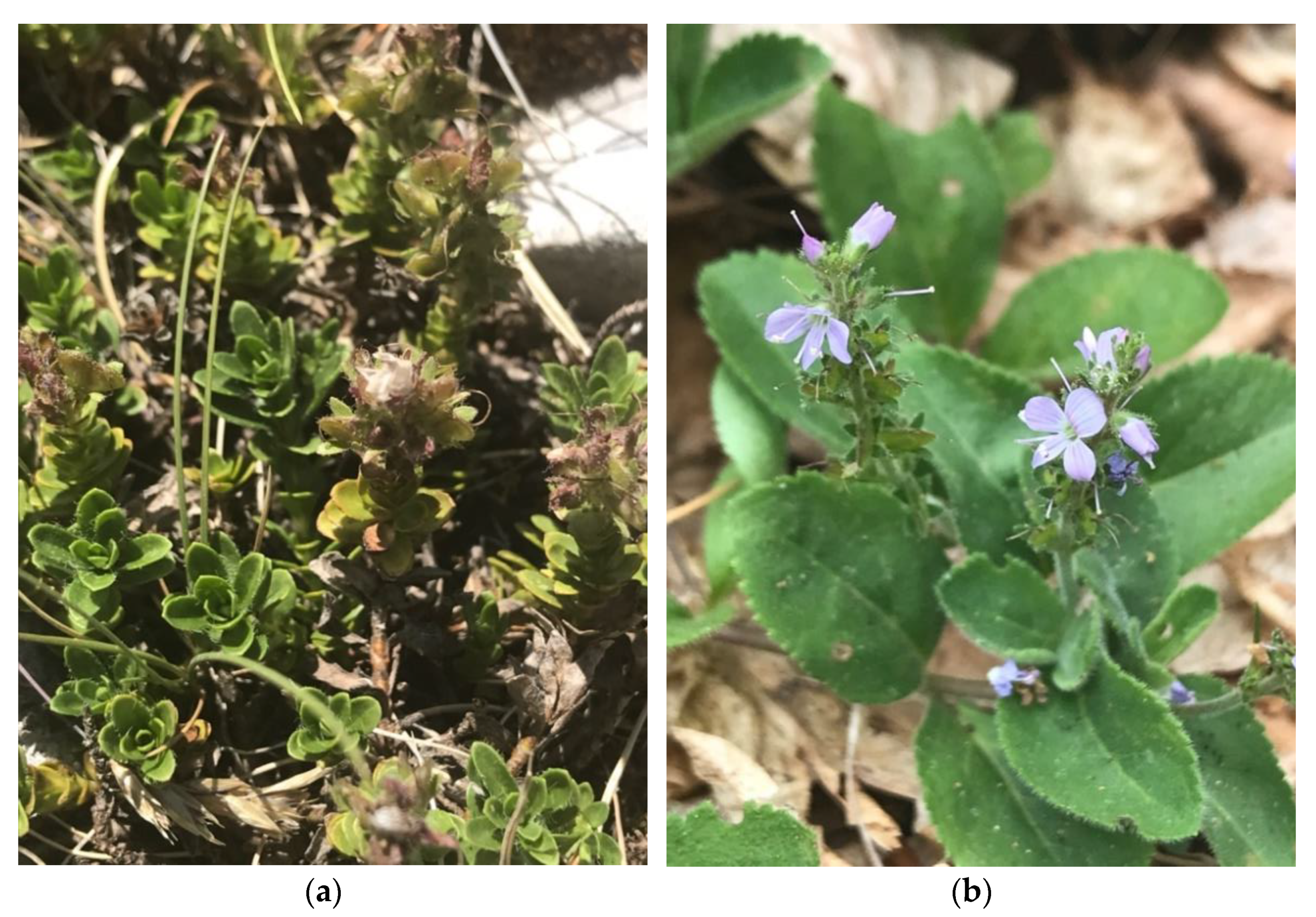
2.1. Plant Material

Plant material for V. officinalis and V. saturejoides was collected from the sites at Dinara Massif (Table 1, Figure 1). The voucher specimens were deposited in the Faculty of Science Herbarium (PMFST-HR), University of Split, Croatia. For GC and GC-MS analyses, the samples were air dried in a single layer and protected from direct sunlight for three weeks. The dried plant material was kept in the dark in double paper bags labeled with the sample number and stored in a dry place.
Above ground plant parts (stems, leaves, and flowers) of seven plants of V. officinalis were fixed in solution made from formalin, 96% ethanol, acetic acid and water in a volume ratio of 5:70:5:20. After three days, the samples were transferred to 70% ethanol and stored in the refrigerator. The study of the trichomes for V. saturejoides was carried out in the previous work [28].

2.2. GC and GC-MS Analyses

Dried above ground parts (50 g) for each sample were hydrodistilled for 3 h in a Clevenger-type apparatus. Three separate extractions were performed for each species and volatile compounds were collected from the lipophilic layer (pentane/diethyl ether) and water residues (hydrosols). Both phases were analyzed by gas chromatography (GC), and gas chromatography and mass spectrometry (GC-MS). GC was performed by gas chromatograph (model 3900, Varian Inc., Lake Forest, CA, USA) that is supplied with a flame ionization detector (FID), mass spectrometer (model 2100T; Varian Inc., Palo Alto, CA, USA), non-polar capillary column VF-5ms (30 m × 0.25 mm inside diameter, coating thickness 0.25 µm, Palo Alto, CA, USA) and polar capillary column CP-Wax 52 CB (30 m × 0.25 mm i.d., coating thickness 0.25 µm, Palo Alto, CA, USA). The chromatographic conditions for the analysis of lipophilic fraction (essential oils) were: FID detector temperature 300 °C, injector temperature 250 °C. The gas carrier was helium at 1 mL min−1. The conditions for the VF-5ms column were: temperature 60 °C isothermal for 3 min, and then increased to 246 °C at a rate of 3 °C min−1, and held isothermal for 25 min. Conditions for the CP Wax 52 column were: temperature 70 °C isothermal for 5 min, and then increased to 240 °C at a rate of 3 °C min−1 and held isothermal for 25 min. The injected volume was 2 µL and the split ratio was 1:20. The MS conditions were: ion source temperature 200 °C, ionization voltage 70 eV, mass scan range 40–350 mass units [9,28]. The individual peaks for all samples were identified by comparison of their retention indices of n-alkanes to those of authentic samples and literature [29,30], comparing it to our libraries from previous work [9,28] and to other previously published material for Veronica species [10,12,31,32]. The results are given as the mean of three extractions with standard deviation.

2.3. Micromorphological Traits

For micromorphological investigations internodes of stem, central parts of leaves (left or right from the main axis), and the whole calyx were used. Before the scanning electron microscopic (SEM) analysis, the samples were transferred from 70% (v/v) ethanol to 70% (v/v) acetone. Thereafter, the samples were subjected to a dehydration procedure, i.e., they were transferred from 70% (v/v) to 90% (v/v), and 100% (v/v) acetone. Each transfer from one to another concentration of acetone was repeated twice and lasted 15 min each at room temperature. With the use of CO2 as the drying medium, the dehydrated samples were subjected to a process of critical point drying in CPD030 device (Bal-tec, Balzers, Liechtenstein). The samples thus prepared were sputter coated with gold in Sputter Coater device (Agar Scientific Ltd., Essex, UK) and researched under the scanning electron microscope XL30 ESEM (FEI Company, Eindhoven, Netherlands) with an acceleration voltage of 20 kV in high vacuum mode [33]. Common terminology [34] was used to describe the trichomes.

2.4. Antiphytoviral Activity

2.4.1. Virus and Plant Hosts

Leaves of Nicotiana tabacum L. cv. Samsun systemically infected with tobacco mosaic virus were used to prepare the virus inoculum as described by Vuko et al. [25]. Leaves of the native host Datura stramonium L. were pollinated with carborundum (Sigma-Aldrich, St. Louis, MO, USA) before virus inoculation, and the inoculum was diluted with inoculation buffer to obtain 5–30 lesions per inoculated leaf. Experiments were carried out when the plants had reached the 5–6 leaf stage. Care was taken to ensure that the experimental plants were as uniform in size as possible.

2.4.2. Antiphytoviral Activity Assay

Essential oil (final concentration adjusted to 500 ppm) or hydrosol (undiluted) were applied as a spray solution to the leaves of local host plants for three consecutive days prior to virus inoculation. Plants were then rubbed with the virus inoculum and treated once with the same spray solutions immediately after inoculation. The antiviral activity of the essential oil and hydrosol was evaluated by the percentage inhibition of the number of local lesions on the leaves of the treated and control plants as described by Vuko et al. [25].

2.5. Cell Culture

Three cancer cell lines, Cervical Cancer Cell Line (HeLa), Human Colon Cancer Cell Line (HCT116) and Human Osteosarcoma Cell Line (U2OS), were grown in an incubator under humidified conditions with 5% CO2 and 37 °C, in a Dulbecco’s modified Eagle’s medium (DMEM, EuroClone, Milan, Italy) containing 4.5 g/L glucose, 10% fetal bovine serum (FBS), and 1% antibiotics (penicillin and streptomycin, EuroClone).

2.6. Cell Proliferation Assay

The antiproliferative capacity of EO of V. officinalis L. and V. saturejoides Vis. ssp. saturejoides was determined on HeLa, HCT116 and U2OS cancer cells using the MTS-based CellTiter 96® Aqueous Assay (Promega). Cells were grown in an incubator at 37 °C and 5% CO2 until they reached 80% confluence. They were counted using a handheld automated cell counter (Sceptre, Merck), and 5000 cells/well were seeded in 96-well plates with a serial dilution of EOs and hydrosols. The cells were then cultured for an additional 48 h. Thereafter, 20 μL MTS tetrazolium reagent (Promega) was added to each well and left in the incubator for an additional 3 h. Then, absorbance was measured at 490 nm using a microplate reader (Bio-Tek, EL808). IC50 values were calculated from three independent experiments using GraFit 6 data analysis software (Erithacus, East Grinstead, UK).

2.7. Antioxidant Activity of Essential Oils and Hydrosols

2.7.1. ORAC

The assay was performed in a Perkin–Elmer LS55 spectrofluorimeter, using 96-well white polystyrene microtiter plates (Porvair Sciences, Leatherhead, UK) following a method described by Fredotović et al. [35], with some adjustments based on different extracts. Hydrophilic assay was performed for hydrosols and lipophilic assay for EOs. Adjustments were made for the EO antioxidant assay. EOs were dissolved in acetone (10 mg in 1 mL acetone). The EO acetone dilutions were further dissolved 40× and 80× in the phosphate buffer for the experiments. For the hydrosol assays total undiluted hydrosols were used. All measurements were performed in triplicate according to the method described in Nazlić et al. [28].

2.7.2. DPPH

The antioxidant capacity of the extracts was determined using the DPPH method already described by Mensor et al. and Payet et al. [36,37] and adapted to tested plant extracts. Plant extracts as described in the ORAC method were used (acetone-dissolved essential oils and absolute hydrosols) for the assay. An amount of 100 µL of methanol (Kemika, Zagreb, Croatia) and 200 µL of sample was pipetted into each well. Serial dilutions of samples were prepared by pipetting 100 µL from the first row with a multichannel pipette into the wells in the second row and so on to the last row, where 100 µL of the solution was ejected after mixing. In the first column, in 96-well plates, a blank sample was always added. For EOs, the acetone and methanolic solution were used as blank and for hydrosols, water and methanolic solution were used as blank. The calculation and presentation of the results were performed according to the method described in the previous research by Nazlić et al. [28].

2.8. Statistical Analyses

Statistical analysis was performed in GraphPad Prism Version 9. All data are presented as mean ± SD (n ≥ 3). Statistical significance for free volatile compounds and antioxidant activity was assessed by multiple t-test, p < 0.05. Statistical tests were performed separately for lipophilic (essential oils) and hydrophilic fractions (hydrosols).

3. Results and Discussion

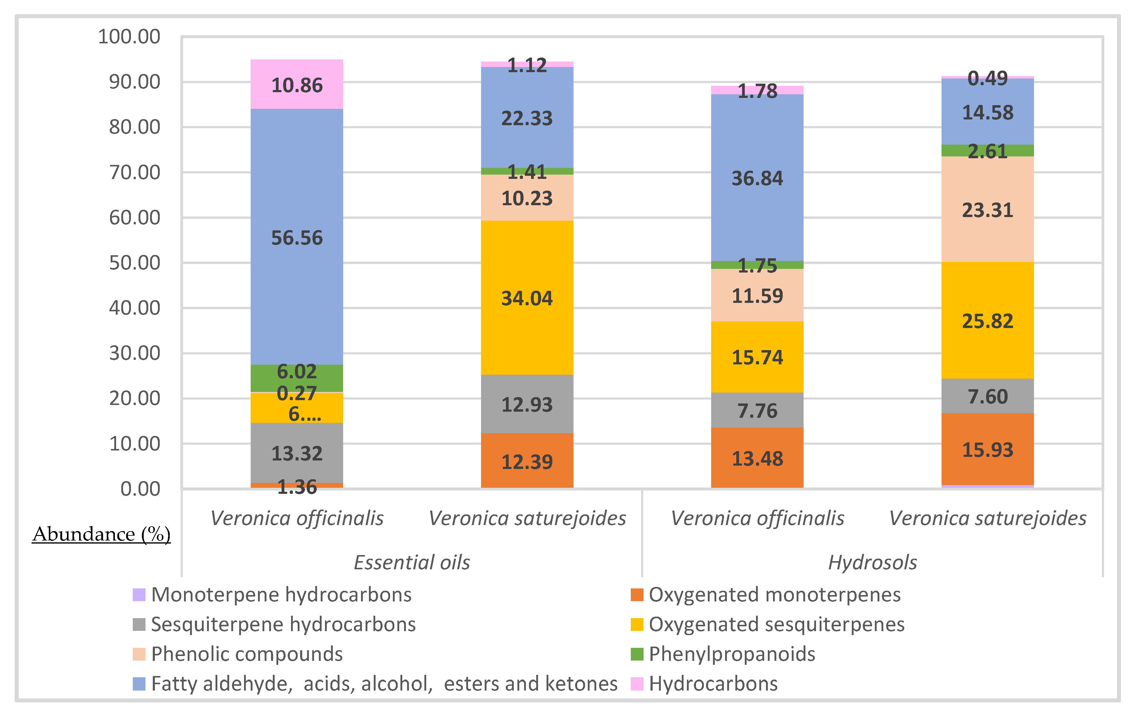
3.1. Composition of Free Volatile Components

The chemical composition of the free volatile compounds of Veronica officinalis L. and Veronica saturejoides Vis. ssp. saturejoides is shown in Table 2. The free volatile compounds were analyzed from the lipophilic-essential oil and aqueous-hydrosol fractions.
The major compounds in the lipophilic fraction of V. officinalis are: hexadecanoic acid (20.62%), β-ionone (17.88%), hexahydrofarnesyl acetone (13.92%) and E-caryophyllene (6.78%). These compounds were also identified in the hydrosol fraction of V. officinalis but with much lower proportions, except for β-ionone, which has a high proportion of 10.74%. Moreover, the phenolic compound p-vinyl guaiacol (11.59%) is the most represented compound in the hydrosol fraction of V. officinalis, followed by benzaldehyde (9.25%), caryophyllene oxide (7.52%) and (E)-β-damascone (6.69%).
The oxygenated sesquiterpenes are the major group in both fractions of V. saturejoides ssp. saturejoids (Table 1, Figure 2) with a predominant caryophyllene oxide compound (23.65% in EO and 21.28% in H). Moreover, among the phenolic compounds, only methyl eugenol was identified as the most abundant compound, in the EO-fraction with 20.23% and in the hydrosol fraction it with 23.31%. Among the oxygenated monoterpenes in Veronica saturejoides ssp. saturejoids in both fractions, trans-1(7),8-p-mentadien-2-ol was the most abundant compound (10.62% in EO and 11.75% in H).
In the previous studies, composition of EO and hydrosol for the species V. saturejoides ssp. saturejoides was analyzed but from two different sites (Kamešnica and Prenj Mountain) [28]. In hydrosols from these two locations trans-1(7),8-p-mentadien-2-ol was the most abundant compound, with a percentage of 31.75% for Prenj Sample and 36.63% for the Kamešnica sample. Caryophyllene oxide was also present in both fractions of V. saturejoides (essential oil and hydrosol) which is consistent with the results of this current research. The composition of the EO of V. spicata was also previously analyzed and the study found that the most abundant compound was phytol (21.13%) [9]. Not many studies have been carried out on the composition of the volatile compounds of other Veronica species. Valyova et al. studied extracts of the Bulgarian species V. officinalis by GC-MS and identified in ethanol extracts terpinen-4-ol, neophytadiene, hexahydrofarnesyl acetone, vitamin E, phytol and squalene for the first time in the genus Veronica [12]. Other research on the EOs of the genus Veronica were carried on V. thymoides subsp. pseudocinerea [10], V. linariifolia [31] and Veronica sp. [32]. Ertas et al. found that the most abundant constituent of the essential oil from Veronica thymoides subsp. pseudocinerea was hexatriacontene (21%) which belongs to the hydrocarbon compound group [10]. In another research of the essential oil composition of Veronica linariifolia Pall. ex Link the major constituents were cyclohexene (25.83%), β-pinene (11.61%), 1S-α-pinene (10.65%), β-phellandrene (10.49%), β-myrcene (10.42%), and germacrene D (4.99%) (monoterpene and sesquiterpene hydrocarbons) [31]. Çelik et al. studied the essential oils extracted from Veronica sp. and found that the main components were mainly linalool (4.18%) and carvacrol (7.28%) [32].

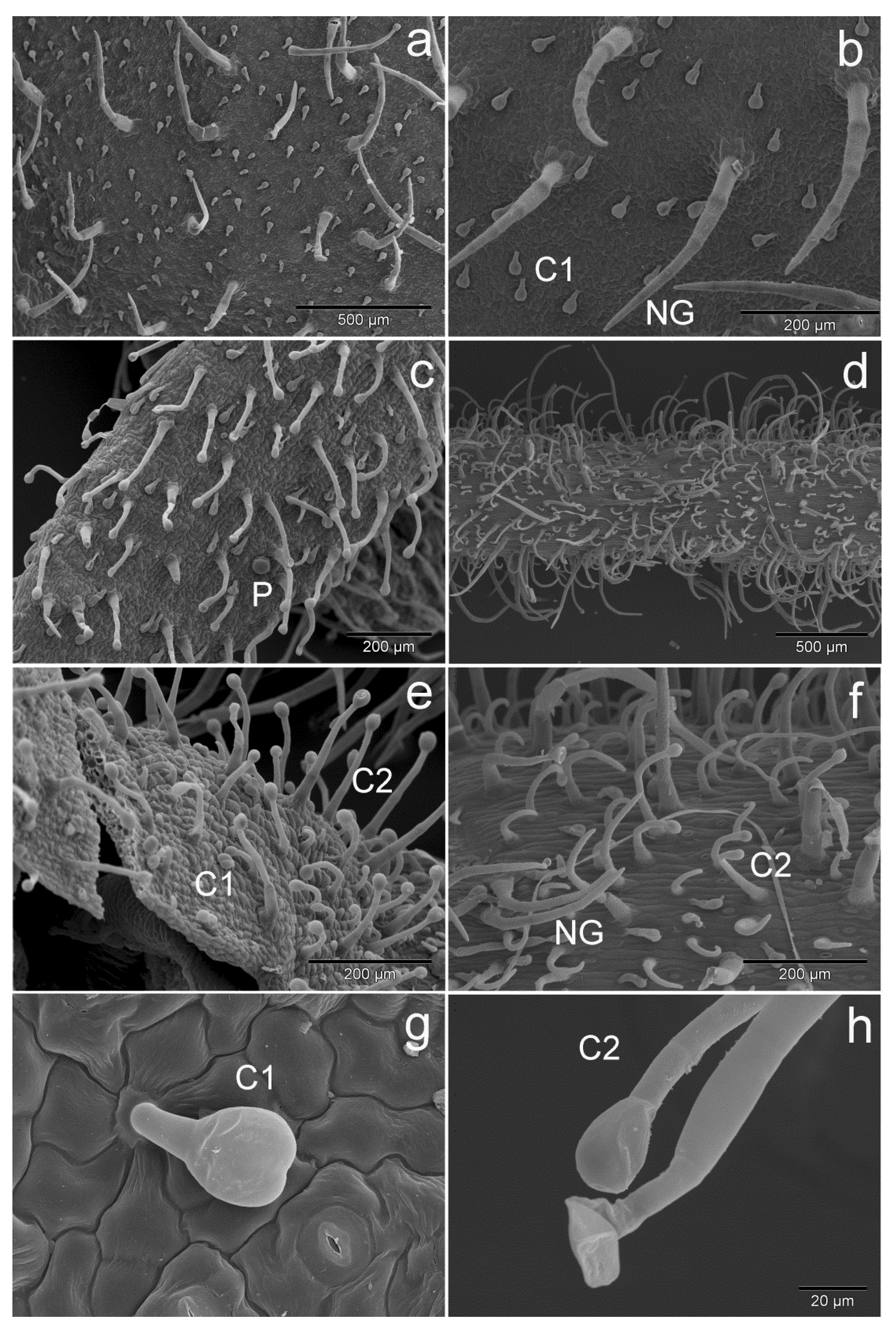
3.2. Glandular and Non-Glandular Trichomes

Glandular trichomes are ‘bio-factories’ of volatile compounds that are part of EOs. They play an important role in protecting plants from herbivores and pathogens and in attracting pollinators [38]. Two types of glandular trichomes can be seen on the surface of the studied plants of V. officinalis: peltate and capitate (Figure 3). Peltate glandular trichomes are composed of a basal cell, a very short stalk cell and a multicellular head with a fairly large subcuticular space (Figure 3c). These trichomes are very rare and they were noticed only on the calyx of V. officinalis. Nazlić et al. [28] did not describe peltate trichomes in Veronica saturejoides ssp. saturejoides. On the other hand, peltate trichomes are common in some other plant species, especially in Lamiaceae [39,40]. Capitate glandular trichomes could be further divided in two subtypes. Subtype 1 (C1) capitate trichomes consist of a stalk cell and two elliptically shaped head cells with a subcuticular space (Figure 3g). They are present on the adaxial and abaxial sides of the leaf, on the calyx and on the stem. Subtype 2 (C2) capitate trichomes are uniseriate, unbranched, multicellular, long and folded at different levels (Figure 3e,h). They consist of several (most often four to five) stalk cells and a head cell with a subcuticular space. They are present on the stem and calyx of V. officinalis. Only C1 trichomes are present on the leaf surface and they appear to be denser than on the stem and calyx. C1 trichomes were also observed on the surface of stem, leaves and calyx of V. saturejoides ssp. saturejoides. On the other hand, the C2 trichomes were not found in V. saturejoides ssp. saturejoides [28]. According to available data, C1 capitate trichomes were previously observed in Veronica beccabunga L. [41]. Comparable, but more or less upright capitate trichomes with a short stalk and two-celled head were observed in Marrubium vulgare L. [39,40] and in endemic Salvia smyrnea L. from Turkey [39,40].
Non-glandular (NG) trichomes also have a protective function in plants. They can protect plants from herbivores and prevent greater water loss through transpiration [38]. NG trichomes were observed on the calyxes, leaves and stems of V. officinalis. According to SEM investigation the NG trichomes are unbranched and uniseriate. It can also be observed that they are short (two-celled) or longer (multicellular) trichomes (Figure 3b,f). These trichomes are folded at different levels. The NG trichomes are denser on the stem surface than on leaves and calyxes (Figure 3). The presence of the same type of NG trichomes was noted before on aerial parts of V. saturejoides [28], on the flower parts of Veronica sp. [42] and for V. persica Poir. [43].

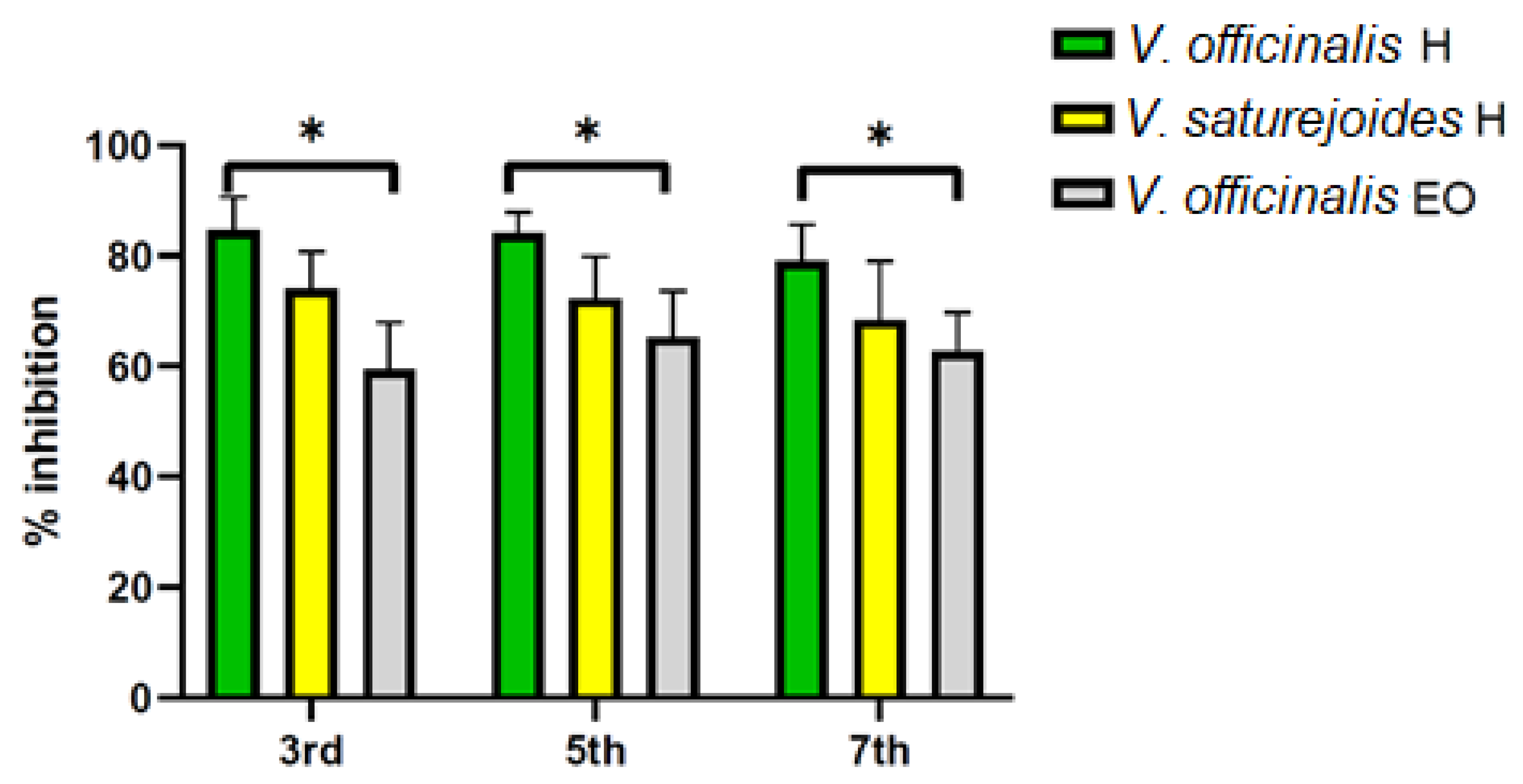
3.3. Antiphytoviral Activity

The numerous biological activities of essential oils known to date and their role in the interaction of plants with their biotic and abiotic environment suggest that these specialized plant metabolites are much more than just plant fragrances. Hydrosols are a by-product of essential oil distillation, making the usability of all products of this process ecologically and biologically desirable. Therefore, the antiphytoviral activity is imposed as a continuation of the study of the biological activities of this genus, especially since this activity of the volatiles of the genus Veronica has never been tested before.
The results show that plants treated with hydrosol (H) of V. officinalis and V. saturejoides prior to TMV infection significantly reduced the number of local lesions compared to control plants. In the essential oil (EO) treated plants, the number of local lesions decreased only on the leaves of the plants treated with V. officinalis EO (Table 3), while the plants treated with V. saturejoides EO in the preliminary experiment developed similar or even more severe infection than the control plants. On the third day post inoculation, the inhibition of lesions on the leaves of the plants treated with V. officinalis H and V. saturejoides H was 84.69% and 74.11%, respectively, while it was 59.43% on the leaves of the plants treated with V. officinalis EO (Figure 4). Until the seventh day after inoculation, the inhibition of local lesions was still pronounced in all treated groups, with 79.09% and 68.38% on the leaves of plants treated with V. officinalis H and V. saturejoides H, respectively, and 62.70% on the leaves of plants treated with V. officinalis EO. We compared these results with the antiphytoviral activity of plant volatiles reported in some of our previous studies and studies by other authors [19,20,21,22,23,24,25,26,27]. Thus, essential oils isolated from the aromatic species Satureja montana ssp. variegata and Teucrium arduini inhibited TMV infection by 29.2% and 25.7%, and cucumber mosaic virus (CMV) infection by 24.1% and 21.9%, respectively [20]. The essential oils of species of the genus Micromeria (M. graeca, M. fruticulosa and M. croatica) showed antiphytoviral activity on plants infected with satellite RNA associated cucumber mosaic virus (satCMV) with an activity of 59.3%, 43.6% and 34.5%, respectively [19,24,44]. The essential oils extracted from four species of the genus Teucrium (T. polium, T. flavum, T. montanum and T. chamaedrys) were shown to reduce the number of lesions in the host plants infected with CMV, with the essential oil of T. polium having the strongest effect with an activity of 41.4% [23]. The essential oils of Hypericum perforatum ssp. veronense, Eryngium alpinum and E. amethystinum showed promising antiviral activity rates of 88%, 77.8% and 80.5%, respectively [22,25]. In addition, among the essential oils extracted from 29 indigenous Chinese aromatic plants, the oils of ginger, lemon, tea tree, tangerine peel, artemisia, and lemongrass caused more than 50% inhibition of TMV at the concentrations tested [26,27]. In addition to plant extracts, inhibitors of plant viruses derived from metabolites of microbes are also considered as potential alternatives to chemical pesticides [45]. Ningnanmycin isolated from the fermentation broth of Streptomyces noursei var. Xichangensis exhibits comprehensive antiphytoviral activity and is characterized by increased resistance, excellent efficiency and low toxicity in host plants by acting through expression of pathogenesis-related proteins, increasing salicylic acid biosynthesis and inducing systemic resistance in host plants [46,47,48].
This and previous studies dealing with essential oils and hydrosols of aromatic plant species [20,21,22,23,24,25] show that volatile plant compounds can increase the resistance of plants to viral pathogens. The activity of hydrosol of both Veronica species is even more promising than that of essential oils, and the presented results suggest the need for further studies on the antiviral activity of volatiles of Veronica species. The activity of the tested extracts is probably related to their chemical composition, where a synergistic effect of the volatiles could activate plant signaling pathways and lead to increased resistance to viral infections. Among the major constituents of the essential oil (Table 2), β-ionone and hexadecanoic acid are abundant in V. officinalis EO (17.88% and 20.62%, respectively), in contrast to V. saturejoides EO, where these constituents are less abundant (hexadecanoic acid) or absent (β-ionone). In addition, δ-selinene (3.32%), benzyl benzoate (4.56%), docosane (2.13%) and pentacosane (2.71%) are noteworthy, which are also detected only in V. officinalis EO, but not in V. saturejoides EO. Comparing the essential oil composition with that of hydrosol, α-terpineol, allo-aromadendrene, benzene acetaldehyde, n-nonanal and hexyl 2-methylbutanoate are listed in similar abundance in both V. saturejoides H and V. officinalis H, while they are less abundant or absent in V. saturejoides EO. The above similarities and differences in the composition of the tested extracts may be useful for future predictions of their efficacy in plant protection.
The reported antiphytoviral activity of Veronica species should be analyzed in more detail in the future, including further studies to evaluate their efficacy against viral diseases under field conditions.

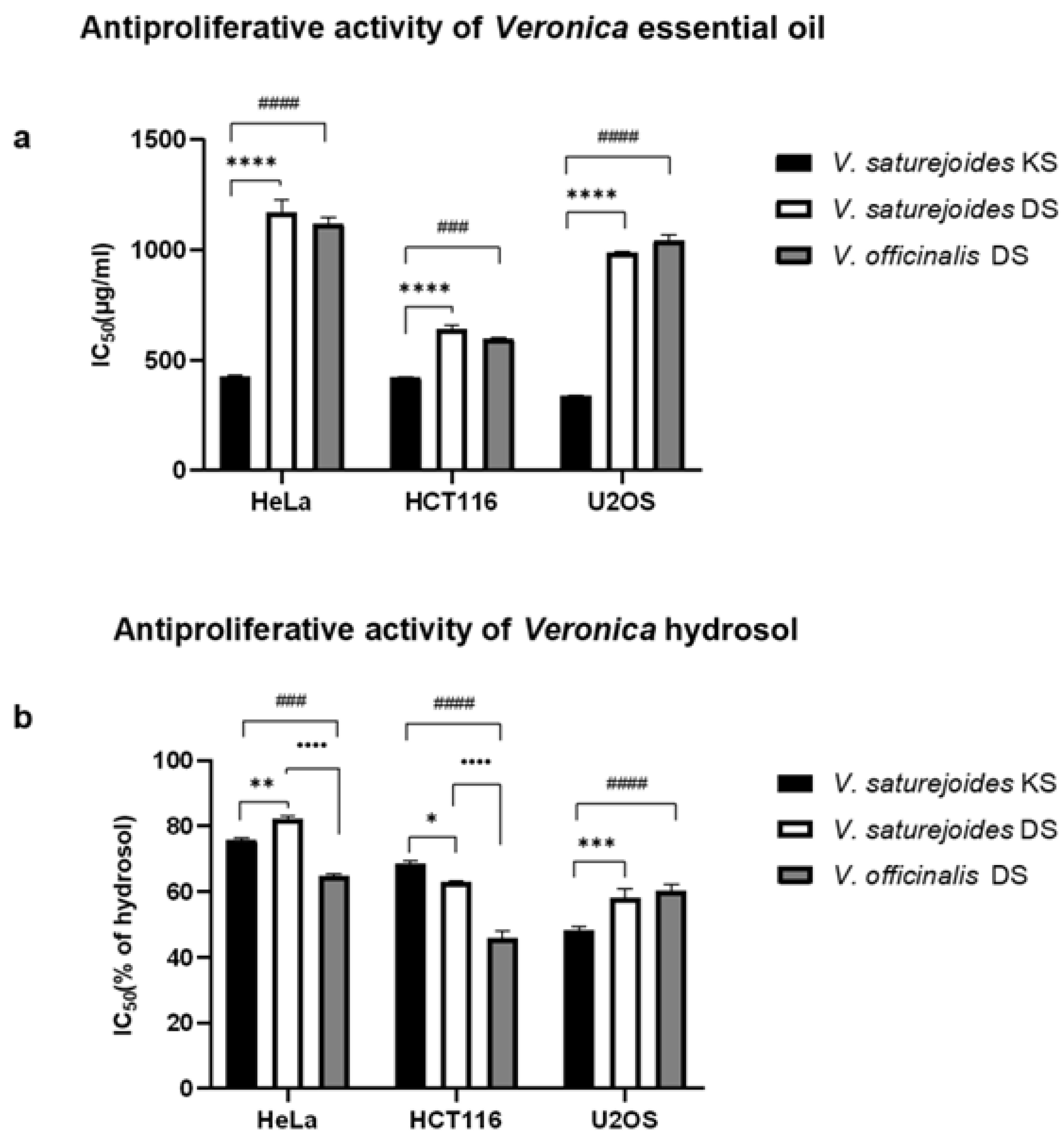
3.4. Antiproliferative Activity

In the present study antiproliferative activity of the essential oil (EO) and hydrosol of two Veronica species was analyzed. This was of particular interest to us because there are no data in the literature on the antiproliferative activity of V. saturejoides and V. officinalis. The results of this study showed significant antiproliferative activity of the oil and hydrosol of these species. Essential oil and hydrosol of V. saturejoides collected from two different locations (Kamešnica sample (KS*) and Dinara sample (DS)) and V. officinalis (Kamešnica sample (KS)) were tested on three cancer cell lines: HeLa (human cervical cancer cell line), HCT116 (colon cancer cell line) and U2OS (osteosarcoma cell line). The best antiproliferative activity on all three cell lines tested was shown by the essential oil of V. saturejoides KS (Figure 5a). A particularly significant difference in the antiproliferative power of V. saturejoides KS compared to V. saturejoides DS and V. officinalis KS was observed in the HeLa and U2OS cell lines. Interestingly, the hydrosol showed slightly different results in contrast to the essential oil (Figure 5b). The hydrosol of V. saturejoides KS and DS showed similar antiproliferative effect on all the tested cancer cells. The hydrosol of V. officinalis DS showed the strongest antiproliferative activity on HeLa and HCT116 cell lines with IC50 values of 64.93% and 45.93%, respectively. V. saturejoides KS hydrosol had the best growth inhibition on the U2OS cell line with IC50 value of 48.23%.
Species of the genus Veronica have long been used in traditional medicine to treat a number of diseases including cancer, influenza, hernia, cough, respiratory diseases and many others [49,50,51]. Studies have shown that these plants are a source of secondary metabolites that are largely responsible for their excellent biological activity, such as antimicrobial, antioxidant and anti-inflammatory properties. Although various Veronica extracts have been used in folk medicine for the treatment of cancer, very few species have been studied for their cytotoxic and anticancer activity [7]. Previous research on various Veronica species has mainly led to the isolation of biologically active compounds such as iridoid glucosides, which have been found to be excellent natural anticancer agents [52,53,54,55,56,57]. Moreover, methanolic and aqueous extracts of several Veronica species have been tested on different cancer cell lines. The cytotoxic activity of methanolic extracts of five Veronica species (V. cymbalaria, V. hederifolia, V. pectinata var. glandulosa, V. persica and V. polita) was tested against KB (human epidermoid carcinoma) and B16 (mouse melanoma) cells. All species showed similar dose-dependent effects against tested cells [54]. Methanolic extract of V. americana managed to stop the division of the two cancer cells of HF-6 (colon) and PC -3 (prostate) cancer cell lines [58]. Feng et al. showed that flavonoids isolated from V. sibirica (Vtfs) induced dose-dependent apoptosis in breast cancer cells MCF-7 with IC50 of 42 µg/mL [59]. Aqueous extracts of V. cuneifolia subsp. cuneifolia and V. cymbalaria showed moderate cytotoxic activity against Hep-2 (human epidermoid carcinoma), RD (human rhabdomyosarcoma) and L-20B (transgenic murine L-cells) [60]. Water fractions of the methanolic extracts of V. persica and V. crista-galli effectively inhibited proliferation of HeLa and MCF-7 cells but showed no toxicity against normal fibroblast cell line [61]. These water fractions contained a high concentration of flavonoids and phenols, which is why they exhibited significant antioxidant activity in addition to their cytotoxic activity.
The results of this study show for the first time that both the essential oil and hydrosol of the species V. saturejoides KS, V. saturejoides DS and V. officinalis KS have significant antiproliferative activity and thus possible chemotherapeutic properties, which is why they deserve further investigation. It is necessary to conduct further studies with the essential oil and hydrosol of V. saturejoides and V. officinalis and their major constituents on different cancer cell lines to determine the mechanism of antiproliferative action and to evaluate the possibility of their use in medicine and pharmacology.
*Note: Chemical composition of the EO and hydrosol of V. saturejoides ssp. saturejoides from the Kamešnica location (KS) and plant material information was reported in the previous work by Nazlić et al. [28].

3.5. Antioxidant Activity

The antioxidant activity of the extracted volatile compound was analyzed by two methods, ORAC and DPPH, and the results are presented in the Table 4. The essential oil of V. saturejoides ssp. saturejoides showed higher antioxidant activity than the EO of V. officinalis by both methods. This could be due to a much higher content of oxygenated sesquiterpenes, especially the compound caryophyllene oxide. The hydrosols showed slightly different results. In the DPPH method, hydrosol of V. saturejoides showed higher activity, and in the ORAC method, hydrosol of V. officinalis showed higher activity (Table 4). Hydrosols of V. saturejoides also have higher content of oxygenated sesquiterpenes. Sesquiterpenes have been previously reported to possess numerous biological activities [62,63]. Comparing these results with those previously reported for V. saturejoides ssp. saturejoides from two other locations (Kamešnica and Prenj Mountain), it can be seen that the antioxidant activity of EO of V. saturejoides (Dinara sample) showed similar ORAC activity to the other two previously reported EOs, but the DPPH activity seems to be lower than the EOs of V. saturejoides from Kamešnica and Prenj Mountain [28]. The sample from Kamešnica seems to have the highest antioxidant activity, as was also shown for antiproliferative activity. V. officinalis EO has lower antioxidant activity than the V. saturejoides EO sample from this research report (Dinara sample) and also lower than the two previously reported EOs. Therefore, the conclusion can be drawn that V. officinalis EO has lower antioxidant activity than V. saturejoides EO. Comparing the results for the hydrosols, it is evident from Table 4 and previous results for V. saturejoides [28] that both the hydrosols of V. officinalis and V. saturejoides have lower antioxidant activity than the previously reported hydrosols from the two locations of V. saturejoides. Comparing these results with reports on other plants, it appears that V. officinalis hydrosol has higher antioxidant ORAC activity than Salvia officinalis hydrosol [64]. Reports on the antioxidant activity of other extracts (phenolic compounds and iridoid glycosides) from Veronica species indicate that these plants have significant antioxidant potential. Valyova et al. studied phenolic extracts of V. officinalis and found that ethyl acetate extracts exhibited excellent antioxidant activity based on DPPH and ABTS assays [11]. Harput et al. investigated antioxidant activity for four Veronica species (V. orientalis, V. baranetzkii, V. officinalis and V. peduncularis) and reported the strongest antioxidant activity for aqueous extracts of V. officinalis (IC50 54.19 µg/mL). This could be due to the highest reported total phenolic content being for this species (200.20 mg/g) [65]. In another study, Harput et al. reported antioxidant activity against SO for V. chamaedrys (IC50) to be higher than the standards (BHA and quercetin) and the highest IC50 value for V. officinalis against DPPH radical to be 40.93 µg/mL [66]. Živković et al. investigated antioxidant activity of three Veronica species and reported highest antioxidant activity for V. teucrium 70% aqueous acetone extracts (IC50 12.58 µg/mL) [67]. Dunkić et al. reported even higher antioxidant activity for methanol extracts of flowers of V. spicata with IC50 8.21 µg/mL [9]. Sharifi-Rad et al. reported DPPH antioxidant activity for methanol extract of aboveground parts of V. persica to be IC50 30 µg/mL. [68]. All these results show that speedwells should be further researched for their in vivo antioxidant activities or for potential usage in food preservations.

4. Conclusions

In this study, the free volatile compounds and their biological activities of two speedwells, V. officinalis and V. saturejoides ssp. saturejoides, from the Dinaric Massif, were analyzed. The most abundant compound in the extracts was caryophyllene oxide for EO and hydrosol of V. saturejoides. Hexadecanoic acid and p-vinyl guaiacol were the most abundant compounds in the EO and hydrosols of V. officinalis, respectively. The volatile compounds extracted from the essential oils and hydrosols showed significant antiphytoviral activity, antiproliferative and antioxidant activity. The results for antiphytoviral activity show that hydrosol of V. officinalis and V. saturejoides reduced the number of local lesions on the plants infected with the TMV virus compared to control plants. Essential oil of V. officinalis also showed results against TMV infection, while EO of V. saturejoides showed no antiphytoviral activity. The best antiproliferative activity on all three cell lines tested was shown by the essential oil of V. saturejoides from the Kamešnica location. However, for the hydrosols the results showed that V. officinalis had the strongest antiproliferative activity against HeLA and HCT116 cell lines. Antioxidant activity of the essential oil of V. saturejoides ssp. saturejoides appeared to be higher than the activity of the V. officinalis EO by both tested methods. Hydrosols gave different results, as hydrosols from the V. officinalis showed higher activity by the ORAC method and hydrosols from V. saturejoides showed higher activity by the DPPH method. These results show that plants of the genus Veronica are more than just a weed, as they are often considered, but a valuable source of biologically active compounds for human use and for neighboring plants if used in horticulture. Further research could be focused on in vivo studies. Furthermore, it would be valuable to grow these plants from seed in a controlled environment and compare chemical composition and biological activity to the species growing in the wild.

Author Contributions

Conceptualization, V.D. and M.N.; Methodology, V.D. and M.N.; Software, M.N.; Validation, V.D., D.K. and M.N.; Formal Analysis, M.N. V.D., Ž.F., D.K. and E.V.; Investigation, V.D., M.N., E.S., D.K., Ž.F., M.R., E.V. and L.F.; Data Curation, M.N. and V.D.; Writing—Original Draft Preparation, M.N.; Writing—Review and Editing, M.N. and V.D.; Visualization, M.N.; Supervision, V.D.; Funding Acquisition, V.D. All authors have read and agreed to the published version of the manuscript.

Funding

This research was supported by the project “Croatian Veronica species: Phytotaxonomy and Biological Activity”, CROVeS-PhyBA, funded by the Croatian Science Foundation. Project number IP-2020-02-8425.

Institutional Review Board Statement

Not applicable.

Informed Consent Statement

Not applicable.

Acknowledgments

The authors thank Janoš Terzić from the School of Medicine, University of Split for providing cell material. The plate reader Infinite M Plex (Tecan) used for the measurement of the DPPH activity was procured through the project “Functional integration of the University of Split through the development of scientific research infrastructure in the building of three faculties” funded by the European Commission through the European Regional Development Fund.

Conflicts of Interest

The authors declare no conflict of interest. The founding sponsors had no role in the collection, analyses, or interpretation of data, or in the writing of the manuscript.

References

  1. Albach, D.C.; Martinez-Ortega, M.M.; Chase, M.W. Veronica: Parallel morphological evolution and phylogeography in the Mediterranean. Plant Syst. Evol. 2004, 246, 177–194. [Google Scholar] [CrossRef]
  2. Albach, D.C.; Martínez-Ortega, M.M.; Fischer, M.A.; Chase, M.W. A new classification of the tribe Veroniceae-problems and a possible solution. Taxon 2004, 53, 429–452. [Google Scholar] [CrossRef]
  3. Sharifi-Rad, M.; Tayeboon, G.S.; Sharifi-Rad, J.; Iriti, M.; Varoni, E.M.; Razazi, S. Inhibitory activity on type 2 diabetes and hypertension key-enzymes, and antioxidant capacity of Veronica persica phenolic-rich extracts. Cell. Mol. Biol. 2016, 62, 80–85. [Google Scholar] [CrossRef]
  4. Nikolić, T. Flora Croatica Database. Available online: http://hirc.botanic.hr/fcd (accessed on 22 May 2021).
  5. Reynolds, T. The evolution of chemosystematics. Phytochemistry 2007, 68, 2887–2895. [Google Scholar] [CrossRef] [PubMed]
  6. Loreto, F.; Dicke, M.; Schnitzler, J.P.; Turlings, T.C.J. Plant volatiles and the environment. Plant Cell Environ. 2014, 37, 1905–1908. [Google Scholar] [CrossRef]
  7. Salehi, B.; Shetty, M.S.; Anil Kumar, N.V.; Živković, J.; Calina, D.; Docea, A.O.; Emamzadeh-Yazdi, S.; Kılıç, C.S.; Goloshvili, T.; Nicola, S.; et al. Veronica plants—Drifting from farm to traditional healing, food application, and phytopharmacology. Molecules 2019, 24, 2454. [Google Scholar] [CrossRef] [Green Version]
  8. Xue, H.; Chen, K.X.; Zhang, L.Q.; Li, Y.M. Review of the Ethnopharmacology, Phytochemistry, and Pharmacology of the Genus Veronica. Am. J. Chin. Med. 2019, 47, 1193–1221. [Google Scholar] [CrossRef]
  9. Dunkić, V.; Kosalec, I.; Kosir, I.; Potočnik, T.; Cerenak, A.; Koncic, M.; Vitali, D.; Muller, I.; Kopricanec, M.; Bezić, N.; et al. Antioxidant and antimicrobial properties of Veronica spicata L. (Plantaginaceae). Curr. Drug Targets 2015, 16, 1660–1670. [Google Scholar] [CrossRef]
  10. Ertas, A.; Boga, M.; Kizil, M.; Ceken, B.; Goren, A.C.; Hasimi, N.; Demirci, S.; Topcu, G.; Kolak, U. Chemical profile and biological activities of Veronica thymoides subsp. pseudocinerea. Pharm. Biol. 2015, 53, 334–339. [Google Scholar] [CrossRef] [Green Version]
  11. Mocan, A.; Vodnar, D.C.; Vlase, L.; Crișan, O.; Gheldiu, A.M.; Crișan, G. Phytochemical characterization of Veronica officinalis L., V. teucrium L. and V. orchidea Crantz from Romania and their antioxidant and antimicrobial properties. Int. J. Mol. Sci. 2015, 16, 21109–21127. [Google Scholar] [CrossRef]
  12. Valyova, M.; Hadjimitova, V.; Stoyanov, S.; Ganeva, Y.; Petkov, I. Free radical scavenging activity of extracts from Bulgarian Veronica officinalis L. and GC-MS analysis of ethanol extract. Internet J. Aesthetic Antiaging Med. 2008, 2, 2–6. [Google Scholar] [CrossRef]
  13. Kwak, J.H.; Kim, H.J.; Lee, K.H.; Kang, S.C.; Zee, O.P. Antioxidative iridoid glycosides and phenolic compounds from Veronica peregrina. Arch. Pharm. Res. 2009, 32, 207–213. [Google Scholar] [CrossRef] [PubMed]
  14. Ishibashi, K.; Ishikawa, M. Replication of Tobamovirus RNA. Annu. Rev. Phytopathol. 2016, 54, 55–78. [Google Scholar] [CrossRef] [Green Version]
  15. Othman, B.A.; Shoman, S.A. Antiphytoviral Activity of the Plectranthus tenuiflorus on Some Important Viruses. Int. J. Agric. Biol. 2004, 6, 844–849. [Google Scholar]
  16. Rusak, G.; Krajačić, M.; Krsnik-Rasol, M.; Gutzeit, H.O. Quercetin influences response in Nicotiana megalosiphon infected by satellite-associated cucumber mosaic virus/Quercetin beeinf lusst die Reaktion von Nikotiana magalosiphon gegenüber Satelliten-assoziierten Gurkenmosaikvirus-Infektionen. J. Plant Dis. Prot. 2007, 114, 145–150. [Google Scholar] [CrossRef]
  17. Krcatović, E.; Rusak, G.; Bezić, N.; Krajačić, M. Inhibition of tobacco mosaic virus infection by quercetin and vitexin. Acta Virol. 2008, 52, 119–124. [Google Scholar]
  18. Moeini, A.; van Reenen, A.; Van Otterlo, W.; Cimmino, A.; Masi, M.; Lavermicocca, P.; Valerio, F.; Immirzi, B.; Santagata, G.; Malinconico, M.; et al. α-costic acid, a plant sesquiterpenoid from Dittrichia viscosa, as modifier of Poly (lactic acid) properties: A novel exploitation of the autochthone biomass metabolite for a wholly biodegradable system. Ind. Crops Prod. 2020, 146, 112134. [Google Scholar] [CrossRef]
  19. Vuko, E.; Dunkić, V.; Bezić, N.; Ruščić, M.; Kremer, D. Chemical Composition and Antiphytoviral Activity of Essential Oil of Micromeria graeca. Nat. Prod. Commun. 2012, 7, 1227–1230. [Google Scholar] [CrossRef] [Green Version]
  20. Dunkić, V.; Bezić, N.; Vuko, E.; Cukrov, D. Antiphytoviral activity of Satureja montana L. ssp. variegata (host) P. W. Ball essential oil and phenol compounds on CMV and TMV. Molecules 2010, 15, 6713–6721. [Google Scholar] [CrossRef] [Green Version]
  21. Dunkić, V.; Bezić, N.; Vuko, E. Antiphytoviral activity of essential oil from endemic species Teucrium arduini. Nat. Prod. Commun. 2011, 6, 1385–1388. [Google Scholar] [CrossRef] [Green Version]
  22. Dunkić, V.; Vuko, E.; Bezić, N.; Kremer, D.; Ruščić, M. Composition and antiviral activity of the essential oils of Eryngium alpinum and E. amethystinum. Chem. Biodivers. 2013, 10, 1894–1902. [Google Scholar] [CrossRef] [PubMed]
  23. Bezić, N.; Vuko, E.; Dunkić, V.; Ruščić, M.; Blažević, I.; Burčul, F. Antiphytoviral Activity of Sesquiterpene-Rich Essential Oils from Four Croatian Teucrium species. Molecules 2011, 16, 8119–8129. [Google Scholar] [CrossRef] [PubMed] [Green Version]
  24. Vuko, E.; Rusak, G.; Dunkić, V.; Kremer, D.; Kosalec, I.; Rada, B.; Bezić, N. Inhibition of satellite RNA associated cucumber mosaic virus infection by essential oil of Micromeria croatica (Pers.) Schott. Molecules 2019, 24, 1342. [Google Scholar] [CrossRef] [Green Version]
  25. Vuko, E.; Dunkić, V.; Ruščić, M.; Nazlić, M.; Mandić, N.; Soldo, B.; Šprung, M.; Fredotović, Ž. Chemical Composition and New Biological Activities of Essential Oil and Hydrosol of Hypericum perforatum L. ssp. veronense (Schrank) H. Lindb. Plants 2021, 10, 1014. [Google Scholar] [CrossRef]
  26. Lu, M.; Han, Z.; Xu, Y.; Yao, L. In Vitro and In Vivo Anti-Tobacco Mosaic Virus Activities of Essential Oils and Individual Compounds. J. Microbiol. Biotechnol. 2013, 23, 771–778. [Google Scholar] [CrossRef] [Green Version]
  27. Bishop, C.D. Antiviral Activity of the Essential Oil of Melaleuca alternifolia (Maiden amp; Betche) Cheel (Tea Tree) Against Tobacco Mosaic Virus. J. Essent. Oil Res. 1995, 7, 641–644. [Google Scholar] [CrossRef]
  28. Nazlić, M.; Kremer, D.; Jurišić-Grubešić, R.; Soldo, B.; Vuko, E.; Stabentheiner, E.; Ballian, D.; Bogunić, F.; Dunkić, V. Endemic Veronica saturejoides Vis. ssp. saturejoides–Chemical Composition and Antioxidant Activity of Free Volatile Compounds. Plants 2020, 9, 1646. [Google Scholar] [CrossRef]
  29. Adams, R.P. Identification of Essential Oil Components by Gas Chromatography/Mass Spectrometry, 4th ed.; Allured Publishing: Carol Stream, IL, USA, 2017; ISBN 978-1-932633-21-4. [Google Scholar]
  30. Babushok, V.I.; Linstrom, P.J.; Zenkevich, I.G. Retention Indices for Frequently Reported Compounds of Plant Essential Oils. J. Phys. Chem. Ref. Data 2011, 40, 1–47. [Google Scholar] [CrossRef] [Green Version]
  31. Li, F. Analysis of chemical constituents of essential oil in Veronica linariifolia by gas chromatography-mass spectrometry. Chin. J. Anal. Chem. 2002, 30, 822–825. [Google Scholar]
  32. Çelik, E.; Çelik, G.Y.; Meysun, A. Essential Oil Composition and Antibacterial Activity of Some Plant Species. J. Appl. Biol. Sci. 2010, 1, 45–48. [Google Scholar] [CrossRef] [Green Version]
  33. Kremer, D.; Müller, I.D.; Dunkić, V.; Vitali, D.; Stabentheiner, E.; Oberländer, A.; Bezić, N.; Kosalec, I. Chemical traits and antimicrobial activity of endemic Teucrium arduini L. from Mt Biokovo (Croatia). Cent. Eur. J. Biol. 2012, 7, 941–947. [Google Scholar] [CrossRef] [Green Version]
  34. Payne, W.W. A glossary of plant hair thermonology. Brittonia 1978, 30, 239–255. [Google Scholar] [CrossRef]
  35. Fredotović, Ž.; Šprung, M.; Soldo, B.; Ljubenkov, I.; Budić-Leto, I.; Bilušić, T.; Cikeš-Čulić, V.; Puizina, J. Chemical composition and biological activity of Allium cepa L. and Allium × cornutum (Clementi ex Visiani 1842) methanolic extracts. Molecules 2017, 22, 448. [Google Scholar] [CrossRef] [Green Version]
  36. Mensor, L.L.; Menezes, F.S.; Leitão, G.G.; Reis, A.S.; dos Santos, T.C.; Coube, C.S.; Leitão, S.G. Screening of Brazilian plant extracts for antioxidant activity by the use of DPPH free radical method. Phyther. Res. 2001, 15, 127–130. [Google Scholar] [CrossRef]
  37. Payet, B.; Sing, A.S.C.; Smadja, J. Assessment of antioxidant activity of cane brown sugars by ABTS and DPPH radical scavenging assays: Determination of their polyphenolic and volatile constituents. J. Agric. Food Chem. 2005, 53, 10074–10079. [Google Scholar] [CrossRef]
  38. Hazzoumi, Z.; Moustakime, Y.; Joutei, K.A. Essential Oil and Glandular Hairs: Diversity and Roles. In Essential Oils—Oils of Nature; El-Shemy, H.A., Ed.; IntechOpen: London, UK, 2019; pp. 1–16. [Google Scholar]
  39. Haratym, W.; Weryszko-Chmielewska, E. Ultrastructural and histochemical analysis of glandular trichomes of Marrubium vulgare L. (Lamiaceae). Flora Morphol. Distrib. Funct. Ecol. Plants 2017, 231, 11–20. [Google Scholar] [CrossRef]
  40. Baran, P.; Aktaş, K.; Özdemir, C. Structural investigation of the glandular trichomes of endemic Salvia smyrnea L. S. Afr. J. Bot. 2010, 76, 572–578. [Google Scholar] [CrossRef] [Green Version]
  41. Kristen, U.; Lockhausen, J. The leaf glands of Veronica beccabunga L.: Ultrastructure and a possible pathway of secretion. Isr. J. Plant Sci. 2015, 34, 147–156. [Google Scholar] [CrossRef]
  42. Kurer, G.A. Kutikularfalten und Protuberanzen an Haaren und Epidermen; Druck, Gebr. Leeman: Dresden, Germany, 1917. [Google Scholar]
  43. Kraehmer, H.; Baur, P. Weed Anatomy; Wiley-Blackwell: Hoboken, NJ, USA, 2013; ISBN 978-0-470-65986-1. [Google Scholar]
  44. Ruščić, M.; Vuko, E.; Bezić, N.; Kremer, D.; Dunkić, V. Croatian Micromeria fruticulosa-essential oil composition and antiphytoviral activity. Nat. Prod. Commun. 2017, 12, 1133–1135. [Google Scholar] [CrossRef] [Green Version]
  45. An, M.; Zhou, T.; Guo, Y.; Zhao, X.; Wu, Y. Molecular Regulation of Host Defense Responses Mediated by Biological Anti-TMV Agent Ningnanmycin. Viruses 2019, 11, 815. [Google Scholar] [CrossRef] [PubMed]
  46. Han, Y.; Luo, Y.; Qin, S.; Xi, L.; Wan, B.; Du, L. Induction of systemic resistance against tobacco mosaic virus by Ningnanmycin in tobacco. Pestic. Biochem. Physiol. 2014, 111, 14–18. [Google Scholar] [CrossRef] [PubMed]
  47. Fan, H.; Song, B.; Bhadury, P.S.; Jin, L.; Hu, D.; Yang, S. Antiviral activity and mechanism of action of novel thiourea containing chiral phosphonate on tobacco mosaic virus. Int. J. Mol. Sci. 2011, 12, 4522–4535. [Google Scholar] [CrossRef] [PubMed] [Green Version]
  48. Gao, D.; Wang, D.; Chen, K.; Huang, M.; Xie, X.; Li, X. Activation of biochemical factors in CMV-infected tobacco by ningnanmycin. Pestic. Biochem. Physiol. 2019, 156, 116–122. [Google Scholar] [CrossRef] [PubMed]
  49. Tomassini, L.; Brkic, D.; Serafini, M.; Nicoletti, M. Constituents of Veronica hederifolia and Veronica polita. Fitoterapia 1995, 66, 382. [Google Scholar]
  50. Su, B.N.; Zhu, Q.X.; Jia, Z.J. Aquaticol, a novel bis-sesquiterpene from Veronica anagallis-aquatica. Tetrahedron Lett. 1999, 40, 357–358. [Google Scholar] [CrossRef]
  51. Graham, J.G.; Quinn, M.L.; Fabricant, D.S.; Farnsworth, N.R. Plants used against cancer—An extension of the work of Jonathan Hartwell. J. Ethnopharmacol. 2000, 73, 347–377. [Google Scholar] [CrossRef]
  52. Chari, V.M.; Grayer-Barkmeijer, R.J.; Harborne, J.B.; Osterdahl, B.-G. An acylated allose-containing 8-hydroxyflavone glycoside from Veronica filiformis. Phytochemistry 1981, 20, 1977–1979. [Google Scholar] [CrossRef]
  53. Taskova, R.; Handjieva, N.; Peev, D.; Popov, S. Iridoid glucosides from three Veronica species. Phytochemistry 1998, 49, 1323–1327. [Google Scholar] [CrossRef]
  54. Harput, U.S.; Saracoglu, I.; Inoue, M.; Ogihara, Y. Anti-inflammatory and cytotoxic activities of five Veronica species. Biol. Pharm. Bull. 2002, 25, 483–486. [Google Scholar] [CrossRef] [Green Version]
  55. Saracoglu, I.; Harput, U.S.; Inoue, M.; Ogihara, Y. New phenylethanoid glycosides from Veronica pectinata var glandulosa and their free radical scavenging activities. Chem. Pharm. Bull. 2002, 50, 665–668. [Google Scholar] [CrossRef] [Green Version]
  56. Saracoǧlu, I.; Harput-Hudaverdi, U.Ş.; Nagatsu, A.; Varel, M. Cytotoxic Activity and Structural Aspects of Iridoids Isolated from Veronica thymoides subsp. pseudocinerea. In Proceedings of the Natural Products with Pharmaceutical, Nutraceutical, Cosmetic, and Agrochemical Interest, 7th Joint Meeting of AFERP, ASP, GA, and PSE (49th Annual Meeting of the American Society of Pharmacognosy), Athens, Greece, 8 July 2008; p. 1046. [Google Scholar]
  57. Saracoǧlu, I.; Harput-Hudaverdi, U.Ş. In vitro Anticancer Activity and Structure-Activity Relationships of Iridoid Glucosides. In Proceedings of the 9th International Symposium on Pharmaceutical Sciences, Ankara, Turkey, 23–26 June 2009. [Google Scholar]
  58. Moreno-Escobar, J.A.; Bazalda, S.; Villarreal, M.L.; Bonilla-Barbosa, J.R.; Mendoza, S.; Rodríguez-López, V. Cytotoxic and antioxidant activities of selected Lamiales species from Mexico. Pharm. Biol. 2011, 49, 1243–1248. [Google Scholar] [CrossRef] [PubMed]
  59. Feng, K.; Jiang, R.; Sun, L.W. Studies on the anti-tumor activity in vitro of the flavonoid extract in round leaf speedwell. In Proceedings of the First International Conference on Cellular, Molecular Biology, Biophysics and Bioengineering, Lushan, China, 17–18 September 2010. [Google Scholar]
  60. Saracoglu, I.; Oztunca, F.H.; Nagatsu, A.; Harput, U.S. Iridoid content and biological activities of Veronica cuneifolia subsp. cuneifolia and V. cymbalaria. Pharm. Biol. 2011, 49, 1150–1157. [Google Scholar] [CrossRef] [PubMed] [Green Version]
  61. Mohadjerani, M.; Asadollahi, S. Veronica crista-galli Steven and Veronica persica Poir. as anticancer and antioxidant plants in-vitro. Trends Phytochem. Res. 2019, 3, 61–66. [Google Scholar] [CrossRef]
  62. Assaeed, A.; Elshamy, A.; El Gendy, A.E.N.; Dar, B.; Al-Rowaily, S.; Abd-ElGawad, A. Sesquiterpenes-rich essential oil from above ground parts of Pulicaria somalensis exhibited antioxidant activity and allelopathic effect on weeds. Agronomy 2020, 10, 399. [Google Scholar] [CrossRef] [Green Version]
  63. Chappell, J.; Coates, R.M. Sesquiterpenes. Compr. Nat. Prod. II Chem. Biol. 2010, 1, 609–641. [Google Scholar] [CrossRef]
  64. Aazza, S.; Lyoussi, B.; Miguel, M. Antioxidant activity of some Morrocan hydrosols. J. Med. Plants Res. 2011, 5, 6688–6696. [Google Scholar] [CrossRef]
  65. Harput, U.S.; Saracoglu, I.; Genc, Y. Comparative Bioactivity Studies on Four Veronica species. Fabad J. Pharm. Sci. 2009, 34, 67–72. [Google Scholar]
  66. Harput, U.Ş.; Genç, Y.; Khan, N.; Saracoglu, I. Radical scavenging effects of different Veronica species. Rec. Nat. Prod. 2011, 5, 100–107. [Google Scholar]
  67. Živković, J.; Ćebović, T.; Maksimović, Z. In vivo and in vitro antioxidant effects of three Veronica species. Cent. Eur. J. Biol. 2012, 7, 559–568. [Google Scholar] [CrossRef]
  68. Sharifi-Rad, J.; Sharifi-Rad, M.; Salehi, B.; Iriti, M.; Roointan, A.; Mnayer, D.; Soltani-Nejad, A.; Afshari, A. In vitro and in vivo assessment of free radical scavenging and antioxidant activities of Veronica persica Poir. Cell. Mol. Biol. 2018, 64, 57–64. [Google Scholar] [CrossRef] [Green Version]
Figure 1. Investigated plants in their natural habitat: (a) Veronica saturejoides ssp. saturejoides on Dinara Mountain; (b) Veronica officinalis L. on Kamešnica Mountain.
Horticulturae 07 00295 g001
Figure 2. Volatile compounds distribution by categories for all samples of essential oils and hydrosols for the investigated species V. officinalis and V. saturejoides ssp. saturejoides.
Horticulturae 07 00295 g002
Figure 3. V. officinalis SEM micrographs showing different types of trichomes on adaxial (a,g) and abaxial (b) leaf surface, on the sepal (c,e) and stem (d,f,h). Non-glandular trichomes (NG), peltate trichomes (P), subtype 1 (C1) and subtype 2 (C2) of capitate trichomes.
Horticulturae 07 00295 g003
Figure 4. Percentage of inhibition of lesions on leaves of treated Datura stramonium plants inoculated with tobacco mosaic virus compared to control plants on the third, fifth and seventh day post inoculation. Treated plants were sprayed with hydrosol (H) and essential oil (EO) of V. officinalis or V. saturejoides for three consecutive days prior and once immediately after inoculation. Error bars show standard deviation of triplicate analyses; significant differences were determined by t- test and marked with * (p ˂ 0.05).
Horticulturae 07 00295 g004
Figure 5. Antiproliferative activity of essential oil (a) and hydrosol (b) of V. saturejoides KS, DS and V. officinalis determined by MTS-based cell proliferation test. Results are expressed as the mean of three independent experiments ± SD (presented as error bars). Statistical analyzes were performed using the two-way ANOVA method followed by the Tukey’s multiple comparisons test, and significant differences were labeled as * (# or ) p < 0.05; ** (## or ••) p < 0.01; *** (### or •••) p < 0.001; and **** (#### or ••••) p < 0.0001.
Horticulturae 07 00295 g005
Table 1. Locations of the plant material collection (for volatile compounds extraction).
LocalityCoordinatesAltitude
a.s.l. (m)
Date of
Collection
Veronica officinalis L.Kamešnica Mountain43°43′03.1″ N 16°50′34.1″ E1445 mJuly 2021
Veronica saturejoides Vis. ssp. saturejoidesDinara Mountain44°3′2.6″ N; 16°22′52.9″ E1650 mJuly 2021
Table 2. Chemical composition of the essential oil (EO) and hydrosol (H) from the aerial parts of Veronica officinalis and Veronica saturejoides Vis. ssp. saturejoides (Plantaginaceae).
V. officinalis V. saturejoidesV. officinalis V. saturejoides
Essential OilsHydrosols
ComponentRI1RI2Mean ± SD (%)Mean ± SD (%)
Monoterpene hydrocarbons NINI0.150.89
α-Thujene9241012NININI0.23 ± 0.01
α-Pinene *9351017NININI0.66 ± 0.03
β -Phellandrene10021195NINI0.15 ± 0.03-
Oxygenated monoterpenes 1.3612.3913.4815.93
1,8-Cineole10261210NINI0.76 ± 0.01NI
γ-Terpinene10571225NINI2.61 ± 0.01NI
Linalool109515060.52 ± 0.01 b0.89 ± 0.05 a4.72 ± 0.01 a1.39 ± 0.01 b
allo-Ocimene112813900.22 ± 0.15NININI
Camphor11511499NINI0.72 ± 0.01NI
Borneol11761719NINI1.59 ± 0.01NI
α-Terpineol11841660NI0.88 ± 0.013.08 ± 0.03 a2.79 ± 0.01 b
trans-1(7),8-p-Mentadien-2-ol118718030.62 ± 0.0110.62 ± 0.02NI11.75 ± 0.01
Sesquiterpene hydrocarbons 13.3212.937.767.6
α-Copaene137714840.78 ± 0.01NININI
E-Caryophyllene *142415856.78 ± 0.04 b7.63 ± 0.01 a0.56 ± 0.01 b1.39 ± 0.01 a
allo-Aromadendrene146516621.32 ± 0.01 a0.87 ± 0.01 b2.59 ± 0.01 b3.87 ± 0.01 a
β-Chamigrene14781724NINI0.27 ± 0.12NI
Germacrene D14811692NI2.61 ± 0.01NI2.34 ± 0.01
δ-Selinene149217563.32 ± 0.01NI4.34 ± 0.01NI
δ-Cadinene151717451.12 ± 0.01 b1.82 ± 0.01 aNINI
Oxygenated sesquiterpenes 6.5434.0415.7425.82
Spathulenol15772101NI1.8 ± 0.015.25 ± 0.01NI
Caryophyllene oxide *158119551.42 ± 0.01 b23.65 ± 0.01 a7.52 ± 0.01 b21.28 ± 0.01 a
Viridiflorol15922099NININI1.53 ± 0.01
γ-Eudesmol163221751.82 ± 0.01 a0.2 ± 0.03 bNI0.23 ± 0.01
α-Muurolol164521813.30 ± 0.01 b7.86 ± 0.01 a2.38 ± 0.012.37 ± 0.01
α-Cadinol16552208NININI0.41 ± 0.01
α-Bisabolol16852210NINI0.59 ± 0.03NI
α-Bisabolol oxide17482511NI0.53 ± 0.01NINI
Phenolic compounds 0.2710.2311.5923.31
p-Vinyl guaiacol131321560.27 ± 0.03NI11.59 ± 0.01NI
Methyl eugenol14032005NI10.23 ± 0.01NI23.31 ± 0.01
Phenylpropanoids 6.021.411.752.61
Z-Methyl isoeugenol145120451.46 ± 0.03 a1.41 ± 0.01 b1.75 ± 0.01 b2.16 ± 0.03 a
Benzyl benzoate176026134.56 ± 0.01NININI
Fatty aldehyde, acids, alcohol, esters and ketones 56.5622.3336.8414.58
Isopentyl acetate8631127NI6.24 ± 0.010.24 ± 0.09 b0.59 ± 0.01 a
Benzaldehyde95215080.98 ± 0.01 b4.29 ± 0.01 a9.25 ± 0.01 a8.87 ± 0.01 b
Benzene acetaldehyde103616330.48 ± 0.01NI4.75 ± 0.01 a3.68 ± 0.01 b
n-Nonanal110013890.89 ± 0.02NI1.68 ± 0.01 a0.28 ± 0.01 b
Hexyl 2-methyl butanoate12331425NINI0.21 ± 0.01 a0.15 ± 0.03 b
Menthyl acetate12941550NINI1.58 ± 0.02NI
(E)-β-Damascone13841819NINI6.69 ± 0.01NI
β-Ionone1487193517.88 ± 0.01NI10.74 ± 0.01NI
Hexahydrofarnesyl acetone1839211313.92 ± 0.01 a6.18 ± 0.01 b0.25 ± 0.03 b0.39 ± 0.03 a
1-Hexadecanol187423711.79 ± 0.01 a1.51 ± 0.03 bNINI
Hexadecanoic acid1959291220.62 ± 0.01 a4.11 ± 0.01 b1.45 ± 0.01 a0.62 ± 0.02 b
Hydrocarbons 10.861.121.780.49
Eicosane *200020004.21 ± 0.01 a1.12 ± 0.01 b1.51 ± 0.04NI
Heneicosane *210021000.98 ± 0.17NI0.27 ± 0.01NI
Docosane *220022002.13 ± 0.01NININI
Tricosane *23002300NINININI
Tetracosane *240024000.83 ± 0.01NINI0.49 ± 0.01
Pentacosane *250025002.71 ± 0.04NININI
Total identification (%) 94.9395.4589.0991.23
Retention indices (RIs) were determined relative to a series of n-alkanes (C8–C40) on capillary columns VF5-ms (RI1) and CPWax 52 (RI2). Identification method: RI, comparison of RIs with those in a self-generated library reported in the literature [29] and/or with authentic samples; comparison of mass spectra with those in the NIST02 and Wiley 9 mass spectral libraries; * co-injection with reference compounds; NI, not identified; SD, standard deviation of triplicate analysis. Significant differences for every volatile compound present in both species were determined using multiple t-test. a, b Mean values in the same row with different superscript letters indicate a statistically significant difference between data from two species (p < 0.05); a for the higher abundance of the compound, b for the lesser abundance of the compound.
Table 3. Number of local lesions (LLN) on leaves of treated Datura stramonium plants inoculated with tobacco mosaic virus and on leaves of control plants (C) on the third, fifth, and seventh day post inoculation (dpi). Treated plants were sprayed with hydrosol (H) and essential oil (EO) of V. officinalis (V.off) or V. saturejoides (V.sat) for three consecutive days prior to and once immediately after inoculation.
LLN
dpiMean ± SDMean ± SD
3rdC5.86 ± 2.39C6.55 ± 1.24
V. off H0.83 ± 0.31 *V.off EO2.57 ± 0.18 *
V. sat H1.50 ± 0.62 *V.sat EOn.a.
5thC8.95 ± 2.95C8.39 ± 2.20
V. off H1.36 ± 0.58 *V.off EO2.75 ± 0.58 *
V. sat H2.24 ± 0.63 *V.sat EOn.a.
7thC10.55 ± 3.62C9.67 ± 2.24
V. off H2.10 ± 0.87 *V.off EO3.39 ± 0.50 *
V. satH3.02 ± 0.89 *V.sat EOn.a.
SD, standard deviation of triplicate analysis; n.a., no activity; significant differences were determined by t-test; * statistically significant differences between control and EO/H treatment data (p ˂ 0.05).
Table 4. Antioxidant potential of V. officinalis and V. saturejoides ssp. saturejoides of the essential oil and hydrosol determined by ORAC and DPPH method.
Essential Oils (Mean ± SD)
Antioxidant AssayV. officinalisV. saturejoides ssp. saturejoides
ORAC (Trolox eq)58.75 ± 3.42 b263.29 ± 4.89 a
DPPH (% inhibition)15.51 ± 1.6719.11 ± 4.09
DPPH (IC 50)31.34 ± 2.91 b15.99 ± 4.17 a
Hydrosols (mean ± SD)
Antioxidant AssayV. officinalisV. saturejoides ssp. saturejoides
ORAC (Trolox eq)0.307 ± 0.0110.258 ± 0.013
DPPH (% inhibition)27.74 ± 0.77 b38.39 ± 5.83 a
ORAC (oxygen radical absorbance capacity) results for EOs expressed as µmol of Trolox equivalents (TE) per g of EO (10 mg/mL) and for hydrosols as µmol of Trolox equivalents (TE) per g of the total (undiluted) tested hydrosol sample; DPPH, results are expressed in percentage of inhibition for the EO concentration of 10 mg per mL of acetone and for the hydrosols in percentage of inhibition for the absolute (undiluted) hydrosol; for the EOs, results are also expressed in IC50 value in mg/mL; SD = standard deviation of triplicate analysis; significant differences were determined using multiple t-test. a, b Mean values in the same row with different superscript letters indicate a statistically significant difference between data from four locations (p < 0.05), a for the higher activity, b for the lower activity.
Publisher’s Note: MDPI stays neutral with regard to jurisdictional claims in published maps and institutional affiliations.

