Article

Flowering Biology of Rhododendron pulchrum

1
Institute for Forest Resources and Environment of Guizhou, Key Laboratory of Forest Cultivation in Plateau Mountain of Guizhou Province, College of Forestry, Guizhou University, Guiyang 550025, China
2
College of Agriculture, Guizhou University, Guiyang 550025, China
*
Author to whom correspondence should be addressed.
Horticulturae 2021, 7(11), 508; https://doi.org/10.3390/horticulturae7110508
Submission received: 14 October 2021 / Revised: 6 November 2021 / Accepted: 14 November 2021 / Published: 18 November 2021

Abstract

:
To study the flowering biology of Rhododendron pulchrum, we used scanning electron microscopy (SEM) and paraffin sectioning to observe the microstructures of its floral organs, a methyl thiazolyl tetrazolium (MTT) colorimetric assay to detect pollen viability in different periods, continuous observations to study flowering phenology, and artificial pollination and a benzidine-hydrogen peroxide method to determine stigma receptivity. R. pulchrum exhibited a centralized flowering phenology. The protogynous stigmas of R. pulchrum were able to receive pollen before flowering. The pollen grains of R. pulchrum fused into tetrads, the average ratio of the polar axis length to the equatorial axis length (P/E) was 1.05, and the pollen viability was highest in the initial flowering period, reaching 88.98%. The pollen/ovule (P/O) ratio was 266–328, and the outcrossing index (OCI) was 4; the vitality of R. pulchrum pollen remained high in the initial flowering and blooming periods. Compared with the lifespan of a single flower, pollen vitality remained high for most of the experimental period, thereby improving male fitness. The P/O ratio suggests that R. pulchrum may have a facultative outcrossing breeding system. The OCI estimation suggests that R. pulchrum is partially self-compatible, most likely requiring pollinators to complete pollination.

1. Introduction

The flowers of angiosperms vary in numberous ways, especially in two elements: flower structure and floral display [1]. Floral display and arrangement affect the breeding systems of plants, and the study of the reproductive characteristics of a species is indispensable to exploring the mechanisms by which it is endangered [2]. The breeding system is an important but relatively vulnerable link in the life cycle of a species and an intermediate focus for the study of the sexual reproduction of populations [3]; these systems have a great impact on the morphological characteristics and evolutionary directions of plants and have become a hot topic among evolutionary biology studies. At present, researchers often use the outcrossing index (OCI) and pollen/ovule (P/O) ratio as predicators to predict breeding systems [4]. In addition, flowering biology also includes processes, such as flowering phenology, floral display, pollination, and senescence. The flowering phenology of plants can be quantified using indicators, such as the initial flowering period, the end of the flowering period, the number of flowers, and flowering synchrony. Flowering phenology, which is not only subject to genetic factors and intragenus evolutionary relationships but also closely associated with external environmental factors, has a large impact on the reproductive success rate of populations [5] and is an important plant characteristic. Floral displays are crucial to attracting pollinators [6]. In general, a full understanding of floral organ structures and breeding systems is a prerequisite for studying the plant life cycle and lays the foundation for understanding the differences in breeding characteristics of plant populations and foreseeing their evolutionary trends. To understand the flowering phenology, pollination, and other characteristics of plants, it is necessary to systematically investigate the flowering characteristics of plants to provide a theoretical basis for the regulation of the flowering period of landscape plants and the development and application of new cultivars.
China has the highest number of species belonging to the Ericaceae family in the world and represents a rich source of azalea provenances, with approximately 560 species. Nearly 60% of the world’s rhododendron species are found in China, which is considered a central hub for these plants. Rhododendrons are strongly favored by flower enthusiasts because of their diverse cultivars, striking multicolored flowers, rich floral patterns, and ornamental value. In addition, rhododendrons have high economic value, as spices can be extracted from their leaves, and after being processed, their bark can be used to purify air. Many varieties of rhododendrons have medicinal value and can be used for anesthesia, promoting blood circulation, analgesia, etc. However, due to deforestation, land clearing, and forest farmers’ poor awareness of resource protection and development, the potential of azaleas in China has not been fulfilled. Some varieties of rhododendrons have lost their genetic resources or even faced extinction [7]. With the development of the world economy and societies, rhododendrons have gradually attracted attention due to their spectacular clusters of showy blooms. The relatively short flowering periods of these plants do not support the ornamental value desired by people, and there is an urgent need for research on the regulation of the flowering periods of rhododendrons. It is necessary to have a robust understanding of the characteristics of rhododendron plants and their relationships with the environment before further studies on flowering times are conducted. To date, scholars have conducted detailed studies on the flowering characteristics of different varieties [8,9] and pollinating insects; made observations of flower characteristics [10,11,12], executed material extraction from rhododendrons [13], studied flowering phenology and flower morphology [14], performed genome sequencing [15], etc. The results of these studies indicated that there are great differences in the biological characteristics of different species of Rhododendron.
Rhododendron pulchrum, also known as bright azalea, is a famous azalea used in landscaping. This species belongs to the Ericaceae family, sect. Anthodendron, and is a semievergreen shrub with small papery leaves, flamboyant flowers, and lush leaves. It is pruning resistant, and of ornamental value. R. pulchrum, which is distributed and cultivated in Liaoning, Yunnan, Chongqing, and other places in China, is suitable for growing in warm and humid environments. To date, there is rather little literature on the flowering biology of R. pulchrum, which has seriously affected further research on this species. Only some reports on the genome [16,17], cutting propagation [18], pathogenic pathology, and photosynthetic characteristics [19,20] of this species are available. However, the short flowering period of R. pulchrum constrains its ornamental value and market value. To support better utilization of this species, we conducted the current study.
In this research, we studied the floral organ characteristics, flowering dynamics, and how these features may influence the breeding system of R. pulchrum. We intended to clarify the palynological characteristics of R. pulchrum and systematically reveal its flowering characteristics to provide a theoretical basis for future research on the reproductive biology and conservation biology of this species and genus and for the regulation and management of its flowering period and breeding of new cultivars.

2. Materials and Methods

2.1. Experiment Site and Materials

The experimental site (26°45′ N, 106°66′ E) was located on the campus of Guizhou University, Jiaxiu South Road, Huaxi district, Guizhou Province, China. The site has a subtropical monsoon temperate climate, an annual average temperature of 15.3 °C, and an annual precipitation of 1129.5 mm. The site typically receives rainfall at night and is sunny during the day; it is warm in winter and cool in summer [21]. In this experiment, robust five-year-old R. pulchrum without pests or diseases was studied. The experimental plants were grown in an acidic yellow loam and maintained with routine pruning and water and fertilizer management.

2.2. Observation of the Microstructures of Floral Organs

2.2.1. Scanning Electron Microscope

Fresh pollen, stigmas, styles, anthers, ovaries, and ovules were collected during the blooming period of R. pulchrum. The samples were bonded to the sample stage before being placed on an ion sputtering instrument (MSP Mini, USA) for gold sputtering and filming. A scanning electron microscope (SEM) (Hitachi TM4000 Plus, Japan) was used for observation and photography. Ten well-developed samples were selected to measure the stigma radius, ovary radius, anther length and width, germinal aperture length, ovule length and width, and pollen polar axis length (P) and equatorial axis length (E). On this basis, the P/E ratio was calculated.

2.2.2. Paraffin Sectioning

Mature anthers, stigmas, styles, and ovaries were removed from the plants and placed in Carnoy’s fluid (ethanol/acetic acid = 3/1) for 2 days. A syringe was used to exhaust the air in the bottle so that the experimental samples sank to the bottom. Then, Carnoy’s fluid was discarded, and 70% alcohol solution was added before the samples were placed in a refrigerator for storage at a low temperature. During the experiment, the samples stained with hematoxylin for 2 days were washed with water for 0.5 days, passed through a gradient of alcohol concentrations (from low to high), and then treated with alcohol and xylene to ensure that the samples were fully wax-impregnated under different temperature conditions before they were embedded. A paraffin microtome (Leica RM2235, Nussloch, Germany) was used for slicing. Then, the samples were bonded, dried, dewaxed, sealed, dried [22], photographed, and observed under a microscope (Leica DM2500, Germany).

2.3. Observation of Morphological Characteristics of Floral Organs and Opening Dynamics of Individual Flowers

Thirty R. pulchrum plants in the bud stage that were growing well and free of pests and diseases were randomly selected and labeled with tags. From the bud stage to flower withering, the flowering dynamics were observed daily, pictures were taken, and dynamic changes in the floral organs were recorded. Thirty flowers were collected in the blooming period, and indexes, such as the lengths and widths of the styles, filaments, sepals, and pedicels and flowering diameter, were measured and recorded. Excel 2016 was used to perform statistics, and SPSS 25 software was used for calculations and analysis.

2.4. Opening Dynamics of Individual Plants

When the flower buds of R. pulchrum began to appear, 30 healthy plants were randomly selected and labeled with tags to facilitate the observation and recording of the flower opening process on individual plants. The growth of flower buds was observed daily and photographed regularly until the flowers wilted and withered.

2.5. Measurement of Pollen Viability in Different Periods

In this experiment, the methyl thiazolyl tetrazolium (MTT) method was used to measure pollen viability in the initial flowering (5–25% flowers open), blooming (25–75% flowers open), and end of flowering (75% or more of flowers open) periods of R. pulchrum. The MTT staining solution was prepared as follows: 1% 2,5-biphenyl tetrazolium bromide (MTT or thiazolyl blue) was dissolved in 5% sucrose solution. After staining, viable pollen was a dark pink color with irregular black lines on the surface. R. pulchrum pollen samples collected from the initial flowering, blooming, and end of flowering periods were placed on glass slides to which 1–2 drops of MTT solution were added. Three repeated treatments were prepared, and then the slides were observed under the microscope to determine whether there was a reaction, and the results were recorded.

2.6. Measurement of the Stigma Receptivity

In this experiment, two methods were used to measure stigma receptivity. The same treatment was repeated three times. The buds that were ready to open were labeled with tags before the experiment; these buds were observed regularly at the experimental site. After blooming, the flowers were emasculated and bagged immediately, and the bags were labeled with the flowering date.
(a)
Benzidine and hydrogen peroxide method. Stigma collection took place 1 day before flowering and 1, 2, and 3 days after flowering, at which point the flower was no longer receptive. The collected stigmas were placed on concave glass slides containing a benzidine-hydrogen peroxide solution (1% benzidine/3% hydrogen peroxide/water = 4/11/22). Pollen receptivity was observed under a stereomicroscope (Leica KL300 LED, Germany) and confirmed by the generation of bubbles around the material. Changes in the reaction resolution were recorded. If the receptivity was very strong, a blue reaction liquid also appeared around the material.
(b)
Pollen germination observation method after artificial pollination. High viability pollen was used to artificially pollinate the stigmas in different flowering periods. Two days after pollination, the collected stigmas were fixed in Carnoy’s fluid (ethanol/acetic acid = 3/1) for 2 days, placed in 70% ethanol, and stored in a refrigerator. The materials were successively rehydrated with 50% and 30% ethanol and ultrapure water, softened for 8 h with 8 mol/L NaOH solution and kept overnight in ultrapure water; then, they were stained for 3–4 h by adding 0.1% aniline blue solution (0.1% aniline blue and 0.15 mol/L dipotassium phosphate) [23,24]. The materials were pressed into a tablet using standard procedures and observed under a fluorescence microscope (Leica DM3000, Germany) to determine whether the pollen on the stigmas germinated; the results were recorded, and photographs of the findings were taken. The receptivity was determined by the presence or absence of germinated pollen attached to the stigmas and was positively correlated with the amount of germinated pollen attached to the stigmas [25].

2.7. Estimation of the P/O Ratio

The breeding system was evaluated according to the P/O ratio with reference to previously defined standards [26]. According to methods described by Yuan et al. [27], we randomly selected 10 anthers that had no sign of cracking or wizening and were in good condition and placed them in a centrifuge tube. The tube was placed in an oven at 25 °C to expose the pollen in the anthers to heat; then, 1 mL of 1% cellulase solution was added, and the tube was placed on a shaker for 1 day to ensure that the pollen grains were evenly distributed in the cellulase solution. Following the shaking treatment, 5 μL of the cellulase solution (approximately equivalent to 1/200 of the total solution) were drawn using a pipette, placed on a clean glass slide, and observed under a microscope to determine the number of pollen grains in the sample. The pollen quantity (grains/flower) was calculated by multiplying the total number of pollen grains observed by 200. The total number of ovules in a single flower was counted, and the P/O ratio was calculated to evaluate the breeding system of R. pulchrum.

2.8. Estimation of OutCrossing Index (OCI)

First, the flowering characteristics and floral organs of R. pulchrum were observed and measured. Then, the OCI was estimated with reference to the standard described by Dafni (Dafni, 1992).

3. Results

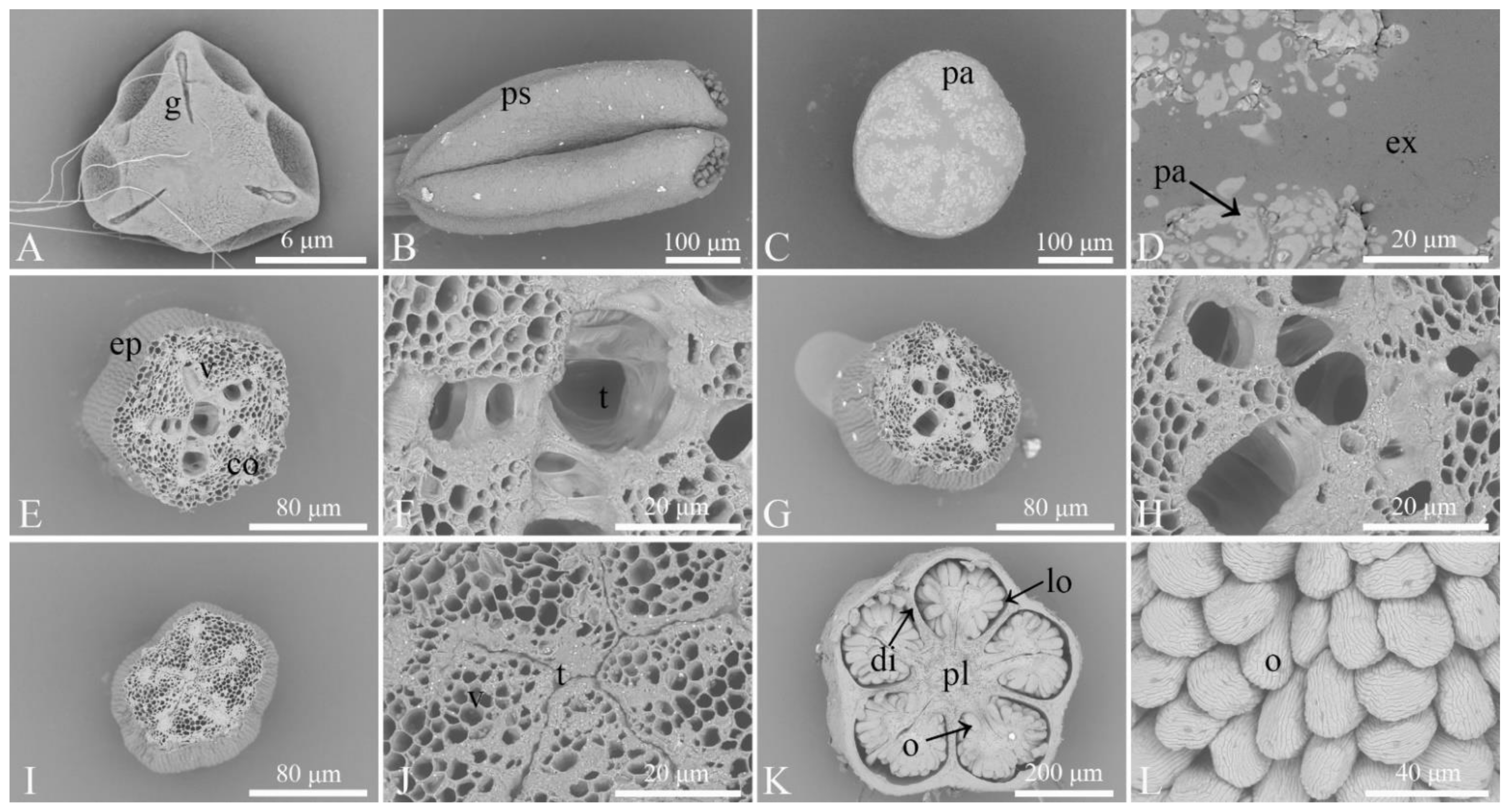
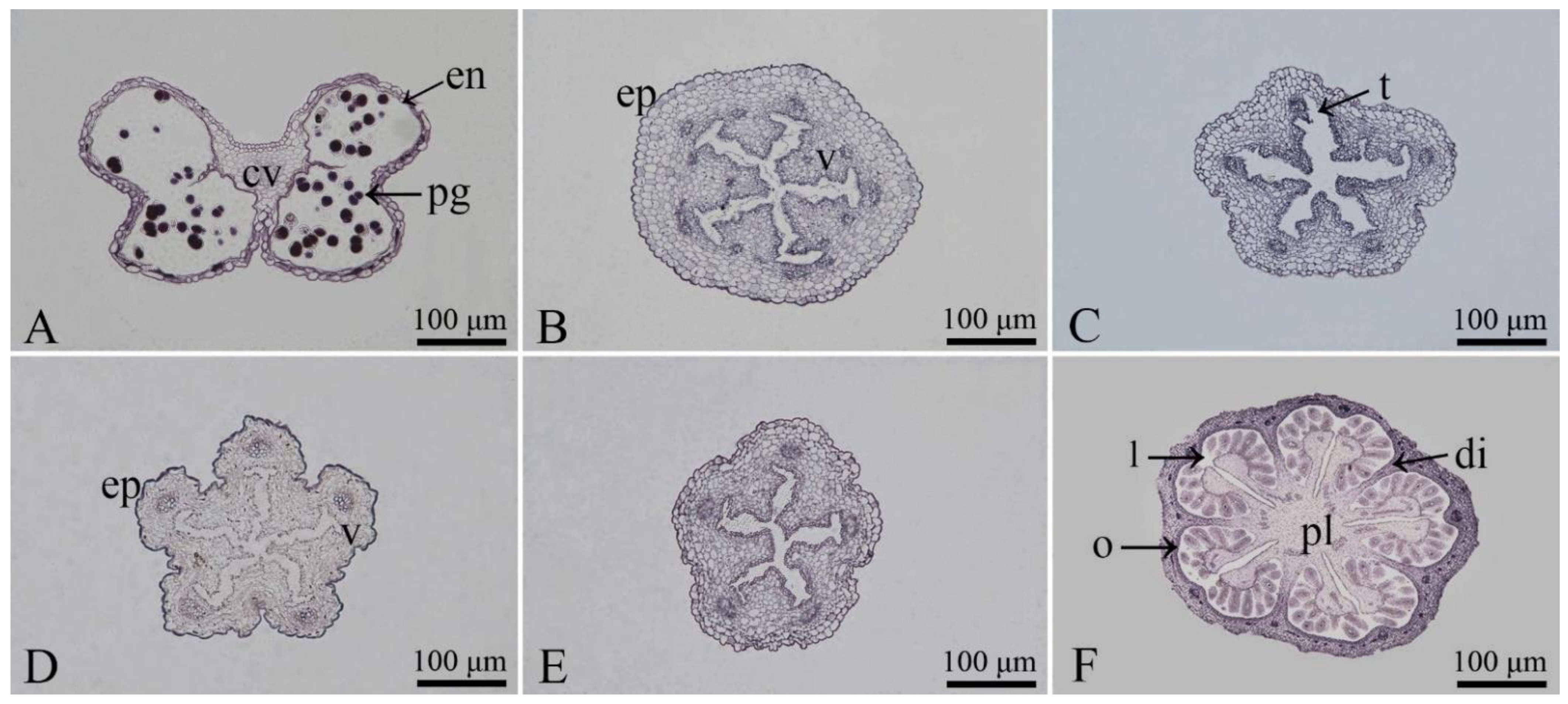
3.1. Observation of the Microstructures of Floral Organs

The pollen tetrad of R. pulchrum (Figure 1A) had sticky filaments on the surface and 3 germinal apertures. It ranged from 11.1 to 17.0 μm in length, with a mean of 14.46 μm. A densely granular pattern was observed near the germinal aperture and the center of the polar face, with the rest of the pollen showing rougher patterns. The polar axis length (P) ranged between 46.2 and 52.9 μm, with a mean of 49.16 μm. The equatorial axis length (E) ranged between 41.1 and 53.0 μm, with a mean of 47.22 μm. The average P/E ratio was 1.05. Under SEM (Figure 1), the top of the stigma of R. pulchrum (Figure 1C,D) appeared hemispherical, with a radius of 519.5–538.6 μm and a mean radius of 527.38 μm. A large number of irregular papillae were present on the surface of the stigma. The central area of the stigma was accompanied by exudate, providing natural conditions for pollen germination. Therefore, the stigma was of a wet type. Paraffin sections (Figure 2B) of the stigma observed under a microscope revealed a neat arrangement of the vascular bundles. The anther (Figure 1B and Figure 2A), consisting of two thecae, was a long cylinder, 781.3–988.3 μm in length, with a mean length of 890.43 μm, and 383.3–457.5 μm in width, with a mean of 422.92 μm. The cross-section of the anther was butterfly shaped. The style was cylindrical and had a smooth surface. The cross-section of the style showed that the style was composed of epidermis and cortex tissue. The upper part of the style (Figure 1E,F and Figure 2C) had a large amount of transmitting tissue, which allowed the pollen tube to grow toward the base, and the middle part of the style (Figure 1G,H and Figure 2D) was narrower than the upper part. Toward the lower section (Figure 1I,J and Figure 2E), the style duct became gradually denser, decreasing in size to form a small gap; in the lower section, the cells were small and compact. The placenta of R. pulchrum was axile. The ovary (Figure 2F) consisted of 5 locules and 10 carpels, with a radius of 1023.8–1055.0 μm and a mean radius of 1043.13 μm. The ovary was made up of the ovary, placenta, ovule, and diaphragm (Figure 1K). The ovule (Figure 1L), approximately 184.50 μm in length and 102.60 μm in width, grew on the funicle and was surrounded by carpels.

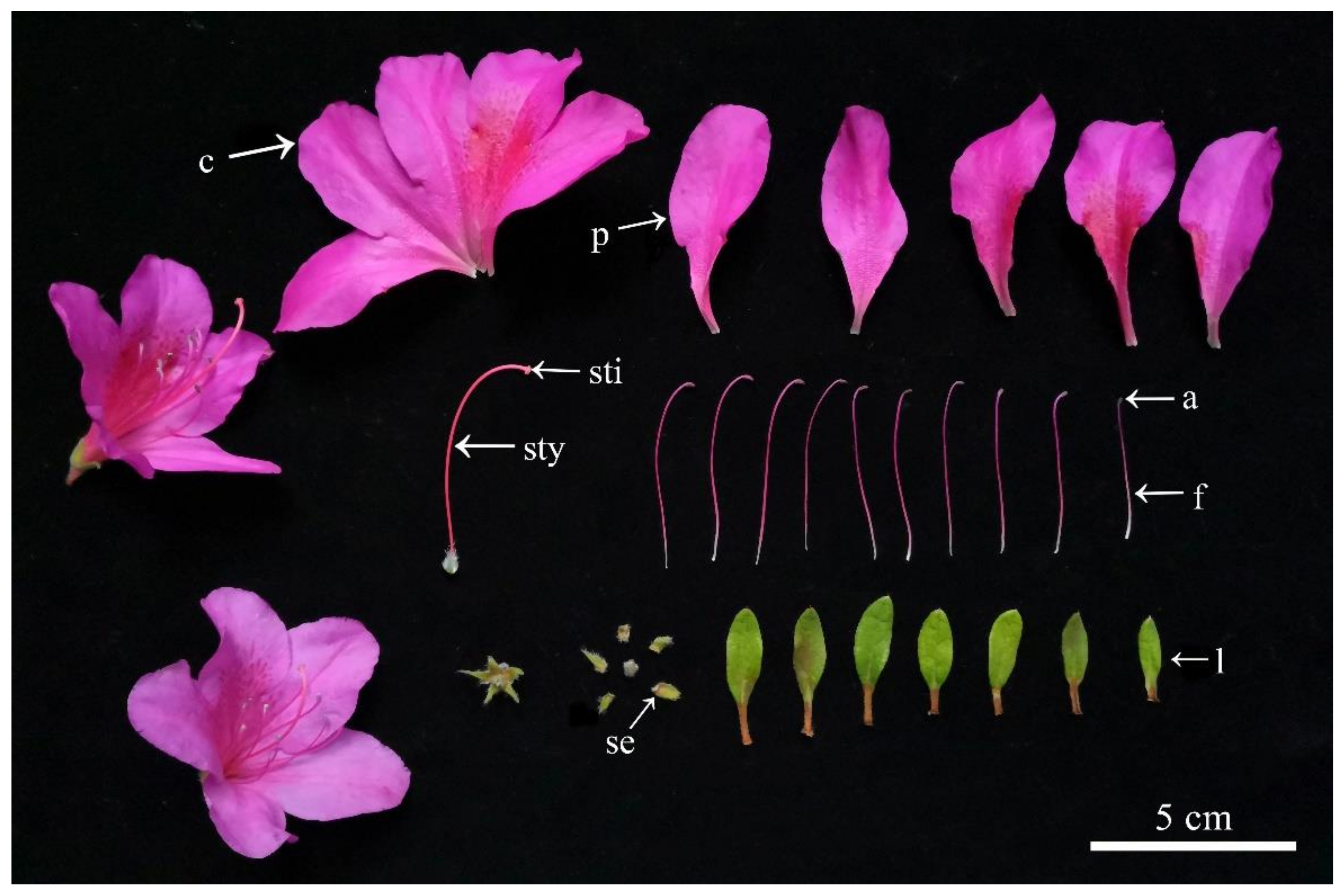
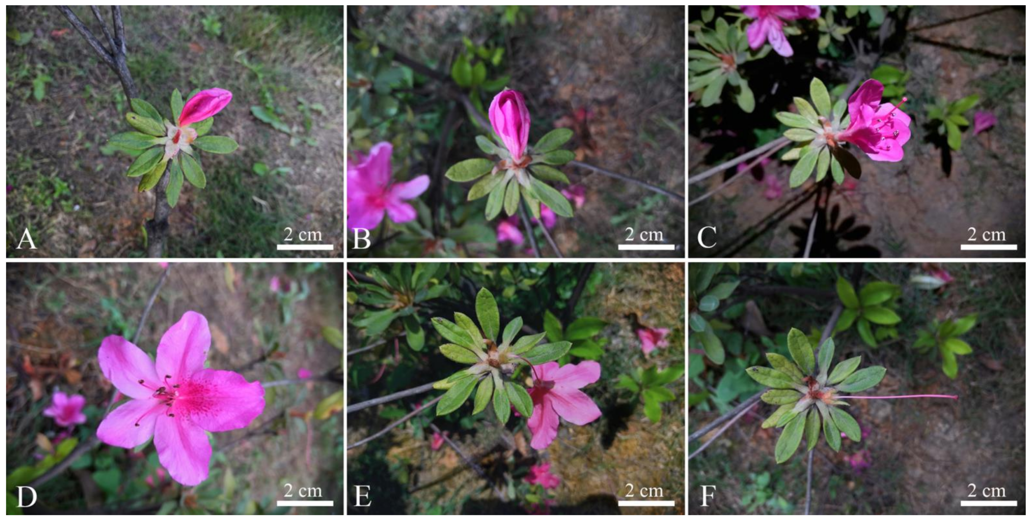
3.2. Observation of the Morphological Characteristics of Floral Organs and Opening Dynamics of Single Flowers

Observation of the characteristics of the floral organs of R. pulchrum (Table 1, Figure 3) revealed that its flowers were hermaphroditic, its flowering period lasted 2–4 months, and its inflorescence was an apical umbel. The style was 53.55 ± 4.71 mm in length and 0.47 ± 0.11 mm in width and was longer than the corolla. There were 10 stamens, the filaments of which ranged between 36.46 and 57.67 mm in length, with a mean length of 47.22 mm. The widths of the filaments varied between 0.06 and 0.48 mm, with a mean width of 0.27 mm. The filaments were smooth, puberulent at the base, and shorter than the styles. The sepals were green, covered with small white hairs, and the calyx was five-lobed. The sepals were 12.79 ± 3.53 mm in length and 3.83 ± 0.58 mm in width. The pedicel was approximately 14.82 mm long and 1.69 mm wide and covered with white hairs of various sizes. The corolla was dark pink and conical, with dark red spots unevenly distributed in three lobes. The maximum diameter of the open flower was 67.63 ± 2.70 mm and the minimum diameter was 65.62 ± 3.88 mm. Detailed information with regard to the measurements of the various parts of the floral organs is summarized in the Supplementary Materials Table S1. The opening dynamics of individual flowers were observed from the time small flower buds grew until they were ready to blossom. During initial flower bud growth, the sepals were stacked close together, and the flower bud was oblong and light pink. Before opening, the flower buds went through a long budding period, during which they developed slowly. Then, the flowers entered the bud period (Figure 4A), during which the buds gradually grew and darkened and the mucus on the stigma disappeared. After a certain period of growth and development, the upper part of the flower bud, that is, the place where the sepal tip tightly closed, began to crack. At this time, R. pulchrum entered the bud cracking period (Figure 4B). As the opening increased, the sepals gradually unfolded. Generally, the flowers opened in the evening. Later, as the flower buds developed, they gradually entered the initial flowering period (Figure 4C), during which the petals continued to expand, mucus was present on the stigma, and the anthers continued to disperse pollen; this period lasted 1–2 days. Then, the blooming period ensued (Figure 4D); during this time, the diameter of the corolla reached a specific extent, after which it no longer expanded. Compared with the initial flowering period, the amount of pollen dispersed by the anthers declined in the blooming period, when the remaining pollen was dispersed. The blooming period lasted for 1–2 days, after which the anthers no longer dispersed pollen, although there was still some mucus on the stigma. The flowers then entered the end of the flowering period (Figure 4E,F), during which petals began to fall off and filaments gradually fell off, leaving only the stigma and sepals, and the stigma began to gradually darken.

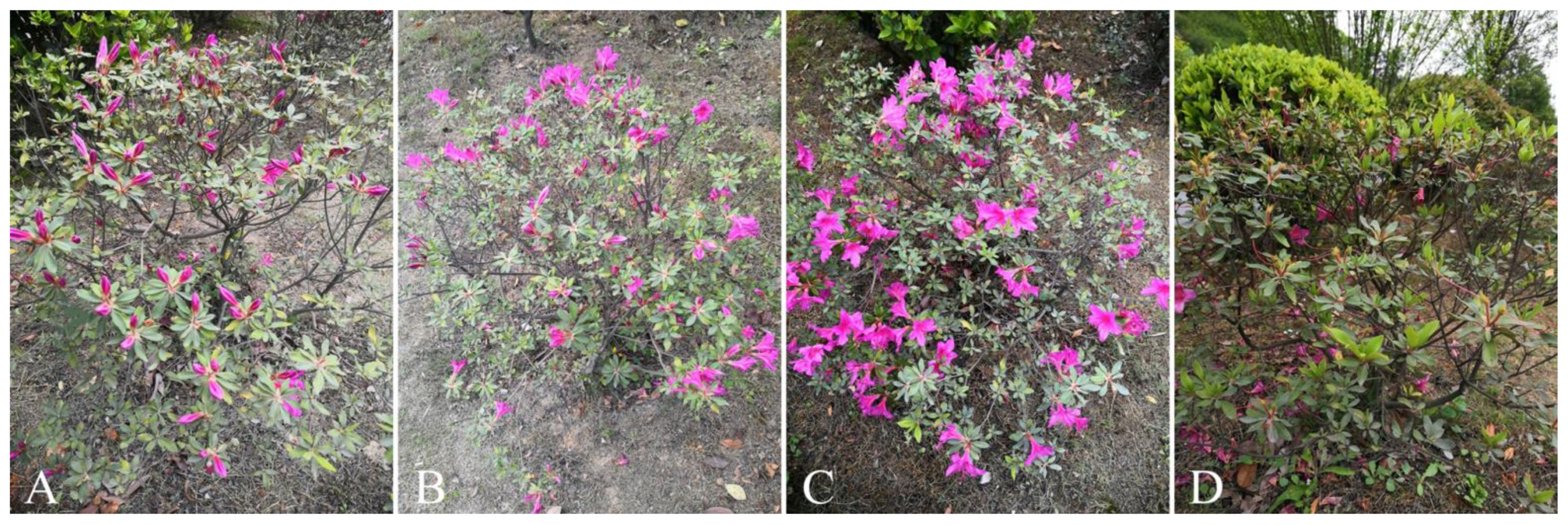
3.3. Opening Dynamics of Individual Plants

R. pulchrum plants first grew vegetatively and entered the squaring period after a period of time (Figure 5A). Flower buds were concentrated on the tops of branchlets. Typically, a whorl of leaves contained two flower buds, and very few whorls contained three buds. Buds on the same plant flowered at approximately the same time. During the development process, the flower buds grew larger, and the color gradually darkened. It took 2–3 days to transition from the squaring period to the initial flowering period (Figure 5B). The initial flowering period was short, generally 1–2 days, during which a small number of flower buds bloomed. During this period, the sepals at the top of most flower buds started to loosen and became ready to bloom. In the blooming period (Figure 5C), most flowers had already blossomed; during this period, which was the best viewing time, flowers appeared bright and vigorous, and various indexes reached their maxima. The blooming period of individual plants lasted for 4–5 days and was followed by the end of the flowering period (Figure 5D), during which some of the flowers gradually withered. When the buds wilted, the entire corolla and 10 stamens fell off together. At the end of the flowering period, all the flowers fell off. The sepals showed no sign of falling off, although their size decreased, and they turned black due to withering.

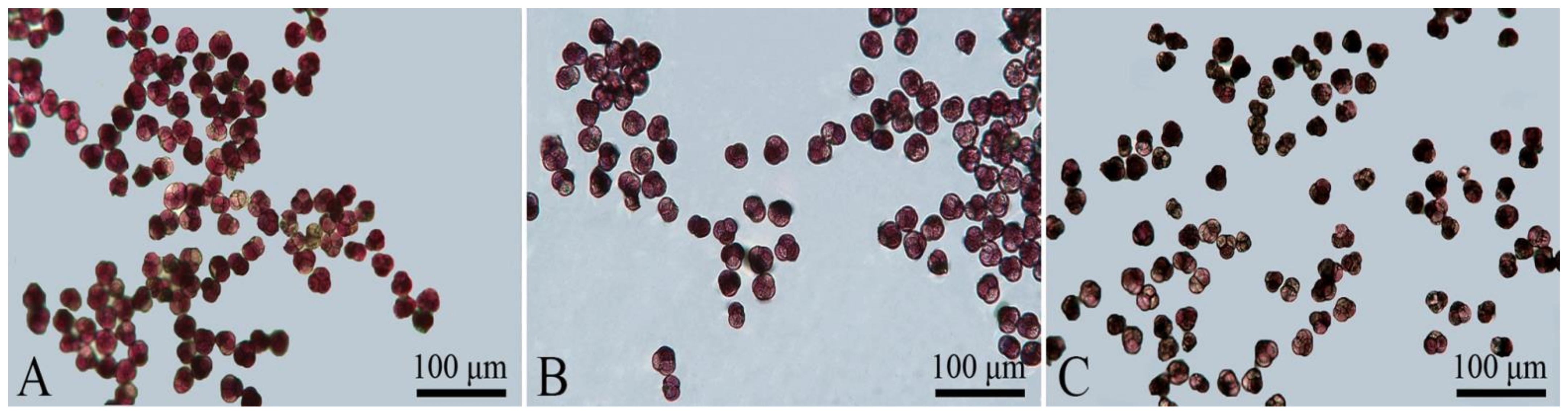
3.4. Measurement of Pollen Vitality in Different Periods

The vitality of R. pulchrum pollen was measured during different flowering periods using the MTT method (Figure 6). The results of microscope observations (Table 2) showed that pollen viability was the highest in the initial flowering period (Figure 6A), reaching 88.98%; pollen viability in the blooming period (Figure 6B) was 77.93%; and pollen viability was low in the end of flowering period (Figure 6C), at only 17.62%. It was concluded that the viability of R. pulchrum pollen was highest when the anthers started to disperse pollen during the initial flowering period. As the flowering process proceeded, pollen viability gradually decreased.

3.5. Detection of Stigma Receptivity

(1)
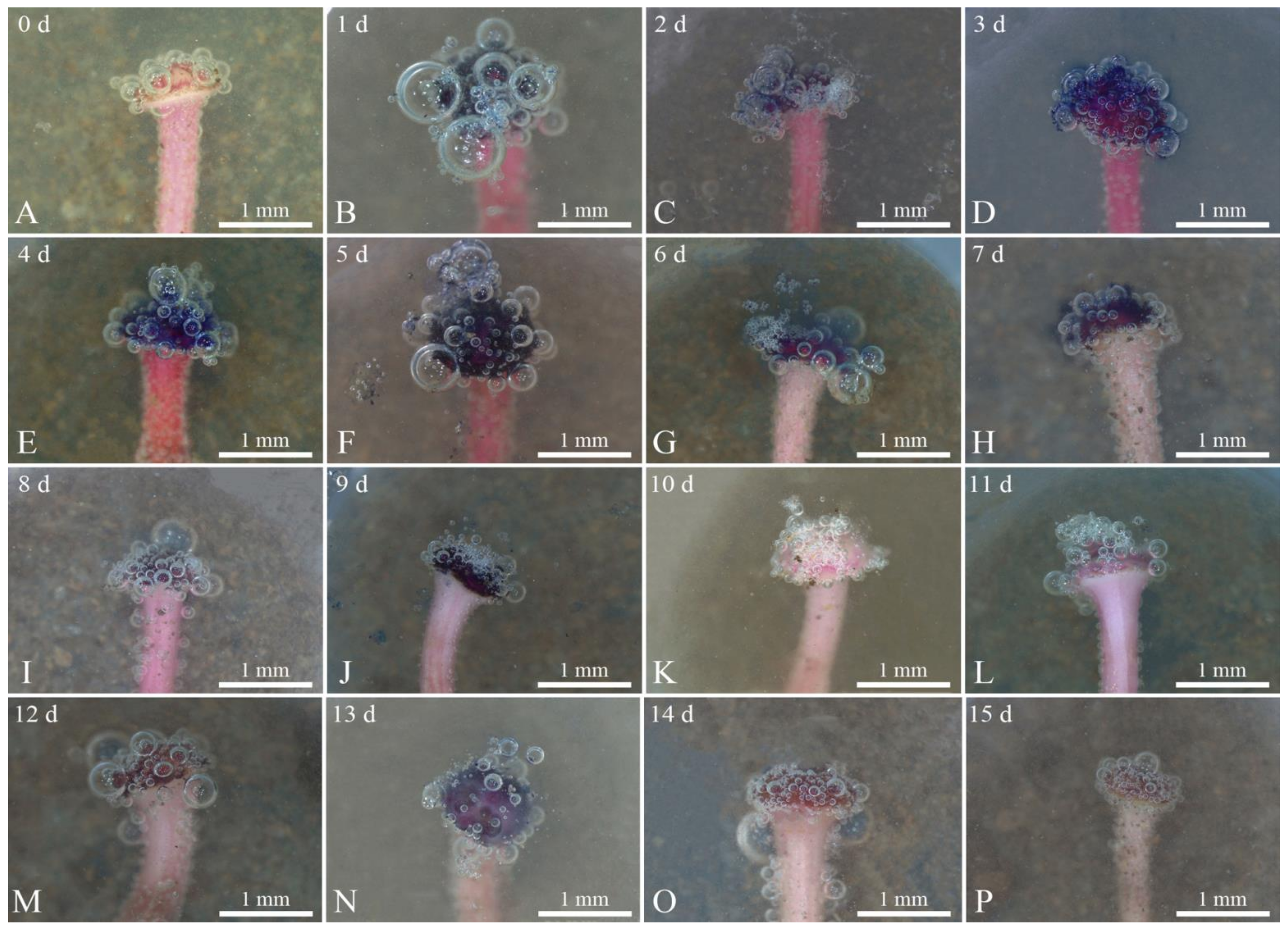
Benzidine-hydrogen peroxide method (Table 3 and Figure 7). Benzidine-hydrogen peroxide solution was used to test the receptivity of R. pulchrum stigmas. Observation under a stereomicroscope revealed that the reactions of stigmas was not intense on the day before flowering (Figure 7A), with only a few bubbles on the stigma, indicating that receptivity was not strong. After 1–2 days (Figure 7B,C) of flowering, the receptivity gradually increased, and the stigma reaction was the most intense on the third and fourth days (Figure 7D,E) after flowering; at this time, there were numerous bubbles of a large radius, and the blue reaction liquid reached its peak, indicating that receptivity was strongest at this time. There was still a small amount of blue reaction liquid on the fifth to seventh day of flowering (Figure 7F–H), indicating that flowers were still receptive during this period, although the reaction was weaker than on the third and fourth days of flowering. The stigma began to turn black from the 8th to the 15th day (Figure 7I–P), and no blue reaction liquid was observed in this time. During this period, the receptivity further declined, with only minute bubbles, showing basically no receptivity. Therefore, we inferred that the period in which the stigma of R. pulchrum were most receptive was the third to fourth day of flowering.
(2)
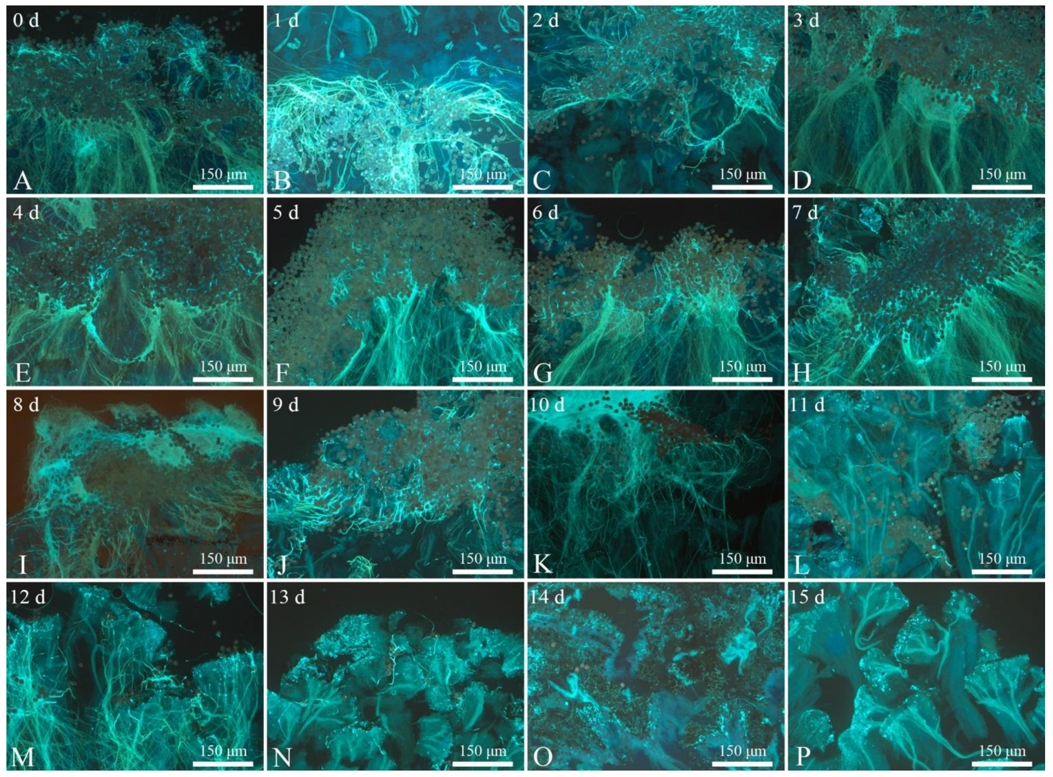
Pollen germination observation method (Figure 8). After regular artificial pollination, we took images with a fluorescence microscope and found that on the day before flowering (Figure 8A), the pollen was attached to the stigma. Large quantities of germinated pollen grains were found attached to the stigma until the ninth day of flowering (Figure 8B–J). On the 10th day, the number of pollen grains attached to the stigma decreased significantly (Figure 8H). Clearly, the stigma had only weak receptivity at this time. There was basically no pollen germination from the 11th day to the 15th day (Figure 8L–P). The results of the two experimental methods were basically consistent.

3.6. Estimation of the P/O Ratio

According to Cruden’s standard [26] and combined with the experimental data (Table 4, Supplementary Materials Table S2), the average P/O ratio of R. pulchrum was determined to be 301.47. The overall P/O ratio ranged between 266 and 328. Therefore, we predict that R. pulchrum has a facultative outcrossing breeding system.

3.7. Estimation of the OCI

Our observations showed that the average diameter of R. pulchrum flowers was 66.07 ± 5.07 mm, far greater than a diameter of 6 mm, for a value of 3. The position of the anther was lower than that of the stigma, and the two organs were not in the same space, for a value of 1. The stigma was already receptive when the pollen was dispersed by the anthers, for a value of 0. According to Dafni’s standard, the OCI of R. pulchrum was equal to 4. Therefore, we surmise that the breeding system of R. pulchrum was partial self-compatibility and that R. pulchrum likely required pollinators to complete pollination.

4. Discussion

For most plants in nature, insects are required for pollination and can play either a dominant or an ancillary role in the process; therefore, flowers need to be appealing enough to attract pollinating insects [28,29]. Research on the characteristics of floral organs provides a theoretical basis for the study of reproductive biology. In this study, we systematically examined the flowering characteristics of R. pulchrum using experiments, observations, and other methods. Pollen is controlled by the male genetic information it carries and is little impacted by external environmental conditions. There is a very low probability of its outer wall patterns, germinal aperture number, structure, size, and other morphological characteristics undergoing gene mutation. It is an extremely important reference index in studying plant evolution and genetic relationships and plays a certain role in the study of plant taxonomy. Previous researchers studied pedicels of R. pulchrum and made paraffin sections of these structures for observation. In this study, we applied paraffin sectioning technology to the anthers, ovaries, styles, and other floral organs of R. pulchrum. Combined with SEM, we observed the morphological characteristics of this species from two perspectives to make the results more scientific and robust [30]. The SEM observations indicated that the pollen of R. pulchrum was with three germinal apertures, consistent with the results of previous studies on pollen of the genus Rhododendron. There were filaments on the surface of the pollen, and the aggregation effect of the filament on pollen has been shown to vary among pollinators in another species of Rhododendron [31]. Bai [32] believed that it was inappropriate to use the outer wall pattern and the length and width of the germinal aperture as the basis for the classification of Rhododendron subgenera. In subsequent research, it was found that even the pollen diameter of the same Rhododendron group varied greatly. Therefore, pollen morphology cannot be used as the only reference when classifying Rhododendron plants.
Pollen vitality plays a decisive role in the success of breeding. Only when viable pollen falls on the stigma due to wind pollination, insect pollination, or other methods during the stigma pollination period can pollination be successful. Therefore, research on pollen viability and stigma receptivity has become an indispensable part of flowering biology. In addition to the impact of genetic information carried by pollen, pollen viability is also affected by external conditions, especially temperature [33]. In this study, we investigated pollen viability under natural conditions in three different periods and determined that pollen viability was the highest during the initial flowering period, reaching 88.98%, and remained high during the blooming period. Compared with the single flower lifespan of approximately 8 days, pollen viability remained high for most of the flowering period, effectively enhancing male fitness [34]. However, in the pollen viability experiment, the influence of the external environment was not taken into consideration. This factor needs to be considered in future experiments to minimize the error caused by external environmental factors. Studies have shown that the stigma of R. pulchrum is able to receive pollen before flowering; that is, the pistil matures earlier than the stamen. This phenomenon, coupled with a longer receptive period, increases the chance of outcrossing for plants [35,36]. The research of Li et al. [37] on Rhododendron longipedicellatum showed that on the first day of flowering, the stigmas of this species were not receptive, which might be due to genetics. The stigmas of R. pulchrum exhibited the strongest receptivity on the 3rd and 4th days of flowering; the receptivity then gradually declined until it was lost on the 13th day of flowering. To a certain extent, the setting rate of plants hinges on the length of the receptive period. Therefore, the length of the receptive period may be a key link in the sexual reproduction process [38]. Some scholars [7,39] studied Rhododendron siderophyllum and Rhododendron polylepis and found that their receptive periods were 9 and 7 days, respectively. In this study, using the benzidine-hydrogen peroxide method and artificial pollination, we found that the receptive period of R. pulchrum was significantly longer than that identified in previous studies on the Rhododendron genus. The reasons for this difference may be as follows: first, the bagging and labeling of the material on the day of flowering might have interfered with the growth and development of the flowers in their natural state, resulting in longer perceived receptive periods; second, when using the benzidine-hydrogen peroxide method to determine receptivity, bubbles might be generated in the area outside stigmas, thereby affecting the evaluation of receptivity. Further study is required to examine whether future studies generate conclusions on the receptive period of R. pulchrum stigma that are consistent with those of the current study.
The flowering period of R. pulchrum is 2–4 months, and the flowering period per flower is 6–8 days; these values are almost the same as those determined by a previous study on the Rhododendron genus [30]. A longer lifespan of individual flowers guarantees the success rate of outcrossing. The flowering periods of individual flowers on the same plant are staggered, which effectively prolongs the period during which the ornamental value of the plant can be appreciated. The flowers of R. pulchrum are bright, with maximum diameter of 67.63 ± 2.70 mm and minimum diameterof 65.62 ± 3.88 mm; therefore, the flowers can attract pollinators, thus increasing the probability of pollination and insemination. R. pulchrum plants have 10 stamens and 1 pistil. The position of the stigma during flowering is higher than that of the stamens, and the stigma is significantly larger than it is in the bud period; that is, R. pulchrum plants are dioecious. Therefore, the probability of selfing within the same flower is reduced. This arrangement is beneficial to cross-pollination and is an effective mechanism to prevent self-pollination [39,40]. Flowering phenology allows plants to adjust the opening time of flowers and the life cycle of flowers according to changes in environmental factors. Studies have shown that alpine plants can prolong their flowering period in response to harsh weather conditions and pollinators to promote their reproductive success. This study shows that the flowering time of individual R. pulchrum plants is approximately 15 days, which is favorable to pollination and insemination in harsh environments [41]. The flowering phenology follows a single-peak curve. Flowers blossom successively within a period of time, enhancing attraction to pollinators. After the individual flowering ratio reaches the maximum, flowers gradually wither, displaying centralized flowering, which is basically consistent with the results of a previous study [7]. The breeding system of plants plays a key role in their evolutionary development. Plants evolve biological characteristics compatible with local environmental conditions to survive [26]. The results obtained in this study show that the P/O ratio of R. pulchrum is relatively low, which may be closely related to pollen aggregation. The pollen tetrad of R. pulchrum is a form of pollen aggregation. Coupled with the OCI of 4, we conclude that R. pulchrum is probably partially self-compatible and requires the participation of pollinators to complete its pollination. Previous research [42] has shown that compared to short-lived herbaceous plants, long-lived woody plants with multiple flowering periods are more prone to pollen restrictions. There is a spatial separation between the stigma and anthers of R. pulchrum. As a result, most pollen grains do not fall on the stigma, resulting in a loss of pollen and pollen limitation if flowers are not visited. In this study, we did not conduct an artificial pollination experiment with bagging. Such experiments are needed to truly determine the extent of self-compatibility and self-pollination in this species. In addition to those experiments, the following questions should be addressed: What are the most suitable storage conditions for the pollen of R. pulchrum, and what features are shared among pollinators visiting flowers of R. pulchrum? Is it advisable to continue to research how R. pulchrum pollen tubes grow in the style after the most receptive period is determined? How should we guide the introduction and domestication of new cultivars according to reproductive biology? Such issues remain unresolved.

Supplementary Materials

The following are available online at https://www.mdpi.com/article/10.3390/horticulturae7110508/s1, Table S1: Measurements of the parameters of floral organs, Table S2: Estimation of the P/O ratio of R. pulchrum.

Author Contributions

Conceptualization, J.Q. and C.G.; methodology, C.G.; software, H.W. and B.W.; validation, Y.H.; formal analysis, Z.G. and H.L.; investigation, J.Q., H.W., Z.G., L.L. and L.Y.; resources, C.G.; data curation, L.Y.; writing—original draft preparation, J.Q.; writing—review and editing, C.G.; visualization, Y.H.; supervision, C.G.; project administration, C.G.; funding acquisition, C.G. All authors have read and agreed to the published version of the manuscript.

Funding

This work was supported by National Natural Science Foundation of China (32060331, 31800516), Science and Technology Plan Project of Guizhou Province of China (Qian Ke He(2019)2310, Qian Ke He(2022)zhongdian 017, Qian Ke He(2020)1Y057, Qian Ke He Fu Qi(2020)4011), Science and Technology Project of Guizhou Education Department (Qian jiao He KY(2018)097), Cultivation Project of Guizhou University((2019)35) and Research Project of Introducing Talents in Guizhou University((2017)41).

Institutional Review Board Statement

Not applicable.

Informed Consent Statement

Not applicable.

Data Availability Statement

The data used for the analysis in this study are within the article and supplementary files.

Conflicts of Interest

The authors declare no conflict of interest. The funders had no role in the design of the study; in the collection, analyses, or interpretation of data; in the writing of the manuscript, or in the decision to publish the results.

References

  1. Mitchell, R.J.; Karron, J.D.; Holmquist, K.G.; Bell, J.M. The influence of Mimulus ringens floral display size on pollinator visitation patterns. Func. Ecol. 2004, 18, 116–124. [Google Scholar] [CrossRef]
  2. Zhang, H.; Wu, H.; Zhou, Q.; Zhao, R.; Sheng, Q.; Zhu, Z. Flowering characteristics and reproductive biology of Nymphaea hybrid, a precious water lily. Sci. Hortic. 2021, 287, 110268. [Google Scholar] [CrossRef]
  3. Grant, V. Plant Speciation; Columbia University Press: New York, NY, USA; Chichester, UK, 1981. [Google Scholar]
  4. Xian, C. Study on Reproductive Biology of Lilium Davidii Varunicolor Salish; Shenyang Agricultural University: Shenyang, China, 2019; (In Chinese with English Abstract). [Google Scholar]
  5. Hirst, P.M. Advances in Understanding Flowering and Pollination in Apple Tree; Achieving Sustainable Cultivation of Apples; Burleigh Dodds Science Publishing: Cambridgeshire, Great Britain, 2017; pp. 109–126. [Google Scholar]
  6. Christopher, D.A.; Karron, J.D.; Semski, W.R.; Smallwood, P.A.; Trapnell, D.W.; Mitchell, R.J. Selfing rates vary with floral display, pollinator visitation and plant density in natural populations of Mimulus ringens. J. Evol. Biol. 2021, 34, 803–815. [Google Scholar] [CrossRef]
  7. Tao, R. Reproductive Ecology of Rhododendron Polylepis in Huanxi Sub-Alpine Botanical Garden; Sichuan Agricultural University: Chengdu, China, 2016; (In Chinese with English Abstract). [Google Scholar]
  8. Bowers, C.G. Rhododendrons and Azaleas: Their Origins, Cultivation and Development; Macmillan Co.: New York, NY, USA, 1960. [Google Scholar]
  9. Stevenson, J.B. The Species of Rhododendron; Royal Horticultural Society: London, UK, 1947. [Google Scholar]
  10. Leppik, E.E. Evolutionary interactions between Rhodo-dendrons, pollinating insects and rust fungi. Quart. Bull. Am. Rhod. Soc. 1974, 28, 70–89. [Google Scholar]
  11. Stevens, P.F. The altitudinal and geographical distribution in flower types in Rhododendron section Vireya, especially in the Papuasian species, and their significance. Bot. J. Linn. Soc. 1976, 72, 1–33. [Google Scholar] [CrossRef]
  12. Argent, G.C.G. Notes from the Royal Botanic Garden Edinburgh, Vireya rhododendrons in Borneo. Notes R. Bot. Gard. Edinb. 1985, 43, 53–61. [Google Scholar]
  13. Robinson, T. The Organic Constituents of Higher Plants; Cordus Press: North Amherts, MA, USA, 1991. [Google Scholar]
  14. Ng, S.C.; Corlett, R.T. Comparative reproductive biology of the six species of Rhododendron (Ericaceae) in Hong Kong, South China. Can. J. Bot. 2000, 78, 221–229. [Google Scholar] [CrossRef] [Green Version]
  15. Shirasawa, K.; Kobayashi, N.; Nakatsuka, A.; Ohta, H.; Isobe, S. Whole-genome sequencing and analysis of two azaleas, Rhododendron ripense and Rhododendron kiyosumense. DNA Res. 2021, 25, dsab010. [Google Scholar] [CrossRef] [PubMed]
  16. Shen, J.S.; Li, X.Q.; Zhu, X.T.; Huang, X.L.; Jin, S.H. Complete chloroplast genome of Rhododendron pulchrum, an ornamental medicinal and food tree. Mitochondrial. DNA B 2019, 4, 3527–3528. [Google Scholar] [CrossRef] [PubMed] [Green Version]
  17. Shen, J.; Li, X.; Zhu, X.; Huang, X.; Jin, S. The complete plastid genome of Rhododendron pulchrum and comparative genetic analysis of Ericaceae Species. Forests 2020, 11, 158. [Google Scholar] [CrossRef] [Green Version]
  18. Si, G.; Zhang, Y.; Gu, X.; Wang, Y.Q.; Zhao, B. Study on cutting propagation technology of Qinling wild Rhododendron calophytum. North. Hortic. 2012, 3, 7. [Google Scholar]
  19. Chen, Z.; Yang, X.; Xu, J.; Jiao, B.; Li, Y.; Xu, Y.; Dai, T. First report of Phytopythium helicoides causing crown and root rot on Rhododendron pulchrum in China. Plant Dis. 2021, 105, 713. [Google Scholar] [CrossRef] [PubMed]
  20. Xu, Y.; Yang, X.; Li, Y.X.; Chen, Z.P.; Dai, T.T. First report of Phytophthora pini causing foliage blight and shoot dieback of Rhododendron Pulchrum in China. Plant Dis. 2021, 105, 1229. [Google Scholar] [CrossRef]
  21. Yang, L.; Gao, C.; Wei, H.L.; Hu, Y. Screening experiment of pollination combination of four improved Camellia oleifera varieties. Seed 2020, 39, 66–69, 74, (In Chinese with English Abstract). [Google Scholar]
  22. Gao, C.; Rui, Y.; Guo, Q.Q.; Yuan, D.Y. Microstructure and ultrastructure characteristics of stigma and style of Camellia Oleifera. For. Res. 2019, 32, 1–7. [Google Scholar]
  23. Burke, J.J. Moisture sensitivity of cotton pollen. Agron. J. 2002, 94, 883–888. [Google Scholar]
  24. Hirasuka, S.; Zhang, S.L.; Nakagawa, E.; Kawai, Y. Selective inhibition of the growth of incompatible pollen tubes by S-protein in the Japanese pear. Sex Plant Reprod. 2001, 13, 209–215. [Google Scholar] [CrossRef]
  25. Li, Y.H.; Kang, X.Y. Stigma receptivity and its detection methods of white poplars. Acta Bot. Boreali-Occident. Sin. 2007, 2007, 864–870, (In Chinese with English Abstract). [Google Scholar]
  26. Cruden, R.W. Pollen-ovule ratios: A conservative indicator of breeding systems in flowering plants. Evolution 1977, 31, 32–46. [Google Scholar] [CrossRef] [PubMed]
  27. Yuan, D.Y.; Tan, X.F.; Hu, Q.S.; Zou, F. Study on Camellia pollen characteristics and the vitality under different storage conditions. J. Zhejiang For. Sci. Technol. 2008, 5, 66–69, (In Chinese with English Abstract). [Google Scholar]
  28. Heiduk, A.; Pramanik, D.; Spaans, M.; Gast, L.; Dorst, N.; van Heuven, B.J.; Gravendeel, B. Pitfall flower development and organ identity of Ceropegia sandersonii (Apocynaceae-Asclepiadoideae). Plants 2020, 9, 1767. [Google Scholar] [CrossRef] [PubMed]
  29. Costa, M.S.; Silva, R.J.; Paulino-Neto, H.F.; Pereira, M.J.B. Beetle pollination and flowering rhythm of Annona coriacea Mart.(Annonaceae) in Brazilian cerrado:Behavioral features of its principal pollinators. PLoS ONE 2017, 12, e0171092. [Google Scholar] [CrossRef] [PubMed]
  30. Tian, X.L. Research on Reproductive Biology of Rhododendron Excellens Hemsl. et. Wils; Nanjing Forestry University: Nanjing, China, 2011; (In Chinese with English Abstract). [Google Scholar]
  31. Song, Y.P.; Huang, Z.H.; Huang, S.Q. Pollen aggregation by viscin threads in Rhododendron varies with pollinator. New Phytol. 2019, 221, 1150–1159. [Google Scholar] [CrossRef] [PubMed] [Green Version]
  32. Bai, Y.Q. Reproductive Biology of Rhododendron Moulmainense Hook f. and Its Adaptability of Introduction at Low Altitude Areas; Beijing Forestry University: Beijing, China, 2017; (In Chinese with English Abstract). [Google Scholar]
  33. Li, Y.; Xu, J.; Shi, L.; Li, F. Examination of pollen germination and vitality of different cultivars of Ziziphus mauritiana in greenhouse. J. Fruit Sci. 2005, 6. Available online: http://en.cnki.com.cn/Article_en/CJFDTOTAL-GSKK200506030.htm (accessed on 2 November 2021).
  34. Hufford, K.M.; Hamrick, J.L. Viability selection at three early life stages of the tropical tree, Platypodium elegans (Fabaceae, Papilionoideae). Evolution 2003, 57, 518–526. [Google Scholar] [CrossRef]
  35. Wada, N.; Uemura, S. Size-dependent flowering behavior and heat production of a sequential hermaphrodite, Symplocarpus renifolius (Araceae). Am. J. Bot. 2000, 87, 1489–1494. [Google Scholar] [CrossRef]
  36. Wang, X.R.; Ding, G.J. Reproductive biology characteristic of Jatropha curcas (Euphorbiaceae). Rev. De Biol. Trop. 2012, 60, 1525–1533. [Google Scholar] [CrossRef] [Green Version]
  37. Li, T.; Liu, X.; Li, Z.; Ma, H.; Wan, Y.; Liu, X.; Fu, L. Study on reproductive biology of Rhododendron longipedicellatum: A newly discovered and special threatened plant surviving in limestone habitat in Southeast Yunnan, China. Front. Plant Sci 2018, 9, 33. [Google Scholar] [CrossRef] [Green Version]
  38. Barrett, S.C.H. The evolution of mating strategies in flowering plants. Trends Plant Sci. 1998, 3, 335–341. [Google Scholar] [CrossRef]
  39. Bai, T.; Guan, W.L.; Song, J.; Xie, W.J.; Li, S.F. Flowering characteristics and breeding system of Rhododendron siderophyllum. J. West China For. Sci. 2014, 43, 47–53. [Google Scholar]
  40. Webb, C.J.; Lloyd, D.G. The avoidance of interference between the presentation of pollen and stigmas in angiosperms II. Herkogamy. N. Z. J. Bot. 1986, 24, 163–178. [Google Scholar] [CrossRef]
  41. Torres-Díaz, C.; Gomez-Gonzalez, S.; Stotz, G.C.; Torres-Morales, P.; Paredes, B.; Pérez-Millaqueo, M.; Gianoli, E. Extremely long-lived stigmas allow extended cross-pollination opportunities in a high Andean plant. PLoS ONE 2011, 6, e19497. [Google Scholar] [CrossRef] [PubMed]
  42. Rambuda, T.D.; Johnson, S.D. Breeding systems of invasive alien plants in South Africa: Does Baker’s rule apply? Divers. Distrib. 2004, 10, 409–416. [Google Scholar] [CrossRef]
Figure 1. SEM of various parts of floral organs. (A): pollen tetrad; (B): anther; (C): overhead shot of the stigma; (D): central area of the stigma; (E): upper part of the style; (F): upper central area of the style; (G): middle part of the style; (H): central area of the middle part of the style; (I): lower part of the style; (J): central area of the lower part of the style; (K): ovary; (L): ovule. g: germinal aperture; ps: pollen sac; pa: papilla; ex: exudate; ep: epidermis; v: vascular bundle; co: cortex tissue; t: transmitting tissue; di: diaphragm; o: ovule; pl: placenta; lo: locule.
Horticulturae 07 00508 g001
Figure 2. Paraffin section of floral organs. (A): anther; (B): stigma; (C): upper part of the style; (D): middle part of the style; (E): lower part of the style; (F): ovary. cv: connective vascular bundle; en: endothecium; pg: pollen grains; ep: epidermis; v: vascular bundle; t: transmitting tissue; l: locule; o: ovary wall; pl: placenta; di: diaphragm.
Horticulturae 07 00508 g002
Figure 3. Morphology and composition of floral organs and the leaves. c: corolla; p: petal; sti: stigma; sty: style; se: sepal; a: anther; f: filament; l: leaves.
Horticulturae 07 00508 g003
Figure 4. Opening dynamics of individual flowers. (A): bud period; (B): bud cracking period; (C): initial flowering period; (D): blooming period; (E,F): end of flowering period.
Horticulturae 07 00508 g004
Figure 5. Opening dynamics of individual plants. (A): Squaring period; (B): initial flowering period; (C): blooming period; (D): end of flowering period.
Horticulturae 07 00508 g005
Figure 6. Pollen tetrads, stained in different periods. (A): in the initial flowering period; (B): in the blooming period; (C): in the end flowering period.
Horticulturae 07 00508 g006
Figure 7. Detection of stigma receptivity under a stereomicroscope using the benzidine-hydrogen peroxide method. (A): The day before flowering; (B): 1st day of flowering; (C): 2nd day of flowering; (D): 3rd day of flowering; (E): 4th day of flowering; (F): 5th day of flowering; (G): 6th day of flowering; (H): 7th day of flowering; (I): 8th day of flowering; (J): 9th day of flowering; (K): 10th day of flowering; (L): 11th day of flowering; (M): 12th day of flowering; (N): 13th day of flowering; (O): 14th day of flowering; (P): 15th day of flowering.
Horticulturae 07 00508 g007
Figure 8. Observation of pollen tube growth on the stigma after artificial pollination under a fluorescence microscope. (A): The day before flowering; (B): 1st day of flowering; (C): 2nd day of flowering; (D): 3rd day of flowering; (E): 4th day of flowering; (F): 5th day of flowering; (G): 6th day of flowering; (H): 7th day of flowering; (I): 8th day of flowering; (J): 9th day of flowering; (K): 10th day of flowering; (L): 11th day of flowering; (M): 12th day of flowering; (N): 13th day of flowering; (O): 14th day of flowering; (P): 15th day of flowering.
Horticulturae 07 00508 g008
Table 1. Measurements of the various parts of floral organs.
Measurement ParameterMinimum (mm)Maximum (mm)Mean (mm)SDNumber of Samples (Flowers)
Filament length36.4657.6747.225.0230
Filament width0.060.480.270.1230
Style length44.9764.2353.554.7130
Style width0.280.780.470.1130
Sepal length6.3919.2812.793.5330
Sepal width2.295.093.830.5830
Pedicel length9.7517.614.821.8730
Pedicel width1.281.991.690.1730
Maximum diameter of the open flower61.3771.9867.632.7030
Minimum diameter of the open flower56.9472.3065.623.8830
Table 2. Measurement of stained pollen tetrads in different periods.
Flowering PeriodSNNumber of Stained Pollen Tetrads Total Number of Pollen TetradsProportion of the Stained Pollen TetradsMean
Initial flowering period19410391.26%88.98%
2546188.52%
39510987.16%
Blooming period18910683.96%77.93%
28010874.07%
310013275.76%
End of flowering period12410423.08%17.62%
2107812.82%
32011816.95%
Table 3. Receptivity of R. pulchrum stigmas.
Time (Day of Flowering)Number of BubblesMucus SecretionReceptivity
The day before flowering+++
1st ++++++
2nd ++++++
3rd +++++++++
4th +++++++++
5th +++++++
6th++++++
7th+++
8th +++
9th +++
10th +-+/-
11th +-+/-
12th +-+/-
13th ---
14th ---
15th---
Receptivity was assessed based on the number of bubbles and mucus secretion. +++, extremely strong receptivity; ++, strong receptivity; +, moderate receptivity; +/-, weak receptivity; -, no receptivity.
Table 4. Estimation of the P/O ratio of R. pulchrum.
Observed FactorMinMaxMeanSD
Number of pollen tatrads per flower499,600.00572,200.00532,400.0022,593.93
Ovule number per flower1584.001902.001768.8792.64
P/O ratio266.00328.00301.4718.29
Publisher’s Note: MDPI stays neutral with regard to jurisdictional claims in published maps and institutional affiliations.

