Article

Morpho-Anatomical and Biochemical Characterization of Embryogenic and Degenerative Embryogenic Calli of Phoenix dactylifera L.

by
Amal F. M. Zein El Din
1,
Ola H. Abd Elbar
2,*,
Saleh M. Al Turki
3,
Khaled M. A. Ramadan
4,5,
Hossam S. El-Beltagi
6,7,
Hemmat A. Ibrahim
5,
Ezzeldin G. Gadalla
1,
Ibrahim M. Shams El-Din
1,
Ibrahim S. Ibrahim
2,
Reham Farag
2,
Mohamed F. M. Ibrahim
2,
Mona S. Abd El-Aal
2,
Ahmed Abou El-Yazied
8,
Mohamed M. El-Mogy
9,*,
Mina S. F. Samaan
8 and
Yasmin M. R. Abdellatif
2
1
The Central Laboratory for Date Palm Researches and Development, Agricultural Research Center (ARC), Giza 12619, Egypt
2
Department of Agricultural Botany, Faculty of Agriculture, Ain Shams University, Cairo 11566, Egypt
3
Arid Land Agriculture Department (Horticulture Program), College of Agriculture and Food Sciences, King Faisal University, P.O. Box 420, Al-Ahsa 31982, Saudi Arabia
4
Central Laboratories, Department of Chemistry, King Faisal University, P.O. Box 420, Al-Ahsa 31982, Saudi Arabia
5
Department of Biochemistry, Faculty of Agriculture, Ain Shams University, Cairo 11566, Egypt
6
Agricultural Biotechnology Department, College of Agriculture and Food Sciences, King Faisal University, P.O. Box 420, Al-Ahsa 31982, Saudi Arabia
7
Biochemistry Department, Faculty of Agriculture, Cairo University, Gamma st, Giza 12613, Egypt
8
Department of Horticulture, Faculty of Agriculture, Ain Shams University, Cairo 11566, Egypt
9
Vegetable Crops Department, Faculty of Agriculture, Cairo University, Giza 12613, Egypt
*
Authors to whom correspondence should be addressed.
Horticulturae 2021, 7(10), 393; https://doi.org/10.3390/horticulturae7100393
Submission received: 26 August 2021 / Revised: 29 September 2021 / Accepted: 5 October 2021 / Published: 12 October 2021
(This article belongs to the Collection Application of Tissue Culture to Horticulture)

Abstract

:
The study of morpho-anatomical aspects, metabolic changes of proteins, antioxidant substances, as well as phenolic compounds in embryogenic callus (EC) and degenerative embryogenic callus (DEC) was the aim of the present investigation. Ability to form somatic embryos (SEs) was associated with the softness of the EC, which exhibited a white or creamy color and was composed of isodiametric cells containing dense cytoplasm, conspicuous nuclei and minimal vacuoles with observed mitotic activity. Furthermore, protein, reduced glutathione (GSH) and ascorbic acid (ASC) concentrations and the ratio between ASC and dehydroascorborbic acid (DHA) were increased significantly in the EC in comparison to the DEC. In addition, the phenolic extract of the EC was proved to have higher scavenging activity than the extract from the DEC. A loss of embryogenic competence in the DEC was correlated with the presence of more rigid clumps and such calli had a yellowish to brown color and no cell division could be observed in the cells of such aggregates as the cells had large vacuoles and they have very thick walls. Moreover, these morphological and anatomical observations of the DEC were accompanied by accumulations of the oxidized form of ascorbic acid (DHA), H2O2, total soluble phenolic compounds and overaccumulation of naringenin. Alternations in cellular metabolism can affect and regulate the morphogenesis of somatic embryos.

1. Introduction

Somatic embryogenesis is an effective biotechnological tool used in mass production of date palm (Phoenix dactylifera L.) in vitro [1]. Moreover, this technique established an attractive system to study the cell morphology and biochemical, cytological and genetic factors that control the ontogeny and the development of somatic embryos (SEs) [2]. The induction of somatic embryos depends on the concept of cellular totipotency, which means the ability of a single cell to reprogram, divide and differentiate into any cell type that forms an entire embryo [3]. This process is highly possible if the somatic cells are both competent and cultured in the appropriate induction conditions [4].
There are several factors implicated in the success or failure of the somatic embryogenesis response such as the plant genotype, the origin of the explant, the media compositions, the types and concentrations of the added plant growth regulators and the environmental conditions [5]. Additionally, the culturing tissues include multiple endogenous molecules that have been implicated in the acquisition or modulation of cellular competence. The role and function of these molecules have not been fully established. Such molecules include proteins, which affect embryo morphogenesis and are used as a marker of different developmental stages of somatic embryos [6], and some molecules like H2O2, which is considered one of the reactive oxygen species (ROS) and plays a role in inducing embryogenic cells [7]. Plants introduced into in vitro conditions are subjected to a stressful environment, which can lead to overproduction of ROS [8], causing a disturbance in the intercellular redox homeostasis and negatively affecting the normal physiological activates [7]. On the other hand, plants have developed defensive strategies to eliminate the excess of ROS and maintain its balance [9]. Previous studies have found a high correlation between changes in concentrations and forms of ascorbic acid, reduced glutathione and the elimination of ROS in different somatic embryogenesis stages of date palm [10] and other species [11].
Phenolic compounds constitute an important group of secondary metabolites that affect plant development by interfering with IAA metabolism [12] or being implicated in some morphogenetic processes during in vitro plant propagation, such as organogenesis [13] and somatic embryogenesis [14]. Moreover, phenols have the ability to increase the rigidity of plant cell walls via forming molecular bridges between cell wall components [15]. The high formation of phenolic bridges causes a loss of cell extension, consequently triggering the cessation of cell growth [4].
The acquisition of cellular competence is still difficult in several cultivars of date palm due to the embryogenic callus gained after a long period (6 to 12 months) of culture [16]. Furthermore, during the same induction conditions both embryogenic callus (EC) and degenerative embryogenic callus (DEC) could be formed from the same explant. Both tissues exhibit different cellular behaviors. Thus, it is necessary to check precise biochemical and histological events associated with the morphogenesis of each tissue. There are several studies concerned with the histological and biochemical components of date palm embryogenic callus initiated from different explants [17,18,19,20]. However, little information is available about the histological structure and the biochemical components, in particular the phenolic compounds in the date palm non-embryogenic callus.
The aim of the present work was to examine DEC and EC biochemically and histologically for a good understanding of the possible relationships which affect date palm somatic embryogenesis positively or negatively. Furthermore, this will help in resolving the problem associated with the formation of such non-regenerative calli in the future.

2. Material and Methods

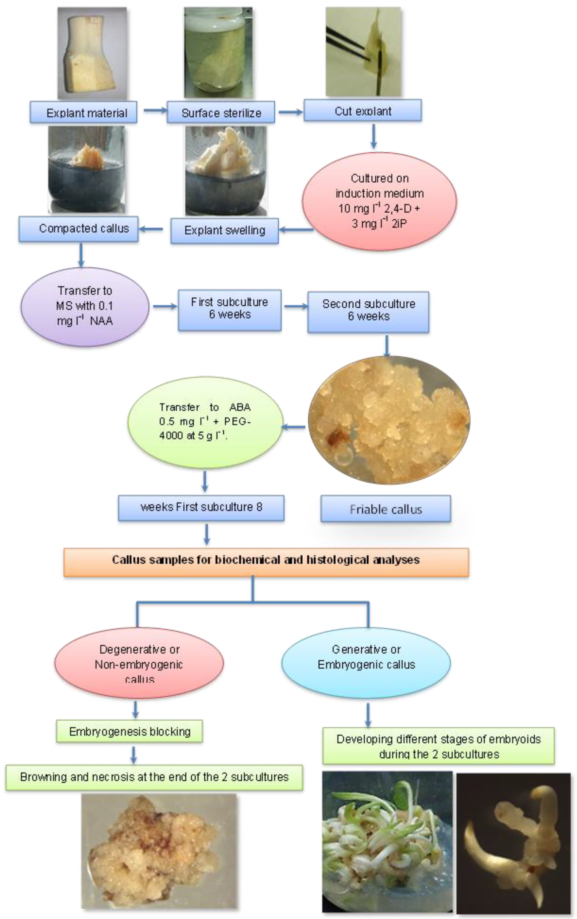
2.1. Experimental Layout

This study was carried out during the period 2019 to 2021. Firstly, to induce callogenesis from date palm shoot tip explants, the procedure that has been described by Zein Eldin and Ibrahim [10] was followed. After that, friable calli were allowed to be more viable for embryogenesis by culturing on MS medium supplemented with 0.1 mg L−1 NAA for two subcultures (6 weeks for each) [21]. Then, further development of friable calli was performed when it was placed in MS maturation medium containing 0.5 mg L−1 ABA and 5 g L−1 polyethylene glycol (PEG-4000) for another three subcultures (8 weeks for each). Embryogenic callus (EC) and degenerative embryogenic callus (DEC) of the same cultivar were harvested for histological and biochemical analyses after the first subculture (8 weeks) from culturing on maturation medium. See Figure 1.

2.2. Histological Analyses

Both degenerative embryogenic and embryogenic calli were fixed in 3% glutaraldehyde rinsed in 0.1 M phosphate buffer (pH = 7.6) for 6 h at 4 °C, according to Frankl and Mari [22], dehydrated in a graded ethanol series and infiltrated with EPON 812 epoxy resin (Sigma-Aldrich, St. Louis, MA, USA,). Semi-thin sections (1 µm) were made using a glass knife and stained with 0.03% toluidine blue. The successive morphogenesis stages (globular, bipolar and FDE) of somatic embryos were fixed in FAA solution (formaldehyde, acetic acid and 50% ethyl alcohol, 5:5:90 by volume) for 24 h. Progressive dehydration using ascending concentrations of ethanol was performed and then embedding in Paraplast (Sigma Aldrich, St. Louis, MA, USA, Paraplast plus p. 3688), according to Abdelbar [23]. Transverse and longitudinal sections 8 µm in thickness were made using a LEICA rotary microtome, model RM 2125 RTS, and stained with a safranin–fast green combination [24]. Observations were made using a LEICA light microscope, model DM-500 (Leica Microsystems, Wetzlar, Germany).

2.3. Biochemical Analyses

All biochemical determinations were carried out on 0.2 g fresh tissue as follows:

2.3.1. Soluble Proteins, Hydrogen Peroxide and Malondialdehyde Determination

Soluble proteins were quantified using the method of Bradford [25] and calculated as mg protein g−1 F.W using bovine serum albumin as a standard. Hydrogen peroxide was determined in tissues using the method of Zhou et al. [26]. Hydrogen peroxide concentration was calculated as µg H2O2 g−1 F.W. Lipid peroxidation was measured via determination of malondialdehyde (MDA) in tissues according to Heath and Packer [27] and calculated using an extinction coefficient of 155 mM−1 cm−1. Malondialdehyde concentration was calculated as mmol MDA g−1 F.W.

2.3.2. Reduced Glutathione, Ascorbic Acid, Dehydroascorbate, Total Phenolic Compounds and Antioxidant Activity Determination

Reduced glutathione on reaction with DTNB (5,5’-dithiobis nitro benzoic acid) was determined according to Moron et al. [28]. Reduced glutathione value was expressed as nmoles GSH/g. F.W. Ascorbic acid (ASC) and dehydroascorbate (DHA) concentrations were measured by using 2,4 dinitrophenylhydrazine according to Kapour et al. [29]. ASC and DHA values were calculated as µg−1 F.W.
To determine total phenolic compounds and antioxidant activity (scavenging activity on DPPH radical and reducing power), 0.2 g of fresh tissue was extracted with 3 mL 80% ethanol for at least 24 h at 5 °C, filtered, and the remaining tissue re-extracted about three times. Collected extract was completed to 10 mL using 80% ethanol. Total phenolic compounds were determined using the Folin-Ciocalteu colorimetric method according to Shahidi and Naczk [30] and expressed as mg g−1 F.W using gallic acid as standard. Antioxidant activity of ethanolic extract was determined using two methods, the first method by measuring the ethanolic extract ability to scavenge 2,2-diphenyl-1-picrylhydrazyl (DPPH) according to Brand-William et al. [31]. The percentage of scavenging activity (% SA) was calculated by the following equation: % SA = (AB − AS)/AB × 100, where AB (blank absorbance) and AS (sample absorbance). The second method of antioxidant activity determination was carried out by measuring the reducing power of ethanolic extract according to the method of Oyaizu [32]. Increased absorbance value of the reaction mixture at 700 nm indicated increased reducing power ethanolic extract. Reducing power was expressed as µg g−1 F.W using ascorbic acid as standard.

2.3.3. Polyphenolic Compounds Analysis by HPLC

To measure total phenolic compounds via HPLC, 0.2 g of fresh tissue was soaked in 1 mL methanol HPLC grade then centrifuged, and the supernatant was used in HPLC analysis. HPLC analysis was conducted using an Agilent 1260 series. The separation was conducted using an Eclipse C18 column (4.6 mm × 250 mm i.d., 5 μm). The mobile phase consisted of water (A) and 0.05% trifluoroacetic acid (Sigma-Aldrich, Merck, Germany) in acetonitrile (B) (HPLC grade 99.9%) at a flow rate of 1 mL/min. The mobile phase was programmed consecutively in a linear gradient as follows: 0 min (82% A); 0–5 min (80% A); 5–8 min (60% A); 8–12 min (60% A); 12–15 min (82% A) and 15–16 min (82% A). The multi-wavelength detector was monitored at 280 nm. The injection volume was 10 μL for each of the sample solutions. The column temperature was maintained at 35 °C [33].

2.4. Statistical Analysis

Experiments were conducted in a complete randomized design with five replicates and data were analyzed using L.S.D test at 5% level of significance by Statistix software, version 9 program [34].

3. Results

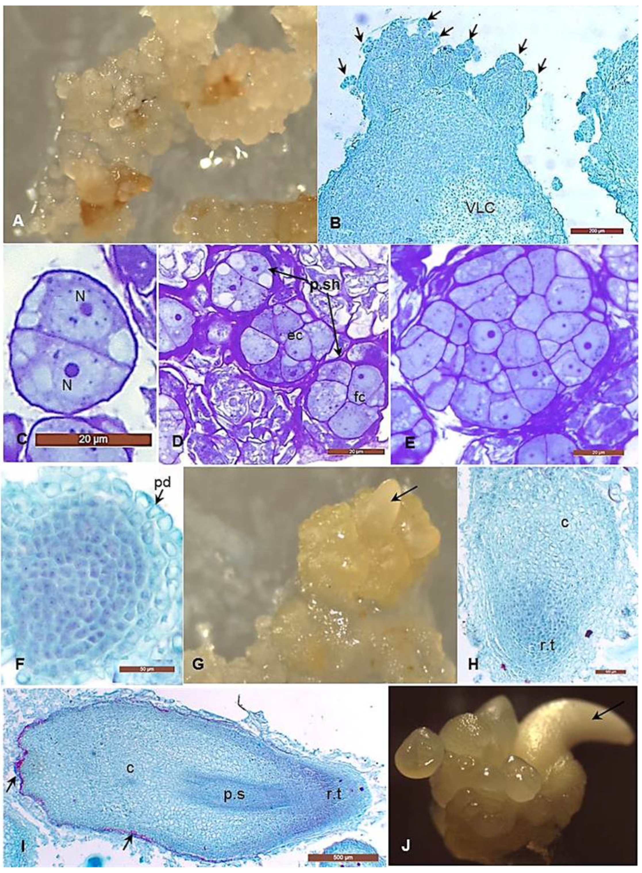
3.1. Morphological and Histological Analyses of Degenerative Embryogenic Callus (DEC)

The degenerative embryogenic callus was firm, compacted, hard to break, yellowish to brown in color and exhibited more rigid opaque clumps of different sizes (Figure 2A). The histological observations showed that it was composed of coherent cellular aggregates representing the first ontogenesis of the somatic embryos (Figure 2B); most of these aggregates were in the globular stage, which exhibited a cessation of cell division, characterized by more vacuolated cells, rich in phenolic compounds and covered with very thick walls (Figure 2C). Some other cellular aggregates were degenerated causing necrosis spots in the DEC masses and had acquired brown color (Figure 2D).

3.2. Morphological and Histological Analyses of the Embryogenic Callus and the Successive Developmental Stages of Somatic Embryos

The embryogenic calli (EC) were smooth, friable masses, white or creamy in color and exhibited more organized translucent structures which form rounded clumps of different sizes (Figure 3A). This callus consists of two distinguished types of cells; the inner zone comprises more loosely, vacuolated disorganized masses bordered by numerous small aggregates that organize the outer or the peripheral zone (Figure 3B). These peripheral cellular aggregates are composed of isodiametric cells that contain dense cytoplasm, conspicuous nuclei and minimal vacuoles. These features characterized the meristmatic cells and indicated high metabolic activity.
The first embryonic stages can be noticed in these aggregates: the two-celled pro-embryo (Figure 3C), the four-celled, the 8–16 celled spherical pro-embryos (Figure 3D) as well as the globular pro-embryo (Figure 3E). These pro-embryos have definite fragmentation lines formed by thick walls or sheaths surrounding each developmental stage. The rapid mitotic divisions continue in the globular pro-embryo and give rise to the formation of the early bipolar structures, which consist of pear-shaped meristematic cells surrounded by a protoderm initial (Figure 3F) protruding from the callus surface (Figure 3G). The structure of bipolar embryos is characterized by the presence of a more meristematic pole which forms the precursors of the shoot-root tips and the more vacuolated pole which forms the cotyledon (Figure 3H). A few days later, the bipolar embryos become more cylindrical, elongated and have a well-differentiated procambium strand (Figure 3I). The developed somatic embryos in date palm are asynchronized (Figure 3J). Then, the last stage “the fully developed somatic embryo” is obtained at the end of 4th to 6th week of the second subculture (Figure 3I,J). In this stage we can observe the cotyledon and the well-developed procambium strand which forms the root-shoot axis, in addition to the tannin cells, which are in the outer layers of the embryo.

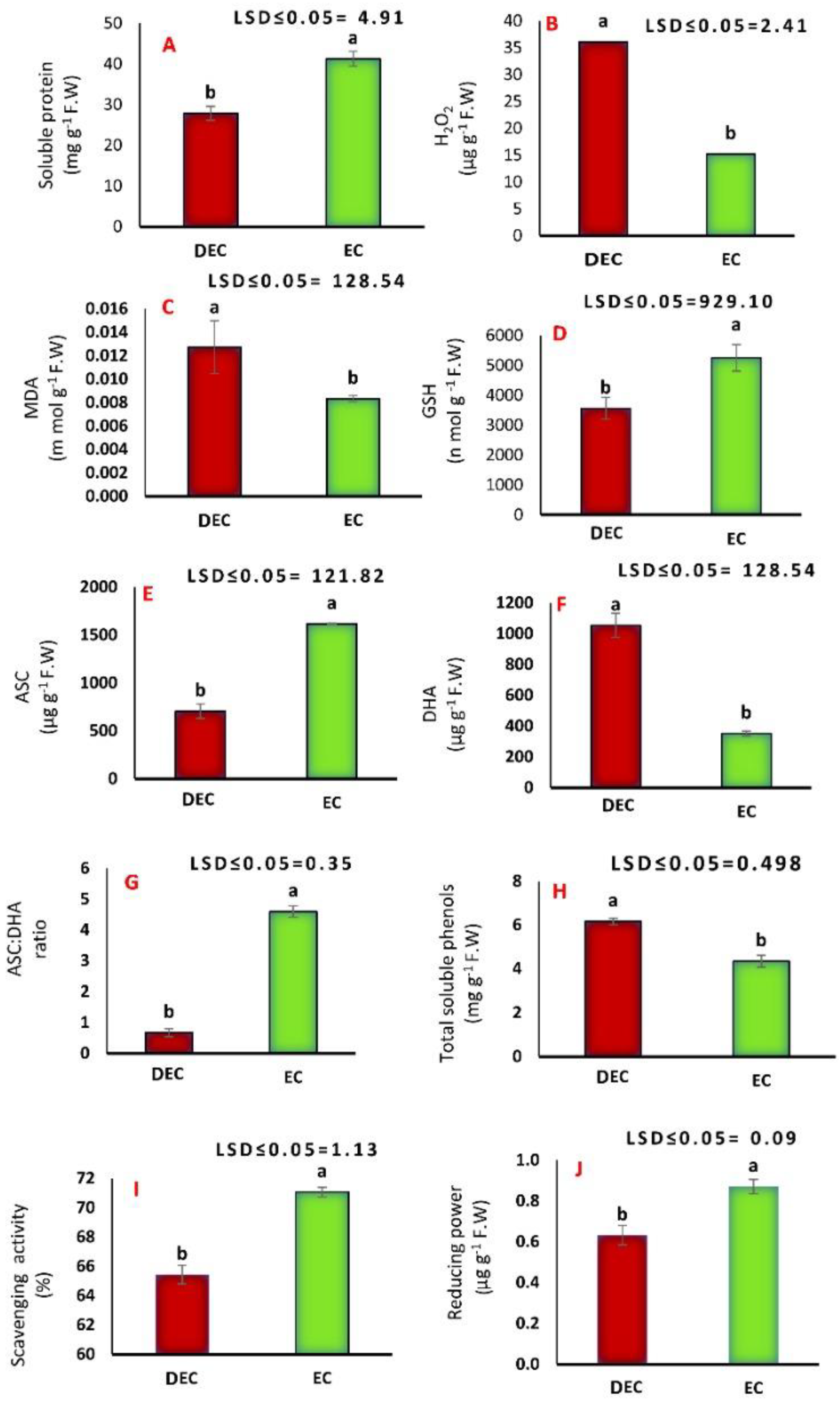
3.3. Biochemical Analyses

Data presented in Figure 4 indicate that the soluble protein concentration in the EC was higher than in the DEC, with significant differences (p ≤ 0.05). The physiological analyses showed differences in both H2O2 and MDA concentrations between the two types of calli. A significant increase in H2O2 and MDA concentrations was observed in the DEC compared to the EC. The reduced forms of both GSH and ASC were increased in the EC compared to the DEC. As for the oxidized form of ascorbate (DHA), its concentration was significantly increased in the DEC compared to the EC, which reached more than a two-fold increase. This reduces the negative effect of oxidized DHA in the embryogenic callus and this was confirmed when the ratio ASC: DHA was higher in the EC than in the DEC. When assessing the concentration of total soluble phenols, the highest significant concentration was determined in the DEC. The phenolic extracts of the different types of calli showed scavenging activity on the DPPH radical. The EC phenolic extract was found to have higher scavenging activity potential than the DEC. In addition, the reducing power of both phenolic extracts was increased in the same manner as before.

3.4. Fractionation of Polyphenolic Compounds

Eleven phenolic compounds were examined: 5 belonging to hydroxycinnamic acids “cinnamic, coumaric, ferulic, chlorogenic and caffeic”, 3 belonging to the hydroxybenzoic acids “gallic, vanillic and ellagic” and 3 belonging to flavonoids “naringenin, catechin and rutin”. The phenolic profiles of the DEC and EC were significantly different (Table 1). Chlorogenic and gallic acids were the most abundant hydroxycinnamic and hydroxybenzoic acids in both calli. The type and concentration of the phenolic compounds were different according to the callus tissue. Cinnamic acid was absent in the EC compared to the DEC. Coumaric and vanillic acids were absent in the DEC, while these phenolic acids have significant values (p ≤ 0.05) in the EC. Concerning ferulic acid, the DEC contained an approximately 2-fold increase compared to the EC. Ellagic showed higher levels in the EC than in the DEC. Data presented an overaccumulation of flavone naringenin in the DEC compared to the EC. Moreover, catechin was detected in the DEC, while rutin was detected in the EC.

4. Discussion

4.1. Histological Analysis

The initiation and maintenance of the embryogenic callus of date palm is the principal step for obtaining somatic embryos. This step requires the presence of auxin, which is necessary for cell dedifferentiation and callus induction and maintenance [18]. However, the induced callus of date palm (Barhee cv.) contained some cells that were able to develop into somatic embryos and are described as embryogenic callus (EC) and others lost their embryogenic competence and are described as degenerative embryogenic callus (DEC).
The two types of calli arose and grew side by side. This indicates that the differentiation process can occur through different pathways under the same culture conditions. Even after transferring the DEC into free auxin media for further development, in spite of some cells already being in the globular stage, these cells were unable to develop into the further stages of somatic embryos. Their cells were compacted, hard to break and somewhat yellowish to brown in color compared to the EC. The microscopic examination proved that they consisted of vacuolated cells, rich in phenolic compounds and covered with very thick walls. These features were associated with accumulation of some metabolites such as DHA and total soluble phenols. López Arnaldos et al. [35] pointed out that the presence of phenolic compounds leads to the formation of bridges between phenolic polysaccharide chains as wells between polysaccharides and either lignins or structural proteins and increases the phenolic deposition within the cell wall leading to a decrease in cell wall extensibility and thereby interrupting cell division and cell growth. A previous study found that callus growth is mainly due to cell expansion, but the accumulation of phenolic compounds has been described as being inversely correlated with cell growth [36]. This result may explain our observations regarding the cessation of cell division and cell growth in the DEC, which affects their ability to regenerate the further developmental stages of date palm somatic embryos (SEs).
The histological analysis of the EC indicated that it has embryogenic characteristics such as small isodiametric cells containing dense cytoplasm, conspicuous nuclei and minimal vacuoles and observed mitotic divisions. These features were consistent and have been observed in date palm in vitro culture [17,18]. The presented features of the EC and the associated metabolites (high values of proteins, ASC, GSH, scavenging activity and low values of MDA and H2O2) can explain the active cell growth and maintain the sequential morphogenesis stages successfully.

4.2. Biochemical Analyses

Protein is the main factor responsible for growth and morphogenesis of SEs [6]. In this study, the content of protein is higher in the EC than the DEC. Similarly, Fki’s [37] findings indicated that total soluble proteins were found to be higher in EC than in DEC counterparts. This increase was related to the synthesis of embryogenic-specific proteins responsible for growth and development. It has been suggested that this increase was related to the synthesis of late embryogenic proteins which protect embryos from dehydration [38]. Moreover, the embryogenesis-specific proteins are not synthesized in non-embryogenic callus, which indicates that these proteins may have a key role in the somatic embryogenesis process [39]. These studies showed that the accumulation of protein was the material substance for EC transformation into SEs compared with DEC. According to other studies, the activity of peroxidase is much higher in the EC than in the non-embryogenic callus; three active isoforms of the enzyme were present in the crude extract from the EC, whereas only a single band appeared in the DEC [40].
Hydrogen peroxide in the DEC of date palm was higher than those in the EC, which showed that the differentiation of the EC was affected by the levels of H2O2. Exogenous treatment with H2O2 (200 µM) promoted the formation of embryogenic cells, while a higher concentration of H2O2 (300 µM) inhibited the differentiation of somatic embryos [9,39]. Therefore, the significant high concentration of H2O2 (p ≤ 0.05) obtained in the DEC is the main reason for the undifferentiating of these cells. Another study indicated that moderate concentrations of H2O2 elevated the antioxidant status and enhanced stress tolerance, while higher levels caused oxidative stress that damaged the cell structure, harmed cell membranes and made cells lose their function [41]. Hydrogen peroxide acts as an oxidative stressor. This has been reported before by Pasternak et al. [42]; accumulation of high levels of intracellular H2O2 makes cells in a less differentiated status. In addition, similar results have been reported in Lycium barbarum L.; the frequency of SEs reaches its maximum value after treatment with low levels of H2O2 [39]. It has been suggested that the accumulation of low levels of H2O2, a secondary ROS product, is involved in cell signal transferring material which regulates gene expression capable of protein synthesis and promotes SEs induction and development. These results are consistent with our results, where an increase in the EC protein content with the presence of a low H2O2 concentration was observed. Low H2O2 potent induced gene expression that related by enhancing antioxidant metabolism [43]. However, H2O2 has a beneficial role in the establishment of polarity at early developmental stages of SEs [44]. Concerning MDA, it accumulated in a high level in the DEC. Our findings are in agreement with results obtained by Adams et al. [45] who reported that exogenous application of MDA inhibited the proliferative growth and embryogenesis in Daucus carota cultures due to its ability of cytotoxicity and reaction with DNA, protein and phospholipids. Moreover, our obtained data reflected that MDA and H2O2 accumulate in high levels in the degenerative embryogenic callus, which lost its totipotency, causing embryogenesis blocking.
In the present results, the significant increase in the reduced forms of ASC and GSH with the decreasing in the oxidized form of DHA in the EC maintain the reduced state in its cells and then enhance the early stages of somatic embryos to further development. ASC and GSH have vital roles in protecting plant cells from harmful effects of ROS [46]. Furthermore, Mapson and Moustafa [47] assumed that ASC and GSH might play a significant role in the respiration of embryonic plant tissues in which cell division is probably frequent. It is well known that the EC is characterized by high aerobic metabolic and dividing activities, resulting in H2O2 formation. One of the important roles of ASC is its ability to inhibit the oxidation processes. The accumulated phenolics in the presence of H2O2 were oxidized by soluble apoplastic peroxidase, which bound to cell walls, resulting in the accumulation of phenoxyl radicals, and ASC partially reduced these radicals to yield DHA and phenols to reach the redox state [48]. In response to the increase of GSH, Abdellatif and Ibrahim [49] illustrated that endogenous glutathione in reduced form scavenges ROS and activates stress signaling molecules that enhance cell division of proliferative tissues. The exogenous addition of GSH reversed the inhibition in the cell division that occurred via its oxidized form glutathione disulfide (GSSH). Consequently, in the present results, accumulation of endogenous components of ASC and GSH in the EC of date palm enables it to be competent for development into somatic embryos. Moreover, exogenous application of ASC increases the conversion and germination of SEs [50].
Concerning the free radical scavenging activity of phenolic extract, the EC possessed higher scavenging activity and reduced power compared to the DEC. Here, we assumed that the scavenging antioxidant activity and reducing power capacity of the EC extracts were compatible with the high levels of GSH, ASC and phenolic compounds, which can act as free radical scavengers. Considerable amounts of polyphenols and ascorbic acid have been reported as scavengers for the DPPH radical due to their ability to transfer electrons or donate hydrogen [51]. The antioxidant potent extracts could be served by inhibiting the interaction of free radicals with DNA [52]. The scavenging activity in date palm depends on the concentration of phenolic compounds and also on the degree of hydroxylation and polymerization [53]. The reaction mixtures of the embryogenic cell extracts remove the free radicals and prevent cellular abnormalities by breaking down the chain reaction of peroxiding lipid moieties, beside its consumption of H2O2 as a signal transferring material in protein synthesis as mentioned previously. Thus, the reduced forms of GSH, ASC and phenolic compound antioxidants may be the major contributors to the embryogenic cells division, differentiation and maturation.

4.3. Fractionation of Polyphenolic Compounds

The phenolic compounds constitute a wide range of plant substances; they are usually regarded as harmful compounds during in vitro culture via causing browning and necrosis of the explant, especially during the propagation of the date palms [1]. However, previous studies showed that phenolic compounds have an essential role for controlling some morphogenic processes during in vitro plant propagation, such as stimulating organogenesis [13] and somatic embryogenesis [1,14]. In addition, Reis et al. [12] pointed out that these phenolics could also interfere with auxin metabolism in feijoa, keeping auxin at concentrations particularly favorable for somatic embryogenesis induction. Alternation in phenolic acid concentrations in DEC and EC suggest the role of these secondary metabolites in the date palm somatic embryogenesis process.
In our results, cinnamic acid was absent in the EC compared to the DEC. A previous study indicated that cinnamic acid is considered one of the most active inhibitors of IAA polar transport, particularly in globular somatic embryos [35]. Consequently, lack of cinnamic acid in the EC cells has a positive effect by enhancing the successive development stages of somatic embryos, since the EC including the first embryogenic stages and the globular embryos (Figure 3C) exhibited the ability to develop into somatic embryos successfully. In contrast, coumaric acid was detected in the EC and was lacking in the DEC. Coumaric acid has an important role in enhancing cell division and stimulates the early stage of SEs [54]. The authors considered the presence of coumaric acid as a sign that the EC would be activated to further develop somatic embryos. Our results showed an overaccumulation of naringenin in the DEC (16.24 µg) while the EC contained lower concentrations of this compound. Naringenin is considered the precursor of most flavonoids [55]. Previous reports support the suggestion that naringenin and cis-cinnamic acid are negative regulators of auxin transport [56]. Exogenous application of naringenin at 0.1 mM reduced soybean root growth causing a loss of cell viability, an increase in cell wall-bound peroxidase (POD) and a decrease in soluble POD [55]. Therefore, the authors assumed that naringenin has a destructive influence on cell growth as recorded in the DEC. Soluble POD acts as an antioxidant enzyme and protects plant cells from the oxidative damage of H2O2 and other reactive oxygen species. Moreover, cell wall-bound peroxidase is involved in lignin formation, which may be a reason for the presence of the very thick cell wall of the DEC compared to the EC (Figure 2C,D and Figure 3D), and as a result of the indirect relationship between increase in naringenin concentration and cell wall lignification, the regeneration ability in the DEC might be altered.
The present results show the lower concentration of ferulic is recorded in the EC rather than the DEC. This was supported by recent studies which suggest an interconnection between auxin (the essential plant hormone for SE induction) and the phenylpropanoid pathway [57]. Previous studies indicated that some phenolic metabolites such as ferulic and sinapic at low concentrations can inhibit the IAA oxidation [58]. Thus, by maintaining the endogenous auxin, the further development of the EC may be influenced negatively. This could be an explanation for the presence of a low concentration of free ferulic in the EC. However, presence of ferulic acid in both types of calli may be explained by its consumption in forming the cross-links between polysaccharides in the cell wall around the cells (Figure 2C,D and Figure 3D). This result agrees with Sane et al. [18] who observed an EC defined by a thick wall built mainly from polysaccharides called sheath. Mnich et al. [59] mentioned that the polysaccharide composite is a dynamic structure, subject to turnover while the cell is expanding. Ferulic acid and its oligomers have an essential role in plant cell wall rigidity by forming cross-links between polysaccharides, polysaccharide chains and lignin [60] and polysaccharides and structural proteins [61]. Oligomerization of the feruloylated polysaccharide links all cell wall components together via phenolic bridges with an effect on cell wall extensibility, thus leading to the cessation of cell growth. Our results illustrated that the cell wall was thicker in the DEC. This could be explained by the over formation of phenolic bridges, which causes loss of cell wall extensibility leading to termination of cell growth and blocking of the successive morphogenesis process.
Concerning the chlorogenic and caffeic acids, they were detected in both the DEC and the EC without significant differences. El Bellaj and El Hadrami [1] indicated that these compounds were the most prominent among the other phenolic acids in the date palm embryogenic callus. The present study showed a higher concentration of gallic acid in the EC than the DEC, whereas vanillic acid was not detected in the DEC and was detected in the EC with significant difference. Gallic acid is a widespread phenolic compound in date palm tissues that has a significant antioxidant effect [62]. In addition, Cvikrová et al. [54] reported that increasing contents of gallic and vanillic acids enhanced the cell divisions, promoting the early stage of somatic embryos. According to the present results, low concentration of gallic and lacking vanillic acid in the DEC may be considered as a marker to explain why this cellular mass stopped developing to further stages compared to the EC.
Furthermore, the significant increase in ellagic acid content in the EC compared to the DEC may be due to the properties of this EC itself, as this EC was distinguished by observed mitotic divisions and higher metabolism and these were associated with producing ROS. However, these cells possess a system of antioxidant defense scavenge ROS, thereby minimizing the oxidative damage [10]. Khan et al. [63] pointed out that ellagic acid is considered one of the best phenolic compounds in scavenging ROS. Therefore, the presence of ellagic in the EC is necessary via its antioxidant role in this type of callus.
The flavone naringenin can transform into taxifolin, catechin, kaempferol and rutin [64]. These flavonoids have an antioxidant activity and inhibit the formation of trans-trans hydroperoxide isomers which are produced during auto-oxidation of lipids. Polyphenols act as a hydrogen donor to proxy radicals via inhibiting lipid auto-oxidation and keeping membrane integrity [65]. This was consistent with our results, in which a significant increase in rutin was observed in the EC, while it was absent in the DEC. In spite of catechin acting primarily as an antioxidant, it also has other physiological roles. The obtained results indicated that catechin was absent in the EC. This result was recommended as catechin can regulate the auxin level by delaying IAA degradation [66], and this was unsuitable for the EC, which needs a decreasing in IAA level for further development. In contrast, the presence of catechin in the DEC causes delay in the development the following stages.

5. Conclusions

Date palm somatic embryogenesis is a complicated process occurring over a long period (6–12 months) of culture. The cellular mechanisms for this process are reflected in changes in biochemical and histological structures and their interaction with each other. During the sub-culturing, both the EC and DEC could be obtained and grown side by side under the same conditions, and both exhibited different cellular behaviors. Thus, it is necessary to check precise biochemical and histological events associated with the morphogenesis of each tissue. The EC displayed a high content of proteins, GSH, ASC, reducing power and scavenging activity, which have a positive effect on cell division and modulate ROS metabolism, thus regulating morphogenesis and developing somatic embryos successfully. These can be used as biomarkers of embryogenic competences. The formation of the DEC was tightly linked with low concentrations of the previous components and high values of H2O2, MDA, DHA, total soluble phenols, and accumulation of some phenolic compounds (naringenin and cinnamic acids), which all inhibited the subsequent development of SEs. Thus, this undesired tissue could be eliminated from the sub-culturing routine to save cost and time or could be separated for the recovery process. Further experiments are currently in progress to recover the DEC of date palm and uncover the role of certain compounds (i.e., cinnamic and naringenin) in date palm somatic embryogenesis.

Author Contributions

Conceptualization, A.F.M.Z.E.D., I.S.I. and H.A.I.; methodology, A.F.M.Z.E.D., O.H.A.E., Y.M.R.A., E.G.G. and H.A.I.; software, S.M.A.T., M.F.M.I., M.S.F.S. and H.S.E.-B.; validation, M.F.M.I., O.H.A.E., I.M.S.E.-D., M.S.F.S. and H.A.I.; formal analysis, Y.M.R.A., R.F., I.M.S.E.-D. and M.M.E.-M. investigation, E.G.G., R.F., Y.M.R.A., O.H.A.E., M.S.A.E.-A. and H.S.E.-B.; resources, I.M.S.E.-D., R.F. and H.S.E.-B.; data curation, S.M.A.T., A.A.E.-Y., Y.M.R.A., M.F.M.I. and E.G.G.; writing—original draft preparation, A.F.M.Z.E.D., O.H.A.E., M.S.A.E.-A. and Y.M.R.A.; writing—review and editing, I.S.I., M.F.M.I., O.H.A.E., M.S.A.E.-A., M.M.E.-M. and I.M.S.E.-D.; visualization, I.S.I., K.M.A.R., M.M.E.-M. and A.A.E.-Y.; supervision, I.S.I. and A.F.M.Z.E.D.; project administration, K.M.A.R., A.A.E.-Y., I.M.S.E.-D., R.F. and M.S.F.S.; funding acquisition, M.M.E.-M., K.M.A.R., E.G.G., S.M.A.T., A.A.E.-Y. and H.S.E.-B. All authors have read and agreed to the published version of the manuscript.

Funding

This research received no external funding.

Institutional Review Board Statement

Not applicable.

Informed Consent Statement

Not applicable.

Acknowledgments

Authors would like to appreciate The Central Laboratory for Date Palm Researches and Development, Agricultural Research Center, Giza, Egypt and the Department of Agricultural Botany, Faculty of Agriculture, Ain Shams University, Shoubra El-Kheima, Egypt for providing the facilities to carry out the research work. The authors wish to thank Sami, A. Habib for helpful discussions on the results of the histological changes and critical reading of the manuscript.

Conflicts of Interest

The authors declare no conflict of interest.

References

  1. Bellaj, E.M.; Hadrami, E.I. Characterization of two constitutive hyroxycinnamic acids derivatives in date palm (Phoenix dactylifera L.) callus in relation with tissue browning. Biotechnology 2004, 3, 155–159. [Google Scholar] [CrossRef]
  2. Morel, A.; Trontin, J.F.; Corbineau, F.; Lomenech, A.M.; Beaufour, M.; Reymond, I.; Le Metté, C.; Ader, K.; Harvengt, L.; Cadene, M.; et al. Cotyledonary somatic embryos of Pinus pinaster Ait. most closely resemble fresh, maturing cotyledonary zygotic embryos: Biological, carbohydrate and proteomic analyses. Planta 2014, 240, 1075–1095. [Google Scholar] [CrossRef] [PubMed] [Green Version]
  3. Nic-Can, G.I.; Galaz-Ávalos, R.M.; De-la-Peña, C.; Alcazar-Magaña, A.; Wrobel, K.; Loyola-Vargas, V.M. Somatic Embryogenesis: Identified Factors that Lead to Embryogenic Repression. A Case of Species of the Same Genus. PLoS ONE 2015, 10, e0126414. [Google Scholar] [CrossRef]
  4. Silveira, V.; de Vita, A.M.; Macedo, A.F.; Dias, M.F.R.; Floh, E.I.S.; Santa-Catarina, C. Morphological and polyamine content changes in embryogenic and non-embryogenic callus of sugarcane. Plant Cell Tissue Organ Cult. 2013, 114, 351–364. [Google Scholar] [CrossRef]
  5. Al-Khayri, J.M.; Naik, P.M. Date palm micropropagation: Advances and applications. Ciência Agrotecnologia 2017, 41, 347–358. [Google Scholar] [CrossRef] [Green Version]
  6. Cangahuala-Inocente, G.C.; Silveira, V.; Caprestano, C.A.; Floh, E.I.S.; Guerra, M.P. Dynamics of physiological and biochemical changes during somatic embryogenesis of Acca sellowiana. In Vitro Cell. Dev. Biol. Plant. 2014, 50, 166–175. [Google Scholar] [CrossRef] [Green Version]
  7. Cui, K.R.; Xing, G.S.; Liu, X.M.; Xing, G.M.; Wang, Y.F. Effect of the hydrogen peroxide on somatic embryogenesis of Lycium barbatum L. Plant Sci. 1999, 146, 9–16. [Google Scholar] [CrossRef]
  8. Fatima, S.; Mujib, A.; Samaj, J. Anti-oxidant enzyme responses during in vitro embryogenesis in Catharanthus roseus. J. Hortic. Sci. Biotechnol. 2011, 86, 569–574. [Google Scholar] [CrossRef]
  9. Manivannan, A.; Jana, S.; Soundararajan, P.; Ko, C.H.; Jeong, B.R. Antioxidant enzymes metabolism and cellular differentiation during the developmental stages of somatic embryogenesis in Torilis japonica (Houtt.) DC. Plant Omics J. 2015, 8, 461–471. [Google Scholar]
  10. Eldin, Z.A.F.M.; Ibrahim, H.A. Some biochemical changes and activities of antioxidant enzymes in developing date palm somatic and zygotic embryos in vitro. Ann. Agric. Sci. 2015, 60, 121–130. [Google Scholar] [CrossRef] [Green Version]
  11. Pullman, G.S.; Zeng, X.; Copeland-Kamp, B.; Crockett, J.; Lucrezi, J.; May, S.W.; Bucalo, K. Conifer somatic embryogenesis: Improvements by supplementation of medium with oxidation–reduction agents. Tree Physiol. 2015, 35, 209–224. [Google Scholar] [CrossRef] [PubMed]
  12. Reis, E.; Batista, M.T.; Canhoto, J.M. Effect and analysis of phenolic compounds during somatic embryogenesis induction in Feijoa sellowiana Berg. Protoplasma 2008, 232, 193–202. [Google Scholar] [CrossRef] [PubMed] [Green Version]
  13. Lorenzo, J.C.; de los Angeles Blanco, M.; Peláez, O.; González, A.; Cid, M.; Iglesias, A.; González, B.; Escalona, M.; Espinosa, P.; Borroto, C. Sugarcane micropropagation and phenolic excretion. Plant Cell Tissue Organ Cult. 2001, 65, 1–8. [Google Scholar] [CrossRef]
  14. Cvikrová, M.; Hrubcová, M. The role of phenolic substances in the processes of differentiation and morphogenesis. In Advances in Regulation of Plant Development; Strnad, M., Pec, P., Beck, E., Eds.; Peres Publications: Prague, Czech Republic, 1999; pp. 213–220. [Google Scholar]
  15. Fry, S.C. Cross-Linking of Matrix Polymers in the Growing Cell Walls of Angiosperms. Annu. Rev. Plant Physiol. 1986, 37, 165–186. [Google Scholar] [CrossRef]
  16. Othmani, A.; Bayoudh, C.; Drira, N.; Marrakchi, M.; Trifi, M. Somatic embryogenesis and plant regeneration in date palm Phœnix dactylifera L., cv. Boufeggous is significantly improved by fine chopping and partial desiccation of embryogenic callus. Plant Cell Tissue Organ Cult. 2009, 97, 71–79. [Google Scholar] [CrossRef]
  17. Tisserat, B.; Demason, D.A. A Histological Study of Development of Adventive Embryos in Organ Cultures of Phoenix dactylifera L. Ann. Bot. 1980, 46, 465–472. [Google Scholar] [CrossRef]
  18. Sane, D.; Aberlenc-Bertossi, F.; Gassama-Dia, Y.K.; Sagna, M.; Trouslot, M.F.; Duval, Y.; Borgel, A. Histocytological Analysis of Callogenesis and Somatic Embryogenesis from Cell Suspensions of Date Palm (Phoenix dactylifera). Ann. Bot. 2006, 98, 301–308. [Google Scholar] [CrossRef]
  19. Abd El Bar, O.H.; El Dawayati, M.M. Histological changes on regeneration in vitro culture of date palm 457 (Phoenix dactylifera) leaf explants. Aust. J. Crop. Sci. 2014, 8, 848–855. [Google Scholar]
  20. Zayed, E.M.M.; Abdelbar, O.H. Morphogenesis of immature female inflorescences of date palm in vitro. Ann. Agric. Sci. 2015, 60, 113–120. [Google Scholar] [CrossRef] [Green Version]
  21. Murashige, T.; Skoog, F. A Revised Medium for Rapid Growth and Bio Assays with Tobacco Tissue Cultures. Physiol. Plant. 1962, 15, 473–497. [Google Scholar] [CrossRef]
  22. Frankl, A.; Mari, M. Electron microscopy for ultrastructural analysis and protein localization in Saccharomyces cerevisiae. Microb. Cell 2015, 2, 412–428. [Google Scholar] [CrossRef] [PubMed] [Green Version]
  23. Abdelbar, O.H. Histological Analysis of the Developmental Stages of Direct Somatic Embryogenesis Induced from in Vitro Leaf Explants of Date Palm. Methods Mol. Biol. 2017, 1637, 145–162. [Google Scholar] [CrossRef]
  24. Berlyn, G.P.; Miksche, J.P.; Sass, J.E. Botanical Microtechnique and Cytochemistry; Iowa State University Press: Ames, IA, USA, 1976; pp. 24–100. [Google Scholar]
  25. Bradford, M.M. A rapid and sensitive method for the quantitation of microgram quantities of protein utilizing the principle of protein-dye binding. Anal. Biochem. 1976, 72, 248–254. [Google Scholar] [CrossRef]
  26. Zhou, B.; Wang, J.; Guo, Z.; Tan, H.; Zhu, X. A simple colorimetric method for determination of hydrogen peroxide in plant tissues. Plant Growth Regul. 2006, 49, 113–118. [Google Scholar] [CrossRef]
  27. Heath, R.L.; Packer, L. Photoperoxidation in isolated chloroplasts: I. Kinetics and stoichiometry of fatty acid peroxidation. Arch. Biochem. Biophys. 1968, 125, 189–198. [Google Scholar] [CrossRef]
  28. Moron, M.S.; Depierre, J.W.; Mannervik, B. Levels of glutathione, glutathione reductase and glutathione S-transferase activities in rat lung and liver. Biochim. Et Biophys. Acta (BBA) Gen. Subj. 1979, 582, 67–78. [Google Scholar] [CrossRef]
  29. Kapour, A.; Haskovic, A.; Copra Janicijevic, A.; Klepo, L.; Topcagic, A.; Tahirovic, I.; Sofic, E. Spectrophotometric analysis of total ascorbic content in various fruits and vegetables. Bull. Chem. Technol. Bosnia Herzeg. 2012, 38, 39–42. [Google Scholar]
  30. Shahidi, F.; Naczk, M. Methods of analysis and quantification of phenolic compounds. In Food Phenolic: Sources, Chemistry, Effects and Applications; Technomic Publishind Company, Inc.: Lancaster, PA, USA, 1995; pp. 287–293. [Google Scholar]
  31. Brand-Williams, W.; Cuvelier, M.E.; Berset, C. Use of a free radical method to evaluate antioxidant activity. LWT Food Sci. Technol. 1995, 28, 25–30. [Google Scholar] [CrossRef]
  32. Oyaizu, M. Studies on products of browning reactions: Antioxidative activities of products of browning reaction prepared from glucosamine. Jananese J. Nutr. 1986, 44, 307–325. [Google Scholar]
  33. Gökbulut, A. Validated RP-HPLC Method for Quantification of Phenolic Compounds in Methanol Extracts of Aerial Parts and Roots of Thymus sipyleus and Evaluation of Antioxidant Potential. Trop. J. Pharm. Res. Oct. 2015, 14, 1871–1877. [Google Scholar] [CrossRef] [Green Version]
  34. Statistix. Statistix 9: Analytical Software; Statistix: Tallahassee, FL, USA, 2009. [Google Scholar]
  35. López Arnaldos, T.; Muñoz, R.; Ferrer, M.A.; Calderón, A.A. Changes in phenol content during strawberry (Fragaria×ananassa, cv. Chandler) callus culture. Physiol. Plant. 2001, 113, 315–322. [Google Scholar] [CrossRef]
  36. Nakamura, M.; Seki, M.; Furusaki, S. Enhanced anthocyanin methylation by growth limitation in strawberry suspension culture. Enzym. Microb. Technol. 1998, 22, 404–408. [Google Scholar] [CrossRef]
  37. Fki, L. Date Palm Micropropagation via Somatic Embryogenesis. In Date Palm Biotechnology; Jain, S., Al-Khayri, J., Johnson, D., Eds.; Springer: Dordrecht, The Netherlands, 2011. [Google Scholar] [CrossRef]
  38. Gomes, H.T.; Bartos, P.M.C.; Silva, C.O.; do Amaral, L.I.V.; Scherwinski-Pereira, J.E. Comparative biochemical profiling during the stages of acquisition and development of somatic embryogenesis in African oil palm (Elaeis guineensis Jacq.). Plant Growth Regul. 2014, 74, 199–208. [Google Scholar] [CrossRef]
  39. Kairong, C.; Ji, L.; Gengmei, X.; Jianlong, L.; Lihong, W.; Yafu, W. Effect of hydrogen peroxide on synthesis of proteins during somatic embryogenesis in Lycium barbarum. Plant Cell Tissue Organ Cult. 2002, 68, 187–193. [Google Scholar] [CrossRef]
  40. El-Hadrami, I.; Baaziz, M. Somatic embryogenesis and analysis of peroxidases in Phoenix dactyfifera L. Biol. Plant. 1995, 37, 196–203. [Google Scholar]
  41. Nurnaeimah, N.; Mat, N.; Suryati Mohd, K.; Badaluddin, N.A.; Yusoff, N.; Sajili, M.H.; Mahmud, K.; Mohd Adnan, A.F.; Khandaker, M.M. The Effects of Hydrogen Peroxide on Plant Growth, Mineral Accumulation, as Well as Biological and Chemical Properties of Ficus deltoidea. Agronomy 2020, 10, 599. [Google Scholar] [CrossRef] [Green Version]
  42. Pasternak, T.; Potters, G.; Caubergs, R.; Jansen, M.A. Complementary interactions between oxidative stress and auxins control plant growth responses at plant, organ, and cellular level. J. Exp. Bot. 2005, 56, 1991–2001. [Google Scholar] [CrossRef] [PubMed]
  43. Hossain, M.A.; Bhattacharjee, S.; Armin, S.-M.; Qian, P.; Xin, W.; Li, H.-Y.; Burritt, D.J.; Fujita, M.; Tran, L.-S.P. Hydrogen peroxide priming modulates abiotic oxidative stress tolerance: Insights from ROS detoxification and scavenging. Front. Plant Sci. 2015, 6, 420. [Google Scholar] [CrossRef] [PubMed] [Green Version]
  44. Hao, L.; Zhou, L.; Xu, X.; Cao, J.; Xi, T. The role of salicylic acid and carrot embryogenic callus extracts in somatic embryogenesis of naked oat (Avena nuda). Plant Cell Tissue Organ Cult. 2006, 85, 109–113. [Google Scholar] [CrossRef]
  45. Adams, L.K.; Benson, E.E.; Staines, H.J.; Bremnerl, D.H.; Millam, S.; Deighton, N. Effects of the Lipid Peroxidation Products 4-Hydroxy-2- Nonenal and Malondialdehyde on the Proliferation and Morphogenetic Development of in vitro Plant Cells. J. Plant Physiol. 1999, 155, 376–386. [Google Scholar] [CrossRef]
  46. Taqi, A.K.; Mazid, M.; Mohammad, F. A review of ascorbic acid potentialities against oxidative stress induced in plants. J. Agrobiol. 2011, 28, 97–111. [Google Scholar] [CrossRef]
  47. Mapson, L.W.; Moustafa, E.M. Ascorbic acid and glutathione as respiratory carriers in the respiration of pea seedlings. Biochem. J. 1956, 62, 248–259. [Google Scholar] [CrossRef] [Green Version]
  48. Takahama, U.; Oniki, T. The Association of Ascorbate and Ascorbate Oxidase in the Apoplast with IAA-Enhanced Elongation of Epicotyls from Vigna angularis. Plant Cell Physiol. 1994, 35, 257–266. [Google Scholar] [CrossRef]
  49. Abdellatif, Y.M.R.; Ibrahim, M.T.S. Non-enzymatic anti-oxidants potential in enhancing Hibiscus sabdariffa L. tolerance to oxidative stress. Int. J. Bot. 2018, 14, 43–58. [Google Scholar] [CrossRef]
  50. Stasolla, C.; Yeung, E.C. Ascorbic acid improves conversion of white spruce somatic embryos. In Vitro Cell. Dev. Biol. Plant 1999, 35, 316–319. [Google Scholar] [CrossRef]
  51. Huang, D.; Ou, B.; Prior, R.L. The Chemistry behind Antioxidant Capacity Assays. J. Agric. Food Chem. 2005, 53, 1841–1856. [Google Scholar] [CrossRef] [PubMed]
  52. Rahman, M.M.; Islam, M.B.; Biswas, M.; Khurshid Alam, A.H. In vitro antioxidant and free radical scavenging activity of different parts of Tabebuia pallida growing in Bangladesh. BMC Res. Notes 2015, 8, 621. [Google Scholar] [CrossRef] [PubMed] [Green Version]
  53. Al-Mamary, M.; Al-Habori, M.; Al-Zubairi, A.S. The in vitro antioxidant activity of different types of palm dates(Phoenix dactylifera) syrups Arab. J. Chem. 2014, 7, 964–971. [Google Scholar]
  54. Cvikrová, M.; Malá, J.; Hrubcová, M.; Eder, J.; Zoń, J.; Macháčková, I. Effect of inhibition of biosynthesis of phenylpropanoids on sessile oak somatic embryogenesis. Plant Physiol. Biochem. 2003, 41, 251–259. [Google Scholar] [CrossRef]
  55. Bido, G.D.; Ferrarese, M.D.L.; Marchiosi, R.; Ferrarese, O. Naringenin inhibits the growth and stimulates the lignification of soybean root. Braz. Arch. Biol. Technol. 2010, 53, 533–542. [Google Scholar] [CrossRef] [Green Version]
  56. Steenackers, W.; Klíma, P.; Quareshy, M.; Cesarino, I.; Kumpf, R.P.; Corneillie, S.; Araújo, P.; Viaene, T.; Goeminne, G.; Nowack, M.K.; et al. cis-Cinnamic Acid Is a Novel, Natural Auxin Efflux Inhibitor That Promotes Lateral Root Formation. Plant Physiol. 2017, 173, 552–565. [Google Scholar] [CrossRef] [Green Version]
  57. Kurepa, J.; Shull, T.E.; Karunadasa, S.S.; Smalle, J.A. Modulation of auxin and cytokinin responses by early steps of the phenylpropanoid pathway. BMC Plant Biol. 2018, 18, 278. [Google Scholar] [CrossRef]
  58. Volpert, R.; Osswald, W.; Elstner, E.F. Effects of cinnamic acid derivatives on indole acetic acid oxidation by peroxidase. Phytochemistry 1995, 38, 19–22. [Google Scholar] [CrossRef]
  59. Mnich, E.; Bjarnholt, N.; Eudes, A.; Harholt, J.; Holland, C.; Jørgensen, B.; Larsen, F.H.; Liu, M.; Manat, R.; Meyer, A.S.; et al. Phenolic cross-links: Building and de-constructing the plant cell wall. Nat. Prod. Rep. 2020, 37, 919–961. [Google Scholar] [CrossRef]
  60. Bunzel, M. Phenolic Compounds as Cross-Links of Plant Derived Polysaccharides. Czech J. Food Sci. Potravinářské Vědy 2004, 22, 64. [Google Scholar] [CrossRef]
  61. Oudgenoeg, G.; Hilhorst, R.; Piersma, S.R.; Boeriu, C.G.; Gruppen, H.; Hessing, M.; Voragen, A.G.; Laane, C. Peroxidase-mediated cross-linking of a tyrosine-containing peptide with ferulic acid. J. Agric. Food Chem. 2001, 49, 2503–2510. [Google Scholar] [CrossRef] [PubMed]
  62. Alturki, S.M.; Wael, F.S.; Mohammed, I.A. Influence of Nutrient Medium on Antioxidants Production of Date Palm (Phoenix dactylifera L.) Cultivars in vitro. Asian J. Plant Sci. 2013, 12, 119–127. [Google Scholar] [CrossRef] [Green Version]
  63. Khan, A.; Nazar, S.; Lang, I.; Nawaz, H.; Hussain, M.A. Effect of ellagic acid on growth and physiology of canola (Brassica napus L.) under saline conditions. J. Plant Interact. 2017, 12, 520–525. [Google Scholar] [CrossRef] [Green Version]
  64. Hammerbacher, A.; Kandasamy, D.; Ullah, C.; Schmidt, A.; Wright, L.P.; Gershenzon, J. Flavanone-3-Hydroxylase Plays an Important Role in the Biosynthesis of Spruce Phenolic Defenses Against Bark Beetles and Their Fungal Associates. Front. Plant Sci. 2019, 10, 208. [Google Scholar] [CrossRef]
  65. Torel, J.; Cillard, J.; Cillard, P. Antioxidant activity of flavonoids and reactivity with peroxy radical. Phytochemistry 1986, 25, 383–385. [Google Scholar] [CrossRef]
  66. Mathesius, U. Flavonoids induced in cells undergoing nodule organogenesis in white clover are regulators of auxin breakdown by peroxidase. J. Exp. Bot. 2001, 52, 419–426. [Google Scholar] [CrossRef] [PubMed]
Figure 1. A scheme illustrating the experimental layout.
Horticulturae 07 00393 g001
Figure 2. (A) Morphology of the degenerative embryogenic callus (DEC) of Phoenix dactylifera cv. Barhee showed the compacted yellow firm masses. (B) A semi-thin section of the DEC illustrated the occurrence of cellular groups aggregated together forming disorganized vacuolated masses with distinguished fragmentation lines. (C) High magnification for one cellular group (early globular stage) showed more vacuolated cells, rich in phenolic compounds, exhibiting a cessation in cell division and surrounded by very thick walls, i.e., polysaccharide sheath (p.sh). (D) Degenerated cellular groups caused necrosis (n.) and acquired brown color in the DEC masses.
Horticulturae 07 00393 g002
Figure 3. Morphological and histological analyses of embryogenic callus (EC) and the successive developmental stages of somatic embryos of Phoenix dactylifera cv. Barhee. (A) Morphology of embryogenic callus. (B) Transverse section of EC showed several disorganized callus masses composed of two types of cells: more vacuolated large cells (VLC) occur in the central part and multiple meristematic clumps (arrows) with visible fragmentation lines. (CE) semi-thin sections in the meristematic aggregates showed the presence of the first embryogenic stages. (C) The two-celled pro-embryo with two large nuclei (N), (D) The four-celled (fc) and the eight-celled spherical (ec) pro-embryos. Note the clear, visible polysaccharide sheath (p.sh) surrounding these pro-embryonic stages. (E) The active cell divisions yield the multicellular globular embryo. (F) Early stage of the bipolar embryo; note the protoderm initial (pd). (G) The bipolar embryo protrudes from the embryogenic callus (arrow). (H) Longitudinal section in the early bipolar embryo illustrates the polarization feature due to the presence of the more meristematic pole, which forms the root tip (r.t.), and the more vacuolated pole, which forms the cotyledon (c.). (I) Longitudinal section in the FDE illustrates the cotyledon (c.) and the well-developed procambium strand (p.s.), which form the root-shoot axis. Note the tannin cells in the outer layer (arrows). (J) A cluster of synchronized stages illustrates one fully differentiated embryo (arrow) and its root pole embedded within the globular embryos.
Horticulturae 07 00393 g003
Figure 4. Variations in the levels of different endogenous soluble protein (A), hydrogen peroxide (H2O2) (B), malondialdehyde (MDA) (C), reduced glutathione (GSH) (D), ascorbic acid (ASC) (E), dehydroascorbate (DHA) (F), ASC: DHA ratio (G), total soluble phenols (H), scavenging activity on DPPH radical (I) and reducing power (J) in DEC and EC of Phoenix dactylifera cv. Barhee after 8 weeks on a culture medium containing ABA at 0.5 mg L−1 and PEG at 5 g L−1. Bars represent the mean ± standard deviation of three replicates; bars referred to using the same letter are not significantly different using Tukey’s test at p ≤ 0.05.
Horticulturae 07 00393 g004
Table 1. Variations in the levels of different endogenous phenolic compounds in degenerative embryogenic callus (DEC) and embryogenic callus (EC) of Phoenix dactylifera cv. Barhee after 8 weeks on culture media containing ABA at 0.5 mg L−1 and PEG at 5 g L−1.
Phenolic Compound
µg mg−1 F.W
Degenerative Embryogenic CallusEmbryogenic CallusLSD ≤ 0.05
Cinnamic0.014 a ± 0.0020.000 b ± 0.004.241 × 10−3
P-Coumaric0.000 b ± 0.000.603 a ± 0.030.0485
Ferulic0.271 a ± 0.030.133 b ± 0.000.0514
Chlorogenic43.991 a ± 14.7637.082 a ± 2.1323.897
Caffeic1.351 a ± 0.321.656 a ± 0.430.8483
Gallic43.323 b ± 10.0958.820 a ± 9.6814.808
Vanillic0.000 b ± 0.000.146 a ± 0.030.040
Ellagic1.016 b ± 0.082.226 a ± 0.130.2504
Naringenin16.247 a ± 0.772.527 b ± 0.401.3894
Catechin0.743 a ± 0.0880.000 b ± 0.000.1401
Rutin0.000 b ± 0.000.413 a ± 0.000.0515
Data revealed significant differences between treatments if means were marked with different letters according to Duncan’s multiple range test at p ≤ 0.05.
Publisher’s Note: MDPI stays neutral with regard to jurisdictional claims in published maps and institutional affiliations.

