Article

Colchicine-Induced Polyploidy in Edible Cactus [Opuntia ficus-indica (L.) Mill.]: Impact on Biological Traits and Bioactive Compound Accumulation


by
Natthakitti Boonnak
1,
Sirithon Siriamornpun
2,3,
Pranom Yangkhamman
4 and
Kriangsuk Boontiang
1,3,*
1
Department of Agricultural Technology, Faculty of Technology, Mahasarakham University, Maha Sarakham 44150, Thailand
2
Department of Food Technology, Faculty of Technology, Mahasarakham University, Maha Sarakham 44150, Thailand
3
Research Unit of Thai Food Innovation (TFI), Mahasarakham University, Kantarawichai, Maha Sarakham 44150, Thailand
4
Faculty of Agricultural Production, Maejo University, Chiang Mai 50290, Thailand
*
Author to whom correspondence should be addressed.
Horticulturae 2026, 12(8), 1044; https://doi.org/10.3390/horticulturae12081044
Submission received: 24 July 2026 / Revised: 16 August 2026 / Accepted: 17 August 2026 / Published: 21 August 2026

Abstract

Opuntia ficus-indica (OFI) is a cactus species of considerable economic importance in agriculture and various nutritional applications. We aimed to establish a feasible method for polyploid induction in OFI using colchicine treatment, as well as to identify and characterize the resulting polyploid plants. Diploid cladodes were treated with 0.5% and 1.0% colchicine for 24 or 48 h to induce polyploidization. The results demonstrated that colchicine concentration and exposure duration influenced tetraploid induction, leading to statistically significant differences (p < 0.05) in cladode size, shoot proliferation, and stomatal guard cell length and density. Higher concentrations of colchicine resulted in larger cladode dimensions and expanded guard cell sizes, along with increased shoot sprouting. In contrast, prolonged exposure durations caused reduced cladode dimensions and lower plant survival rates. Among the tested treatments, 1.0% colchicine for 48 h produced pronounced morphological responses, including increased cladode growth and reduced spine-to-spineless phenotype, although this treatment was also associated with reduced plant survival. Furthermore, induced tetraploid plants exhibited treatment-dependent increases in bioactive compounds and antioxidant activities. Specifically, 0.5% colchicine for 24 h yielded the highest total flavonoid content and antioxidant capacities (DPPH and FRAP), whereas 1.0% colchicine for 48 h significantly enhanced total phenolic content compared to control plants. Overall, the results indicate that colchicine-induced polyploidization can generate OFI variants with altered morphological characteristics and treatment-dependent bioactive profiles, providing potential breeding material for further evaluation.

1. Introduction

Climate change has intensified global drought conditions, leading to a significant decline in precipitation and expanding drought-affected areas by approximately 41% [1,2,3,4]. This severe environmental shift threatens global food security and impacts agricultural systems, particularly for staple crops such as rice (Oryza sativa), maize (Zea mays), wheat (Triticum aestivum), cassava (Manihot esculenta), potatoes (Solanum tuberosum), and vegetables [5,6]. In response, organizations such as the Food and Agriculture Organization (FAO) and the United Nations Sustainable Development Goals (SDGs 12 and 13) emphasize the urgent need for climate-resilient agricultural systems and sustainable crop production. Consequently, Opuntia ficus-indica (OFI) has been highlighted as a promising drought-tolerant alternative crop to ensure food and fodder security under water-limited environments [7,8].
OFI is distinguished by its dual function as both a nutritional and medicinal resource, attributable to its remarkable adaptability to arid environments characterized by high temperatures, prolonged drought periods, and nutrient-deficient soils [9,10,11]. Historical evidence indicates that this species has been widely utilized as a source of human food and livestock forage with recognized nutritional and therapeutic value [12]. In recent years, OFI has attained growing commercial significance, with an estimated cultivation area of 2.6 million hectares worldwide [13]. OFI demonstrates exceptional productivity, yielding up to 80 tons per hectare (t/ha) annually, which is approximately 16 times greater than that of alfalfa (Medicago sativa; 4.7 t/ha) [14] and approximately three times higher than that of maize (Zea mays) or beetroot (Beta vulgaris var. alba; 22.6 t/ha) [15]. These characteristics underscore the agronomic and economic importance of OFI, supporting its increasing utilization as a sustainable, climate-resilient crop and a valuable source of bioactive compounds in its cladodes for nutritional and functional applications [16].
Currently, chemical agents such as colchicine, sodium azide, and oryzalin are widely used to induce polyploidy, as the natural mutation rate in plants is relatively low (approximately 0.3%) [17]. Among these, colchicine is the most widely used and effective agent [18]. Colchicine has been used to induce rapid polyploidization in horticultural species, a process that promotes reproductive isolation and may contribute to spontaneous speciation [19]. Polyploid induction by colchicine treatments results in cytological and morphological modifications, thereby serving as an important strategy in plant breeding. Chromosome doubling is the primary cytological outcome of colchicine applications, whereas morphological traits such as stomatal size and density act as indirect markers of polyploidy [20,21,22]. Within the Cactaceae family, colchicine treatment effectively alters key morphological and anatomical traits. In Gymnocalycium mihanovichii, application of colchicine induced novel spineless phenotypes, enlarged guard cell length, and reduced stomatal density [23]. Similarly, in fruit-bearing vine cacti (Hylocereus spp.), in situ application of colchicine and oryzalin onto axillary buds and seeds successfully generated stable autopolyploids ( 4 x , 6 x , and 8 x ), with stomatal density serving as a reliable preliminary marker before flow cytometry and chromosome counting [24]. While these studies confirm the utility of colchicine across diverse cactaceous genera, optimized in vivo polyploidy induction protocols and their subsequent impacts on bioactive compound profiles in OFI remain under investigation.
The present study investigated the effects of varying colchicine concentrations and exposure durations on OFI to induce polyploidy and evaluate their impacts on morphological traits, cytological characteristics, and the accumulation of bioactive compounds. We hypothesized that colchicine-induced polyploidization would alter nuclear genome dosage, thereby inducing favorable morphological variations (such as altered spine traits and stomatal dimensions) and enhancing secondary metabolite accumulation and antioxidant capacity in OFI. This study aimed to elucidate these phenotypic and biochemical responses, providing a foundation for breeding improved, climate-resilient OFI cultivars.

2. Materials and Methods

2.1. Plant Materials

Young cladodes of OFI, approximately 5–6 cm in diameter and 15–17 cm in length (fresh weight approximately 100–150 g), were collected from healthy 3-year-old diploid mother plants grown in the MSU Cactus Greenhouse, Department of Agricultural Technology, Faculty of Technology, Mahasarakham University, Thailand. The selected cladodes were uniform in size and free from visible symptoms of pests or diseases to minimize biological variation during polyploidy induction. After excision, the cut surface of each cladode was washed with clean water and immediately treated with red lime paste (a traditional antimicrobial sealant composed of calcium hydroxide and betel leaf extract) to prevent fungal contamination. The cladodes were partially air-dried under shaded conditions at room temperature (28–32 °C) with adequate ventilation for 14–21 days to promote complete wound healing (suberization) and initial root primordia formation while preventing tissue decay. This pretreatment step was applied to reduce tissue damage, prevent microbial contamination, and enhance survival during subsequent colchicine treatments.
The experiment was arranged as a two-factor factorial in a Randomized Complete Block Design (RCBD) with three replications (blocks). Factor A consisted of three colchicine concentrations: 0.0% (w/v, control), 0.5% (w/v), and 1.0% (w/v). Factor B comprised two exposure durations: 24 and 48 h. The combination of these factors yielded six treatment groups: TA (0.0%, 24 h), TB (0.0%, 48 h), TC (0.5%, 24 h), TD (0.5%, 48 h), TE (1.0%, 24 h), and TF (1.0%, 48 h). Each block contained 20 cladodes per treatment, resulting in 120 cladodes per block and a total of 360 cladodes across the experiment. Blocking was based on physical bench locations within the greenhouse to control for minor microclimatic variations, specifically light intensity and temperature gradients (32–35 °C).

2.2. Colchicine Treatment

The treatments consisted of an untreated control and two colchicine concentrations (0.5% and 1.0%, w/v) applied for two exposure durations (24 and 48 h). These colchicine concentrations and exposure durations were selected based on preliminary optimization trials and previously established protocols for polyploid and mutation induction in Cactaceae species, notably Gymnocalycium mihanovichii, where exposure to 0.25–0.50% colchicine for 24–48 h effectively altered stomatal architecture and induced spine variations without causing lethal tissue damage [23]. For each treatment, approximately one-eighth of each cladode (the basal meristematic region) was immersed in the colchicine solution, while the remaining portion was kept above the solution to minimize excessive tissue damage. During the exposure period, the treated cladodes were maintained in darkness at room temperature (25 ± 2 °C) to prevent colchicine photodegradation and to ensure uniform uptake. A 1.0% (w/v) colchicine stock solution was prepared by dissolving 10 g of colchicine powder (C9754, Sigma-Aldrich, St. Louis, MO, USA) in 1000 mL of sterile distilled water. This stock solution was subsequently diluted with sterile distilled water to obtain the working concentration of 0.50% (w/v).
Colchicine treatments were designated as treatments A–F based on concentration and exposure duration: TA = 0.0% colchicine for 24 h; TB = 0.0% colchicine for 48 h; TC = 0.5% colchicine for 24 h; TD = 0.5% colchicine for 48 h; TE = 1.0% colchicine for 24 h; and TF = 1.0% colchicine for 48 h.
Following colchicine exposure, the cladodes were rinsed three times with sterile distilled water to remove residual colchicine and then air-dried at room temperature (25 ± 2 °C) under shade for 24 h. The treated cladodes were subsequently transplanted into individual 12-inch plastic pots containing a substrate mixture of garden soil, volcanic rock, and coconut flakes (1:1:1, v/v/v), supplemented with 5 g of slow-release fertilizer (14–14–14, N–P–K).
The plants were maintained under nursery conditions in the MSU Cactus Greenhouse under natural and ambient temperature conditions (32–35 °C). Irrigation was applied every five days without supplemental chemical fertilization during the initial 4-week recovery phase to allow tissue stabilization. During the 180 days of post-treatment, newly developed cladodes originating from the treated meristems were sampled strictly according to the two-factor factorial in an RCBD, where cladodes were randomly selected from each of the three independent blocks per treatment group to eliminate spatial bias.

2.3. Flow Cytometry Analysis of Plant Ploidy

2.3.1. Flow Cytometry Analysis

Ploidy levels of OFI samples were determined at the Apomixis Laboratory, Center for Agricultural Biotechnology, Kasetsart University, Thailand. Nuclear DNA content was analyzed using flow cytometry (FCM) with a Quantum Analysis Flow Cytometer (Model QAFCM QP 2; serial no. QP1919058; Quantum Analysis GmbH, Münster, Germany).

2.3.2. Nuclei Isolation and Staining

Young cladode tissue (approximately 0.1–0.5 g) was excised and placed in a 90 × 15 mm plastic Petri dish. Nuclei were released by finely chopping the tissue with a sharp razor blade in 500 µL of Quantum Stain NA UV2 buffer A (M Quantum Stain NA UV2 Set), supplemented with approximately 0.01 g of polyvinylpyrrolidone (PVP) to minimize interference from secondary metabolites. The resulting nuclear suspension was filtered through a 30 µm CellTrics nylon mesh to remove cellular debris.
An additional 500 µL of Quantum Stain NA UV2 buffer B, containing 4′,6-diamidino-2-phenylindole (DAPI) as the fluorescent fluorochrome for nuclear DNA labeling, was added to the filtered suspension, and the mixture was gently vortexed to ensure uniform staining. The DAPI-stained nuclei were then immediately subjected to flow cytometric analysis under UV excitation.

2.3.3. Flow Cytometry Measurement and Calibration

Flow cytometric measurements were performed using UV excitation, and fluorescence signals were recorded as histograms of relative fluorescence intensity (representing ploidy level). For instrument calibration, nuclei isolated from control plants (0% colchicine for 24 h; diploid level (2n = 2x = 22)) were used as an external reference standard. The G1 peak of the control nuclei was adjusted to channel 200. Calibration was conducted in triplicate and periodically verified throughout the analysis to minimize run-to-run variation and ensure measurement consistency.
For each sample, two independent flow cytometric measurements were performed, acquiring a minimum of 5000–10,000 nuclei per sample. Ploidy levels were evaluated based on relative G 1 peak channel positions and the DNA Index (DI), which was calculated as follows:
DNA   Index   ( DI ) = Mean   G 1   peak   channel   position   of   treated   sample Mean   G 1   peak   channel   position   of   diploid   control
The diploid control was assigned a reference DI of 1.00, whereas a theoretical DI of approximately 2.00 corresponds to a tetraploid genome. In this study, observed DI values were interpreted alongside relative fluorescence intensity and overall histogram patterns rather than requiring an absolute value of 2.00. Histograms with a peak coefficient of variation (CV <   5 % ) were accepted for standard ploidy determination, whereas histograms with CV <   8 % were accepted for cytochimeric samples due to heterogeneous cell populations. A distinct G 1 population exhibiting higher fluorescence intensity relative to the diploid control, accompanied by a DI shift consistent with genome multiplication, was classified as tetraploid.

2.4. Morphological Analysis

The survival rate of OFI following colchicine exposure was calculated using the following formula:
Survival   rate   ( % ) = Number   of   surviving   plants Total   number   of   treated   plants × 100
In addition, morphological abnormalities were recorded 180 days after treatment.
Morphological characteristics of both control and colchicine-treated OFI plants were evaluated six months after treatment. The following growth parameters were recorded: plant height (cm), stem diameter (cm), number of cladodes, and root length (cm). Plant height was measured from the stem base to the apex of the tallest cladode. Stem diameter was determined at the basal region of the main cladode using a digital vernier caliper. The number of cladodes per plant was recorded by direct counting. All measurements were conducted on fully acclimatized plants to ensure consistent growth conditions across treatments. For spine observation, micrographs were captured using a ZEISS Stemi 2000-C stereo microscope equipped with a digital camera system. The total magnification was set at 50×, achieved using 10× eyepieces and a 5.0× zoom objective setting. Precise focusing was performed manually, and light intensity was standardized across all samples to ensure consistency in image acquisition [23]. Spine traits were assessed qualitatively based on the visible presence, reduction, or absence of spines in the areoles. No quantitative spine measurements were performed in the present study.

2.5. Stomatal Observation and Stomatal Characteristics

For stomatal observations, epidermal tissue samples were isolated from young, fully expanded cladodes of control and colchicine-treated OFI plants. Cladodes of OFI were sectioned into small segments measuring approximately 0.5 × 1.0 cm. The epidermal layer was carefully peeled from the cladode surface using a sterile razor blade. These epidermal peel samples were surface-sterilized by immersion in 5% (v/v) sodium hypochlorite (Clorox) solution for 10 min and subsequently rinsed with sterile distilled water for 5 min.
Prepared epidermal samples were mounted on glass slides and examined using scanning electron microscopy (SEM; Hitachi TM4000Plus, Tokyo, Japan). Quantitative assessments of stomatal traits, including guard cell length and stomatal density, were conducted. Guard cell length was measured from twenty stomata per treatment at 400× magnification, with four stomata measured along a diagonal transect of each epidermal peel to ensure uniform spatial sampling.
Stomatal density was calculated as the mean number of stomata per mm2 of epidermal surface. Detailed stomatal morphology, including guard cell structure and stomatal pore characteristics, was further documented using SEM at 1500× magnification [23].

2.6. Bioactive Compounds

2.6.1. Extraction of Cactus Samples

The extraction of OFI cladode samples was performed following a modified method described by Boonarsa et al. [25]. Young, fully expanded cladodes were harvested at 180 days post-treatment from both control and colchicine-treated plants, washed, sliced, and freeze-dried. Freeze-dried cladode tissue (5 g) was mixed with 25 mL of 80% (v/v) ethanol and incubated in an orbital incubator shaker at room temperature for 18 h. After extraction, the mixture was filtered through Whatman No. 1 filter paper, and the filtrate was collected and stored in amber bottles at 4 °C until further analysis of bioactive compounds.

2.6.2. Total Phenolic Content (TPC) Analysis

TPC was determined according to the method of Thammapat et al. [26] with slight modifications. Briefly, 0.5 mL of the extract was mixed with 2.5 mL of 10% (v/v) Folin–Ciocalteu reagent and vortexed thoroughly to ensure homogeneity. Subsequently, 2.25 mL of 7% (w/v) Na2CO3 solution was added, and the reaction mixture was incubated at room temperature for 90 min in the dark. The absorbance was measured using a UV–visible spectrophotometer (DR 2800, Hach, Loveland, CO, USA) at 765 nm. Total phenolic content was expressed as milligrams of gallic acid equivalent per 100 g of dry weight (mg GAE/100 g DW).

2.6.3. Total Flavonoid Content (TFC) Analysis

TFC was determined following the method described by Wanyo et al. [27] with slight modifications. An aliquot of 1.0 mL of the extract was mixed with 2.25 mL of distilled water, followed by the addition of 150 µL of 5% (w/v) NaNO2 solution. After 6 min, 300 µL of 10% (w/v) AlCl3·6H2O solution was added. The mixture was allowed to stand for another 6 min before adding 1.0 mL of 1 M NaOH. The final mixture was thoroughly vortexed, and the absorbance was measured at 510 nm using a UV–visible spectrophotometer (DR 2800, Hach, Loveland, CO, USA). Total flavonoid content was expressed as milligrams of quercetin equivalents per 100 g of dry weight (mg QE/100 g DW).

2.6.4. DPPH Free Radical Scavenging Assay Analysis

DPPH radical scavenging activity was determined according to the method of Siriamornpun et al. [28] with slight modifications. Briefly, 0.5 mL of the extract was mixed with 4.5 mL of 0.06 mM DPPH solution. The mixture was vortexed and incubated in the dark at room temperature for 30 min. The absorbance was measured at 517 nm using a UV–visible spectrophotometer (DR 2800, Hach, Loveland, CO, USA). Antioxidant activity was expressed as milligrams of L-ascorbic acid equivalent per 100 g of dry weight (mg AA/100 g DW).

2.6.5. Ferric Reducing Antioxidant Power (FRAP) Assay Analysis

FRAP was assessed following the method described by Boonarsa et al. [25] with slight modifications. Briefly, 0.06 mL of extract was mixed with 0.18 mL of distilled water and 1.8 mL of freshly prepared FRAP reagent. The FRAP reagent consisted of 0.3 M acetate buffer (pH 3.6), 10 mM 2,4,6-tripyridyl-s-triazine (TPTZ) solution in 40 mM HCl, and 20 mM FeCl3 solution in methanol at a volumetric ratio of 10:1:1. After vortexing, the reaction mixture was incubated in a water bath at 37 °C for 4 min. Absorbance was recorded at 593 nm using a UV–visible spectrophotometer. FRAP values were expressed as milligrams of FeSO4 equivalents per 100 g of dry weight (mg FeSO4/100 g DW).

2.7. Data Analysis

In this study, all data were statistically analyzed using IBM SPSS Statistics version 29.0 (IBM Corp., Armonk, NY, USA). The effects of colchicine concentration and exposure duration were evaluated by Analysis of Variance (ANOVA), appropriate for a factorial experiment arranged in a Randomized Complete Block Design (RCBD). When significant differences were detected, mean separation was performed using Duncan’s multiple range test (DMRT). Differences were considered statistically significant at p < 0.05. All data are presented as mean ± standard deviation (SD), and each measurement was conducted in three independent replicates.

3. Results and Discussion

3.1. Flow Cytometric Analysis and Morphological Traits

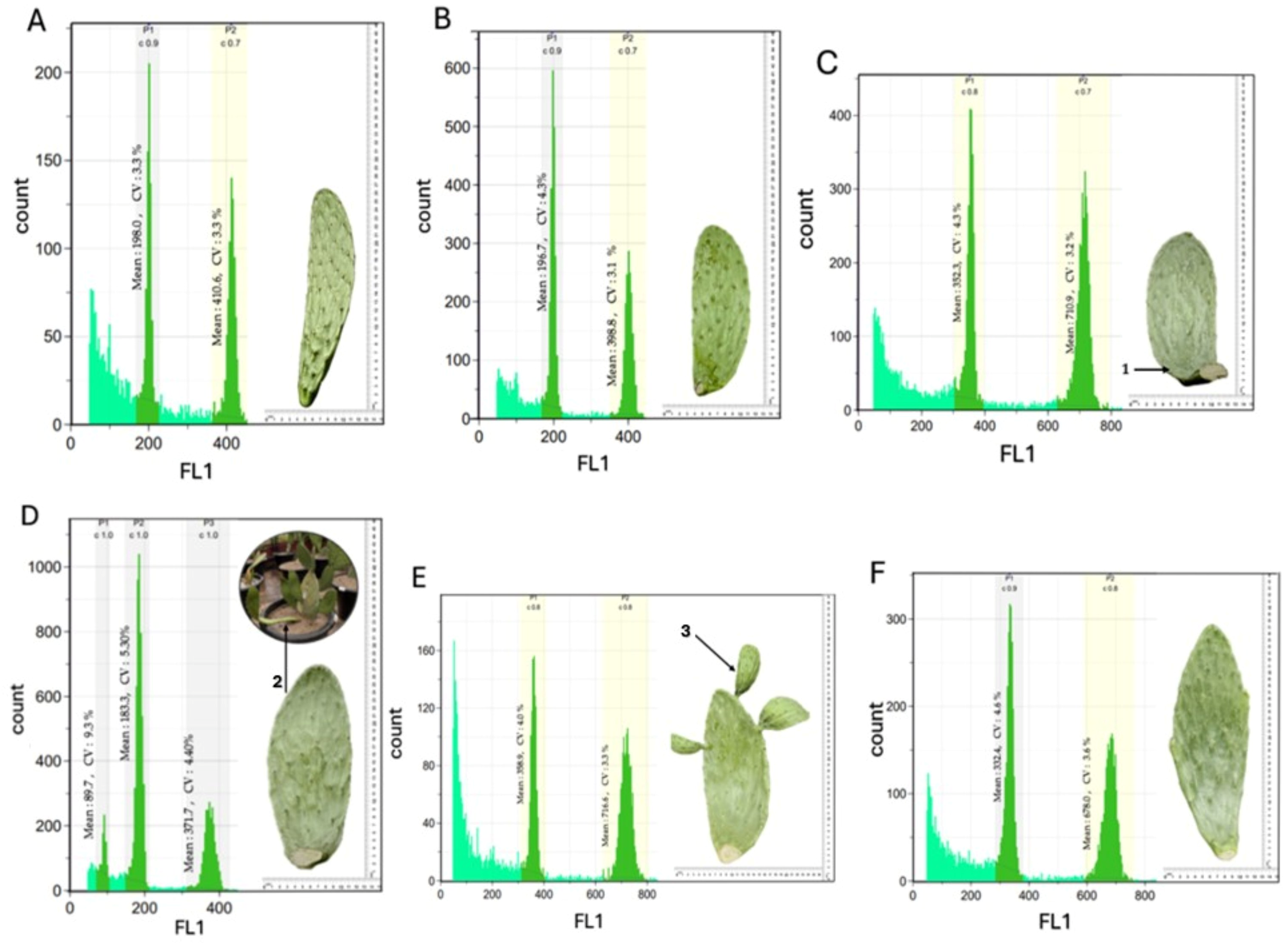
Flow cytometric analysis was performed to determine the ploidy status of the treated cladodes. As shown in Figure 1A–F, the samples could be classified into diploid (2n = 2x), putative tetraploid (2n = 4x), and cytochimeric groups based on their G1 peak positions and relative fluorescence patterns. The diploid control plants exhibited a mean G1 peak channel position of 197.35 ± 0.92 (Figure 1A,B), corresponding to a DNA Index (DI) of 1.00 ± 0.00. The plants classified as tetraploid exhibited a higher fluorescence G1 peak with a mean channel position of 347.87 ± 13.80 (Figure 1C,E,F), corresponding to a mean DI of 1.76 ± 0.07. Although this value is slightly lower than the theoretical DI of 2.00 expected for a duplicated genome, such proportional deviations are commonly observed in plant flow cytometry. This slight reduction in fluorescence intensity can be attributed to increased chromatin condensation in polyploid nuclei, potential endopolyploid background, or minor stoichiometric fluorochrome quenching caused by residual cytosolic secondary metabolites (such as mucilage and polyphenols) binding to DAPI despite the inclusion of PVP in the isolation buffer. Nevertheless, the distinct rightward shift in the G1 peak and the stable single peak profiles confirmed successful genome duplication. Cytochimeric individuals displayed three distinct fluorescence peaks at mean channel positions of 89.7, 183.3, and 371.7 (Figure 1D), corresponding to DI values of 0.45, 0.93, and 1.88, respectively. The coexistence of multiple distinct fluorescence populations within the same sample was therefore interpreted as evidence of cytological chimerism/mixoploidy. The corresponding histograms showed acceptable coefficients of variation (CV < 8%). In terms of morphology, larger tetraploid plants were obtained, as is typically observed in polyploid plants. This study found that cytochimeric cladodes exhibited distinct cytological chimeric characteristics (Figure 1D, Arrow 2), whereas tetraploid cladodes produced multiple newly developing cladodes on a single plant (Figure 1E, Arrow 3). Regarding phytotoxicity and induction efficiency, colchicine exposure exhibited a dose- and duration-dependent phytotoxic effect on plant survival (Table 1). While control groups (TA and TB) maintained 100% survival, survival rates significantly declined ( p < 0.05 ) to 91.67% and 75.00% under 0.5% colchicine (TC and TD) and reached a minimum of 58.34–58.35% under 1.0% colchicine treatments (TE and TF). However, despite this physiological cost, treatments with 1.0% colchicine demonstrated high polyploid induction efficiency among surviving individuals, promoting superior vegetative development, including significantly greater plant height (up to 36.33 ± 0.69 cm in TF), enhanced stem diameter (up to 16.62 ± 1.44 cm in TE), and increased proliferation of new cladodes (1.75–2.25 per plant) compared to controls.
Colchicine is a widely utilized antimitotic agent for the induction of polyploidy in plants [29]. Its effectiveness depends on the optimization of key parameters, including concentration, exposure duration, explant type, and method of application, in order to achieve a balance between induction efficiency and plant viability [30]. The optimal conditions for colchicine-induced polyploidization vary among plant species, reflecting differences in physiological and genetic responsiveness [31]. In the present study, the occurrence of chimeric mixoploid tissues suggests partial genome duplication induced by colchicine treatment. Such mixoploidy may arise from incomplete chromosome doubling during mitotic inhibition. Furthermore, tetraploid plants exhibited enhanced growth performance, characterized by increased biomass accumulation, larger organ size, and the production of multiple cladodes [32]. These traits are of particular significance, as they may contribute to improved yield potential and resource use efficiency.

3.2. Induction of Spineless Phenotype

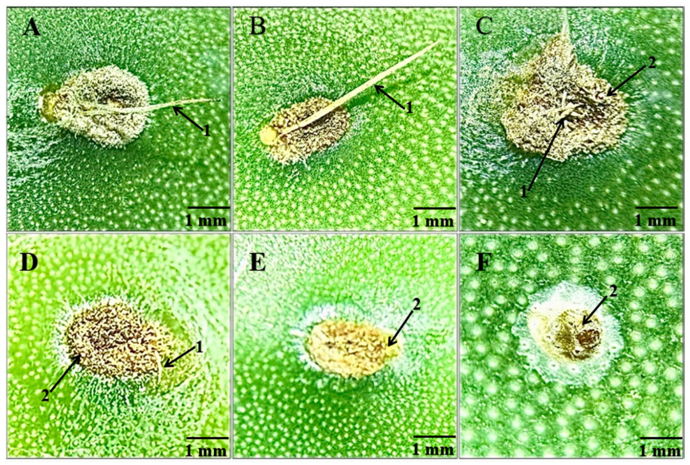
As shown in Figure 2, qualitative morphological observations indicated that colchicine concentration and exposure duration visibly induced spine reduction and spinelessness in the areoles of OFI cladodes. A visual reduction in spine length on the cladodes was first observed at a colchicine concentration of 0.5% with a 24 h exposure (Figure 2C), with progressively shorter spines evident under higher treatment intensities (Figure 2D–F). The spineless phenotype, a distinct qualitative trait characterized by the absolute absence of spines while maintaining normal areole structure, was achieved at a higher concentration of 1.0% colchicine with a 48 h exposure (Figure 2F). This qualitative screening provides a fundamental baseline for selecting a desirable spineless phenotype in early developmental stages following polyploidization.
In OFI, areoles and spines originate from lateral meristematic tissues on the surface of cladodes and represent modified leaf structures formed during shoot ontogeny [33]. The presence of spines constitutes a major limitation for agricultural, food, and industrial utilization, as they can cause mechanical injury, skin irritation, and persistent discomfort due to their ease of detachment and tendency to embed in tissues [34,35,36]. The spineless phenotype observed in this study following colchicine treatment is likely associated with disruptions in early shoot development. These morphological alterations suggest that colchicine-induced changes may interfere with meristematic activity and organ differentiation, thereby suppressing spine formation [23].
The development of spineless or reduced-spine OFI may provide practical advantages for handling, harvesting, and postharvest processing. These traits may also increase the suitability of OFI for utilization as a fresh vegetable crop, livestock fodder, and functional food resource, thereby supporting its potential agricultural and commercial value.

3.3. Stomatal Characteristics

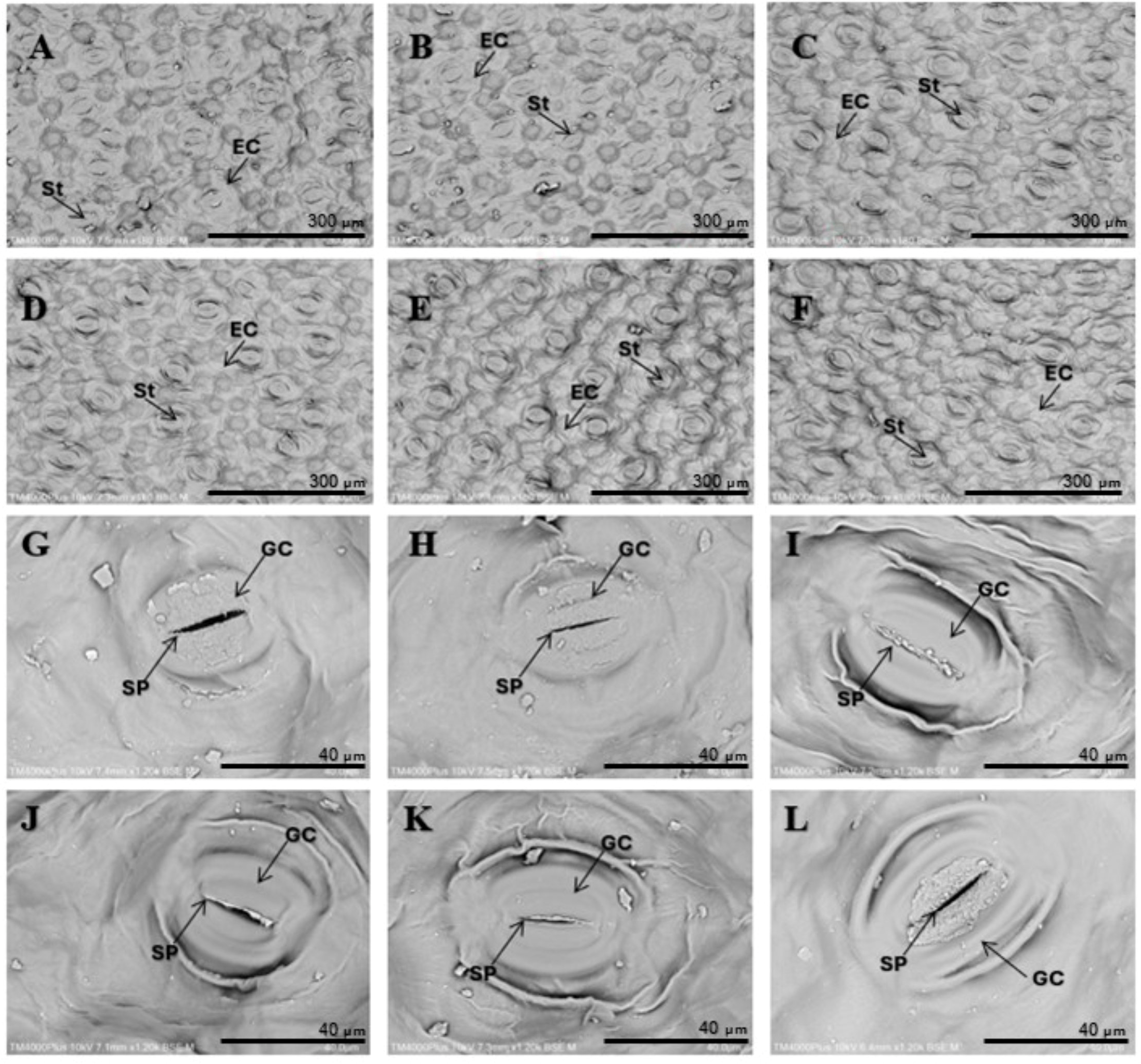
Significant differences in density of stomatal guard cells (DSG) and length of stomatal guard cells (LSG) were observed among treatments and ploidy levels (Table 2). In general, LSG increases with polyploidization, whereas DSG exhibits an inverse relationship [37,38,39]. In our study, LSG in TE increased significantly ( p < 0.05 ) compared to that of the control (31.2% and 27.9% higher than TA and TB, respectively), consistent with previous reports. Concurrently, tetraploid plants in TD, TE, and TF exhibited significantly lower DSG than diploid controls. These epidermal variations are directly linked to the structural expansion of underlying tissues; specifically, the increased genome copy number induces the ‘gigas effect’, leading to enlarged mesophyll cell volume and altered internal tissue packing within the cladode. Consequently, larger guard cells serve as a reliable proxy reflecting overall cellular expansion and increased mesophyll volume in induced tetraploids [24]. However, treatment TC (0.5% colchicine for 24 h) showed no significant difference in DSG compared to diploid plants, maintaining a relatively higher DSG approaching control levels. These observations were further supported by scanning electron microscopy (SEM) (Figure 3). The micrographs clearly illustrate key surface structures, including stomata, epidermal cells, guard cells, and stomatal pores. While DSG decreased in most tetraploid treatments (TD–TF), the presence of treatment TC demonstrates that DSG was not uniformly negatively correlated with ploidy level across all colchicine treatments applied in induced tetraploids. Similar inconsistencies have been reported in Chinese chive (Allium tuberosum) by Yao et al. [40]. These findings suggest that the relationship between stomatal density and ploidy level is species-specific and may vary depending on genetic background or developmental regulation.
Furthermore, stomatal density has been associated with plant water use efficiency and drought tolerance, with lower stomatal density generally linked to reduced transpiration and improved water retention [41,42]. In cases where DSG did not decrease, such as in treatment TC, it is possible that other factors, such as stomatal size or stomatal regulation, play a more significant role in determining physiological performance in the induced tetraploids.

3.4. Bioactive Compounds

Significant differences in bioactive compounds were observed among the experimental groups (Table 3). Total phenolic content (TPC) ranged from 367.29 to 1241.52 mg GAE/100 g DW, with the highest value recorded in the TF treatment, representing approximately 3.4-fold higher than the control (TA). In contrast, total flavonoid content (TFC) was markedly enhanced in the TC treatment, reaching 4932.02 mg QE/100 g DW, which was approximately 2.9-fold greater than the control. Antioxidant activity also varied significantly among treatments. The TC treatment exhibited the strongest DPPH radical scavenging activity (354.56 mg AA equivalent/100 g DW) and ferric-reducing antioxidant power (1947.84 mg FeSO4/100 g DW), corresponding to approximately 1.2- and 1.1-fold increases over the control, respectively. In contrast, the TB treatment showed the lowest antioxidant activity despite exhibiting higher TPC and TFC than the control. To further clarify these relationships, a correlation analysis was performed (Table S1, Figure S1). TFC showed strong positive correlations with DPPH (p = 0.00011) and FRAP (p = 0.00065), whereas TPC showed no significant correlation with DPPH (p = 0.174) or FRAP (p = 0.311), confirming that flavonoids are the main contributors to antioxidant activity.
The enhanced antioxidant activity observed in the TC treatment coincided with a substantial increase in flavonoid accumulation, suggesting that flavonoids may have contributed to the antioxidant capacity of the cladode extracts. Phenolic compounds can contribute to antioxidant activity through their electron- and hydrogen-donating properties; however, the relationship between TPC, TFC, and antioxidant activity was not strictly proportional among treatments. For example, although TF exhibited the highest TPC, its DPPH and FRAP values were significantly lower than those of TC. This observation indicates that antioxidant capacity may depend not only on the total amount of phenolic compounds but also on the composition, chemical structure, and relative abundance of individual phenolic and flavonoid constituents. Differences in other antioxidant constituents not quantified in the present study may also have contributed to the observed responses [43,44,45,46]. Cinnamic acid is subsequently metabolized through reactions involving cinnamate 4-hydroxylase (C4H) and 4-coumarate-CoA ligase (4CL), generating intermediates such as p-coumaroyl-CoA that serve as precursors for various phenolic metabolites [45,46,47]. Within the flavonoid branch, chalcone synthase (CHS) catalyzes the condensation of p-coumaroyl-CoA with malonyl-CoA to form naringenin chalcone, which is subsequently converted by chalcone isomerase (CHI) and other downstream enzymes into diverse flavonoid compounds [46,48]. Thus, variation in the regulation or activity of enzymes associated with this pathway, including PAL and CHS, represents one potential biochemical basis for differences in phenolic and flavonoid accumulation.
Polyploidization may also influence secondary metabolism through changes in gene dosage and transcriptional regulation. Chromosome doubling increases gene copy number and can be accompanied by transcriptional and epigenetic reorganization; consequently, changes in gene dosage do not necessarily result in proportional changes in gene expression [49,50,51]. Such genomic changes may affect the regulation of metabolic pathways and thereby alter the abundance or composition of specialized metabolites. Previous studies have reported changes in phenolic, flavonoid, terpenoid, and other secondary metabolites following induced polyploidization, although the direction and magnitude of these responses vary among plant species, genotypes, metabolite classes, and experimental conditions [16,52,53]. Similarly, colchicine-induced polyploidization in Wedelia chinensis was associated with increased phenolic and flavonoid accumulation and enhanced antioxidant activity [54]. Within this context, the contrasting responses of TC and TF are noteworthy. TC (0.5% colchicine for 24 h) exhibited the highest TFC together with the highest DPPH and FRAP values, whereas TF (1% colchicine for 48 h) exhibited the highest TPC but substantially lower TFC and antioxidant activities than TC. These treatment-dependent responses suggest that colchicine concentration and exposure duration may be associated with differential changes in phenolic and flavonoid accumulation rather than a uniform increase in all classes of phenylpropanoid-derived metabolites. One possible interpretation, based on the established organization of the phenylpropanoid pathway, is that different treatments may influence the relative allocation of metabolites among its downstream branches. However, this possibility cannot be confirmed from the present data because individual phenolic and flavonoid compounds, pathway-specific metabolic flux, gene expression, and enzyme activities were not determined.
Therefore, the involvement of PAL, CHS, and polyploidy-associated transcriptional regulation discussed above should be regarded as a literature-supported hypothesis rather than a mechanism demonstrated by the present study. Further studies integrating ploidy verification with targeted gene expression or transcriptomic analyses of PAL, C4H, 4CL, CHS, CHI, F3H, and FLS, together with enzyme activity assays and targeted metabolomic profiling, would be required to determine whether the observed changes in phenolic and flavonoid accumulation are associated with altered regulation of the phenylpropanoid pathway.

4. Conclusions

This study established an efficient in vivo protocol for polyploidy induction in OFI using colchicine. Colchicine treatments at 0.5% for 24 h (TC), 1.0% for 24 h (TE), and 1.0% for 48 h (TF) successfully induced tetraploid plants, resulting in significant morphological modifications, including increased cladode size and altered stomatal architecture. Higher colchicine concentrations and extended exposure durations (specifically 1.0% for 48 h) reduced plant survival compared to the control. Anatomically, guard cell length (LSG) was significantly greater in tetraploids than in diploid controls, whereas stomatal density (DSG) was generally lower, except in treatment TC (0.5%, 24 h), which maintained a DSG comparable to the control.
At the biochemical level, induced tetraploids exhibited enhanced accumulation of secondary metabolites, dependent on treatment conditions. Notably, TC yielded the highest total flavonoid content along with superior antioxidant capacities (DPPH and FRAP), whereas TF exhibited the highest total phenolic content. To build upon these findings, future work should incorporate standardized quantitative characterizations of spine traits including spine length, spine density per areole, and the proportion of spineless areoles to evaluate the degree and longitudinal stability of spine reduction across vegetative generations. Such quantitative profiling will provide a more objective evaluation of the degree and stability of spine reduction, thereby facilitating precise comparisons among colchicine treatments as well as across subsequent generations and clonally propagated lineages. Overall, these results demonstrate that colchicine-induced polyploidization is a viable breeding strategy for generating novel OFI germplasms featuring spineless phenotypes, enlarged morpho-anatomical traits, and enriched bioactivity profiles for future crop improvement programs.

Supplementary Materials

The following supporting information can be downloaded at: https://www.mdpi.com/article/10.3390/horticulturae12081044/s1, Figure S1: Correlation analysis between bioactive compounds and antioxidant activities; Table S1: Correlation coefficients between bioactive compounds and antioxidant activities.

Author Contributions

Conceptualization, K.B.; methodology, K.B., N.B., S.S. and P.Y.; formal analysis, K.B., N.B., S.S. and P.Y.; investigation, K.B. and S.S.; resources, K.B.; data curation, K.B., N.B., S.S. and P.Y.; writing—original draft preparation, N.B.; writing—review and editing, N.B., S.S., P.Y.; visualization, N.B.; supervision, K.B.; project administration, K.B.; funding acquisition, K.B. All authors have read and agreed to the published version of the manuscript.

Funding

This research project was financially supported by Mahasarakham University.

Data Availability Statement

The data presented in this study are available on request from the corresponding author. The data are not publicly available because they are part of an ongoing research dataset and require appropriate contextual information for interpretation. The underlying data supporting the findings of this study can be made available by the corresponding author upon reasonable request.

Acknowledgments

This research project was financially supported by Mahasarakham University. The authors thank the Laboratory Equipment Center of Mahasarakham University for their cooperation and scientific assistance. During manuscript preparation, QuillBot (web-based version, https://quillbot.com/, accessed 16 August 2026) was used solely for language-related assistance, including paraphrasing and grammar correction to improve the clarity and readability of the English text. The tool was not used for experimental design, data collection, data generation, statistical analysis, or scientific interpretation of the results. All AI-assisted revisions were subsequently reviewed and verified by the authors, who take full responsibility for the accuracy and integrity of the final manuscript.

Conflicts of Interest

The authors declare no conflicts of interest.

References

  1. Moss, R.H.; Edmonds, J.A.; Hibbard, K.A.; Manning, M.R.; Rose, S.K.; van Vuuren, D.P.; Carter, T.R.; Emori, S.; Kainuma, M.; Kram, T.; et al. The next Generation of Scenarios for Climate Change Research and Assessment. Nature 2010, 463, 747–756. [Google Scholar] [CrossRef] [Scilit] [PubMed]
  2. Dubeux, J.C.B.; Santos, M.V.F.D.; Cunha, M.V.D.; Santos, D.C.D.; Souza, R.T.D.A.; Mello, A.C.L.D.; Souza, T.C.D. Cactus (Opuntia and Nopalea) Nutritive Value: A Review. Anim. Feed Sci. Technol. 2021, 275, 114890. [Google Scholar] [CrossRef] [Scilit]
  3. Katz, R.W. Sir Gilbert Walker and a Connection between El Niño and Statistics. Stat. Sci. 2002, 17, 97–112. [Google Scholar] [CrossRef] [Scilit]
  4. Soni, P.; Taewichit, C.; Salokhe, V.M. Energy Consumption and CO2 Emissions in Rainfed Agricultural Production Systems of Northeast Thailand. Agric. Syst. 2013, 116, 25–36. [Google Scholar] [CrossRef] [Scilit]
  5. He, D.; Wang, E.; Wang, J.; Robertson, M.J. Data Requirement for Effective Calibration of Process-Based Crop Models. Agric. For. Meteorol. 2017, 234–235, 136–148. [Google Scholar] [CrossRef] [Scilit]
  6. Wongkhunkaew, P. Effect of Climate Variability on Rice Production of Northeastern Thailand. Int. J. GEOMATE 2020, 18, 129–134. [Google Scholar] [CrossRef] [Scilit]
  7. United Nations. The Sustainable Development Goals Report; United Nations: New York, NY, USA, 2019. [Google Scholar]
  8. Mondragón-Jacobo, C.; Pérez-González, S. Cactus (Opuntia spp.) as Forage; Food & Agriculture Org: Rome, Italy, 2011; Volume 169. [Google Scholar]
  9. Prisa, D. Opuntia ficus-indica the Key Plant in Climate Change: Characteristics, Cultivation and Uses. GSC Biol. Pharm. Sci. 2021, 17, 94–105. [Google Scholar] [CrossRef] [Scilit]
  10. Sipango, N.; Ravhuhali, K.E.; Sebola, N.A.; Hawu, O.; Mabelebele, M.; Mokoboki, H.K.; Moyo, B. Prickly Pear (Opuntia spp.) as an Invasive Species and a Potential Fodder Resource for Ruminant Animals. Sustainability 2022, 14, 3719. [Google Scholar] [CrossRef] [Scilit]
  11. Vastolo, A.; Calabrò, S.; Cutrignelli, M.I.; Raso, G.; Todaro, M. Silage of Prickly Pears (Opuntia spp.) Juice By-Products. Animals 2020, 10, 1716. [Google Scholar] [CrossRef] [Scilit] [PubMed]
  12. Daniloski, D.; D’Cunha, N.M.; Speer, H.; McKune, A.J.; Alexopoulos, N.; Panagiotakos, D.B.; Petkoska, A.T.; Naumovski, N. Recent Developments on Opuntia spp., Their Bioactive Composition, Nutritional Values, and Health Effects. Food Biosci. 2022, 47, 101665. [Google Scholar] [CrossRef] [Scilit]
  13. Alary, V.; Nefzaoui, A.; Jemaa, M.B. Promoting the Adoption of Natural Resource Management Technology in Arid and Semi-Arid Areas: Modelling the Impact of Spineless Cactus in Alley Cropping in Central Tunisia. Agric. Syst. 2007, 94, 573–585. [Google Scholar] [CrossRef] [Scilit]
  14. Yahia, E.M.; Sáenz, C. Cactus Pear (Opuntia Species). In Postharvest Biology and Technology of Tropical and Subtropical Fruits; Elsevier: Amsterdam, The Netherlands, 2011; pp. 290–331. [Google Scholar]
  15. Rodrigues, A.M.; Pitacas, F.I.; Reis, C.M.G.; Blasco, M. Nutritional Value of Opuntia ficus-indica Cladodesfrom Portuguese Ecotypes. Bulg. J. Agric. Sci. 2016, 22, 40–45. [Google Scholar]
  16. Boontiang, K.; Chaikwang, T.; Chumroenphat, T.; Siriamornpun, S. Characterization of Bioactive Composition and Structural Properties of Epidermis and Cortex Tissues in Edible Cactus [Opuntia ficus-indica (L.) Mill.] Cladodes at Different Maturity Stages. Foods 2026, 15, 2520. [Google Scholar] [CrossRef] [Scilit] [PubMed]
  17. Ramsey, J.; Schemske, D.W. Pathways, Mechanisms, and Rates of Polyploid Formation in Flowering Plants. Annu. Rev. Ecol. Syst. 1998, 29, 467–501. [Google Scholar] [CrossRef] [Scilit]
  18. Mo, L.; Chen, J.; Lou, X.; Xu, Q.; Dong, R.; Tong, Z.; Huang, H.; Lin, E. Colchicine-Induced Polyploidy in Rhododendron fortunei Lindl. Plants 2020, 9, 424. [Google Scholar] [CrossRef] [Scilit] [PubMed]
  19. Liu, R.; Gao, C.; Jin, J.; Wang, Y.; Jia, X.; Ma, H.; Zhang, Y.; Zhang, H.; Qi, B.; Xu, J. Induction and Identification of Tetraploids of Pear Plants (Pyrus bretschneideri and Pyrus betulaefolia). Sci. Hortic. 2022, 304, 111322. [Google Scholar] [CrossRef] [Scilit]
  20. Eng, W.-H.; Ho, W.-S. Polyploidization Using Colchicine in Horticultural Plants: A Review. Sci. Hortic. 2019, 246, 604–617. [Google Scholar] [CrossRef] [Scilit]
  21. Dhooghe, E.; Van Laere, K.; Eeckhaut, T.; Leus, L.; Van Huylenbroeck, J. Mitotic Chromosome Doubling of Plant Tissues in Vitro. Plant Cell Tissue Organ Cult. 2011, 104, 359–373. [Google Scholar] [CrossRef] [Scilit]
  22. Wu, H.; Jiang, L.; Li, J.; Lu, M.; An, H. Polyploid Induction and Identification of Rosa roxburghii f. eseiosa. Plants 2023, 12, 2194. [Google Scholar] [CrossRef] [Scilit] [PubMed]
  23. Boonyawiwat, N.; Siritrakulsak, T.; Senakun, C.; Boontiang, K. Effect of Colchicine on Morphological and Anatomical Traits of Gymnocalycium mihanovichii (Frič & Gürke) Britton & Rose. Trends Sci. 2023, 20, 6597. [Google Scholar] [CrossRef] [Scilit]
  24. Tel-Zur, N.; Dudai, M.; Raveh, E.; Mizrahi, Y. In Situ Induction of Chromosome Doubling in Vine Cacti (Cactaceae). Sci. Hortic. 2011, 129, 570–576. [Google Scholar] [CrossRef] [Scilit]
  25. Boonarsa, P.; Bunyatratchata, A.; Chumroenphat, T.; Thammapat, P.; Chaikwang, T.; Siripan, T.; Li, H.; Siriamornpun, S. Nutritional Quality, Functional Properties, and Biological Characterization of Watermeal (Wolffia globosa). Horticulturae 2024, 10, 1171. [Google Scholar] [CrossRef] [Scilit]
  26. Thammapat, P.; Meeso, N.; Siriamornpun, S. Effects of the Traditional Method and an Alternative Parboiling Process on the Fatty Acids, Vitamin E, γ-Oryzanol and Phenolic Acids of Glutinous Rice. Food Chem. 2016, 194, 230–236. [Google Scholar] [CrossRef] [Scilit] [PubMed]
  27. Wanyo, P.; Kaewseejan, N.; Meeso, N.; Siriamornpun, S. Bioactive Compounds and Antioxidant Properties of Different Solvent Extracts Derived from Thai Rice By-Products. Appl. Biol. Chem. 2016, 59, 373–384. [Google Scholar] [CrossRef] [Scilit]
  28. Butsat, S.; Siriamornpun, S. Antioxidant Capacities and Phenolic Compounds of the Husk, Bran and Endosperm of Thai Rice. Food Chem. 2010, 119, 606–613. [Google Scholar] [CrossRef] [Scilit]
  29. Narukulla, V.; Lahane, Y.; Fiske, K.; Pandey, S.; Ziogas, V. Induction of Polyploidy in Citrus Rootstocks through In Vitro Colchicine Treatment of Seed-Derived Explants. Agronomy 2023, 13, 1442. [Google Scholar] [CrossRef] [Scilit]
  30. Salma, U.; Kundu, S.; Mandal, N. Artificial Polyploidy in Medicinal Plants: Advancement in the Last Two Decades and Impending Prospects. J. Crop Sci. Biotechnol. 2017, 20, 9–19. [Google Scholar] [CrossRef] [Scilit]
  31. Singh, B.; Yun, S.; Gil, Y.; Park, M.-H. The Role of Colchicine in Plant Breeding. Int. J. Mol. Sci. 2025, 26, 6743. [Google Scholar] [CrossRef] [Scilit] [PubMed]
  32. Kumar, K.; Thakur, L.; Thakur, M.; Bhardwaj, V.; Gupta, M. In Vitro Polyploidy Induction in Ginger (Zingiber officinale Rosc.) Cv. Himgiri: Morpho-Physiological, Biochemical, and Molecular Evaluation of Regenerants. In Vitro Cell. Dev. Biol.-Plant 2024, 60, 693–710. [Google Scholar] [CrossRef] [Scilit]
  33. Ahmed, F.A.; Ibrahim, M.A.; El-Azab, M.M.; Fahmy, W.G.E.; Fahmy, D.M. A Review: Opuntia ficus-indica as a Source of Bioactive Compound Ingredients for Functional Foods, Nutrition, Human Disease and Health. Univ. J. Pharm. Res. 2024, 9. [Google Scholar] [CrossRef] [Scilit]
  34. Xia, Z.; Gong, G.; Huang, R.; Goossens, T.; Lemâle, O.; Cardoso, D.; Mallmann, B.; Zhao, L.; Wei, J.; Deng, J. Butyric Acid-Based Products, Alone or in Combination with Hydroxy-Selenomethionine, Improve Performance of Laying Hens in Post-Peak Period by Modulating Their Antioxidant, Metabolic and Immune Status. Poult. Sci. 2025, 104, 104840. [Google Scholar] [CrossRef] [Scilit] [PubMed]
  35. Humphries, T.; Campbell, S.; Florentine, S. Challenges Inherent in Controlling Prickly Pear Species; a Global Review of the Properties of Opuntia Stricta, Opuntia ficus-indica and Opuntia monacantha. Plants 2022, 11, 3334. [Google Scholar] [CrossRef] [Scilit] [PubMed]
  36. Corrales-García, J.; González-Martínez, P. Effect of Gibberellic Acid and (2-Chloroethane) Phosphonic Acid on Glochid Abscission in Cactus Pear Fruit (Opuntia amyclaea Tenore). Postharvest Biol. Technol. 2001, 22, 151–157. [Google Scholar] [CrossRef] [Scilit]
  37. Lourkisti, R.; Oustric, J.; Quilichini, Y.; Froelicher, Y.; Herbette, S.; Morillon, R.; Berti, L.; Santini, J. Improved Response of Triploid Citrus Varieties to Water Deficit Is Related to Anatomical and Cytological Properties. Plant Physiol. Biochem. 2021, 162, 762–775. [Google Scholar] [CrossRef] [Scilit] [PubMed]
  38. Yuan, S.; Liu, Y.; Fang, Z.; Yang, L.; Zhuang, M.; Zhang, Y.; Sun, P. Study on the Relationship Between the Ploidy Level of Microspore-Derived Plants and the Number of Chloroplast in Stomatal Guard Cells in Brassica Oleracea. Agric. Sci. China 2009, 8, 939–946. [Google Scholar] [CrossRef] [Scilit]
  39. Ye, Y.M.; Tong, J.; Shi, X.P.; Yuan, W.; Li, G.R. Morphological and Cytological Studies of Diploid and Colchicine-Induced Tetraploid Lines of Crape Myrtle (Lagerstroemia indica L.). Sci. Hortic. 2010, 124, 95–101. [Google Scholar] [CrossRef] [Scilit]
  40. Yao, P.-Q.; Chen, J.-H.; Ma, P.-F.; Xie, L.-H.; Cheng, S.-P. Stomata Variation in the Process of Polyploidization in Chinese Chive (Allium tuberosum). BMC Plant Biol. 2023, 23, 595. [Google Scholar] [CrossRef] [Scilit] [PubMed]
  41. Caine, R.S.; Yin, X.; Sloan, J.; Harrison, E.L.; Mohammed, U.; Fulton, T.; Biswal, A.K.; Dionora, J.; Chater, C.C.; Coe, R.A.; et al. Rice with Reduced Stomatal Density Conserves Water and Has Improved Drought Tolerance under Future Climate Conditions. New Phytol. 2019, 221, 371–384. [Google Scholar] [CrossRef] [Scilit] [PubMed]
  42. Dunn, J.; Hunt, L.; Afsharinafar, M.; Meselmani, M.A.; Mitchell, A.; Howells, R.; Wallington, E.; Fleming, A.J.; Gray, J.E. Reduced Stomatal Density in Bread Wheat Leads to Increased Water-Use Efficiency. J. Exp. Bot. 2019, 70, 4737–4748. [Google Scholar] [CrossRef] [Scilit] [PubMed]
  43. Ali, M.B.; McNear, D.H. Induced Transcriptional Profiling of Phenylpropanoid Pathway Genes Increased Flavonoid and Lignin Content in Arabidopsisleaves in Response to Microbial Products. BMC Plant Biol. 2014, 14, 84. [Google Scholar] [CrossRef] [Scilit] [PubMed]
  44. Dong, N.; Lin, H. Contribution of Phenylpropanoid Metabolism to Plant Development and Plant–Environment Interactions. J. Integr. Plant Biol. 2021, 63, 180–209. [Google Scholar] [CrossRef] [Scilit] [PubMed]
  45. Lee, H.; Lee, S.B.; Park, S.; Song, J.; Kim, B.-G. Biochemical Evaluation of Molecular Parts for Flavonoid Production Using Plant Synthetic Biology. Front. Plant Sci. 2025, 16, 1528122. [Google Scholar] [CrossRef] [Scilit] [PubMed]
  46. Wang, X.; Hu, H.; Wu, Z.; Fan, H.; Wang, G.; Chai, T.; Wang, H. Tissue-Specific Transcriptome Analyses Reveal Candidate Genes for Stilbene, Flavonoid and Anthraquinone Biosynthesis in the Medicinal Plant Polygonum Cuspidatum. BMC Genom. 2021, 22, 353. [Google Scholar] [CrossRef] [Scilit] [PubMed]
  47. Nabavi, S.M.; Šamec, D.; Tomczyk, M.; Milella, L.; Russo, D.; Habtemariam, S.; Suntar, I.; Rastrelli, L.; Daglia, M.; Xiao, J.; et al. Flavonoid Biosynthetic Pathways in Plants: Versatile Targets for Metabolic Engineering. Biotechnol. Adv. 2020, 38, 107316. [Google Scholar] [CrossRef] [Scilit] [PubMed]
  48. Liu, W.; Feng, Y.; Yu, S.; Fan, Z.; Li, X.; Li, J.; Yin, H. The Flavonoid Biosynthesis Network in Plants. Int. J. Mol. Sci. 2021, 22, 12824. [Google Scholar] [CrossRef] [Scilit] [PubMed]
  49. Doyle, J.J.; Coate, J.E. Polyploidy, the Nucleotype, and Novelty: The Impact of Genome Doubling on the Biology of the Cell. Int. J. Plant Sci. 2019, 180, 1–52. [Google Scholar] [CrossRef] [Scilit] [PubMed]
  50. Otto, S.P. The Evolutionary Consequences of Polyploidy. Cell 2007, 131, 452–462. [Google Scholar] [CrossRef] [Scilit] [PubMed]
  51. Van De Peer, Y.; Mizrachi, E.; Marchal, K. The Evolutionary Significance of Polyploidy. Nat. Rev. Genet 2017, 18, 411–424. [Google Scholar] [CrossRef] [Scilit] [PubMed]
  52. Madani, H.; Escrich, A.; Hosseini, B.; Sanchez-Muñoz, R.; Khojasteh, A.; Palazon, J. Effect of Polyploidy Induction on Natural Metabolite Production in Medicinal Plants. Biomolecules 2021, 11, 899. [Google Scholar] [CrossRef] [Scilit] [PubMed]
  53. Zargoosh, Z.; Ghavam, M.; Bacchetta, G.; Tavili, A. Effects of Ecological Factors on the Antioxidant Potential and Total Phenol Content of Scrophularia Striata Boiss. Sci. Rep. 2019, 9, 16021. [Google Scholar] [CrossRef] [Scilit] [PubMed]
  54. Tsai, Y.-T.; Chen, P.-Y.; To, K.-Y. Induction of Polyploidy and Metabolic Profiling in the Medicinal Herb Wedelia Chinensis. Plants 2021, 10, 1232. [Google Scholar] [CrossRef] [Scilit] [PubMed]
Figure 1. Flow cytometric histograms and corresponding cladode morphological alterations of OFI evaluated at 180 days post-treatment following colchicine exposure: (A) TA (control, 0.0% colchicine for 24 h); (B) TB (control, 0.0% colchicine for 48 h); (C) TC (0.5% colchicine for 24 h); (D) TD (0.5% colchicine for 48 h); (E) TE (1.0% colchicine for 24 h); and (F) TF (1.0% colchicine for 48 h). In flow cytometry histograms, the x-axis represents green fluorescence intensity (FL1, arbitrary units) and the y-axis represents nucleus count. Insets show representative cladode morphologies from three independent blocks. Each treatment consisted of 20 cladodes per block. Arrow 1 indicates broadened and thickened cladodes in colchicine-induced plants; Arrow 2 highlights a chimeric/mixoploid individual displaying transitional morphological traits during plant recovery; and Arrow 3 indicates enhanced shoot proliferation with multiple newly emerged cladodes.
Horticulturae 12 01044 g001
Figure 2. Comparison of areole and spine micro morphological variations in newly emerged OFI cladodes evaluated at 180 days post-treatment: (A) TA (control, 0.0% colchicine for 24 h); (B) TB (control, 0.0% colchicine for 48 h); (C) TC (0.5% colchicine for 24 h); (D) TD (0.5% colchicine for 48 h); (E) TE (1.0% colchicine for 24 h); and (F) TF (1.0% colchicine for 48 h). Arrow 1: Spine morphology, showing distinct developmental differences and reduced presence in treated groups. Arrow 2: Areole characteristics. Scale bars = 1 mm.
Horticulturae 12 01044 g002
Figure 3. Stomatal characteristics of OFI under colchicine treatments (TA–TF) evaluated at 180 days post-treatment. (AF) Light microscopy micrographs and (GL) scanning electron microscopy (SEM) micrographs corresponding to TA (control, 0.0% for 24 h) (A,G), TB (control, 0.0% for 48 h) (B,H), TC (0.5% for 24 h) (C,I), TD (0.5% for 48 h) (D,J), TE (1.0% for 24 h) (E,K), and TF (1.0% for 48 h) (F,L). Abbreviations: St: stomata; EC: epidermal cells; GC: guard cells; SP: stomatal pore. Scale bars: (AF) = 300 µm; (GL) = 40 µm. Micrographs represent consistent observations from three independent replications (n = 20).
Horticulturae 12 01044 g003
Table 1. Effects of colchicine concentration and exposure duration on survival rate and morphological characteristics of OFI evaluated at 180 days post-treatment.
TreatmentColchicine Concentration (%)Time of TreatmentSurvival
Rate (%)
Plant Height
(cm)
Stem Diameter
(cm)
No. of
New Cladodes
TA0.024100.00 ± 0.00 a27.70 ± 1.09 c8.70 ± 0.98 d0
TB48100.00 ± 0.00 a22.06 ± 0.39 e9.12 ± 0.53 d0
TC0.52491.67 ± 16.67 ab23.95 ± 0.80 d14.30 ± 0.16 b0.75 ± 0.50 b
TD4875.00 ± 16.67 bc27.90 ± 0.51 c12.03 ± 0.61 c0
TE1.02458.34 ± 16.67 c34.29 ± 0.41 b16.62 ± 1.44 a2.25 ± 0.96 a
TF4858.35 ± 16.67 c36.33 ± 0.69 a14.25 ± 0.84 b1.75 ± 1.50 ab
Values are expressed as mean ± standard deviation (SD) of three replications (n = 12 total plants per treatment, 3 plants per block). Means within the same column followed by different lowercase letters (a–e) are statistically significantly different according to Analysis of Variance (ANOVA) followed by Duncan’s Multiple Range Test (DMRT) at p < 0.05. Survival rate was recorded at 4 weeks post-treatment, whereas morphological parameters were evaluated at 180 days post-treatment. TA = 0.0% colchicine for 24 h (control); TB = 0.0% colchicine for 48 h (control); TC = 0.5% colchicine for 24 h; TD = 0.5% colchicine for 48 h; TE = 1.0% colchicine for 24 h; TF = 1.0% colchicine for 48 h. Identical standard deviation values for survival rates resulted from the discrete nature of survival data and equal sample size across treatments.
Table 2. Effect of colchicine treatments on stomatal characteristics of newly emerged OFI cladodes evaluated at 180 days post-treatment.
TreatmentDensity of Stomatal
Guard Cell (mm−2)
Length of Stomatal Guard Cell (µm)
TA24.75 ± 1.50 a36.20 ± 1.33 d
TB24.50 ± 1.29 a37.13 ± 2.87 d
TC23.25 ± 0.96 a40.20 ± 0.32 c
TD17.50 ± 0.58 b41.58 ± 0.79 b
TE17.75 ± 0.50 b47.48 ± 0.59 a
TF16.75 ± 0.50 b42.18 ± 0.38 b
Each value represents the mean ± SD of three independent experiments with at least 20 replicates per treatment. Means followed by different letters within the same column indicate statistically significant differences ( p < 0.05 ) according to Duncan’s Multiple Range Test (DMRT). TA = 0% colchicine for 24 h, TB = 0% colchicine for 48 h, TC = 0.5% colchicine for 24 h, TD = 0.5% colchicine for 48 h, TE = 1% colchicine for 24 h, and TF = 1% colchicine for 48 h.
Table 3. Effect of colchicine treatments on potential bioactive compounds and antioxidant activities in newly emerged cladodes of OFI evaluated at 180 days post-treatment.
TreatmentTPC
(mg GAE/
100 g DW)
TFC
(mg QE/
100 g DW)
DPPH
(mg AA/
100 g DW)
FRAP
(mg FeSO4/
100 g DW)
TA367.29 ± 7.37 e1693.25 ± 75.69 d290.76 ± 4.33 b1760.99 ± 5.86 b
TB386.76 ± 13.91 e2382.75 ± 91.07 b224.50 ± 2.66 f1421.18 ± 23.54 c
TC1069.47 ± 18.70 b4932.02 ± 47.70 a354.56 ± 4.21 a1947.84 ± 16.47 a
TD702.44 ± 3.84 c1893.53 ± 51.85 c262.31 ± 11.27 c1416.08 ± 15.97 c
TE428.20 ± 7.86 d1897.73 ± 40.12 c252.52 ± 3.49 d1354.15 ± 30.95 d
TF1241.52 ± 12.16 a1845.61 ± 20.06 c244.22 ± 2.32 e1446.81 ± 9.44 c
Values are expressed as mean ± standard deviation (SD) of three independent replications ( n = 12 total plants per treatment, 4 plants per block). Means with different lowercase letters (a–f) within the same column are statistically significantly different according to Analysis of Variance (ANOVA) followed by Duncan’s Multiple Range Test (DMRT) at p < 0.05 . TA = 0.0% colchicine for 24 h (control); TB = 0.0% colchicine for 48 h (control); TC = 0.5% colchicine for 24 h; TD = 0.5% colchicine for 48 h; TE = 1.0% colchicine for 24 h; TF = 1.0% colchicine for 48 h. TPC = Total Phenolic Content; TFC = Total Flavonoid Content; DPPH = 2,2-diphenyl-1-picrylhydrazyl radical scavenging activity; FRAP = ferric reducing antioxidant power; GAE = gallic acid equivalent; QE = quercetin equivalent; AA = ascorbic acid equivalent; DW = dry weight.
Disclaimer/Publisher’s Note: The statements, opinions and data contained in all publications are solely those of the individual author(s) and contributor(s) and not of MDPI and/or the editor(s). MDPI and/or the editor(s) disclaim responsibility for any injury to people or property resulting from any ideas, methods, instructions or products referred to in the content.