Article

Exogenous 2-(3,4-Dichlorophenoxy) Trimethylamine (DCPTA) Alleviates Copper Toxicity in Cucumber Seedlings via Coordinated Regulation of Root Architecture, Cell Wall Composition, and Nitrogen Metabolism


College of Horticulture, Henan Agricultural University, Zhengzhou 450002, China
*
Authors to whom correspondence should be addressed.
Horticulturae 2026, 12(5), 549; https://doi.org/10.3390/horticulturae12050549
Submission received: 21 March 2026 / Revised: 26 April 2026 / Accepted: 28 April 2026 / Published: 29 April 2026

Abstract

The toxicity of copper (Cu) severely affects the growth and physiological metabolism of plants. 2-(3,4-Dichlorophenoxy) triethylamine (DCPTA) is a plant growth regulator known to enhance plant tolerance to various abiotic stresses; however, its specific role in mitigating Cu toxicity via cell wall modulation and nitrogen metabolism remains unclear. “Zhongnong 26” (Cucumis sativus L.) seedlings were subjected to a randomized block design with four treatments: control (CK), 0.25 mg/L DCPTA, 50 μM Cu, and 50 μM Cu + 0.25 mg/L DCPTA, with three biological replicates per treatment. The results indicated that DCPTA application significantly alleviated Cu-induced growth inhibition. Specifically, DCPTA improved root system architecture by markedly increasing total root length (68.8%), surface area (68.7%), and the number and length of secondary lateral roots (69.6%, 173.2%). Furthermore, DCPTA enhanced the biosynthesis of cell wall polysaccharides—including pectin (24.3%), hemicellulose 1 (22.4%), hemicellulose 2 (23.7%) and cellulose (33.1%) in roots. Fourier Transform Infrared (FTIR) spectroscopy analysis revealed that DCPTA modified functional groups (e.g., –OH, –COOH) within the cell wall, enhancing their metal-chelating capacity. Consequently, DCPTA promoted the immobilization of Cu in the root cell wall fractions (particularly pectin and HC2) and shifted Cu into less toxic, pectate- and protein-bound forms, thereby reducing its translocation to leaves. Additionally, DCPTA restored the activities of key nitrogen metabolism enzymes in leaves and roots. Compared with Cu treatment alone, nitrate reductase (NR) activity increased by 77.7% and 90.6%, while glutamine synthetase (GS) activity remained stable, and glutamate synthase (GOGAT) activity increased by 10.3% and 71.3% in leaves and roots, respectively. In conclusion, DCPTA enhances copper sequestration in roots by coordinating the regulation of root structure and cell wall strengthening (with an increase in pectin and hemicellulose content). This is crucial for protecting the nitrogen metabolism within the cells (including the enzymes that drive the nitrate–ammonium reduction pathway) to maintain metabolic balance under Cu stress.

1. Introduction

Soil heavy metal pollution has emerged as a critical global environmental issue [1,2]. Copper (Cu) is an essential micronutrient, yet its excessive accumulation in soils primarily from industrial discharge, mining, and the overuse of Cu-based fungicides induces severe phytotoxicity [3,4]. Excessive Cu adversely affects plant growth and metabolism by interfering with development and nutrient absorption [5,6]. Specifically, Cu accumulation not only impairs root growth and root system architecture (RSA) [7,8], but also induces imbalances in key nutrients, including nitrogen (N), phosphorus (P), calcium (Ca), and iron (Fe), ultimately inhibiting overall plant growth [9,10]. Additionally, Cu exposure promotes the accumulation of reactive oxygen species (ROS) in roots, leading to lipid peroxidation and increased membrane permeability [11,12].
Under conditions of Cu excess, plants have evolved diverse strategies to mitigate toxicity. Externally, root exudates (such as organic acids, amino acids and phenolics) chelate Cu and reduce its uptake [13,14]. Internally, detoxification is achieved through vacuolar compartmentalization and binding with phytochelatins, metallothioneins, and glutathione [15,16]. Plants also enhance their antioxidant systems to scavenge ROS and regulate Cu transporter genes (e.g., COPT, HMA, ZIP) to control Cu uptake and translocation [17,18]. Additionally, hormonal signaling and secondary metabolism are modulated to improve tolerance and maintain cellular homeostasis under Cu stress [19].
Notably, the plant cell wall serves as the primary physical barrier against heavy metal stress, playing a critical role in Cu immobilization [15]. Composed mainly of cellulose, hemicellulose, and pectin, the cell wall can chelate Cu ions via carboxyl groups in pectin and other polysaccharides, thereby limiting Cu entry into the cytoplasm [20]. Pectin demethylesterification, mediated by pectin methylesterase (PME), increases free carboxyl groups and enhances the wall’s chelating capacity [21]. Under Cu stress, plants often elevate pectin and hemicellulose content in root cell walls to improve metal retention [20]. Furthermore, the rigidity and porosity of the cell wall are modulated to restrict apoplastic Cu diffusion [22].
While the cell wall acts as a physical barrier, maintaining metabolic homeostasis is equally vital for stress adaptation. Nitrogen (N) metabolism is essential for plant growth, involving the uptake of nitrate (NO3) and ammonium (NH4+) and their assimilation via enzymes, including nitrate reductase (NR), glutamine synthetase (GS), and glutamate synthase (GOGAT) [23,24]. Copper excess severely disrupts this process by inhibiting nitrate uptake and reducing the activities of key N-assimilating enzymes [25,26]. Such disturbances decrease nitrogen assimilation efficiency, promote toxic ammonium accumulation, and reduce amino acid and protein biosynthesis [27]. The GS/GOGAT cycle is particularly sensitive to copper stress, while the alternative glutamate dehydrogenase (GDH) pathway may be induced as a compensatory response [28]. Consequently, nitrogen uptake and utilization are compromised, hindering plant growth in copper-contaminated environments. Despite extensive evidence of copper-induced disruptions to nitrogen metabolism, whether exogenous regulators can protect nitrogen assimilation capacity under copper stress remains largely unexplored.
The application of chemical regulation technology has been widely recognized as a highly effective approach for mitigating heavy metal pollution [29,30]. Among exogenous agents, DCPTA plays a significant role in enhancing plant tolerance to various abiotic stresses by regulating key physiological and molecular processes. Moreover, DCPTA is widely regarded as a plant biostimulant with low environmental toxicity and good compatibility with the environment [31]. Under waterlogging stress, DCPTA sustains root activity and improves leaf photosynthesis in spring maize by enhancing the ascorbate–glutathione cycle, thereby mitigating oxidative damage and maintaining water transport capacity [31]. Under salinity stress, DCPTA alleviates growth inhibition in maize seedlings by improving photosynthetic capacity, maintaining water status, and regulating ion homeostasis through the modulation of genes such as ZmSOS1, ZmHKT1, ZmNHX1, and ZmSKOR [32]. Under drought conditions, DCPTA enhances nitrogen metabolism, antioxidant enzyme activities, and photosynthetic efficiency, thereby promoting growth and yield in maize [33,34]. Additionally, DCPTA improves carbon metabolism and sucrose synthesis in mung bean, leading to increased yield [35]. These findings collectively demonstrate that DCPTA is a versatile plant growth regulator that enhances stress resilience through multifaceted physiological and molecular mechanisms.
Despite these advances, a critical knowledge gap remains: it is unknown whether and how DCPTA alleviates Cu toxicity, particularly through the coordinated modulation of cell wall composition and nitrogen metabolism. Given the critical roles of cell wall immobilization and nitrogen metabolism in heavy metal tolerance, we hypothesized that DCPTA alleviates Cu toxicity by coordinating root architecture modulation, cell wall modification (enhancing Cu immobilization) and nitrogen assimilation (maintaining metabolic function). This hypothesis was partly prompted by preliminary observations from our recent transcriptomic analysis under the same stress conditions, which indicated that DCPTA treatment influenced pathways related to cell wall and nitrogen homeostasis [36]. However, changes in the transcriptomic alterations at the gene level do not necessarily correspond to the physiological outcomes. Therefore, the present study was designed to independently and functionally verify this hypothesis through comprehensive physiological and biochemical analyses.
Cucumber (Cucumis sativus L.) is a significant cash crop, with a global annual production of approximately 91.3 million tons [37]. In recent years, cucumber has become increasingly susceptible to Cu stress following the application of fungicides, pesticides and fertilizers with high Cu2+ [38]. Therefore, in this study, using the variety “Zhongnong 26”, the effects of DCPTA under Cu stress on root structure, cell wall components, and nitrogen metabolism were investigated. Additionally, changes in root Fourier Transform Infrared (FTIR) spectra were analyzed. Under the same Cu stress conditions, our previous work has confirmed that DCPTA effectively mitigates Cu-induced oxidative damage by scavenging reactive oxygen species (H2O2 and O2·) and enhancing antioxidant enzyme systems (SOD, POD, CAT, APX) [36]. Building upon this established foundation of redox homeostasis, the purpose of this study is to verify the following hypothesis: DCPTA can reduce the damage caused by Cu stress to cucumber seedlings through the following aspects: (1) root system architecture (RSA) and growth responses; (2) Cu chemical forms and subcellular distribution; (3) cell wall composition (pectin, cellulose) and structure (SEM); and (4) comprehensive nitrogen metabolism parameters—including nitrate, ammonium contents and key enzyme activities (NR, GS, GOGAT, GDH) in both roots and shoots. Furthermore, principal component analysis (PCA) was employed to integrate these datasets and reveal the coordinated responses underlying DCPTA-mediated Cu tolerance.

2. Materials and Methods

2.1. Plant Materials

The specific methods for cultivating the plant materials and conducting the experimental treatments are as described by Li et al. (2025) [36]. Briefly, “Zhongnong No. 26” (C. sativus L.) seeds were germinated in a Petri dish for 2 days. Then, the seedlings were transferred to hydroponic systems containing 1 L of Yamazaki nutrient solution and cultured hydroponically in a controlled growth chamber: light intensity of 400 μmol m−2 s−1 with a 12 h photoperiod, day/night temperatures of 26/18 °C and a relative humidity of 75% (six seedlings per device). The nutrient solution (pH was adjusted to 5.8) was changed every three days throughout the entire experiment. When the seedlings had grown to a one-leaf-one-heart stage, the experimental treatment was applied to the uniformly sized seedlings. To minimize potential micro-environmental gradients (e.g., light or temperature) within the growth chamber, the experiment was arranged as a randomized block design. Specifically, each layer of the cultivation rack was defined as an independent block. The four treatments were randomly assigned to each layer, ensuring one independent experimental unit per treatment per block. The specific treatments were as follows: (1) CK (Control); (2) Cu (50 μM CuSO4·5H2O) (Sinopharm Chemical Reagent Co., Ltd., Beijing, China); (3) D (0.25 mg/L DCPTA) (Zhengzhou Zhengshi Chemical Co., Ltd., Zhengzhou, China); (4) D + Cu (0.25 mg/L DCPTA + 50 μM CuSO4·5H2O). Consequently, each treatment comprised three independent biological replicates, corresponding to the three blocks. Before collecting the test materials, the root systems of the cucumber seedlings were rinsed three times with deionized water to remove the excess Cu on the surface of the root systems. The harvested samples were placed in a −80 °C freezer for storage so that the subsequent experimental indicators could be measured.

2.2. Analysis of Root System Architecture (RSA) and Biomass Determination

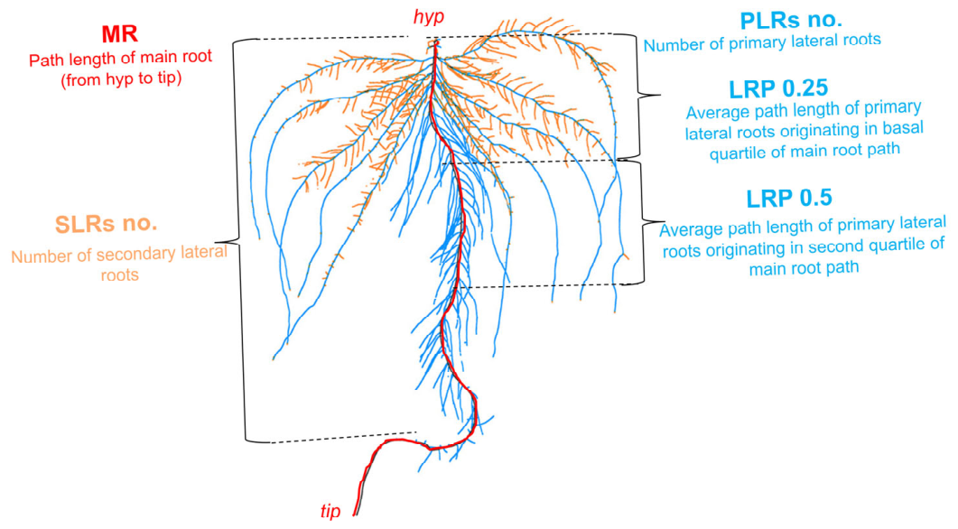
After 6 days of treatment, three plants were randomly selected from each group and separated into shoots and roots. To characterize the RSA, the fresh roots were immediately scanned using an Expression 12000XL scanner (Epson America, Inc., Long Beach, CA, USA). The scanned images were analyzed using Win RHIZO software 2025a (Regent Instruments Inc., Québec City, QC, Canada) to quantify key RSA parameters, including total root length and root surface area. Additional RSA traits (Figure 1) were further evaluated using ImageJ (v. 1.54g) software [39]. Subsequently, the fresh weights of the first true leaf, the second true leaf, and the root were recorded. The first and second true leaves were also scanned for leaf area determination. Finally, all samples (the first true leaf, the second true leaf, the root, and the remaining shoot parts) were oven-dried at 65 °C to constant weight, and their dry weights were recorded.

2.3. Determination of Chemical Forms of Cu

The chemical forms of Cu in the roots and leaves of cucumber seedlings were analyzed using a sequential extraction procedure [40]. Briefly, Cu was fractionated into five operationally defined forms using the following series of extractants: (1) 80% ethanol: intended to extract ethanol-soluble Cu, including inorganic salts and low-molecular-weight organic complexes; (2) deionized water: extracting water-soluble Cu; (3) 1 M NaCl: extracting pectate- and protein-bound Cu; (4) 2% acetic acid (HAc): extracting insoluble phosphate-bound Cu; and (5) 0.6 M HCl: extracting oxalate-bound Cu. The plant samples and the extractant were ground into a homogeneous mixture at a ratio of 1:100 (w/v), and then were subjected to 25 °C constant temperature shaking for 22 h. They were then centrifuged at 5000× g at room temperature for 10 min to collect the supernatant. The precipitate was further extracted with the same extractant twice, shaken for 2 h, and centrifuged at 5000× g at room temperature for 10 min. The three supernatants were combined. The precipitate was successively added with the above extractant and extracted using the same method. The supernatant was dried at 65 °C, and the Cu content of each component was determined using a flame atomic absorption spectrophotometer (ZEEnit700p, Analytic Jena AG, Jena, Germany) [41].

2.4. Subcellular Distribution of Cu in Plants

The subcellular distribution of Cu in the roots of cucumber seedlings and the second true leaf was determined by differential centrifugation [42]. The plant tissues were separated into cell wall fraction (Fcw), organelle fraction (Fco) and soluble fraction (Fs) using differential centrifugation. Briefly, 0.5 g of fresh samples (roots or leaves) were homogenized in 25 mL of an ice-cold extraction buffer containing 0.25 M sucrose, 50 mM Tris-HCl (pH 7.5), and 1 M dithiothreitol. All procedures were performed at 4 °C. The homogenate was first centrifuged at 3000× g for 15 min at 4 °C. The pellet, primarily consisting of cell walls and cell wall fragments, was designated as the Fcw. The supernatant from the first centrifugation was then centrifuged at 20,000× g for 30 min at 4 °C to pellet organelles and membranes. This pellet was collected as the Fco. The resulting supernatant, containing soluble cytosolic and vacuolar components, was designated as the Fs. These three components were dried to constant weight at 65 °C, and then the Cu concentration of each component was determined using a flame atomic absorption spectrophotometer (ZEEnit700p, Analytic Jena AG, Jena, Germany).

2.5. Cell Wall Preparation and Polysaccharide Extraction

The alcohol-insoluble residue (AIR) from plant root and leaf samples was prepared using a modified extraction method with minor modifications [43]. Briefly, frozen tissues were finely ground in liquid nitrogen. The resulting powder was then homogenized in pre-chilled 75% (v/v) ethanol and incubated on ice for 10 min, followed by centrifugation at 5000× g for 10 min at 4 °C. This ethanol extraction step was repeated twice. Subsequently, the pellet was sequentially extracted with the following ice-cold solvents, each for 10 min with centrifugation as above: acetone, a methanol: chloroform mixture (1:1, v/v), and finally pure methanol. The final pellet was dried under vacuum and stored as the crude cell wall fraction (AIR) for subsequent analysis.
Sequential fractionation of the AIR into pectin, hemicellulose, and cellulose components was performed using a sequential extraction procedure [44]. The dried AIR was suspended in 4 mL of deionized water and incubated in a boiling water bath for 1 h with occasional vortexing. After cooling, the suspension was centrifuged at 5000× g for 10 min. The supernatant was collected. The pellet was re-extracted with another 4 mL of deionized water under the same conditions. The combined supernatants from the two extractions were designated as the pectin fraction. The water-insoluble pellet was then subjected to extraction with 4% (w/v) potassium hydroxide (KOH). The pellet was suspended in 3 mL of 4% KOH and shaken on a horizontal shaker (150 rpm) at room temperature for 12 h. The extract was separated by centrifugation (5000× g, 10 min). The extraction was repeated once with a fresh 3 mL aliquot of 4% KOH. The combined alkaline supernatants were neutralized to pH ~7.0 with glacial acetic acid and defined as the hemicellulose 1 (HC1) fraction. The resulting pellet was further extracted with 24% (w/v) KOH using the same protocol (3 mL, 12 h shaking, repeated once). The combined supernatants were neutralized as above and defined as the hemicellulose 2 (HC2) fraction. The final pellet, resistant to the strong alkaline treatment, was washed thoroughly with deionized water and dried. This residue was considered the cellulose-rich fraction. The concentration of Cu in each of the obtained cell wall fractions (pectin, HC1, HC2, and cellulose) was determined using a flame atomic absorption spectrophotometer (ZEEnit700p, Analytic Jena AG, Jena, Germany).

2.6. FTIR Analysis of Root Cell Wall Materials (CWMs)

The extracted cell wall components were analyzed by FTIR spectroscopy [45]. For FTIR analysis, the root CWMs were ground with KBr in a ratio of 1:100 (w:w), and pressed into tablets. The FTIR spectra of 4000–400 cm−1 were obtained using an FTIR Spectrometer (Perkin Elmer Spectrum Two; PerkinElmer, Westford, MA, USA) at 4 cm−1 resolution and 32 scans/sample.

2.7. Scanning Electron Microscopy (SEM) Analysis of Cucumber Seedling Roots

The distribution of Cu in cucumber seedlings was determined using scanning electron microscopy (SEM). In brief, fresh roots were rinsed thoroughly with deionized water and sectioned, then immediately immersed in an electron microscopy fixative and fixed at room temperature for 2 h. The samples were subsequently stored at 4 °C until they sank to the bottom, followed by further preparation steps. The fixed samples were rinsed three times with 0.1 M Phosphate Buffer (PB, pH 7.4) for 15 min each. Then the samples were fixed in 1% osmic acid (prepared with 0.1 M Phosphate-Buffered Saline (PBS), pH 7.4) at room temperature in the dark for 1–2 h, and rinsed another three times with 0.1 M PBS (pH 7.4) for 15 min each. Tissues were dehydrated sequentially in 30%, 50%, 70%, 80%, 90%, 95%, 100%, and 100% ethanol for 15 min at each concentration, followed by treatment with isoamyl acetate for 15 min. The samples were dried in a critical point dryer, then mounted onto conductive carbon double-sided tape and placed on the stage of an ion sputtering coater for gold sputtering for approximately 30 s. Finally, observation and image acquisition were performed under a scanning electron microscope.

2.8. Determination of Cu Concentration in Cucumber Seedlings

After a 6 d Cu stress treatment, the young plant tissues (the first true leaf, the second true leaf, the stem, and the root system) were dried, ground into fine powder, and weighed (each sample 0.1 g, stem 0.05 g). The ground tissues were digested with 5 mL of concentrated HNO3. The Cu concentration (μg/g) was determined using an atomic absorption spectrometer (ZEEnit700p, Analytic Jena AG, Jena, Germany).

2.9. Determination of Nitrogen Metabolism-Related Products and Nitrogen Metabolism Enzyme Activities

The nitrate (NO3) content was determined according to the method described previously [46]. Approximately 100 mg of sample was extracted with 1 mL of deionized water in a water bath at 45 °C for 1 h. After centrifugation at 5000× g for 15 min at 20 °C, the supernatant was collected. A 0.2 mL aliquot of the supernatant was mixed with 0.8 mL of 5% (w/v) salicylic acid and allowed to stand at room temperature for 20 min. Subsequently, 19 mL of 2 M NaOH was added. After the solution cooled to room temperature, the absorbance was measured at 410 nm.
The ammonium (NH4+) content in various tissues of cucumber seedlings was determined following the method described previously [47]. Briefly, 100 mg of sample was homogenized in 1.5 mL of extraction solution (100 mM HCl and 500 μL chloroform) and shaken at 4 °C for 15 min. The mixture was centrifuged at 10,000× g for 10 min at 4 °C. The supernatant was collected, mixed thoroughly, and centrifuged again at 12,000× g for 10 min at 4 °C. The resulting supernatant was mixed with a 1% (w/v) phenol-0.005% (w/v) sodium nitroprusside solution, followed by the addition of a 1% (v/v) sodium hypochlorite-0.5% (w/v) NaOH solution. The mixture was incubated at 37 °C for 30 min, and the absorbance was recorded at 620 nm.
The nitrite (NO2) content was measured according to the method described previously [48]. Approximately 100 mg of sample was homogenized in 1 mL of extraction buffer containing 50 mM Tris-HCl (pH 7.9), 5 mM cysteine, and 2 mM EDTA. The homogenate was centrifuged at 10,000× g for 20 min at 20 °C. A 500 μL aliquot of the supernatant was mixed with 1% sulfanilamide and 0.02% N-1-naphthylethylenediamine dihydrochloride. Following color development, the absorbance of the solution was measured at 540 nm.
The activities of nitrogen metabolism-related enzymes, including nitrate reductase (NR), nitrite reductase (NiR), glutamate dehydrogenase (GDH), glutamate synthase (GOGAT), and glutamine synthetase (GS), were determined using specific assay kits (Beijing Solarbio Technology Co., Ltd., Beijing, China) according to the manufacturer’s instructions.

2.10. Statistical Analysis

One-way (ANOVA) was conducted using SPSS 26.0 software, and Tukey’s HSD test was used for multiple comparisons at p ≤ 0.05. Origin 2022 software was used for drawing figures and principal component analysis (PCA).

3. Results

3.1. DCPTA Alleviated Cu-Induced Inhibition of Leaf Growth and Biomass Accumulation in Cucumber Seedlings

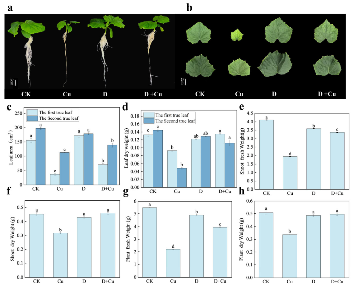
Compared with the CK, Cu stress significantly decreased the fresh and dry weights of both the shoots and whole plants (Figure 2). Specifically, compared with the single Cu treatment, exogenous DCPTA increased the fresh weight of shoot and plant by 74.4% and 78.9%, respectively (Figure 2e,g), and increased the dry weight of shoot and plant by 44% and 46.2%, respectively (Figure 2f,h). Furthermore, DCPTA alleviated the Cu-induced stress by increasing the leaf area of the first and second true leaves by 23% and 91.4%, respectively (Figure 2c), and their dry weight by 45.6% and 129.3%, respectively (Figure 2d). In summary, DCPTA effectively alleviated the copper stress in cucumbers by significantly restoring biomass accumulation and leaf development.

3.2. DCPTA Mitigated the Negative Impact of Cu on RSA Parameters

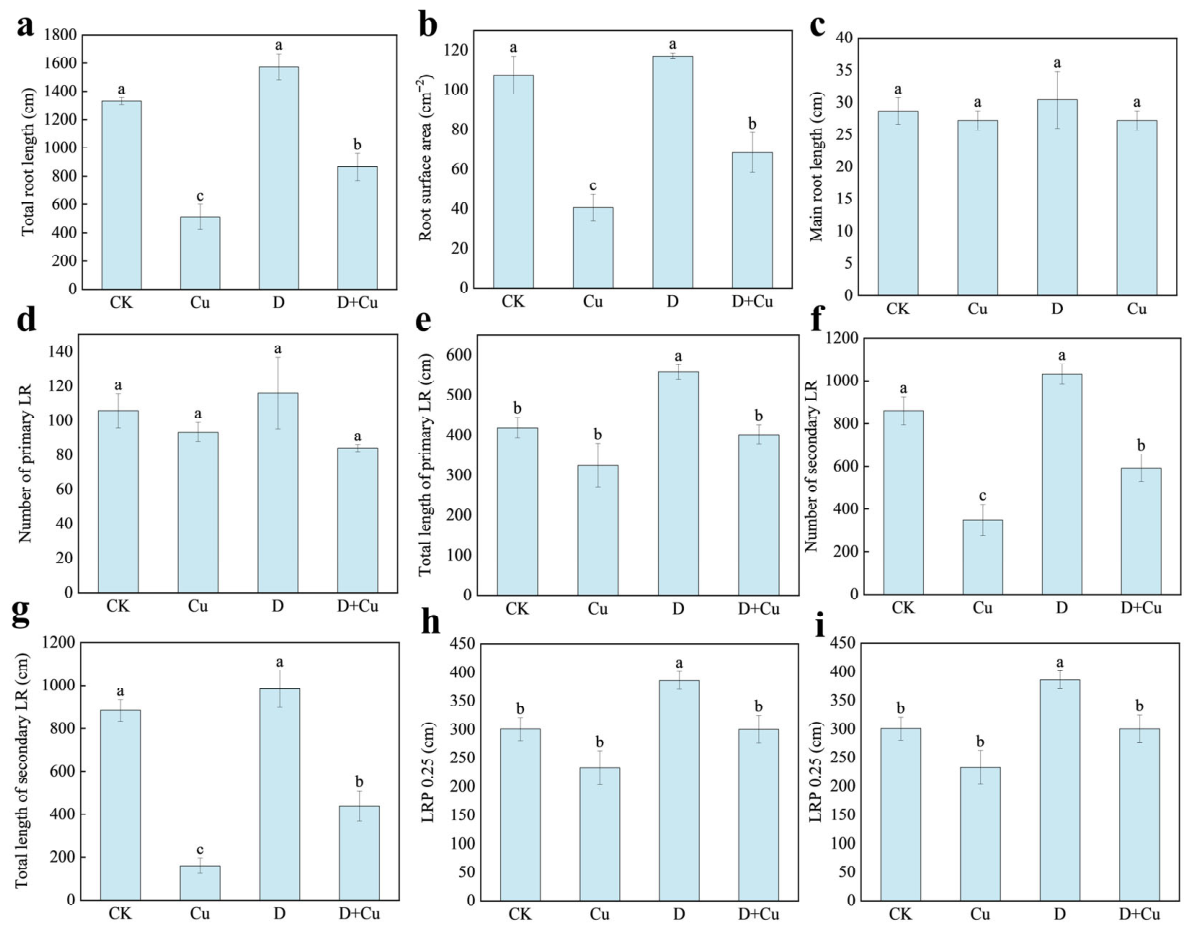
Figure 3 presents the effects of different treatments (CK, Cu, D, and D + Cu) on cucumber root morphological parameters. Compared with CK, Cu treatment notably reduced the total root length, total surface area, the number of secondary lateral roots (LRs), and the total length of secondary LRs. Exogenous application of DCPTA resulted in significant increases of 68.8%, 68.7%, 69.6% and 173.2% in total root length, total surface area, number of secondary roots and total length of secondary roots, respectively, compared to the copper treatment. These results indicate that exogenous DCPTA alleviates root architecture impairment under Cu stress primarily by promoting the development of secondary LRs.
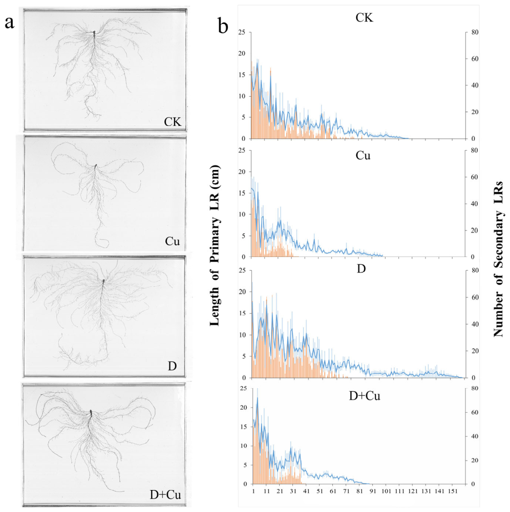
Figure 4 illustrates the distribution of primary and secondary LRs originating from the main root. In general, the length of primary LRs and the number of secondary LRs gradually decreased with the increase in primary LR sequence. All four treatments mainly produced primary LRs in the basal quarter of the main root, with lengths ranging from 0 to 25 cm. In the CK, Cu, D, and D + Cu treatments, the primary LRs at the base (from the 1st to the 81st, 1st to the 31st, 1st to the 71st, and 1st to the 41st) were longer, and served as the main growth areas for the secondary LR system. Compared with CK, the number of secondary LRs in the Cu treatment decreased significantly starting from the 31st position. In contrast, the D + Cu treatment expanded the distribution area of secondary LRs and significantly increased their number. These results are consistent with the quantitative data in Figure 3, indicating that adding DCPTA improves the morphological development of the cucumber root system by expanding the growth range and increasing the number of secondary LRs.

3.3. DCPTA Reduces the Translocation of Copper to the Shoots Under Cu Stress

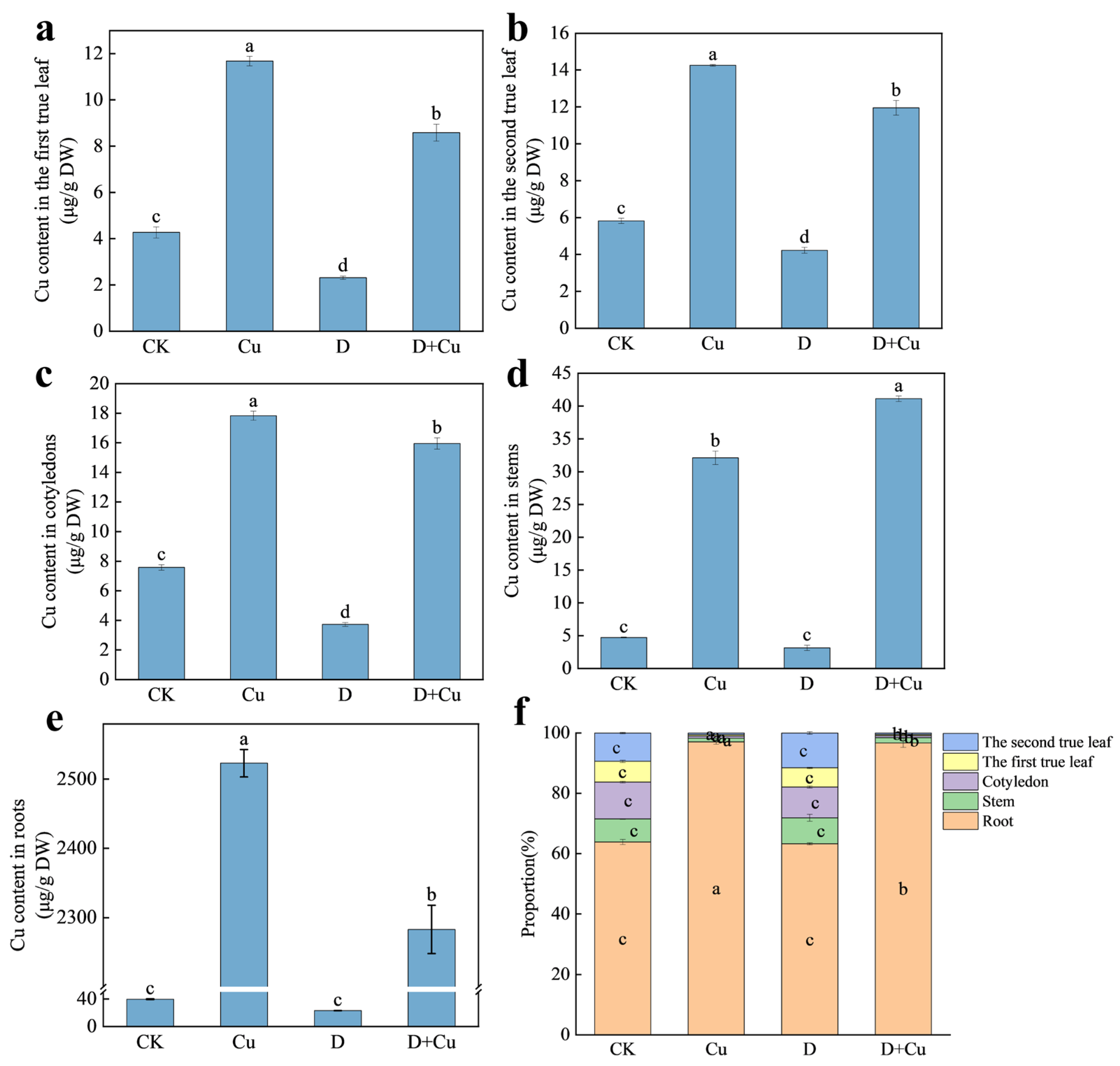
The roots are the main organ for Cu accumulation, and their Cu content is much higher than that of the shoots (leaves, cotyledons, stems). Under Cu stress, after the application of exogenous DCPTA, the Cu concentration in the roots, the first true leaf, and the second true leaf decreased by 9.5%, 26.5%, and 16.1%, respectively, although an unexpected increase was observed in the stems (Figure 5). This indicates that DCPTA can effectively alleviate Cu accumulation.

3.4. DCPTA Alters the Chemical Forms and Subcellular Distribution of Cu, Favoring Its Immobilization in Roots

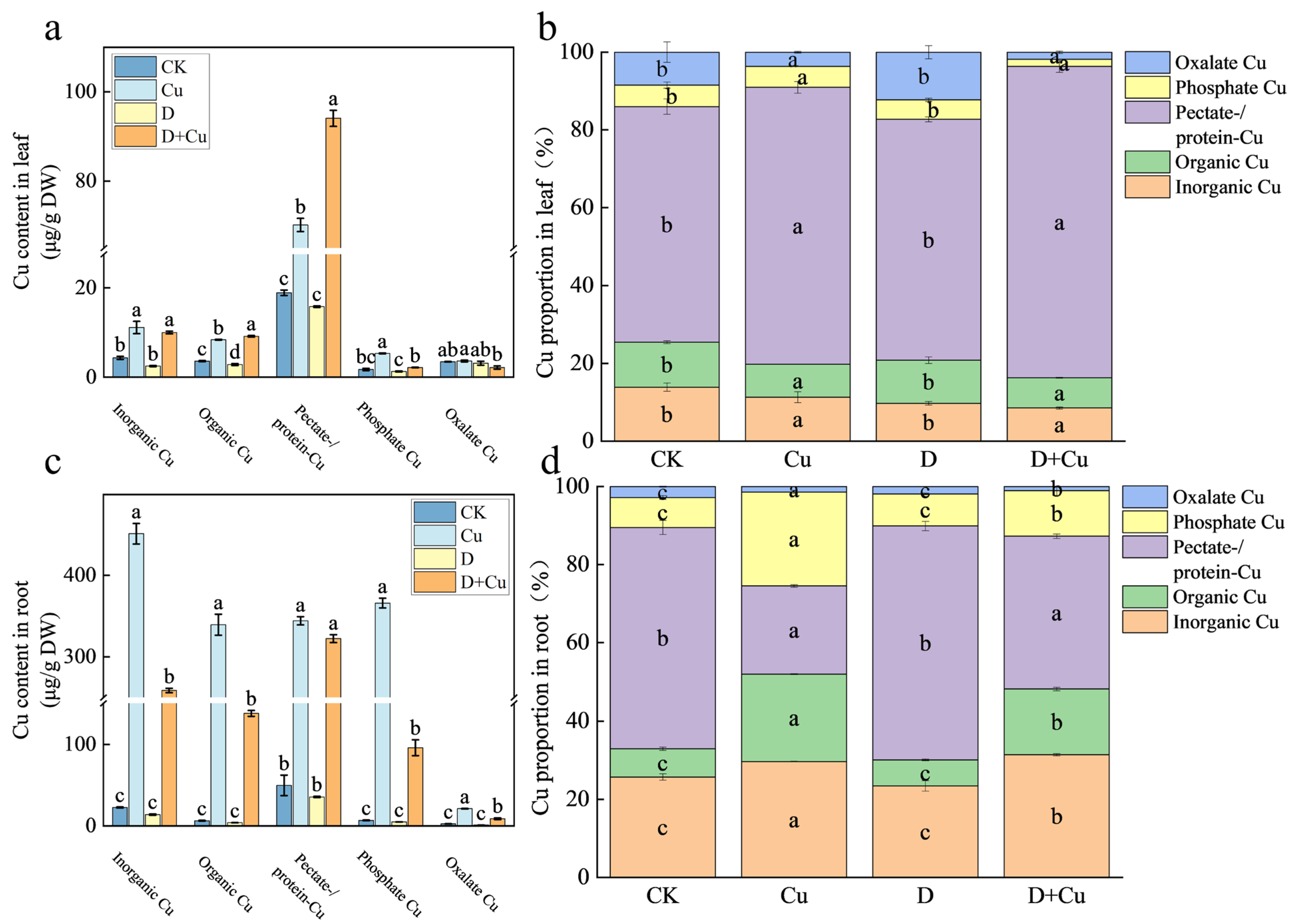
Cu chemical speciation analysis showed that polysaccharide/protein-bound Cu was the predominant form in both leaf and root tissues (Figure 6a,c). In leaves, DCPTA reduced the contents of inorganic Cu, phosphate-bound Cu, and oxalate-bound Cu by 10.1%, 59.7%, and 40%, respectively, while increasing organic Cu and pectate/protein-bound Cu by 9% and 34% (Figure 6a). In roots, DCPTA treatment led to substantial reductions in several Cu forms, with organic Cu, phosphate-bound Cu, and oxalate-bound Cu decreasing by 59.4%, 73.8%, and 58.2%, respectively (Figure 6c).
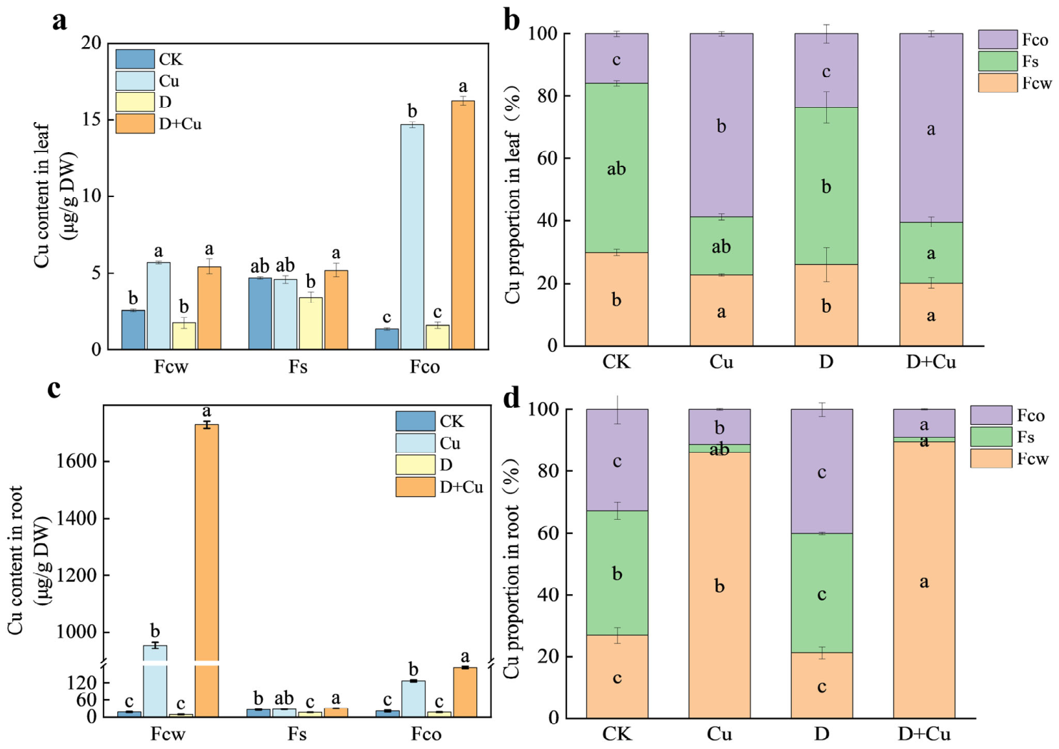
In parallel, analysis of Cu subcellular distribution revealed distinct patterns between leaves and roots. Under Cu stress, Cu accumulation in leaves across subcellular fractions followed the order organelle fraction (Fco) > cell wall fraction (Fcw) > soluble fractions (Fs). Compared to Cu treatment alone, DCPTA increased the Cu concentration in Fs and Fco by 13.1% and 10.7%, respectively (Figure 7a). Conversely, in roots, Cu was predominantly localized in the Fcw, with its concentration significantly exceeding those in Fs and Fco. Under copper stress, exogenous DCPTA increased the copper concentrations in root Fcw, Fs and Fco by 81.3%, 11% and 37.7%, respectively (Figure 7c).
Collectively, these results indicate that DCPTA promotes Cu retention in root cell walls and shifts Cu toward less mobile, organically bound forms, thereby enhancing Cu immobilization in roots and reducing its translocation and toxicity in shoot tissues.

3.5. DCPTA Mitigates Cu-Induced Damage to Root Anatomy and Elemental Composition

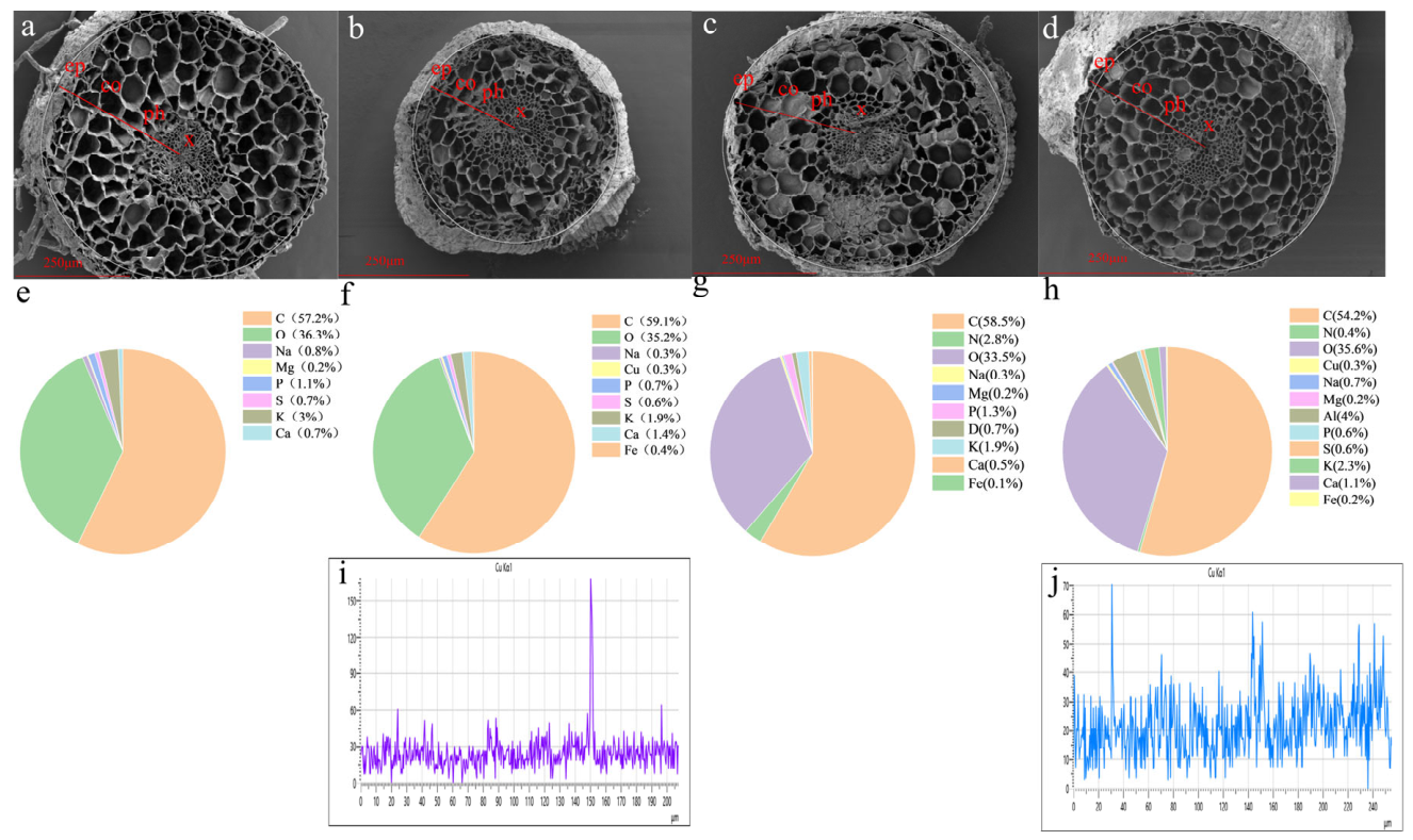
Root cross-sectional scanning electron microscopy (SEM) images revealed that root tissues in the control group exhibited a regular morphology with clearly delineated epidermal (ep), cortical (co), phloem (ph), and xylem (x) structures (Figure 8a). Under Cu stress, slight shrinkage of the epidermis was observed in cucumber roots, accompanied by a decreased cortical tissue ratio and increased proportions of xylem and phloem tissues (Figure 8b). Following DCPTA application, the cellular structure of cucumber seedling roots showed marked improvement. Compared with Cu treatment alone, DCPTA supplementation decreased the C/O ratio in roots and slightly elevated the concentrations of other elements, such as Na and K, indicating that DCPTA can partially restore nutrient element uptake in cucumber seedlings under Cu stress (Figure 8f,h).

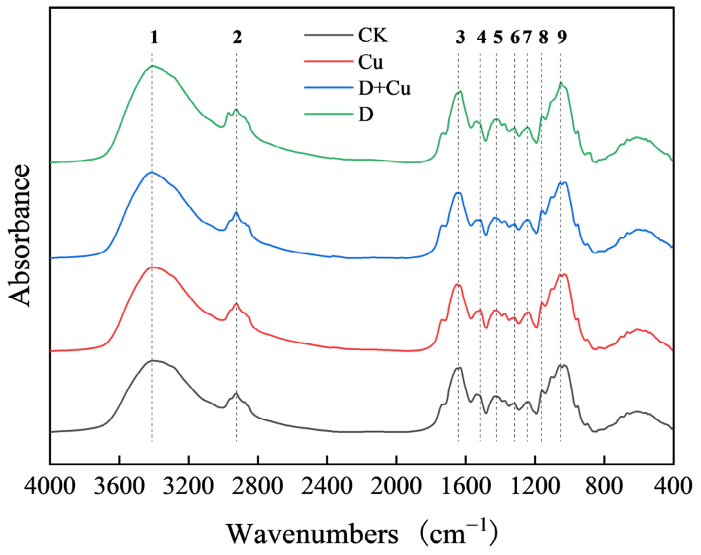
3.6. DCPTA Modifies Functional Groups in Root Cell Walls

Nine characteristic absorption bands were detected in the root, and Cu treatment resulted in significant shifts in the wave numbers, namely 2924 cm−1 (cellulose and pectin), 1651 cm−1 (Amide I), and 1516 cm−1 (Amide II), with shifts of −1, 19, and 19, respectively. These shifts indicated that Cu2+ not only interacted strongly with the amide bonds of cell wall proteins, but also altered the structure of polysaccharides (including cellulose, hemicellulose, and pectin) in the root cell walls. Exogenous application of DCPTA enhanced the absorption of functional groups in the root cell walls of cucumber seedlings, leading to an increase in characteristic peak intensities and a slight shift toward higher wavenumbers (Figure 9 and Table 1). Exogenous DCPTA significantly reversed or alleviated most of the peak shifts caused by Cu stress, indicating that DCPTA alleviates Cu stress by regulating protein amide groups and optimizing the structure of cellulose and pectin.

3.7. DCPTA Enhances Polysaccharide Biosynthesis and Increases Cu Retention in Root Cell Wall Polysaccharides

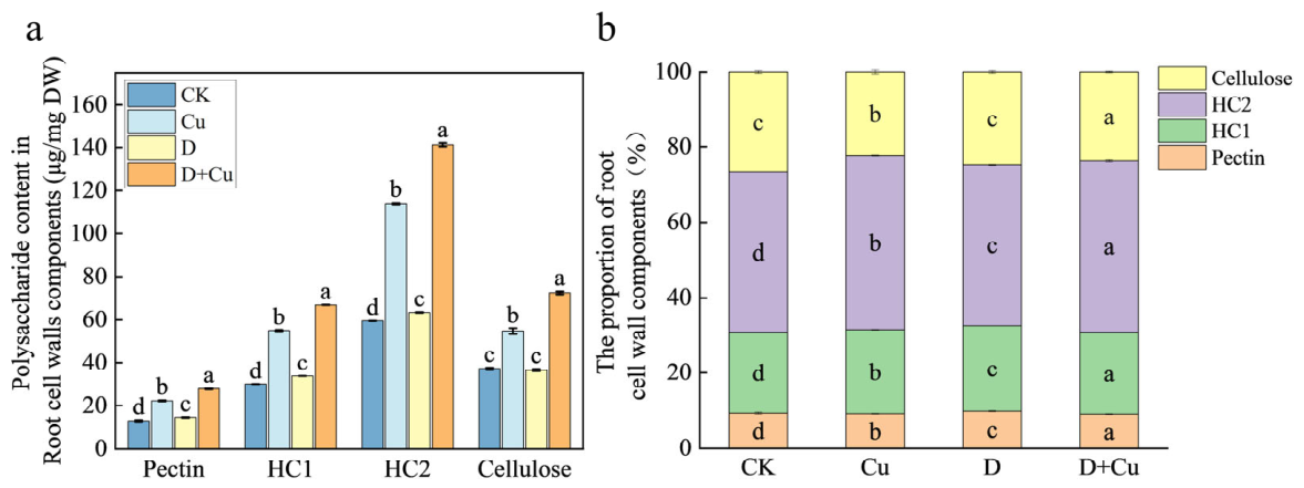
Exposure to Cu stress is known to alter plant cell wall architecture, often by modulating the composition and content of polysaccharides, which are crucial for structural integrity and metal sequestration. In this study, compared to the control, Cu stress significantly increased the total polysaccharide content in the root cell walls of cucumber seedlings, indicating a possible adaptive response to reinforce the cell wall under metal toxicity. Under Cu stress conditions, exogenous application of DCPTA further enhanced the accumulation of key polysaccharide fractions: pectin, hemicellulose (HC1), hemicellulose (HC2), and cellulose increased by 24.3%, 22.4%, 23.7%, and 33.1%, respectively (Figure 10a). Among these components, HC2 constituted the highest proportion in the root cell wall (Figure 10b), suggesting that it may play a particularly significant structural and functional role in mediating Cu stress adaptation. These results imply that DCPTA not only mitigates Cu-induced structural disruption but may also actively promote the biosynthesis and deposition of cell wall polysaccharides, thereby enhancing the mechanical strength and metal-binding capacity of the root cell wall under stress conditions.
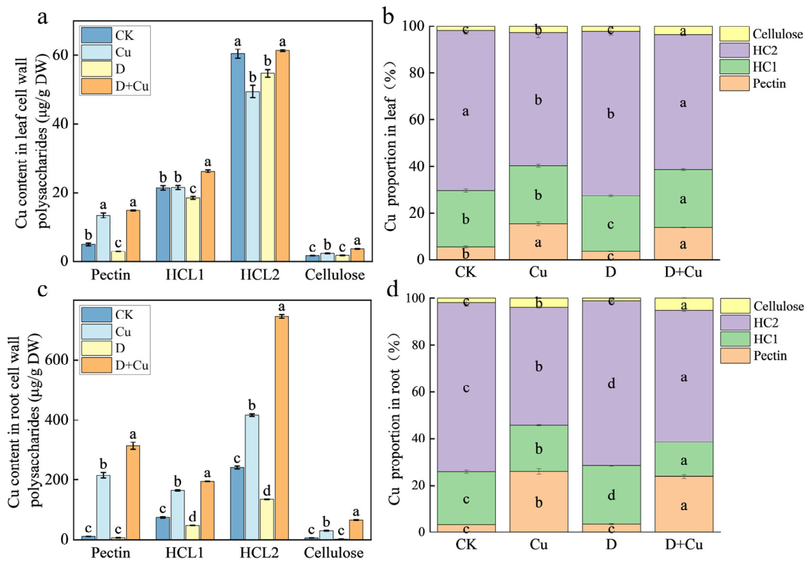
This DCPTA-induced enhancement of polysaccharide biosynthesis was directly correlated with improved Cu retention capacity. Correspondingly, Cu concentrations bound to pectin and HC2 in roots increased substantially by 46.3% and 79.2%, respectively, following DCPTA treatment (Figure 11d). Notably, HC2 constituted the highest proportion among polysaccharide components (Figure 10b) and also showed the highest Cu-binding level (Figure 11b,d), underscoring its pivotal structural and functional role in metal chelation. Collectively, these findings demonstrate that DCPTA actively fortifies the root cell wall by boosting the synthesis of polysaccharides—especially those with high metal-binding affinity—thereby expanding the capacity for Cu sequestration and contributing to reduced metal mobility and toxicity.

3.8. Effects of DCPTA on Nitrogen Metabolism-Related Enzyme Activities and Metabolite Contents in Cucumber Seedlings Under Copper Stress

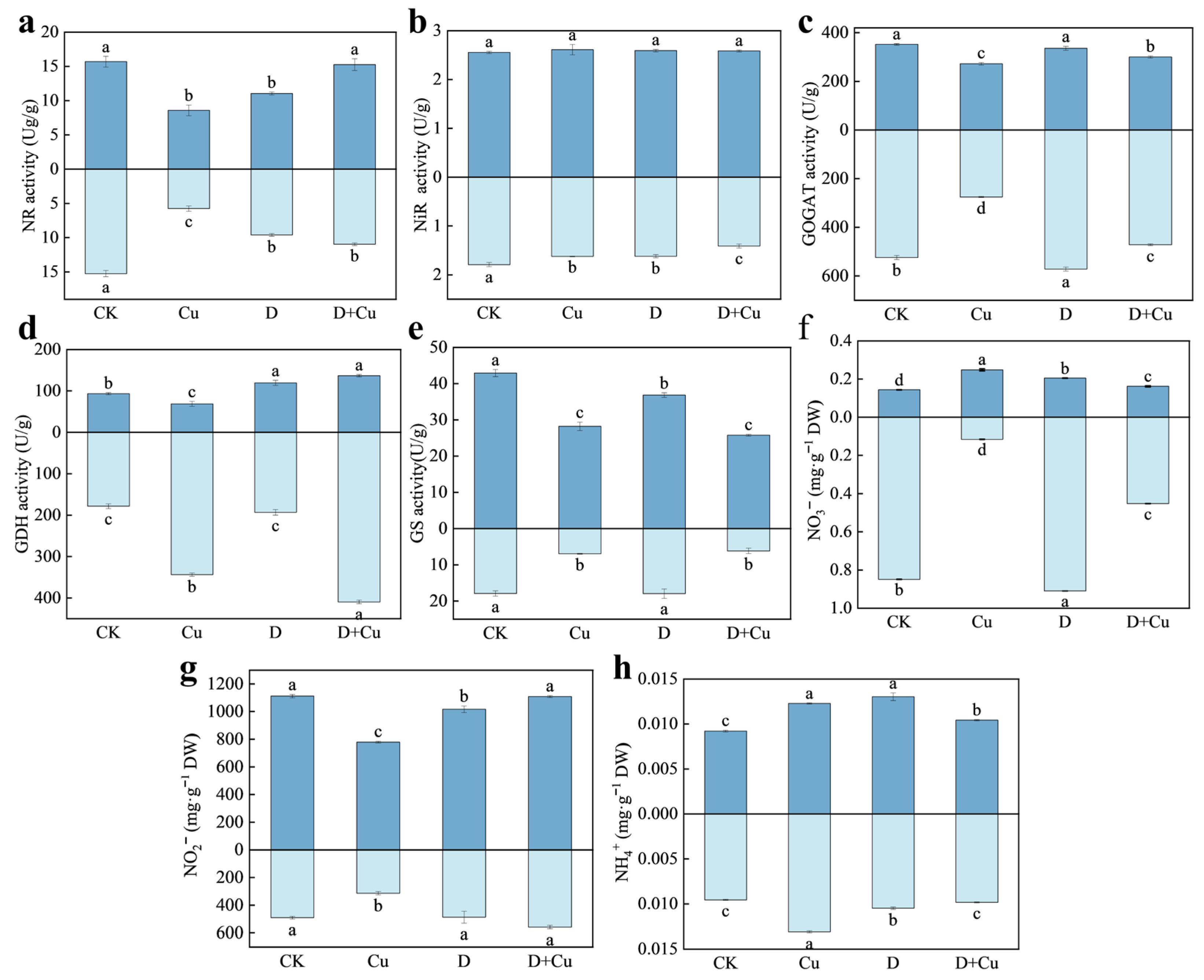
Compared with the control, Cu stress significantly reduced NR activity in leaves and roots by 45.3% and 62.3%, respectively, indicating that Cu toxicity inhibited NR activity. However, compared with Cu treatment alone, NR activity in the D + Cu treatment increased by 77.7% and 90.6% in leaves and roots, respectively, suggesting that exogenous DCPTA alleviated the inhibitory effects of Cu stress on NR activity (Figure 12a). Significant differences were observed in GOGAT (Figure 12c) and GDH (Figure 12d) activities between Cu and D + Cu treatments in both parts. Specifically, DCPTA significantly increased GOGAT and GDH activity in both leaves and roots under Cu stress. Among these, the leaf parts increased by 10.3% and 26.3%, respectively, while the root parts increased by 71.3% and 99.1%, respectively (Figure 12c,d). Cu stress also reduced nitrate and nitrite content in roots by 86.4% and 36.1%, respectively, compared with CK.
Under Cu stress, DCPTA application significantly increased nitrate and nitrite content in both parts. Notably, compared with Cu treatment alone, nitrate content in roots increased by 291.7%, and nitrite content in roots increased by 78.3%. Cu stress increased the nitrate content in the shoot part of the plant by 72.7% and decreased the nitrite content by 42.8%. This indicates that Cu stress mainly inhibited the reduction and utilization of NO3 (as NR activity decreased), leading to its accumulation within the plant. After the addition of exogenous DCPTA, the nitrate content decreased by 53.2%, while the nitrite content increased by 42.4%. This indicates that under Cu stress, the conversion rate of NO3 to NO2 was significantly slowed down due to the reduced NR activity, resulting in insufficient production of NO2. After DCPTA treatment, the NR activity recovered and the generation rate of NO2 increased (Figure 12f,g). Furthermore, NH4+ content was also significantly altered by DCPTA under Cu stress in both parts (Figure 12h).

3.9. PCA of the Morphological and Physiological Indicators of Leaves and Root Systems

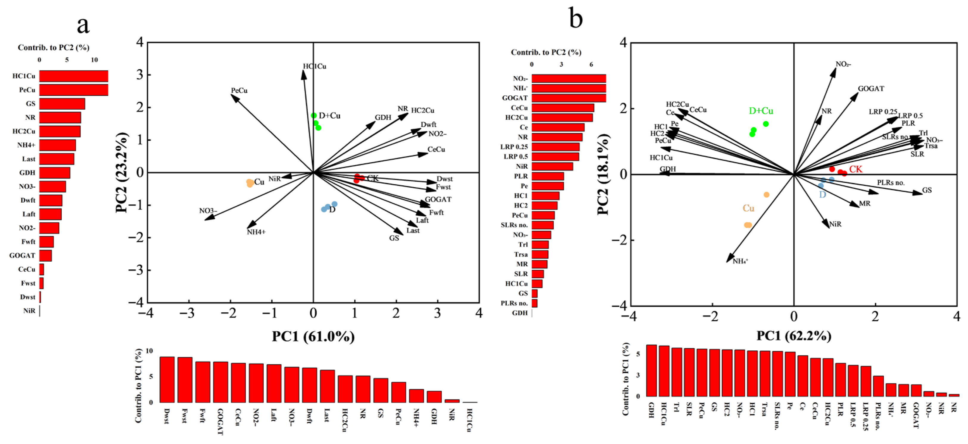
PCA was performed using morphological and physiological indicators from aboveground and underground parts to analyze their responses to Cu and DCPTA treatments. PCA revealed that Cu stress caused significant damage to both leaves and roots (Figure 13). Cu-treated and D + Cu-treated samples were clearly separated along PC1 and PC2. The main indicators contributing to PC1 included Dwst (dry weight of the second true leaf), Fwst (fresh weight of the second true leaf), Fwft (fresh weight of the first true leaf), GOGAT, and CeCu. The main indicators contributing to PC2 included HC1Cu, PeCu, GS, and NR (Figure 13a). For roots, PC1 (PC2) contributed 62.2% (18.1%) of the total variation. Samples treated with Cu and D + Cu showed more distinct separation along PC2. The main indicators contributing to PC2 included nitrite (NO2), ammonium (NH4+), GOGAT, CeCu, HC2Cu, Ce, NR, LRP0.25, and LRP0.5 (Figure 13b).

4. Discussion

The concentration of copper in soil has significantly escalated due to anthropogenic activities, particularly industrial and agricultural production, rendering it a prominent environmental pollutant [63]. Cu pollution in cultivated land has caused devastating effects on human health, the environment, and food security [64]. Cu is a necessary microelement for well-balanced plant growth and development. Excess Cu can limit the plant’s absorption of water and nutrients, thus inhibiting its growth. Restricted root growth can affect overground growth and thus affect various physiological processes. However, the potential regulatory mechanism of DCPTA on Cu-stressed plant growth remains unclear. This study proved that DCPTA alleviated the effects of Cu stress on plants by restoring biomass accumulation and leaf growth, and regulating root development. Mechanistically, DCPTA enhanced Cu immobilization in the root cell wall by upregulating polysaccharide biosynthesis and modifying functional groups, which reduced Cu transport to shoots. In addition, DCPTA effectively mitigated the inhibition of nitrogen metabolism by restoring the activities of key enzymes such as nitrate reductase and glutamate synthase, thereby maintaining nutrient homeostasis. Therefore, DCPTA can alleviate the damage of Cu stress on cucumber (Cucumis sativus L.) seedlings.

4.1. DCPTA Alleviates the Effects of Cu on Cucumber Plant Biomass and Root Morphology

Plant roots are the primary sites for heavy metal perception and are crucial for water and nutrient uptake; thus, root growth inhibition is a well-documented symptom of Cu toxicity [63]. Excess Cu restricts cell elongation and division in the root meristem, leading to a stunted root system and reduced biomass accumulation [64]. In the present study, Cu stress significantly reduced cucumber seedling biomass, including leaf area and dry weight, and severely inhibited root growth (Figure 2). These findings align with previous reports that Cu stress disrupts physiological processes, resulting in growth retardation. However, the application of DCPTA effectively mitigated these inhibitory effects. Our results showed that DCPTA significantly restored the biomass and leaf development of cucumber seedlings under Cu stress (Figure 2), consistent with its known growth-promoting roles in crops like maize under drought stress [34].
The root system architecture (RSA) plays a pivotal role in plant adaptation to environmental stress. Changes in RSA, such as the length and density of lateral roots, can directly affect the plant’s ability to acquire resources [65]. Our analysis indicated that DCPTA significantly altered the RSA of cucumber seedlings under Cu stress. Compared to the Cu treatment, DCPTA application increased the total root length, total length of primary lateral roots, and the number of secondary lateral roots (Figure 3). This morphological adjustment suggests that DCPTA may regulate the allocation of photosynthates and nutrients, prioritizing the elongation of existing lateral roots to expand the absorption surface area rather than initiating new primordia under stress conditions. This strategy is crucial for efficient resource acquisition. This finding resonates with a recent study on silicon nanoparticles (SiNPs), which showed that SiNP supplementation under Cd stress significantly increased root tip and fork numbers in rice, promoting a compensatory architectural adjustment that supports nutrient foraging and stress resilience [66]. Furthermore, the improvement in root morphology was associated with altered Cu distribution. By promoting a more developed root system, DCPTA likely facilitates a more efficient compartmentalization of Cu, thereby reducing its toxic effects on the shoots (Figure 5). Therefore, we conclude that DCPTA alleviates Cu-induced growth inhibition by regulating root morphological plasticity and restoring biomass accumulation.

4.2. DCPTA Alters Cu Subcellular Distribution and Chemical Forms to Enhance Cu Immobilization in Roots

The plant cell walls (CWs) serve as the first physical barrier against heavy metals (HMs) entering the cytoplasm (protoplast) [21,43]. Previous studies have indicated that plant varieties with higher Cu tolerance typically accumulate more Cu in the root CWs, thereby mitigating its toxicity to cellular organelles [52,67]. Consistent with these findings, our subcellular analysis revealed that under Cu stress, a majority of Cu in cucumber roots was compartmentalized in the cell wall fraction (Fcw). Notably, DCPTA application further increased both the concentration and proportion of Cu in the Fcw (Figure 7). This enhanced sequestration in the apoplast limited the entry of Cu into the soluble fraction (Fs) and organelle fraction (Fco). Consequently, DCPTA treatment significantly reduced the translocation of Cu from roots to shoots, as evidenced by the lower Cu concentrations in the stems and leaves compared to the Cu-only treatment (Figure 5). This suggests that DCPTA promotes a root-based exclusion mechanism by reinforcing the cell wall barrier. This is analogous to the role of tomato cell wall pectin in Cd fixation, where the majority of Cd is located in the apoplast, especially in the cell wall, acting as a crucial defense mechanism [68].
In addition to subcellular distribution, the toxicity of heavy metals is closely governed by their chemical forms. Generally, heavy metals extracted by water and ethanol possess the highest mobility and toxicity, while those extracted by NaCl (bound to pectin and proteins) exhibit lower mobility and toxicity [69,70]. Our results demonstrated that DCPTA treatment substantially altered the chemical forms of Cu in roots. Specifically, DCPTA reduced the proportions of highly mobile and toxic forms, such as inorganic Cu and phosphate-bound Cu, while increasing the proportion of Cu bound to pectin and proteins (Figure 6). This shift indicates that DCPTA facilitates the conversion of Cu into less phytotoxic forms, effectively detoxifying the metal within the root tissue. The critical role of these binding sites is directly evidenced by chemical modification assays; for instance, in tomato, esterification and amidation of the cell wall, which reduce -COOH and -OH groups, respectively, significantly decreased Cd adsorption, proving that these functional groups are key binding sites [68].

4.3. DCPTA Enhances Copper Immobilization by Regulating Cell Wall Polysaccharide Synthesis

FTIR analysis confirmed that DCPTA significantly enhanced the characteristic functional groups related to pectin and hemicellulose in cucumber root cell walls, thereby enhancing the capacity of the cell wall to immobilize Cu. The capacity of the CW to immobilize heavy metals is largely determined by the abundance of polysaccharides, particularly pectin and hemicellulose, which provide the primary binding sites. Recent studies have highlighted that the ability of cell walls to bind copper is closely related to the degree of pectin methylation (DPM). Under copper stress, plants upregulate pectin methylesterase (PME) activity to reduce the DPM, thereby exposing more free carboxyl groups. These negatively charged groups non-covalently bind copper ions, immobilizing them within the cell wall matrix. This mechanism is strongly supported by findings in Cd-stressed tomato plants, where PME activity and the expression of its encoding gene (SlPME1) were significantly upregulated, leading to pectin demethylation and increased Cd binding [68]. In this study, DCPTA significantly elevated the contents of key polysaccharide fractions, including pectin, HC1, HC2, and cellulose (Figure 10). This implies that DCPTA may induce “cell wall remodeling” [20], increasing pectin synthesis while potentially optimizing its methylation status to provide more copper-binding sites. Correspondingly, the Cu concentrations bound to pectin and HC2 in roots increased significantly following DCPTA treatment (Figure 11). Notably, HC2 constituted the highest proportion among polysaccharide components and exhibited the highest Cu-binding level. In addition to the carboxyl groups provided by pectin, the hydroxyl groups within the hemicellulose structure are also confirmed to be key sites for adsorbing heavy metal ions. The significant increase in HC2 content and its high affinity for Cu under DCPTA treatment suggest that it plays a synergistic role with pectin in copper fixation. Collectively, these findings demonstrate that DCPTA actively fortifies the cell wall barrier by enhancing polysaccharide synthesis and modification, thereby strengthening metal immobilization. A similar strategy of cell wall reinforcement has been reported for SiNPs, which reduced Cd accumulation in both the cell wall and symplast of rice roots, indicating restricted metal uptake and translocation [66].

4.4. Impact of DCPTA on Nitrogen Metabolism in Response to Cu Toxicity

The present study revealed that Cu stress significantly impaired key nitrogen (N) metabolic processes in cucumber seedlings. As the rate-limiting enzyme in nitrate assimilation, nitrate reductase (NR) is highly sensitive to heavy metal toxicity. Our results showed that Cu exposure reduced NR activity by 45.3% in leaves and 62.3% in roots compared to the control, which is consistent with previous reports that Cu ions disrupt the structure and function of NR, thereby limiting the conversion of nitrate to nitrite [25]. However, exogenous DCPTA effectively mitigated these inhibitory effects. The application of exogenous IAA increased the content of NO3 and the activity of NR. It was involved in the reduction of NO3 and nitrogen metabolism, thus indicating that the application of IAA could enhance the absorption efficiency of NO3 by young plants under copper stress, increase the activity of NR, and enhance the nitrogen reduction capacity [71]. This inhibition directly hindered the entry of nitrate into the organic N pool, as evidenced by the 86.4% and 36.1% decrease in nitrate content in roots and leaves, respectively, which aligns with findings in other plant species under heavy metal stress. PCA further supported these findings, with NR and GOGAT being key contributors to PC1 (61.0% variance) in leaves and PC2 (18.1% variance) in roots, respectively (Figure 13), indicating their central role in nitrogen metabolism under Cu stress.
Notably, DCPTA application restored NR activity by 77.7% in leaves and 90.6% in roots relative to the Cu-only treatment, and increased nitrate content by 291.7% in roots and 78.3% in leaves. These findings suggest that DCPTA may protect NR from Cu-induced oxidative damage or enhance its gene expression, thereby promoting nitrate assimilation. Although no significant changes were observed in NiR, GDH, and GS activities under Cu or D + Cu treatments, DCPTA significantly increased GOGAT activity by 66.9% in roots (Figure 12). This upregulation of GOGAT is crucial, as it drives the GS-GOGAT cycle—the primary pathway for ammonium assimilation into amino acids [72]. PCA revealed that GOGAT was a major contributor to PC1 in leaves and PC2 in roots, further emphasizing its role in DCPTA-mediated nitrogen metabolism regulation.
Cu stress also altered nitrite and ammonium content: nitrite decreased by 36.1% in roots, while ammonium content was significantly altered by DCPTA (Figure 12h). PCA of root nitrogen metabolism indicators showed that NO2 and NH4+ were key contributors to PC2 (18.1% variance), highlighting DCPTA’s modulation of nitrate reduction and ammonium homeostasis under Cu stress (Figure 13b). These results indicate that DCPTA restores nitrogen assimilation by enhancing NR and GOGAT activities, thereby improving nitrate reduction and ammonium utilization, which supports overall plant growth under Cu stress.
The differential responses of N metabolism enzymes between leaves and roots highlight the tissue-specific regulation of DCPTA. The more pronounced restoration of NR and GOGAT activities in roots suggests that DCPTA primarily acts at the site of Cu uptake and accumulation, thereby protecting the root system from toxicity and sustaining N uptake and assimilation. This, in turn, ensures a steady supply of reduced N to the leaves, supporting photosynthesis and overall plant growth under Cu stress.

4.5. An Integrated Mechanism for DCPTA-Mediated Cu Detoxification

It is well established that Cu stress triggers the excessive accumulation of ROS, a primary toxic mechanism. Excess ROS not only causes oxidative damage but also readily inactivates key metabolic enzymes, including those in the nitrogen assimilation pathway (e.g., NR and GOGAT), which are highly sensitive to oxidative stress [73]. In this study, our previous work under the same experimental conditions confirmed that DCPTA effectively mitigates Cu-induced oxidative damage by scavenging H2O2 and O2· and enhancing antioxidant enzyme systems (SOD, POD, CAT, APX), thereby restoring redox homeostasis [36]. Building upon this established antioxidant foundation, the present study reveals the subsequent, distinct physiological strategies employed by DCPTA: rather than merely scavenging ROS, DCPTA actively fortifies the root cell wall to immobilize Cu and specifically protects the nitrogen metabolic machinery to maintain intracellular homeostasis.
Compared with other plant growth regulators such as auxin [74], melatonin [75], and 24-Epibrassinolide [76], which mainly enhance heavy metal tolerance via activating antioxidant systems or signaling pathways, DCPTA exhibits a distinct mechanism. PCA indicates that cell wall polysaccharides and nitrogen metabolism-related enzyme activities contribute significantly to the alleviation of copper stress in cucumber seedlings by DCPTA. Therefore, we believe that DCPTA can promote an increase in pectin and hemicellulose contents to enhance the ability of copper to be fixed in the cell wall, reducing the influx of copper into the cells and thereby protecting the intracellular metabolic system, including various enzymes involved in the nitrate–ammonium reduction pathway. This unique mechanism highlights the potential value of DCPTA as a regulator that is expected to be used to alleviate heavy metal stress in horticultural crops.
One of the main limitations of this study is the relatively short 6-day exposure period. Although these findings highlight the early defense strategies of DCPTA against acute Cu stress, the results should be interpreted with caution and cannot be directly inferred as long-term persistent Cu tolerance. Under prolonged exposure, plants may regulate different physiological or molecular mechanisms, which is worthy of further study.

5. Conclusions

In conclusion, during the initial stress phase, DCPTA effectively alleviates Cu toxicity in cucumber seedlings through coordinated morphological and physiological adaptations. Morphologically, DCPTA promotes the development of secondary lateral roots. Physiologically, DCPTA promotes the accumulation of copper in the root cell walls and shifts Cu into less toxic forms, thereby enhancing root fixation and reducing its transfer to leaf tissues. Ultimately, this early structural reinforcement and root development successfully protect intracellular nitrogen metabolism from Cu-induced disruption, providing a preliminary physiological basis for using DCPTA to mitigate heavy metal stress in horticulture.

Supplementary Materials

The following supporting information can be downloaded at: https://www.mdpi.com/article/10.3390/horticulturae12050549/s1.

Author Contributions

Y.L.: Writing—original draft, Writing—review & editing, Formal analysis, Data curation, Conceptualization. M.H.: Data curation, Formal analysis, Methodology. Y.C.: Data curation. R.J.: Formal analysis. D.C.: Methodology. J.L.: Writing—review & editing, Visualization, Supervision, Project administration, Conceptualization. S.L.: Visualization. All authors have read and agreed to the published version of the manuscript.

Funding

The work was supported by the Bulk Vegetable Industry Technical System Project of Henan Province, China (HARS-22-07-S).

Data Availability Statement

The original contributions presented in this study are included in the article/Supplementary Material. Further inquiries can be directed to the corresponding authors.

Conflicts of Interest

The authors declare no conflicts of interest.

References

  1. Malakoff, D. Great Lakes people among first coppersmiths. Science 2021, 371, 1299. [Google Scholar] [CrossRef]
  2. Liu, H.; Wang, H.; Nie, Z.; Tao, Z.; Peng, H.; Shi, H.; Zhao, P.; Liu, H. Combined application of arbuscular mycorrhizal fungi and selenium fertilizer increased wheat biomass under cadmium stress and shapes rhizosphere soil microbial communities. BMC Plant Biol. 2024, 24, 359. [Google Scholar] [CrossRef]
  3. Li, Z.; Ma, Z.; van der Kuijp, T.J.; Yuan, Z.; Huang, L. A review of soil heavy metal pollution from mines in China: Pollution and health risk assessment. Sci. Total Environ. 2014, 468–469, 843–853. [Google Scholar] [CrossRef]
  4. Wang, K.; Li, S.; Yang, Z.; Chen, C.; Fu, Y.; Du, H.; Sun, H.; Li, J.; Zhao, Q.; Du, C. L-type lectin receptor-like kinase OsCORK1 as an important negative regulator confers copper stress tolerance in rice. J. Hazard. Mater. 2023, 459, 132214. [Google Scholar] [CrossRef]
  5. Xia, J.; Chen, C.; Liu, T.; Liu, C.; Liu, S.; Fang, J.; Shangguan, L. Germplasm resource evaluation and the underlying regulatory mechanisms of the differential copper stress tolerance among Vitis species. Environ. Exp. Bot. 2023, 206, 105198. [Google Scholar] [CrossRef]
  6. Lu, W.; Liu, Y.; Guan, R.; Zhai, Q.; Liu, X.; Zhao, W.; An, S.; Li, X. Effects of copper-based fungicides on the growth and tolerance of Helicoverpa armigera: Implications for pest management. Pestic. Biochem. Physiol. 2025, 208, 106297. [Google Scholar] [CrossRef]
  7. Xin, J.; Huang, B.; Dai, H.; Mu, Y. Characterization of root morphology and root-derived low molecular weight organic acids in two sweet potato cultivars exposed to cadmium. Arch. Agron. Soil Sci. 2016, 63, 723–734. [Google Scholar] [CrossRef]
  8. Zhuang, M.; Li, C.; Wang, J.; Mao, X.; Li, L.; Yin, J.; Du, Y.; Wang, X.; Jing, R. The wheat SHORT ROOT LENGTH 1 gene TaSRL1 controls root length in an auxin-dependent pathway. J. Exp. Bot. 2021, 72, 6977–6989. [Google Scholar] [CrossRef] [PubMed]
  9. Huang, W.Y.; Yang, H.; Chen, X.F.; Lu, F.; Xie, R.R.; Yang, L.T.; Ye, X.; Huang, Z.R.; Chen, L.S. The Ameliorative Effect of Coumarin on Copper Toxicity in Citrus sinensis: Insights from Growth, Nutrient Uptake, Oxidative Damage, and Photosynthetic Performance. Plants 2024, 13, 3584. [Google Scholar] [CrossRef] [PubMed]
  10. Qin, S.; Xu, Y.; Liu, H.; Li, C.; Yang, Y.; Zhao, P. Effect of different boron levels on yield and nutrient content of wheat based on grey relational degree analysis. Acta Physiol. Plant. 2021, 43, 127. [Google Scholar] [CrossRef]
  11. Zhang, J.; Chen, X.Y.; Huang, W.L.; Chen, H.H.; Huang, Z.R.; Ye, X. High pH Alleviated Sweet Orange (Citrus sinensis) Copper Toxicity by Enhancing the Capacity to Maintain a Balance between Formation and Removal of Reactive Oxygen Species and Methylglyoxal in Leaves and Roots. Int. J. Mol. Sci. 2022, 23, 13896. [Google Scholar] [CrossRef]
  12. Zhao, W.; Wang, K.; Chang, Y.; Zhang, B.; Li, F.; Meng, Y.; Li, M.; Zhao, Q.; An, S. OsHyPRP06/R3L1 regulates root system development and salt tolerance via apoplastic ROS homeostasis in rice (Oryza sativa L.). Plant Cell Environ. 2022, 45, 900–914. [Google Scholar] [CrossRef]
  13. Chen, H.H.; Zheng, Z.C.; Chen, W.S.; Rao, R.Y.; Chen, X.F.; Ye, X.; Guo, J.; Yang, L.T.; Chen, L.S. Regulation on copper-tolerance in Citrus sinensis seedlings by boron addition: Insights from root exudates, related metabolism, and gene expression. J. Hazard. Mater. 2023, 459, 132277. [Google Scholar] [CrossRef] [PubMed]
  14. Li, C.; Li, G.; Wang, Y.; Wang, J.; Liu, H.; Gao, W.; Qin, S.; Sui, F.; Fu, H.; Zhao, P. Supplementing two wheat genotypes with ZnSO4 and ZnO nanoparticles showed differential mitigation of Cd phytotoxicity by reducing Cd absorption, preserving root cellular ultrastructure, and regulating metal-transporter gene expression. Plant Physiol. Biochem. 2024, 206, 108199. [Google Scholar] [CrossRef]
  15. Kumar, V.; Pandita, S.; Sidhu, G.P.S.; Sharma, A.; Khanna, K.; Kaur, P.; Bali, A.S.; Setia, R. Copper bioavailability, uptake, toxicity and tolerance in plants: A comprehensive review. Chemosphere 2021, 262, 127810. [Google Scholar] [CrossRef]
  16. Faizan, M.; Alam, P.; Hussain, A.; Karabulut, F.; Tonny, S.H.; Cheng, S.H.; Yusuf, M.; Adil, M.F.; Sehar, S.; Alomrani, S.O.; et al. Phytochelatins: Key regulator against heavy metal toxicity in plants. Plant Stress 2024, 11, 100355. [Google Scholar] [CrossRef]
  17. Wu, J.; Luo, X.; Huang, Y.; Tang, F. Nitric oxide enhances copper tolerance by regulating cell wall composition and copper transporting-related transcripts in cotton roots. Plant Physiol. Biochem. 2025, 221, 109621. [Google Scholar] [CrossRef]
  18. Xu, S.; Hu, C.; Tan, Q.; Qin, S.; Sun, X. Subcellular distribution of molybdenum, ultrastructural and antioxidative responses in soybean seedlings under excess molybdenum stress. Plant Physiol. Biochem. 2018, 123, 75–80. [Google Scholar] [CrossRef]
  19. Huang, W.L.; Huang, W.T.; Chen, X.F.; Wu, T.; Tong, L.Y.; Xia, T.T.; Wu, B.S.; Lu, F.; Lai, N.W.; Yang, L.T.; et al. Exogenous coumarin improves cell wall and plasma membrane stability and function by maintaining copper and calcium homeostasis in citrus roots under copper excess. Plant Physiol. Biochem. 2025, 224, 109949. [Google Scholar] [CrossRef] [PubMed]
  20. Wang, Y.; Chen, X.; Chen, J. Advances of the mechanism for copper tolerance in plants. Plant Sci. 2025, 350, 112299. [Google Scholar] [CrossRef] [PubMed]
  21. Zhang, L.; Gao, C.; Gao, Y.; Yang, H.; Jia, M.; Wang, X.; Zhang, B.; Zhou, Y. New insights into plant cell wall functions. J. Genet. Genom. 2025, 52, 1308–1324. [Google Scholar] [CrossRef] [PubMed]
  22. Han, Q.; Yang, Q.; Guo, B.; Kong, D. Linking root cell wall width with plant functioning under drought conditions. J. Exp. Bot. 2024, 75, 5463–5466. [Google Scholar] [CrossRef]
  23. Zayed, O.; Hewedy, O.A.; Abdelmoteleb, A.; Ali, M.; Youssef, M.S.; Roumia, A.F.; Seymour, D.; Yuan, Z.C. Nitrogen journey in plants: From uptake to metabolism, stress response, and microbe interaction. Biomolecules 2024, 13, 1443. [Google Scholar] [CrossRef]
  24. Singh, M.; Singh, V.P.; Prasad, S.M. Responses of photosynthesis, nitrogen and proline metabolism to salinity stress in Solanum lycopersicum under different levels of nitrogen supplementation. Plant Physiol. Biochem. 2016, 109, 72–78. [Google Scholar] [CrossRef] [PubMed]
  25. Hippler, F.W.R.; Mattos, D., Jr.; Boaretto, R.M.; Williams, L.E. Copper excess reduces nitrate uptake by Arabidopsis roots with specific effects on gene expression. J. Plant Physiol. 2018, 228, 158–165. [Google Scholar] [CrossRef]
  26. Llorens, N.; Arola, L.; Bladé, C.; Mas, A. Effect of copper exposure upon nitrogen metabolism in tissue cultured Vitis vinifera. Plant Sci. 2000, 160, 159–163. [Google Scholar] [CrossRef]
  27. Wang, L.; Zhou, Q.; Ding, L.; Sun, Y. Effect of cadmium toxicity on nitrogen metabolism in leaves of Solanum nigrum L. as a newly found cadmium hyperaccumulator. J. Hazard. Mater. 2008, 154, 818–825. [Google Scholar] [CrossRef]
  28. Gong, Q.; Wang, L.; Dai, T.; Zhou, J.; Kang, Q.; Chen, H.; Li, K.; Li, Z. Effects of copper on the growth, antioxidant enzymes and photosynthesis of spinach seedlings. Ecotoxicol. Environ. Saf. 2019, 171, 771–780. [Google Scholar] [CrossRef]
  29. Cao, Y.; Ma, C.; Chen, G.; Zhang, J.; Xing, B. Physiological and biochemical responses of Salix integra Thunb. under copper stress as affected by soil flooding. Environ. Pollut. 2017, 225, 644–653. [Google Scholar] [CrossRef]
  30. Hoefer, C.; Santner, J.; Puschenreiter, M.; Wenzel, W.W. Localized metal solubilization in the rhizosphere of Salix smithiana upon sulfur application. Environ. Sci. Technol. 2015, 49, 4522–4529. [Google Scholar] [CrossRef] [PubMed]
  31. Xie, T.; Mei, L.; Yang, X.G.; Wang, M.; Zhang, Q.; Li, W.; Zhang, H.; Zhang, M.; Yang, D.; Dou, J.; et al. 2-(3,4-Dichlorophenoxy) triethylamine (DCPTA) Sustains Root Activity Through the Enhancement of Ascorbate-Glutathione in Spring Maize (Zea mays L.) Under Post-Tasseling Waterlogging. Int. J. Mol. Sci. 2025, 26, 3698. [Google Scholar] [CrossRef] [PubMed]
  32. Li, L.; Gu, W.; Zhang, L.; Li, C.; Chen, X.; Qian, C.; Wang, Z.; Li, W.; Zuo, S.; Wei, S. Exogenous 2-(3,4-Dichlorophenoxy) triethylamine alleviates salinity stress in maize by enhancing photosynthetic capacity, improving water status and maintaining K+/Na+ homeostasis. BMC Plant Biol. 2020, 20, 348. [Google Scholar] [CrossRef]
  33. Xie, T.; Gu, W.; Li, C.; Li, J.; Wei, S. Exogenous DCPTA Increases the Tolerance of Maize Seedlings to PEG-Simulated Drought by Regulating Nitrogen Metabolism-Related Enzymes. Agronomy 2019, 9, 676. [Google Scholar] [CrossRef]
  34. Xie, T.; Gu, W.; Meng, Y.; Li, J.; Li, L.; Wang, Y.; Qu, D.; Wei, S. Exogenous DCPTA Ameliorates Simulated Drought Conditions by Improving the Growth and Photosynthetic Capacity of Maize Seedlings. Sci. Rep. 2017, 7, 12684. [Google Scholar] [CrossRef]
  35. Gao, Y.; Zhang, X.; Wang, X.; Zhang, Q.; Tang, H.; Qiu, T.; Zhang, H.; Zhao, B.; Wang, H.; Liang, X.; et al. Exogenous DCPTA Treatment Increases Mung Bean Yield by Improving Carbon Metabolism Pathway and Up-Regulating Photosynthetic Capacity and Antioxidants Capacity and Antioxidants. Front. Plant Sci. 2022, 13, 796694. [Google Scholar] [CrossRef]
  36. Li, Y.; Wang, X.; Huang, M.; Xing, Y.; Wang, X.; Wang, H.; Li, M.; Du, Q.; Xiao, H.; Wang, J.; et al. Morphological, physiological, element absorption, and transcriptomic analysis reveals the mechanism of 2-(3,4-Dichlorophenoxy) trimethylamine alleviating copper stress in cucumber seedlings. Ecotoxicol. Environ. Saf. 2025, 290, 117574. [Google Scholar] [CrossRef]
  37. FAOSTAT-Forestry. Forestry Production and Trade; FAO: Rome, Italy, 2000. [Google Scholar]
  38. Cao, Y.Y.; Qi, C.D.; Li, S.T.; Wang, Z.R.; Wang, X.Y.; Wang, J.F.; Ren, S.X.; Li, X.S.; Zhang, N.; Guo, Y.D. Melatonin alleviates copper toxicity via improving copper sequestration and Ros scavenging in cucumber. Plant Cell Physiol. 2019, 60, 562–574. [Google Scholar] [CrossRef]
  39. Kellermeier, F.; Armengaud, P.; Seditas, T.J.; Danku, J.; Salt, D.E.; Amtmann, A. Analysis of the Root System Architecture of Arabidopsis Provides a Quantitative Readout of Crosstalk between Nutritional Signals. Plant Cell 2014, 26, 1480–1496. [Google Scholar] [CrossRef]
  40. Wang, Q.Y.; Liu, J.S.; Hu, B. Integration of copper subcellular distribution and chemical forms to understand copper toxicity in apple trees. Environ. Exp. Bot. 2016, 123, 125–131. [Google Scholar] [CrossRef]
  41. Li, Y.; Zhang, S.; Bao, Q.; Chu, Y.; Sun, H.; Huang, Y. Jasmonic acid alleviates cadmium toxicity through regulating the antioxidant response and enhancing the chelation of cadmium in rice (Oryza sativa L.). Environ. Pollut. 2022, 304, 119178. [Google Scholar] [CrossRef]
  42. Yang, X.; Lin, R.; Zhang, W.; Xu, Y.; Wei, X.; Zhuo, C.; Qin, J.; Li, H. Comparison of Cd subcellular distribution and Cd detoxification between low/high Cd-accumulative rice cultivars and sea rice. Ecotoxicol. Environ. Saf. 2019, 185, 109698. [Google Scholar] [CrossRef] [PubMed]
  43. Kang, W.; Bao, J.; Zheng, J.; Hu, H.; Du, J. Distribution and chemical forms of copper in the root cells of castor seedlings and their tolerance to copper phytotoxicity in hydroponic culture. Environ. Sci. Pollut. Res. 2015, 22, 7726–7734. [Google Scholar] [CrossRef]
  44. Shi, W.; Zhou, J.; Li, J.; Ma, C.; Zhang, Y.; Deng, S.; Yu, W.; Luo, Z.B. Lead exposure-induced defense responses result in low lead translocation from the roots to aerial tissues of two contrasting poplar species. Environ. Pollut. 2021, 271, 116346. [Google Scholar] [CrossRef] [PubMed]
  45. Yan, L.; Riaz, M.; Wu, X.; Du, C.; Liu, Y.; Jiang, C. Ameliorative effects of boron on aluminum induced variations of cell wall cellulose and pectin components in trifoliate orange (Poncirus trifoliate (L.) Raf.) rootstock. Environ. Pollut. 2018, 240, 764–774. [Google Scholar] [CrossRef]
  46. Patterson, K.; Çakmak, T.; Cooper, A.; Lager, I. Distinct signaling pathways and transcriptome signatures differentiate ammonium and nitrate-supplied plants. Plant Cell Environ. 2010, 33, 1486–1501. [Google Scholar] [CrossRef]
  47. Bräutigam, A.; Gagneul, D.; Weber, A.P. High-throughput colorimetric method for the parallel assay of glyoxylic acid and ammonium in a single extract. Anal. Biochem. 2007, 362, 151–153. [Google Scholar] [CrossRef]
  48. Ogawa, T.; Fukuoka, H.; Yano, H.; Ohkawa, Y. Relationships between nitrite reductase activity and genotype-dependent callus growth in rice cell cultures. Plant Cell Rep. 1999, 18, 576–581. [Google Scholar] [CrossRef]
  49. Liu, Y.; Riaz, M.; Yan, L.; Zeng, Y.; Cuncang, J. Boron and calcium deficiency disturbing the growth of trifoliate rootstock seedlings (Poncirus trifoliate L.) by changing root architecture and cell wall. Plant Physiol. Biochem. 2019, 144, 345–354. [Google Scholar] [CrossRef] [PubMed]
  50. Huang, W.X.; Chen, X.W.; Wu, L.; Yu, Z.S.; Gao, M.Y.; Zhao, H.M.; Mo, C.H.; Li, Y.W.; Cai, Q.Y.; Wong, M.H.; et al. Root cell wall chemistry remodelling enhanced arsenic fixation of a cabbage cultivar. J. Hazard. Mater. 2021, 420, 126165. [Google Scholar] [CrossRef]
  51. Cheng, J.; Riaz, M.; Yan, L.; Zeng, Y.; Jiang, C. Increasing media pH contribute to the absorption of boron via roots to promote the growth of citrus. Plant Physiol. Biochem. 2022, 178, 116–124. [Google Scholar] [CrossRef]
  52. Ren, C.; Qi, Y.; Huang, G.; Yao, S.; You, J.; Hu, H. Contributions of root cell wall polysaccharides to Cu sequestration in castor (Ricinus communis L.) exposed to different Cu stresses. J. Environ. Sci. 2020, 88, 209–216. [Google Scholar] [CrossRef]
  53. Wang, J.; Chen, X.; Chu, S.; Hayat, K.; Chi, Y.; Zhi, Y.; Zhang, D.; Zhou, P. Influence of Cd toxicity on subcellular distribution, chemical forms, and physiological responses of cell wall components towards short-term Cd stress in Solanum nigrum. Environ. Sci. Pollut. Res. 2021, 28, 13955–13969. [Google Scholar] [CrossRef]
  54. Sene, C.; McCann, M.C.; Wilson, R.H.; Grinter, R. Fourier-Transform Raman and Fourier-Transform Infrared Spectroscopy (An Investigation of Five Higher Plant Cell Walls and Their Components). Plant Physiol. 1994, 106, 1623–1631. [Google Scholar] [CrossRef]
  55. Rongpipi, S.; Ye, D.; Gomez, E.D.; Gomez, E.W. Progress and Opportunities in the Characterization of Cellulose-An Important Regulator of Cell Wall Growth and Mechanics. Front. Plant Sci. 2019, 9, 1894. [Google Scholar] [CrossRef] [PubMed]
  56. Zeier, J.; Schreiber, L. Fourier transform infrared-spectroscopic characterisation of isolated endodermal cell walls from plant roots: Chemical nature in relation to anatomical development. Planta 1999, 209, 537–542. [Google Scholar] [CrossRef]
  57. Oh, S.Y.; Yoo, D.I.; Shin, Y.; Kim, H.C.; Kim, H.K.; Chung, Y.S.; Park, W.H.; Youk, J.H. Crystalline structure analysis of cellulose treated with sodium hydroxide and carbon dioxide by means of X-ray diffraction and FTIR spectroscopy. Carbohydr. Res. 2005, 340, 2376–2391. [Google Scholar] [CrossRef]
  58. Ma, Y.; He, J.; Ma, C.; Luo, J.; Li, H.; Liu, T.; Polle, A.; Peng, C.; Luo, Z.B. Ectomycorrhizas with Paxillus involutus enhance cadmium uptake and tolerance in Populus × canescens. Plant Cell Environ. 2014, 37, 627–642. [Google Scholar] [CrossRef] [PubMed]
  59. Shen, C.; Huang, Y.Y.; Liao, Q.; Huang, B.F.; Xin, J.L.; Wang, L.; Fu, H.L. Characterization of cadmium accumulation mechanism between eggplant (Solanum melongena L.) cultivars. Front. Plant Sci. 2023, 13, 1097998. [Google Scholar] [CrossRef]
  60. Riaz, M.; Kamran, M.; Fang, Y.; Yang, G.; Rizwan, M.; Ali, S.; Zhou, Y.; Wang, Q.; Deng, L.; Wang, Y.; et al. Boron supply alleviates cadmium toxicity in rice (Oryza sativa L.) by enhancing cadmium adsorption on cell wall and triggering antioxidant defense system in roots. Chemosphere 2021, 266, 128938. [Google Scholar] [CrossRef]
  61. Liao, J.; Shang, Y.; Han, Z.; Jin, S.; Zhang, W.; He, Z.; Qi, X.; Jiang, Y.; Deng, X.; Pu, X.; et al. Dynamic remodeling of pectin and hemicellulose for contrastive cadmium accumulations with root cell walls in distinct Salvia miltiorrhiza ecotypes. Int. J. Biol. Macromol. 2025, 329, 147782. [Google Scholar] [CrossRef] [PubMed]
  62. Wu, X.; Riaz, M.; Yan, L.; Du, C.; Liu, Y.; Jiang, C. Boron Deficiency in Trifoliate Orange Induces Changes in Pectin Composition and Architecture of Components in Root Cell Walls. Front. Plant Sci. 2017, 8, 1882. [Google Scholar] [CrossRef]
  63. Adrees, M.; Ali, S.; Rizwan, M.; Ibrahim, M.; Abbas, F.; Farid, M.; Zia-Ur-Rehman, M.; Irshad, M.K.; Bharwana, S.A. The effect of excess copper on growth and physiology of important food crops: A review. Environ. Sci. Pollut. Res. 2015, 22, 8148–8162. [Google Scholar] [CrossRef]
  64. Zaheer, I.E.; Ali, S.; Rizwan, M.; Farid, M.; Shakoor, M.B.; Gill, R.A.; Najeeb, U.; Iqbal, N.; Ahmad, R. Citric acid assisted phytoremediation of copper by Brassica napus L. Ecotoxicol. Environ. Saf. 2015, 120, 310–317. [Google Scholar] [CrossRef] [PubMed]
  65. Zhu, L.; Liu, L.; Sun, H.; Zhang, Y.; Liu, X.; Wang, N.; Chen, J.; Zhang, K.; Bai, Z.; Wang, G.; et al. The responses of lateral roots and root hairs to nitrogen stress in cotton based on daily root measurements. J. Agron. Crop Sci. 2022, 208, 89–105. [Google Scholar] [CrossRef]
  66. Hosain, M.T.; Sarkar, M.I.U.; Mia, S.; Rahman, M.M.; Naidu, R.; Rahman, M.M. Silicon nanoparticles mitigate cadmium toxicity in rice by modulating root development and exudate dynamics. Environ. Exp. Bot. 2026, 242, 106318. [Google Scholar] [CrossRef]
  67. Chen, H.H.; Zheng, Z.C.; Hua, D.; Chen, X.F.; Huang, Z.R.; Guo, J.; Yang, L.T.; Chen, L.S. Boron-mediated amelioration of copper toxicity in Citrus sinensis seedlings involved reduced concentrations of copper in leaves and roots and their cell walls rather than increased copper fractions in their cell walls. J. Hazard. Mater. 2024, 467, 133738. [Google Scholar] [CrossRef]
  68. Jia, H.; Wang, X.; Wei, T.; Zhou, R.; Muhammad, H.; Hua, L.; Ren, X.; Guo, J.; Ding, Y. Accumulation and fixation of Cd by tomato cell wall pectin under Cd stress. Environ. Exp. Bot. 2019, 167, 103829. [Google Scholar] [CrossRef]
  69. Weng, B.; Xie, X.; Weiss, D.J.; Liu, J.; Lu, H.; Yan, C. Kandelia obovata (S., L.) Yong tolerance mechanisms to Cadmium: Subcellular distribution, chemical forms and thiol pools. Mar. Pollut. Bull. 2012, 64, 2453–2460. [Google Scholar] [CrossRef] [PubMed]
  70. Yu, H.; Xiang, Z.; Zhu, Y.; Wang, J.; Yang, Z.; Yang, Z.; Yang, Z. Subcellular and molecular distribution of cadmium in two rice genotypes with different levels of cadmium accumulation. J. Plant Nutr. 2012, 35, 71–84. [Google Scholar] [CrossRef]
  71. Gong, Q.; Li, Z.; Wang, L.; Dai, T.; Kang, Q.; Niu, D. Exogenous of Indole-3-Acetic Acid Application Alleviates Copper Toxicity in Spinach Seedlings by Enhancing Antioxidant Systems and Nitrogen Metabolism. Toxics 2019, 8, 1. [Google Scholar] [CrossRef]
  72. Kojima, S.; Minagawa, H.; Yoshida, C.; Inoue, E.; Takahashi, H.; Ishiyama, K. Coregulation of glutamine synthetase1;2 (GLN1;2) and NADH-dependent glutamate synthase (GLT1) gene expression in Arabidopsis roots in response to ammonium supply. Front. Plant Sci. 2023, 14, 1127006. [Google Scholar] [CrossRef] [PubMed]
  73. Rather, B.S.; Masood, A.; Qiao, F.; Jiang, X.; Zafar, M.M.; Cong, H.; Khan, N.A. The role of nitric oxide and nitrogen in mediating copper stress in Brassica juncea L. Plant Sci. 2025, 353, 112414. [Google Scholar] [CrossRef] [PubMed]
  74. Zhu, X.F.; Wang, Z.W.; Dong, F.; Lei, G.J.; Shi, Y.Z.; Li, G.X.; Zheng, S.J. Exogenous auxin alleviates cadmium toxicity in Arabidopsis thaliana by stimulating synthesis of hemicellulose 1 and increasing the cadmium fixation capacity of root cell walls. J. Hazard. Mater. 2013, 263, 398–403. [Google Scholar] [CrossRef] [PubMed]
  75. Huang, R.; Duan, M.; Wen, K.; Jiang, H.; Liang, X.; Mi, Y.; Nian, H. Effects of exogenous melatonin on seedling growth and physiological characteristics of soybean under copper stress. J. South China Agric. Univ. 2023, 44, 780–786. [Google Scholar] [CrossRef]
  76. Zhou, Y.; Huo, S.; Wang, L.; Meng, J.; Zhang, Z.; Xi, Z. Exogenous 24-Epibrassinolide alleviates oxidative damage from copper stress in grape (Vitis vinifera L.) cuttings. Plant Physiol. Biochem. 2018, 130, 555–565. [Google Scholar] [CrossRef]
Figure 1. Schematic of root system architecture (RSA) parameters. Color-coded schematic main root (MR), primary lateral roots (PLRs), and secondary lateral roots (SLRs) are shown in red, blue and yellow, respectively. Root tip (tip); root–hypocotyl border (hyp).
Horticulturae 12 00549 g001
Figure 2. DCPTA alleviated the effects of Cu stress on cucumber seedlings. Plant phenotypes (a), leaf growth (b), leaf area (c), leaf dry weight (d), shoot fresh weight (e), shoot dry weight (f), plant fresh weight (g), and plant dry weight (h). The concentration of D and Cu was 0.25 mg/L DCPTA and 50 μM CuSO4·5H2O. Data are the means ± standard error (SE, n = 4). Different lowercase letters show that mean values are significantly different from one another at p ≤ 0.05. Bar = 2 cm.
Horticulturae 12 00549 g002
Figure 3. Effects of different treatments on root system architecture (RSA) parameters of cucumber seedlings. (a) Total root length; (b) Root surface area; (c); Main root length; (d) Number of primary LR; (e) Total length of primary LR; (f) Number of secondary LR; (g) Total length of secondary LR; (h) LRP 0.25; (i) LRP 0.5; Lateral root (LR); average path length of primary LRs originating in basal quartile of main root path (LRP 0.25); average path length of primary LRs originating in second quartile of main root path (LRP 0.5). The concentration of D and Cu was 0.25 mg/L DCPTA and 50 μM CuSO4·5H2O. Data are the means ± standard error (SE, n = 3). Different lowercase letters show that mean values are significantly different from one another at p ≤ 0.05.
Horticulturae 12 00549 g003
Figure 4. (a) Scanning images of roots under different treatments. (b) Effects of different treatments on lateral root distribution in cucumber seedlings. Orange bars represent primary LR length (left y-axis), blue bars represent number of secondary LRs (right y-axis).The concentration of D and Cu was 0.25 mg/L DCPTA and 50 μM CuSO4·5H2O. Data are the means ± standard error (SE, n = 3).
Horticulturae 12 00549 g004
Figure 5. Cu content in different organs of cucumber seedlings. (a) Cu concent in the first true leaf; (b) Cu concent in the second true leaf; (c) Cu concent in cotyledons; (d) Cu concent in stems; (e) Cu concent in roots; (f) percentage. Data are the means ± standard error (SE, n = 3). Different lowercase letters show that mean values are significantly different from one another at p ≤ 0.05.
Horticulturae 12 00549 g005
Figure 6. Chemical forms of Cu in cucumber seedlings under different treatments. (a) Cu content in leaf; (b) Cu proportion in leaf; (c) Cu content in root; (d) Cu proportion in root. Data are the means ± standard error (SE, n = 3). Different lowercase letters show that mean values are significantly different from one another at p ≤ 0.05.
Horticulturae 12 00549 g006
Figure 7. Subcellular distribution of cucumber seedlings under different treatments. (a) Cu content in leaf; (b) Cu proportion in leaf; (c) Cu content in root; (d) Cu proportion in root. Cell wall fraction (Fcw); soluble fraction (Fs); organelle fraction (Fco). Data are the means ± standard error (SE, n = 3). Different lowercase letters show that mean values are significantly different from one another at p ≤ 0.05.
Horticulturae 12 00549 g007
Figure 8. Scanning electron microscopy–energy dispersive X-ray spectrometry (SEM-EDS) micrographs of cucumber root cross-sections under different treatments. Control (a,e); Cu (b,f,i); D (c,g); D + Cu (d,h,j). Purple represents Cu signal distribution in Cu treatment; blue represents Cu signal distribution in D+Cu treatment. epidermal (ep), cortical (co), phloem (ph), xylem (x). Scale bar = 250 μm. Data are the means ± standard error (SE, n = 3).
Horticulturae 12 00549 g008
Figure 9. FTIR spectra of cell walls in cucumber seedling roots. Numbers 1–9 correspond to the wavenumber positions detailed in Table 1.
Horticulturae 12 00549 g009
Figure 10. (a) Polysaccharide content in root cell wall components; (b) The proportion of root cell wall components. Hemicellulose 1 (HC1); hemicellulose 2 (HC2). Data are the means ± standard error (SE, n = 3). Different lowercase letters show that mean values are significantly different from one another at p ≤ 0.05.
Horticulturae 12 00549 g010
Figure 11. Cu content in each cell wall component. (a) Cu content in leaf cell wall polysaccharides; (b) Cu proportion in leaf; (c) Cu content in root cell wall polysaccharides; (d) Cu proportion in root. Data are the means ± standard error (SE, n = 3). Different lowercase letters show that mean values are significantly different from one another at p ≤ 0.05.
Horticulturae 12 00549 g011
Figure 12. The effects of DCPTA on the activities/contents of nitrogen metabolism-related enzymes under Cu stress, including nitrate reductase ((a), NR); nitrite reductase ((b), NiR); glutamate synthase ((c), GOGAT); glutamate dehydrogenase ((d), GDH); glutamine synthetase ((e), GS). NO3: nitrate (f), NO2: nitrite (g), NH4+: ammonium (h). Data are the means ± standard error (SE, n = 3). Different lowercase letters show that mean values are significantly different from one another at p ≤ 0.05.
Horticulturae 12 00549 g012
Figure 13. Principal component analysis (PCA). (a) Leaf; (b) root. Leaf area of the first true leaf (Laft); leaf area of the second true leaf (Last); fresh weight of the first true leaf (Fwft); fresh weight of the second true leaf (Fwst); dry weight of the first true leaf (Dwft); dry weight of the second true leaf (Dwst); path length of main root (MR); total root length (Trl); total root surface area (Trsa); number of primary lateral roots (PLRs no.); primary lateral root length (PLR); SLR no.: number of secondary lateral roots (SLR no.); secondary lateral root length (SLR); average path length of primary lateral roots originating in basal quartile of main root path (LRP 0.25); average path length of primary lateral roots originating in second quartile of main root path (LRP 0.5).
Horticulturae 12 00549 g013
Table 1. The functional groups and the corresponding wave numbers of the compound sources.
No.CKCuDD + CuBond TypesCompound SourcesReference
Wave Number (cm−1)Wave Number (cm−1)Offset (cm−1)Wave Number (cm−1)Offset (cm−1)Wave Number (cm−1)Offset (cm−1)
134063405−13413734071-OH stretchingProtein,
carbohydrates,
cellulose and
hemicellulose
[49]
229252924−12924−129250C-H stretchingProtein, cellulose
and pectin
[50,51]
316321651191649171630−2C=O stretchingAmide I[49,52]
4153515161915171815361N-H bendingAmide II[51,53]
5143414340143401420−14C-H bendingCellulose[54,55]
613190 1318−11318−1Ar-OH/C-O bendingCellulose[56,57]
712411240−11243212432C=O stretchingAmide III and lignin[58,59]
8115811580115910 C-O stretchingLignin and
carbohydrate chain
[60]
910321032010331105018C-O stretchingCarbohydrate[61,62]
Disclaimer/Publisher’s Note: The statements, opinions and data contained in all publications are solely those of the individual author(s) and contributor(s) and not of MDPI and/or the editor(s). MDPI and/or the editor(s) disclaim responsibility for any injury to people or property resulting from any ideas, methods, instructions or products referred to in the content.