1. Introduction
Gardenia jasminoides Ellis (Rubiaceae), commonly known as gardenia or cape jasmine, is a perennial evergreen shrub of considerable historical and contemporary importance in Southeast Asia and the temperate regions of East Asia. In recent years, research on
G. jasminoides has mainly focused on the extraction and identification of its chemical constituents and the evaluation of its pharmacological properties [
1]. However, functional genomic studies at the cellular level, particularly the molecular mechanisms underlying key trait regulation, remain largely unexplored. As a medicinal and ornamental plant,
G. jasminoides is widely cultivated from subtropical to temperate regions, with its major production concentrated in subtropical areas. With the rapid development of the pharmaceutical, cosmetic and horticultural industries, the global demand for
G. jasminoides has continued to increase, and its economic value and cultural significance have become increasingly prominent. Compared with coffee, a plant in the same family (Rubiaceae) [
2], basic research and studies on the molecular mechanisms of
G. jasminoides are still markedly limited. Although the biosynthetic mechanisms of medicinal compounds such as geniposide and crocin, as well as its physiological responses to stress, have attracted increasing attention [
3], major technical challenges remain in functional gene mining, metabolic pathway analysis, and the establishment of genetic transformation systems.
In recent years, advances in plant protoplast isolation and transformation technologies have provided powerful tools for gene function studies. As wall-less cells, protoplasts are highly suitable materials for transient gene expression, enabling efficient functional analysis and rapid verification of homologous gene functions [
4]. In addition, plants regenerated from fused protoplasts generally exhibit low chimerism and high homozygosity, thus offering new opportunities for crop improvement and breeding [
5]. These characteristics make protoplasts particularly suitable for transient expression assays. Protoplast-based analyses of subcellular localization, protein–protein interactions, and gene regulatory mechanisms have become important approaches in plant molecular biology research [
6]. However, to date, no protoplast isolation or transient expression system has been reported for
G. jasminoides, which has greatly hindered in-depth functional studies and the effective utilization of its genetic resources.
Plant protoplasts are mainly isolated by either mechanical separation or enzymatic digestion. Although the mechanical method is relatively simple, it generally suffers from low yield, strict material requirements, and limited applicability. In contrast, enzymatic digestion degrades the cell wall using a complex enzyme mixture, such as cellulase and macerozyme, thereby efficiently and gently releasing intact and viable protoplasts [
7]. Consequently, it has become the most widely used method for protoplast isolation. The efficiency of enzymatic digestion is influenced by multiple factors, including explant type, physiological status, pretreatment conditions, enzyme composition, the type and concentration of osmotic stabilizers, and digestion time [
8]. Therefore, systematic optimization is required for different plant species and tissues [
9,
10]. In addition, purification procedures and centrifugation conditions directly affect the final yield and viability of protoplasts and are critical for establishing a stable isolation system.
Following successful protoplast isolation, the establishment of an efficient transient transformation system is essential for gene function analysis. Among the available methods, polyethylene glycol (PEG)-mediated transformation has become the most widely used approach for transient gene delivery into plant protoplasts because of its simplicity, low cost, and broad applicability. PEG facilitates the uptake of exogenous DNA by altering the physicochemical properties of the plasma membrane [
11]. Transformation efficiency is affected by several factors, including protoplast density, plasmid amount, PEG type and concentration, and incubation time. Optimization of these parameters is crucial for achieving efficient exogenous gene expression. To date, PEG-mediated transformation has been successfully applied in several plant species, including
Arabidopsis thaliana, banana, and
Malus domestica [
3,
12,
13], providing reliable platforms for gene function characterization.
With the aid of protoplast transient expression systems, researchers can rapidly perform a wide range of functional assays. For example, protoplasts from
Arabidopsis thaliana have been widely used for subcellular localization analyses [
14,
15]. Protoplast-based systems also support protein–protein interaction assays, such as bimolecular fluorescence complementation (BiFC) [
16], as well as the validation of gene-editing efficiency [
17], thereby greatly improving the efficiency and accuracy of functional studies. In particular, the use of green fluorescent protein (GFP) as a reporter has made it more convenient to visualize gene expression and protein localization in living cells [
18]. However, due to the lack of a species-specific protoplast system in
G. jasminoides, most functional analyses still rely on heterologous expression in model plants, which may not fully reflect the biological processes of
G. jasminoides.
Therefore, this study aimed to establish an efficient protoplast isolation system using petals and mesophyll tissues of G. jasminoides, optimize PEG-mediated transient transformation conditions, and ultimately develop a stable and reliable protoplast transient expression system for this species. The establishment of this system provides a useful platform for the preliminary functional characterization of genes in G. jasminoides, particularly the molecular mechanisms underlying flower development, biosynthesis of medicinal compounds, and stress responses. In addition, it will provide important technical support and a theoretical basis for subsequent studies on genetic transformation, gene editing, and cultivar improvement, thereby promoting the molecular breeding and industrial application of G. jasminoides.
2. Materials and Methods
2.1. Plant Materials
The plant materials used in this study included 4-year-old G. jasminoides plants naturally grown in the rooftop experimental plot of the Forestry Building, Central South University of Forestry and Technology. Freshly opened petals of G. jasminoides and leaves from aseptic seedlings regenerated through tissue culture of newly emerged shoots were collected for the experiments. Nicotiana benthamiana seedlings were grown under a 16 h light/8 h dark photoperiod at 23 °C.
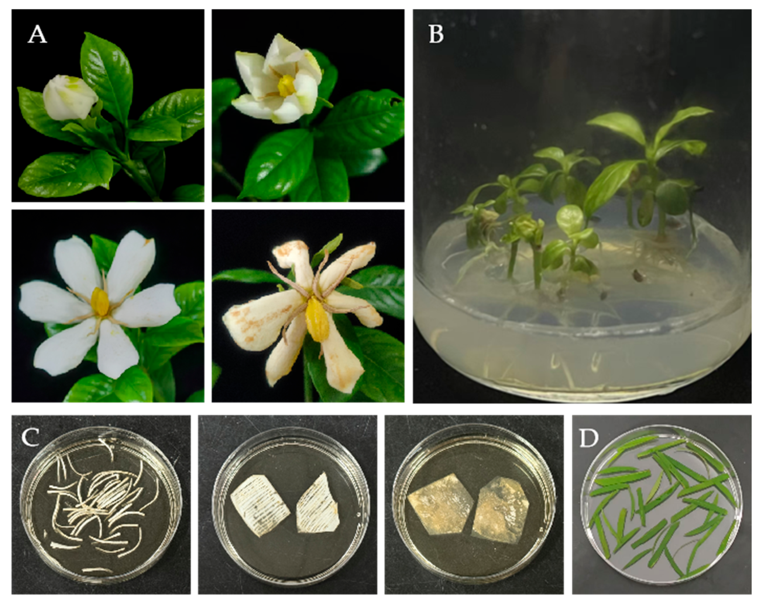
The flowering period of
G. jasminoides generally extends from May to August, lasting approximately 3–4 months. The full-flowering period of a single plant typically lasts 20–30 days, whereas the lifespan of an individual flower is only 3–5 days (
Figure 1A). To establish an efficient method for protoplast isolation from petals and mesophyll tissues, fully expanded petals and fresh leaves from tissue-cultured seedlings were selected as materials for protoplast preparation (
Figure 1A,B). To improve the isolation efficiency and quality of petal protoplasts, three pretreatment methods, namely cutting (The sharp blade rapidly cuts into strips approximately 0.1 mm in width), scratching (Cause epidermal abrasion by slicing petals with a blade), and peeling (Use tape to adhere both sides of the petals and remove the adhesive tape from the lower epidermis), were applied to
G. jasminoides petals (
Figure 1C). In contrast, for mesophyll protoplast isolation, the cutting method was applied to fresh leaves from tissue-cultured seedlings (
Figure 1D), followed by enzymatic digestion.
2.2. Protoplast Isolation
2.2.1. Isolation of Petal Protoplasts from G. jasminoides
Freshly opened petals of G. jasminoides were collected and immediately used for protoplast isolation. The petals were rinsed with sterile water, and surface moisture was removed using filter paper. The abaxial epidermis was then carefully peeled off with adhesive tape to fully expose the inner cells. The treated petals were subsequently incubated in freshly prepared enzyme solution containing 10 mM CaCl2, 20 mM KCl, 20 mM MES (pH 5.7), cellulase R-10 (Yakult, Tokyo, Japan), macerozyme R-10 (Yakult, Tokyo, Japan), and mannitol (Sigma, St. Louis, MO, USA).
Protoplast yield was determined using a hemocytometer under an OLYMPUS BX53 light microscope (Olympus Corporation, Tokyo, Japan). Protoplast viability was assessed by fluorescein diacetate (FDA) (10 μg/mL dissolved in DMSO) staining. Protoplasts exhibiting green fluorescence were regarded as viable. The formulas used to calculate protoplast yield and viability are shown in Equations (1) and (2), respectively.
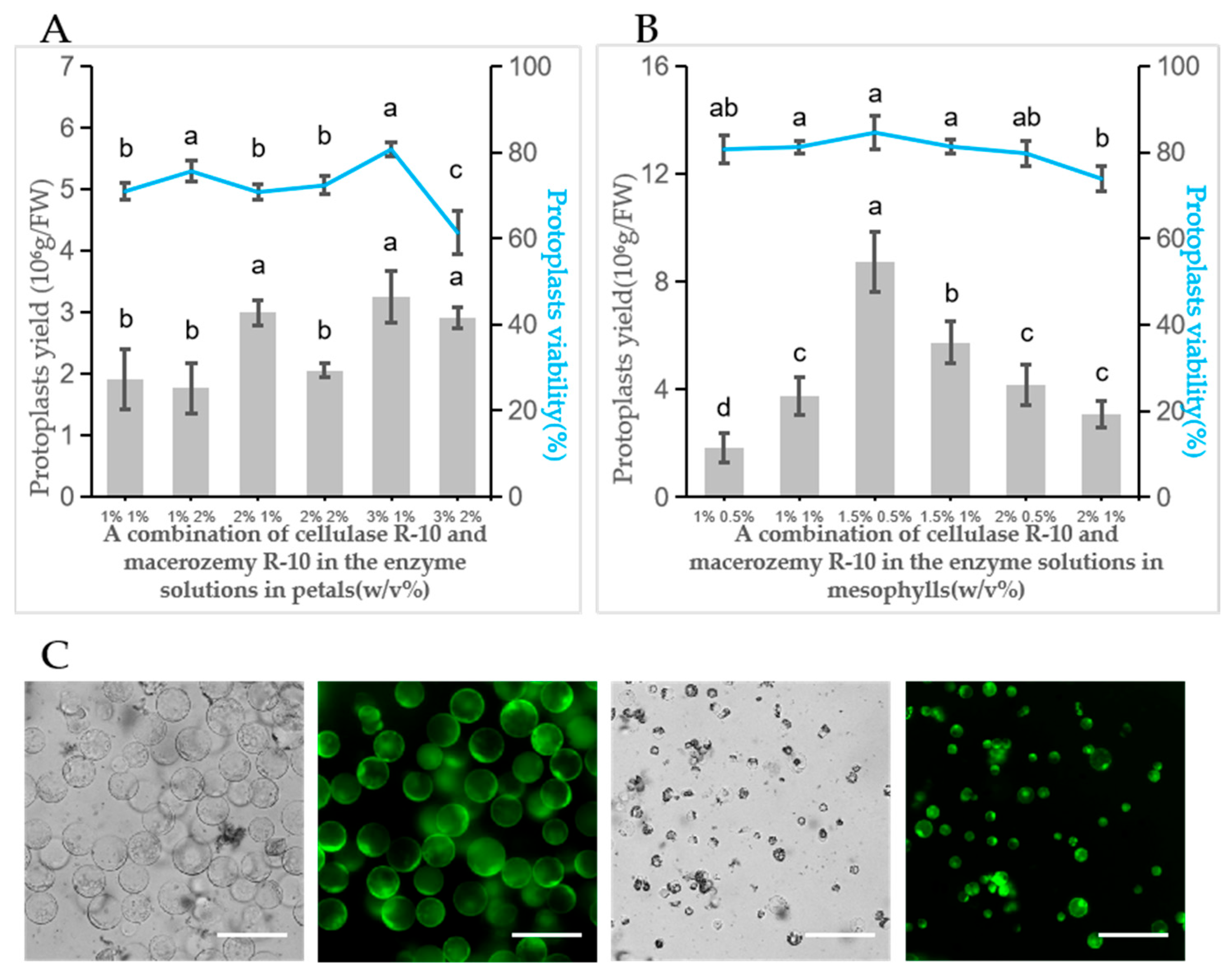
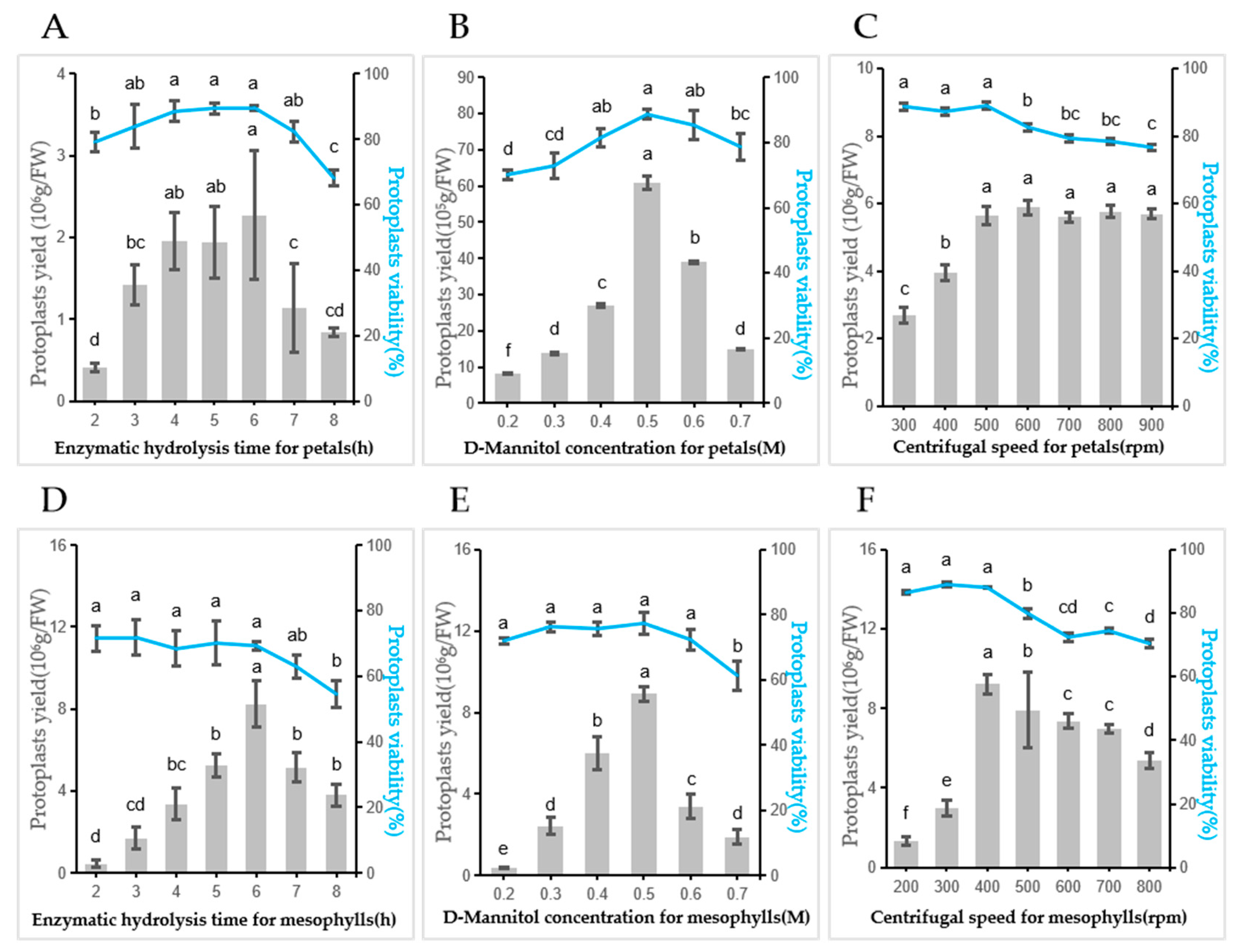
To determine the optimal enzyme combination for petal protoplast isolation, three concentrations of cellulase R-10 (1.0%, 2.0%, and 3.0%, w/v) and two concentrations of macerozyme R-10 (1.0% and 2.0%, w/v) were tested, generating six enzyme treatment combinations in total. In all treatments, 0.4 M D-mannitol was used as the osmotic stabilizer, the enzyme solution pH was adjusted to 5.8, and samples were incubated in the dark at 25 °C with shaking at 40 rpm for 6 h. To further optimize petal protoplast isolation, the effects of digestion time, D-mannitol concentration, and centrifugation speed after enzymatic digestion were evaluated. Seven digestion time points (2, 3, 4, 5, 6, 7, and 8 h), and six D-mannitol concentrations (0.2, 0.3, 0.4, 0.5, 0.6, and 0.7 M) were tested. Centrifugation speeds of 300, 400, 500, 600, 700, 800, and 900 rpm were evaluated for petal protoplasts.
2.2.2. Isolation of Mesophyll Protoplasts from G. jasminoides
Mesophyll protoplasts were isolated from young leaves of in vitro-cultured G. jasminoides seedlings. The leaves were rinsed with sterile water, blotted dry, and then quickly cut into 0.5–1 mm-wide strips before being transferred into freshly prepared enzyme solution. The enzyme solution consisted of 10 mM CaCl2, 20 mM KCl, 20 mM MES (pH 5.7), cellulase R-10, macerozyme R-10, and mannitol. Protoplast yield and viability were assessed using the same methods as those described for petal protoplasts.
To optimize mesophyll protoplast isolation from G. jasminoides leaves, tissue-cultured seedlings bearing young leaves were used as the source material. Cellulase R-10 was tested at 1.0%, 1.5%, and 2.0% (w/v), whereas macerozyme R-10 was tested at 0.5% and 1.0% (w/v), resulting in six enzyme combinations. In all treatments, 0.5 M mannitol was used as the osmotic stabilizer, the enzyme solution pH was adjusted to 5.8, and samples were incubated in the dark at 25 °C with shaking at 50 rpm for 6 h. To further optimize mesophyll protoplast isolation, the same seven digestion time points (2, 3, 4, 5, 6, 7, and 8 h), and six D-mannitol concentrations (0.2, 0.3, 0.4, 0.5, 0.6, and 0.7 M) were tested. Centrifugation speeds of 200, 300, 400, 500, 600, 700, and 800 rpm were tested for mesophyll protoplasts.
Each sample was counted at least three times, and the experiment was independently repeated three times.
2.3. Protoplast Transformation
2.3.1. PEG-Mediated Transient Transformation of Petal Protoplasts
PEG-mediated transient gene expression in
G. jasminoides petal protoplasts was performed based on a previously reported method with modifications [
19]. After enzymatic digestion, the protoplast suspension was filtered into a 50 mL centrifuge tube and centrifuged at 500 rpm for 2 min at room temperature to collect the protoplasts. The supernatant was discarded, and the protoplast pellet was gently resuspended in 5 mL of pre-cooled W5 solution, followed by incubation on ice for 30 min. Although a range of 1 × 10
5–1 × 10
6 cells/mL was tested during optimization, a final protoplast density of 1 × 10
6 cells/mL was used in all subsequent experiments unless otherwise stated.
For transformation, 200 μL of protoplast suspension was gently mixed with plasmid DNA in a 2 mL centrifuge tube. To determine the optimal plasmid amount, 2, 4, 6, 8, and 10 μg DNA were tested (
Figure S2B). Immediately afterward, 220 μL of PEG-CaCl
2 solution containing 20% PEG4000, 100 mM CaCl
2, and 0.2 M mannitol (pH 5.7) was added and gently mixed. To optimize the PEG concentration, 10%, 20%, 30%, 40%, and 50% (
w/
v) PEG4000 were evaluated (
Figure S2A). The mixture was incubated at room temperature in the dark for 20 min, and transformation was terminated by adding 800 μL of W5 solution.
The mixture was then centrifuged at 500 rpm for 2 min, the supernatant was removed, and the pellet was gently resuspended in 200 μL of WI solution containing 4 mM MES, 20 mM KCl, and 0.5 M mannitol (pH 5.7). All solutions were sterilized by filtration through a 0.22 μm membrane filter before use. The transformed protoplasts were incubated in the dark at 25 °C for 12–16 h, and GFP fluorescence was observed using a laser confocal microscope with an excitation wavelength of 488 nm and an emission wavelength of 510–530 nm. Transformation efficiency was calculated according to Equation (3).
At least three independent biological replicates were performed, and more than 200 cells were counted per replicate.
2.3.2. PEG-Mediated Transient Transformation of Mesophyll Protoplasts
PEG-mediated transient transformation of
G. jasminoides mesophyll protoplasts was performed in a similar manner, with conditions optimized for mesophyll-derived protoplasts [
20]. After enzymatic digestion, the protoplast suspension was filtered and centrifuged at 400 rpm for 2 min at room temperature to collect the protoplasts. The protoplast pellet was gently resuspended in 5 mL of pre-cooled W5 solution (200 μL 0.5 M MES, 7.7 mL 1 M NaCl, 6.25 mL 1 M CaCl
2, 500 μL 0.5 M KCl, and ddH
2O were diluted to 50 mL) and incubated on ice for 30 min. The protoplast concentration was then adjusted to approximately 1 × 10
5–1 × 10
6 cells mL
−1 using MMG solution (400 μL 0.5 M MES, 24.9 mL 0.8 M D-Mannitol, 1.5 mL 0.5 M MgCl
2, and ddH
2O were diluted to 50 mL).
For transformation, 200 μL of protoplast suspension was gently mixed with plasmid DNA, and 2, 4, 6, 8, and 10 μg DNA were tested to optimize the plasmid amount (
Figure S2D). Subsequently, 220 μL of PEG-CaCl
2 solution containing 40% PEG4000 (
Figure S2C), 100 mM CaCl
2, and 0.2 M mannitol (pH 5.7) was added and gently mixed. The mixture was incubated in the dark at room temperature for 20 min. Transformation was terminated by adding 800 μL of W5 solution, followed by centrifugation at 400 rpm for 2 min using Low-speed small centrifuge (Scilogex, Rocky Hill, CT, USA). The supernatant was discarded, and the pellet was gently resuspended in 200 μL of WI solution (37.5 mL 0.8 M D-Mannitol, 400 μL 0.5 M MES, 400 μL 0.5 M KCl, and ddH
2O were diluted to 50 mL). The transformed protoplasts were incubated in the dark at 25 °C for 12–16 h, and GFP fluorescence was observed under a laser confocal microscope. Transformation efficiency was calculated as described above. Each treatment was independently repeated three times.
2.4. Plasmid Construction and Protein Subcellular Localization
Total RNA was extracted from fresh flower buds of G. jasminoides using a rapid universal plant RNA extraction kit (Beijing Huayueyang Biotechnology Co., Ltd., Beijing, China). First-strand cDNA was synthesized from 3 μg of total RNA using anchored oligo(dT) primers and TransScript® First-Strand cDNA Synthesis SuperMix (AT301, TransGen Biotech, Beijing, China) according to the manufacturer’s instructions.
Gene-specific primers containing homologous overlapping ends were designed using Primer Premier 5.0 based on the CDS of GjAP3, GjPI, GjSEP, GjNPR1, and GjTGA2 (
Table S1). The target genes were amplified by PCR using Phanta Max Master Mix (Vazyme Biotech, Nanjing, China), 0.4 μL PCR Forward Primer (10 μM), 0.4 μL PCR Reverse Primer (10 μM), 1 μL 1:20 cDNA, and ddH
2O to diluted to 10 μL. PCR amplification was performed using the following cycling conditions: an initial denaturation at 95 °C for 3 min, followed by 37 cycles of denaturation at 95 °C for 15 s, annealing at 56 °C for 15 s, and extension at 72 °C for 30 s per kb, with a final extension at 72 °C for 5 min. The PCR products were purified and introduced into the pDONR207 vector by BP recombination using Gateway™ BP Clonase II Mix (Invitrogen, Carlsbad, CA, USA). After sequence verification, the entry clones were transferred into the Gateway-compatible destination vector pB7WGF2 by LR recombination using Gateway™ LR Clonase II Mix (Invitrogen, Carlsbad, CA, USA) [
21,
22,
23], generating the recombinant plasmids GFP-GjAP3, GFP-GjPI, GFP-GjSEP, GFP-GjNPR1, and GFP-GjTGA2.
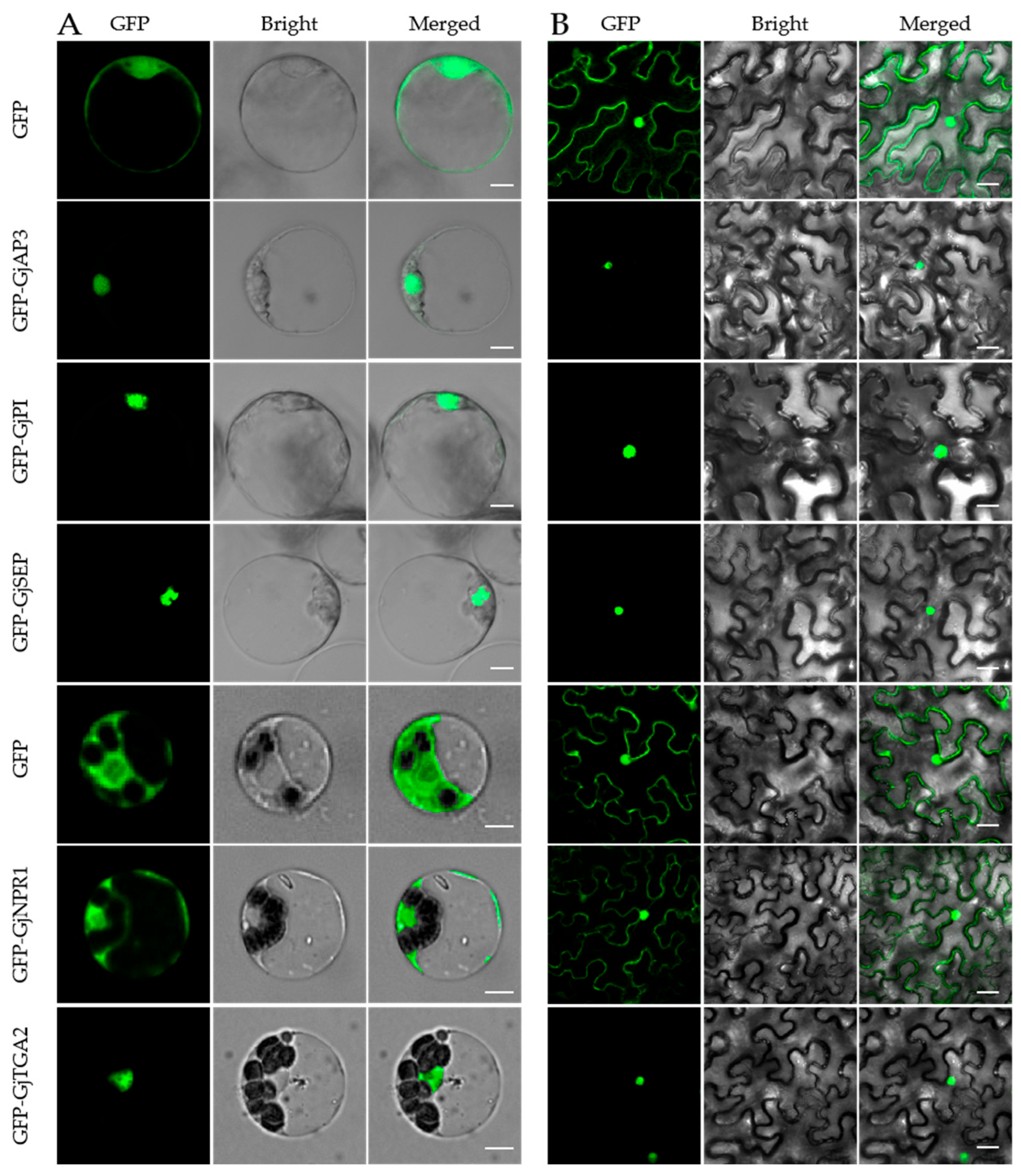
For transient expression in protoplasts, plasmid DNA was prepared on a large scale using a Plasmid Maxprep Kit (Vigorous Biotechnology, Beijing, China). The recombinant plasmids and the empty GFP vector, used as a control, were separately introduced into G. jasminoides petal or mesophyll protoplasts by PEG-mediated transformation. After incubation for 12–16 h, GFP fluorescence was observed and imaged using a laser scanning confocal microscope (Olympus Corporation, Tokyo, Japan).
As a parallel control, subcellular localization was also examined in tobacco leaves by
Agrobacterium tumefaciens-mediated transient expression. The plasmids were introduced into
A. tumefaciens strain GV3101 (pSoup) and cultured overnight at 28 °C. The bacterial cells were collected and resuspended in infiltration buffer containing 10 mM MES (pH 5.7), 10 mM MgCl
2, and 100 μM acetosyringone, adjusted to an OD
600 of 1.0, and incubated at room temperature for 2 h. The suspensions were then infiltrated into
N. benthamiana leaves using a sterile syringe. After 48 h of incubation in the dark, GFP fluorescence was observed under the same confocal microscope. Based on previous studies, the expected subcellular localization of the proteins observed in
G. jasminoides is consistent with that in
N. benthamiana [
23].
2.5. Protein–Protein Interaction in Yeast Cells and G. jasminoides Protoplasts
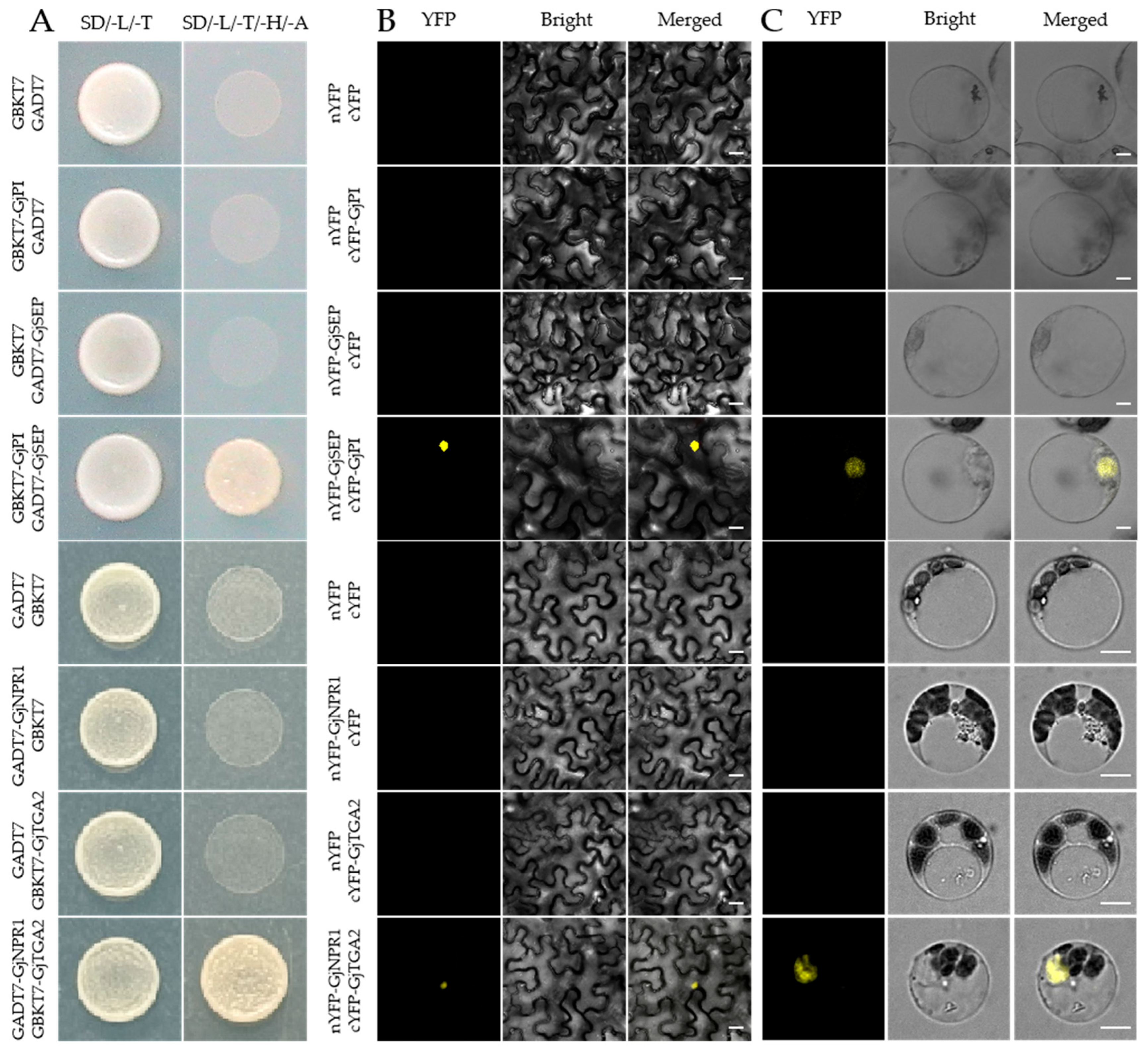
To verify protein–protein interactions, the CDS of GjSEP and GjPI were cloned into the Gateway-compatible destination vectors pGADT7 and pGBKT7, generating pGADT7-GjSEP and pGBKT7-GjPI, respectively. Yeast cells carrying empty pGADT7 and pGBKT7 vectors were used as negative controls, whereas yeast cells carrying pGADT7-GjSEP plus empty pGBKT7 or empty pGADT7 plus pGBKT7-GjPI were used as negative controls. The transformed yeast cells were cultured on SD/-Leu/-Trp medium at 29 °C for 48–96 h. Single colonies were selected, suspended in ddH2O, and spotted onto SD/-Leu/-Trp and SD/-Leu/-Trp/-His/-Ade selective media. After incubation at 29 °C for 48–96 h, colony growth was recorded.
BiFC analysis was performed in G. jasminoides petal protoplasts. Recombinant plasmids nYFP-GjSEP and cYFP-GjPI were generated by cloning the CDS of GjSEP and GjPI into the pCL112 and pCL113 vectors, respectively. nYFP-GjSEP and cYFP-GjPI were co-transformed into petal protoplasts. The negative controls included nYFP + cYFP, nYFP + cYFP-GjPI, and cYFP + nYFP-GjSEP. As a parallel control, the same experiment was performed in tobacco leaves by agroinfiltration.
Similarly, to verify the interaction between GjNPR1 and GjTGA2, the CDS of GjNPR1 and GjTGA2 were cloned into pGADT7 and pGBKT7, generating pGADT7-GjNPR1 and pGBKT7-GjTGA2, respectively. Yeast two-hybrid assays were performed as described above. For BiFC analysis in G. jasminoides mesophyll protoplasts, recombinant plasmids nYFP-GjNPR1 and cYFP-GjTGA2 were constructed and co-transformed. The negative controls included nYFP + cYFP, nYFP + cYFP-GjTGA2, and cYFP + nYFP-GjNPR1. Tobacco leaf agroinfiltration was used as a parallel control.
2.6. Statistical Analysis
Statistical analyses were performed using SPSS 20.0 software (SPSS Inc., Chicago, IL, USA). Each experiment included at least three independent biological replicates, and more than 200 cells were counted per replicate. The data were organized using Excel 2010. Data are presented as the mean ± standard error (SE) of three independent experiments and were plotted using GraphPad Prism 9 (GraphPad Software, La Jolla, CA, USA). Significant differences among groups were analyzed using One-way ANOVA followed by Duncan’s multiple range test at p < 0.05.
4. Discussion
At present, enzymatic digestion is the most commonly used method for protoplast isolation [
7]. In this study, based on previously reported methods, cellulase R-10 and macerozyme R-10 were selected to isolate protoplasts from
G. jasminoides petals and to establish an enzymatic digestion system [
23,
24]. In addition, auxiliary treatments such as vacuum infiltration, hypertonic pretreatment, and gentle shaking have been reported to accelerate enzymatic digestion and improve protoplast yield [
25]. Our results showed that increasing the total enzyme concentration and extending the digestion time could enhance protoplast yield to a certain extent. However, excessively high enzyme concentrations or prolonged digestion times may exert cytotoxic effects on the plasma membrane, thereby reducing protoplast viability. Previous studies have shown that D-mannitol, as an osmotic regulator, can significantly affect both protoplast yield and viability [
26]. In the present study, analysis of the effect of mannitol concentration on protoplast isolation demonstrated that 0.5 M D-mannitol significantly improved the yield of
G. jasminoides protoplasts. Following enzymatic digestion, purification is required to obtain high-quality protoplasts for transient expression assays. Because the flotation and interface methods are relatively difficult to operate [
27,
28], centrifugation-based sedimentation was adopted in this study for protoplast purification. During purification, low centrifugation speeds (300–400 rpm for petal protoplasts and 200–300 rpm for mesophyll protoplasts) resulted in substantial protoplast loss, whereas excessively high speeds (600–900 rpm for petal protoplasts and 500–800 rpm for mesophyll protoplasts) caused protoplast rupture and death. Accordingly, a high-quality protoplast isolation system with high viability was established in this study.
PEG-mediated transient transformation of protoplasts is widely used because of its rapidity, low cost, robustness, high throughput, and lack of requirement for specialized equipment [
29]. Previous studies have described high-throughput protoplast systems for analyzing signaling pathways and transcription factors in
Arabidopsis thaliana [
30]. In the present study, we established, for the first time, a protocol for high-yield isolation of
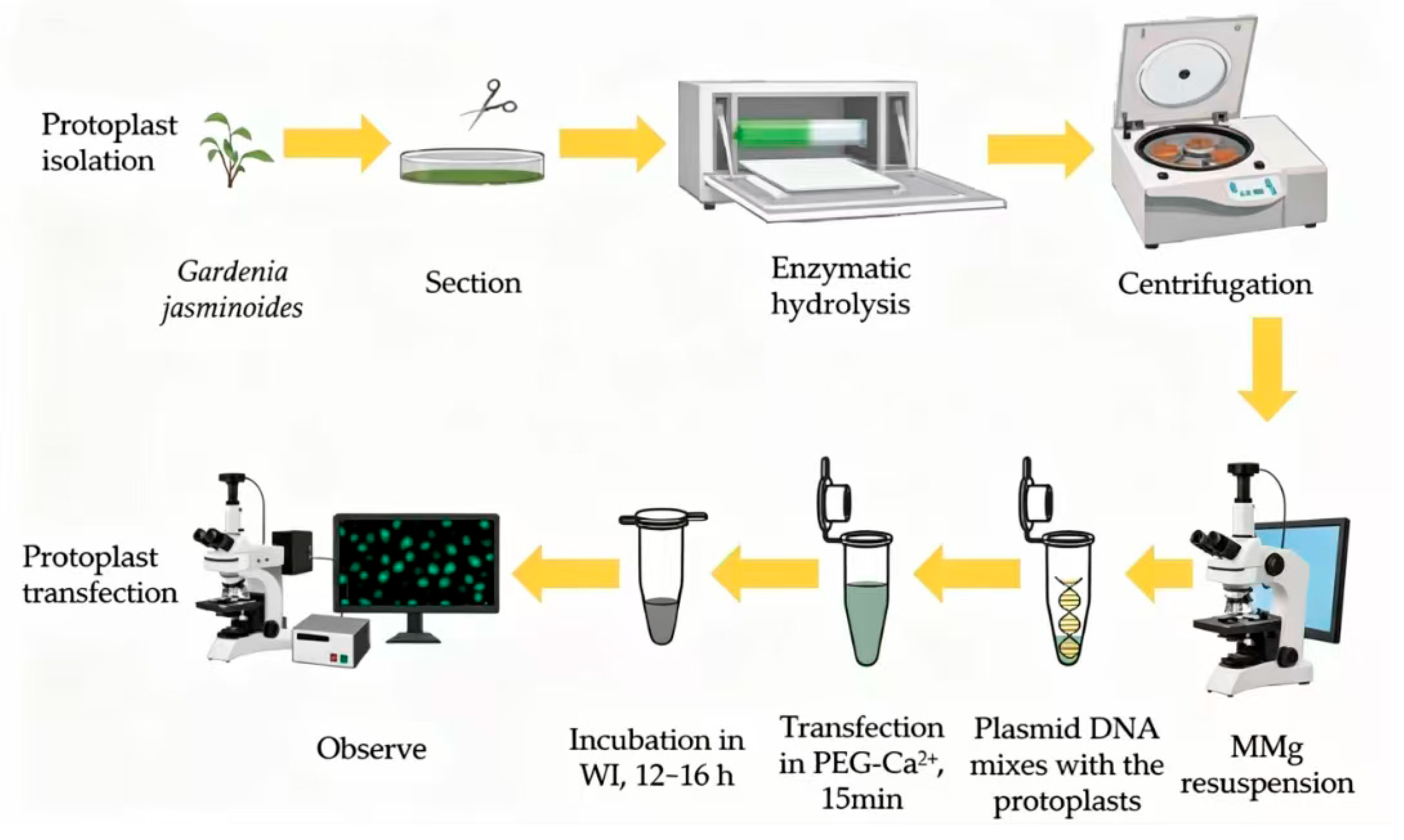
G. jasminoides protoplasts and PEG-mediated transformation of these protoplasts (
Figure 6). The successful use of tissue culture-derived materials highlights the importance of a controlled sterile environment for obtaining uniform starting materials with high viability. This observation is consistent with the general view that young tissues possess easily degradable cell walls and are subject to less interference from secondary metabolites. Protoplast transient expression systems have now been widely applied in plant molecular and cellular biology. Previous studies have shown that PEG4000 concentration, as well as the amount and concentration of plasmid DNA, can markedly affect transformation efficiency in plant protoplasts [
31,
32]. Our results showed that transformation efficiency initially increased with increasing PEG4000 concentration. However, when the PEG concentration reached 20% for petal protoplasts and 40% for mesophyll protoplasts, the number of ruptured protoplasts increased and transformation efficiency declined. In addition, higher plasmid DNA input generally resulted in higher transformation efficiency, consistent with previous findings in protoplasts from other plant species [
24,
29]. Taken together, these results indicate that an effective PEG-mediated transient expression system was successfully established for both petal and mesophyll protoplasts of
G. jasminoides.
Previous studies have shown that members of the SEPALLATA (SEP) subfamily of E-class floral homeotic genes play essential regulatory roles in floral organ development [
33]. SEP proteins can interact with proteins encoded by other classes of floral organ identity genes to form higher-order protein complexes involved in floral organ specification and development. In addition, Nonexpressor of Pathogenesis-Related genes 1 (NPR1) and TGACG-binding transcription factor 2 (TGA2) are both closely associated with plant immune responses and play important roles in defense against pathogen invasion, regulation of growth and development, and adaptation to environmental stress [
34,
35]. In this study, the established transient expression system was applied to the subcellular localization of GjAP3, GjPI, and GjSEP in petal protoplasts, and GjNPR1 and GjTGA2 in mesophyll protoplasts. The observed localization patterns were highly consistent with those obtained by heterologous expression in
N. benthamiana, indicating that this system provides a useful platform for assessing subcellular localization of proteins in
G. jasminoides. Previous reports have demonstrated interactions between SEP and PISTILLATA (PI) proteins [
36], as well as between NPR1 and TGA2 proteins [
37]. In the present study, BiFC was successfully applied to
G. jasminoides protoplasts for the first time, visually confirming the interactions between GjPI and GjSEP, and between GjNPR1 and GjTGA2. These results were further supported by yeast two-hybrid assays. Together, these application cases provide evidence that the established system can be used for transient functional analysis, such as subcellular localization and protein–protein interaction assays.
So far, to the best of our knowledge, this study is the first to establish a relatively efficient protoplast isolation and transient expression system for G. jasminoides. The transient expression platform established here fills a critical technical gap in the functional genomics of G. jasminoides and related woody species. Its major advantage lies in its efficiency and species-specific applicability: it bypasses the lengthy and difficult process of stable genetic transformation in woody plants and enables rapid verification of gene function directly in G. jasminoides cells, thereby reducing the potential discrepancies associated with heterologous systems. Nevertheless, the transient transformation efficiency of mesophyll protoplasts remained relatively low in this study and protoplast-based transient expression systems still have inherent limitations, including the short duration of exogenous gene expression and the inability to achieve stable genetic inheritance.




 ,
,